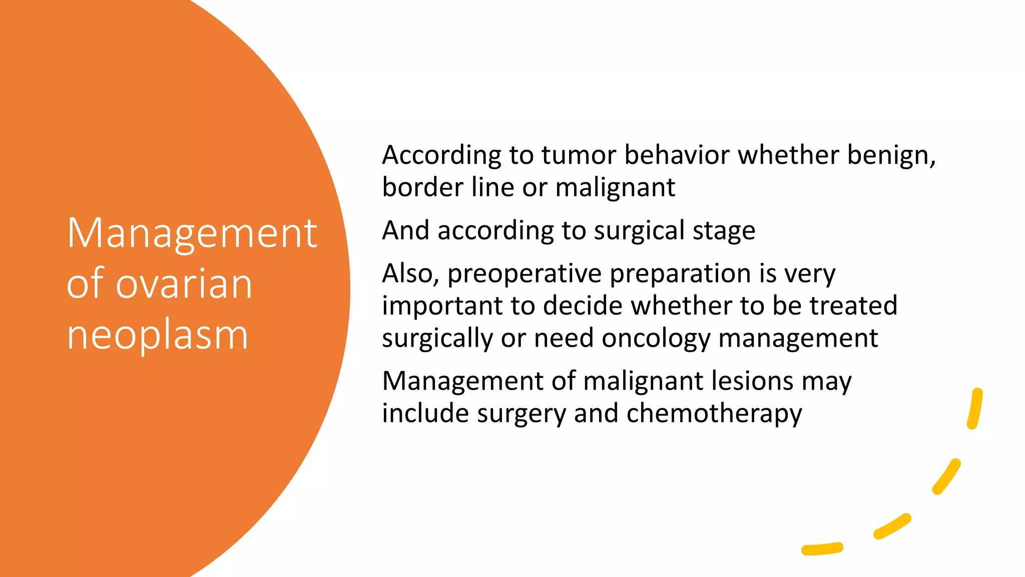
Ovarian
neoplasms
By
Dr. Ahmed Mohamed Nasef
Assistant lecturer in
Obstetrics & Gynecology
Benha University
Incidence
It is the 2nd most common
malignancy of female genital tract
(10-15/100000women)
It the most frequent cause of death
from gynecologic neoplasms
Its incidence increases with age (50-
70 years old)
The disease is more frequent among
whites than blacks
Risk factors
• Women who are related to frequent regular
ovulation at high risk (late menopause,
nulliparity or late childbearing)
• The number of years during which the
women has ovulated
• Talcum powder use over perineum
• Diet high in fat
• BRCA gene mutation
• Use of Ovulation induction drugs
(controversial)
Protective
factors
• Use of OCPs
• Breast feeding
• Pregnancy
• Tubal ligation & salpingectomy
• Hysterectomy with ovarian
preservation also decrease the
risk
Ovarian histology
The ovarian histology is composed of the
following:
Cortex (outer most part)
Covered by mesothelium (single layer of cuboidal
to columnar epithelium)
Composed of stroma and follicles (contain the
germ cells, oocyte)
Medulla (middle part)
Composed of stroma, blood vessels & nerves
Hilum
Where blood vessels enter parenchyma
Classification of ovarian neoplasm
WHO classified it according to site of origin into:
Surface epithelium (65%) most common
from surface epithelium
Germ cells (15%) 2nd most common
Main cause of ovarian malignancy in young women (20s)
From germ cells
Sex cord-stromal cells (10%) 3rd most common
From sex cord & stroma
Secretory ovarian tumors (secrete sex steroid hormones) or may be non secretory
Gonadoblastoma occur in dysgenetic gonads
Consist of germ cells & sex cord stromal elements
Metastases (5%)
From reproductive tract tumors or from distant sites (bowel or stomach)
Miscellaneous
Such as Hemangioma & lipoma
Epithelial tumors
• They resemble normal epithelium in the genitourinary tract
Serous tumors like fallopian tube epithelium
Mucinous tumors like endocervical mucosa
Endometrial tumors like endometrium
Brenner tumors like transitional epithelium of bladder
• many of them present a mixed histological appearance
• May be benign, borderline or malignant
Germ cell tumors
• Occur prior to puberty, in adolescence & early adult life
• They include
Teratoma (mature, immature, solid, cystic)
Monodermal tumors
Dysgerminoma
Yolk sac tumor
Mixed germ cell tumors
• All are malignant except the mature teratoma (dermoid cyst) has little tendency to malignant
transformation
Germ cell tumors
• Every type of them secrete substances in the blood which is used as a tumor marker
for diagnosis & follow up as:
Immature teratoma secrete CA19.9
Dysgerminoma secrete LDH
Endodermal sinus tumor secrete alpha fetoprotein
Sex-cord stromal tumors
• Functioning ovarian tumors as they secrete sex hormones like estrogens & androgens
• Composed of either male or female origin
• They include those containing (granulosa, theca, Sertoli or Leydig cells)
• Also, they include stromal elements (fibroma, fibro thecoma)
Border line
tumors
Criteria include:
Epithelial proliferation
Nuclear atypia
Increased mitotic activity
Absence of true stromal invasion
Diagnosed based on histologic features
Simple
clear cyst
by us
Dermoid cyst
Suspected malignant cyst by us
DD between
different classes
of ovarian
neoplasms
Surface epithelium
tumors
Germ cell tumors Sex cord-stromal
tumors
Cell of origin Coelomic epithelium Germ cells Sex cord & stroma
Incidence 65%
90% of malignant
15%
Most in young
10%
PF Ovulation age Dysgentic gonads
Age group B, BL (30-50)
M (50-80)
Young Any age
Bilaterality Bilateral Mainly unilateral Unilateral
Consistency Cystic, solid,
heterogenous
Most solid Solid white or
yellow
Behaviour B, BL, M All malignant except
dermoid
Granulosa M
Thecoma, fibroma B
Mixed LGM
Meigs syndrome
Criteria for diagnosis
Tumor must be (ovarian, solid, benign)
Both hydrothorax and ascites must be present
Removal of tumor must result in spontaneous and permanent cure
Complications of
ovarian
neoplasms
• Torsion
Dermoid cyst most prone to torsion
• rupture of a cyst
• Hemorrhage into a cyst
Most are self limited
• Incarceration
• Infection
• Intestinal obstruction
• Malignant transformation
• Pain
• Pressure
Constipation, distension, bloating, indigestion & dyspnea
• Pregnancy in presence of ovarian swellings
Clinical presentation
Symptoms
Asymptomatic
Bleeding
Complication
Dyspepsia leading symptom of ovarian tumor
Enlargement abdominal swelling
Pressure edema of legs & vulva
Pain
Discovered accidently in pregnancy
Signs
• General
Cachexia
Weight loss
• abdominal
Ascites
Consistency of the lesion (solid, cystic or
heterogenous)
Mobility, tenderness, bilaterality
• Local
vulval edema, vaginal bleeding
Clinical features suggesting malignancy
• Age
Incidence increase with age
Neoplasm in childhood (usually malignant)
• Bilaterality
• Edema & varicosities of lower limb or vulva
• Solid, nodular, irregular shaped edges
• Fixation
• Ascites
• Tenderness
• Enlargement rate
• Nodules in DP
Causes of
ascites
• Tumor perforated capsule (oozing)
• Peritoneal metastases (oozing)
• Subdiaphragmatic metastases (lymphatic
obstruction)
• Hepatic metastases (portal hypertension)
• Electrolyte imbalance
• Cachexia (hypoproteinemia)
Spread of
ovarian
cancer
• Direct
To pelvic & abdominal organs
• Lymphatic
To paraaortic, pelvic & inguinal lymph nodes
Left supraclavicular lymph node
• Blood
To brain, bone, liver & lung
• Implantation
Staging (FIGO surgical staging)
Stage 1
Limited to organ
A one ovary B both ovaries C (surgical spill, capsule ruptured, malignant cells in ascites or peritoneal washings
Stage 2
Limited to pelvis
A genital tract B (bladder, rectum & DP)
Stage 3
Spread to peritoneum outside pelvis and or metastasis to retroperitoneal lymph nodes
A microscopic involvement of retroperitoneal lymph nodes or extra pelvic peritoneum
B macroscopic peritoneal involvement outside pelvis > 2 cm
C macroscopic peritoneal involvement outside pelvis < 2 cm, or extension to only capsule of liver and spleen
Stage 4
Distant metastases
A pleural effusion B to extra abdominal organs and parenchymal involvement of liver and spleen
Screening
Screening
To whom
General population screening will be a financial
problem
In USA, the cost will exceed 10 billion dollar annually
So preferred to be done only for high-risk population
When to screen
Annually
How to screen
Physical examination (least sensitive)
Biomarkers (CA125)
Ultrasound
Screening by
ultrasound
• If positive screening
Cyst > 7cm, unilocular, thin wall, clear fluid,
premenopausal
Follow up after 2 months
Cyst < 7cm, multilocular, thick wall, turbid
fluid, postmenopausal
IOTA description
B surgical treatment
Uncertain further evaluation
M oncology work up
• If negative screening
Rescreen annually
Diagnosis
Diagnosis is done in 2 steps:
• Preoperative
Physical examination
Ultrasound (IOTA)
Biochemical (tumor markers)
RMI
Advanced imaging (CT, MRI)
• Operative
only diagnosis and staging is surgical
Diagnosis is established by histologic examination of tumor tissue
removed at operation
IOTA (international ovarian
tumor analysis) USA
IOTA (international ovarian
tumor analysis) USA
Ultrasound suggestion of benign or
malignant
Likely benign Likely malignant
B1 Unilocular cyst M1 Solid
B2 No solid
component or
> 7 mm
M2 Ascites
B3 Acoustic
shadows
M3 < 4 papilla
B4 Smooth
Diameter >
10cm
M4 Irregular
Diameter <
10cm
B5 No color flow M5 Strong color
flow
IOTA interpretation
M features B features
Very low risk No 3 or more
Low risk No 2
No 1 B (B1)
Intermediate risk No 1B (but not B1)
Elevated risk M features > B features
Very high risk M features < B features
RMI (Risk of Malignancy
Index) UK
RMI (Risk
of
Malignancy
Index) UK
Product of ultrasound score, menopausal
status & serum CA125 level
RMI= U × M × CA125
RMI final score
• >25 low malignancy risk
• 25 – 200 intermediate malignancy risk
• < 200 high malignancy risk
RMI (U)
Ultrasound result Ultrasound score to be
used in RMI calculation
Multilocular cysts 0 or 1 U 0 For ultrasound score of
0
Solid areas 0 or 1 U 1 For ultrasound score of
1
Metastases 0 or 1 U 3 For ultrasound score < 1
Ascites 0 or 1
Bilateral lesions 0 or 1
RMI (M)
Premenopausal 1
Postmenopausal 3
RMI (CA125 level)
Measured in IU/ml
Management
of ovarian
neoplasm
According to tumor behavior whether benign,
border line or malignant
And according to surgical stage
Also, preoperative preparation is very
important to decide whether to be treated
surgically or need oncology management
Management of malignant lesions may
include surgery and chemotherapy
Surgical rules
Cystectomy
Only remove cyst
Ovariectomy (oophorectomy)
Remove ovary
Salpingoophrectomy
Remove tube and ovary
Cytoreductive surgery
TAH, BSO, complete omentectomy, resection of all
visible tumor, retroperitoneal lymphadenectomy
Additional procedures (diaphragmatic resection,
splenectomy, hepatic resection, bowel and bladder
resection)
Surgical rules
Exploratory staging
Involve
Sampling of any free fluid
All intraabdominal viscera are explored
systematically
Biopsies are taken from suspicious areas
Biopsies from diaphragmatic surface
Retroperitoneal spaces are explored
Omentectomy are performed
Benign ovarian
neoplasm
• Young & need fertility
Cystectomy
• Elderly
TAH & BSO
Malignant
border line and
stage 1 low
grades
No cystectomy
Young need fertility
USO then after complete
childbearing TAH & BSO
All others TAH & BSO
Follow up after treatment
Periodic pelvic examinations &
CA125 level till childbearing end
Stage 1A high
grade
TAH & BSO
Chemotherapy and follow up
Further management according to
result if there is remission may
need maintenance therapy
Stage 2
TAH & BSO
Pelvic & paraaortic lymph nodes
are samples
Omentectomy, appendectomy
Then chemotherapy, follow up
Further management according to
result may need secondary
cytoreduction
Stage 3 & 4
Cytoreductive surgery
Chemotherapy and follow up
Further management according to
result may need trial of mew
modalities of chemotherapy
ovarian neoplasm.pptx

ovarian neoplasm.pptx

  • 1.
    Ovarian neoplasms By Dr. Ahmed MohamedNasef Assistant lecturer in Obstetrics & Gynecology Benha University
  • 2.
    Incidence It is the2nd most common malignancy of female genital tract (10-15/100000women) It the most frequent cause of death from gynecologic neoplasms Its incidence increases with age (50- 70 years old) The disease is more frequent among whites than blacks
  • 4.
    Risk factors • Womenwho are related to frequent regular ovulation at high risk (late menopause, nulliparity or late childbearing) • The number of years during which the women has ovulated • Talcum powder use over perineum • Diet high in fat • BRCA gene mutation • Use of Ovulation induction drugs (controversial)
  • 5.
    Protective factors • Use ofOCPs • Breast feeding • Pregnancy • Tubal ligation & salpingectomy • Hysterectomy with ovarian preservation also decrease the risk
  • 6.
    Ovarian histology The ovarianhistology is composed of the following: Cortex (outer most part) Covered by mesothelium (single layer of cuboidal to columnar epithelium) Composed of stroma and follicles (contain the germ cells, oocyte) Medulla (middle part) Composed of stroma, blood vessels & nerves Hilum Where blood vessels enter parenchyma
  • 7.
    Classification of ovarianneoplasm WHO classified it according to site of origin into: Surface epithelium (65%) most common from surface epithelium Germ cells (15%) 2nd most common Main cause of ovarian malignancy in young women (20s) From germ cells Sex cord-stromal cells (10%) 3rd most common From sex cord & stroma Secretory ovarian tumors (secrete sex steroid hormones) or may be non secretory Gonadoblastoma occur in dysgenetic gonads Consist of germ cells & sex cord stromal elements Metastases (5%) From reproductive tract tumors or from distant sites (bowel or stomach) Miscellaneous Such as Hemangioma & lipoma
  • 9.
    Epithelial tumors • Theyresemble normal epithelium in the genitourinary tract Serous tumors like fallopian tube epithelium Mucinous tumors like endocervical mucosa Endometrial tumors like endometrium Brenner tumors like transitional epithelium of bladder • many of them present a mixed histological appearance • May be benign, borderline or malignant
  • 11.
    Germ cell tumors •Occur prior to puberty, in adolescence & early adult life • They include Teratoma (mature, immature, solid, cystic) Monodermal tumors Dysgerminoma Yolk sac tumor Mixed germ cell tumors • All are malignant except the mature teratoma (dermoid cyst) has little tendency to malignant transformation
  • 13.
    Germ cell tumors •Every type of them secrete substances in the blood which is used as a tumor marker for diagnosis & follow up as: Immature teratoma secrete CA19.9 Dysgerminoma secrete LDH Endodermal sinus tumor secrete alpha fetoprotein
  • 15.
    Sex-cord stromal tumors •Functioning ovarian tumors as they secrete sex hormones like estrogens & androgens • Composed of either male or female origin • They include those containing (granulosa, theca, Sertoli or Leydig cells) • Also, they include stromal elements (fibroma, fibro thecoma)
  • 16.
    Border line tumors Criteria include: Epithelialproliferation Nuclear atypia Increased mitotic activity Absence of true stromal invasion Diagnosed based on histologic features
  • 17.
  • 18.
  • 19.
  • 20.
    DD between different classes ofovarian neoplasms Surface epithelium tumors Germ cell tumors Sex cord-stromal tumors Cell of origin Coelomic epithelium Germ cells Sex cord & stroma Incidence 65% 90% of malignant 15% Most in young 10% PF Ovulation age Dysgentic gonads Age group B, BL (30-50) M (50-80) Young Any age Bilaterality Bilateral Mainly unilateral Unilateral Consistency Cystic, solid, heterogenous Most solid Solid white or yellow Behaviour B, BL, M All malignant except dermoid Granulosa M Thecoma, fibroma B Mixed LGM
  • 21.
    Meigs syndrome Criteria fordiagnosis Tumor must be (ovarian, solid, benign) Both hydrothorax and ascites must be present Removal of tumor must result in spontaneous and permanent cure
  • 22.
    Complications of ovarian neoplasms • Torsion Dermoidcyst most prone to torsion • rupture of a cyst • Hemorrhage into a cyst Most are self limited • Incarceration • Infection • Intestinal obstruction • Malignant transformation • Pain • Pressure Constipation, distension, bloating, indigestion & dyspnea • Pregnancy in presence of ovarian swellings
  • 23.
    Clinical presentation Symptoms Asymptomatic Bleeding Complication Dyspepsia leadingsymptom of ovarian tumor Enlargement abdominal swelling Pressure edema of legs & vulva Pain Discovered accidently in pregnancy Signs • General Cachexia Weight loss • abdominal Ascites Consistency of the lesion (solid, cystic or heterogenous) Mobility, tenderness, bilaterality • Local vulval edema, vaginal bleeding
  • 24.
    Clinical features suggestingmalignancy • Age Incidence increase with age Neoplasm in childhood (usually malignant) • Bilaterality • Edema & varicosities of lower limb or vulva • Solid, nodular, irregular shaped edges • Fixation • Ascites • Tenderness • Enlargement rate • Nodules in DP
  • 25.
    Causes of ascites • Tumorperforated capsule (oozing) • Peritoneal metastases (oozing) • Subdiaphragmatic metastases (lymphatic obstruction) • Hepatic metastases (portal hypertension) • Electrolyte imbalance • Cachexia (hypoproteinemia)
  • 26.
    Spread of ovarian cancer • Direct Topelvic & abdominal organs • Lymphatic To paraaortic, pelvic & inguinal lymph nodes Left supraclavicular lymph node • Blood To brain, bone, liver & lung • Implantation
  • 27.
    Staging (FIGO surgicalstaging) Stage 1 Limited to organ A one ovary B both ovaries C (surgical spill, capsule ruptured, malignant cells in ascites or peritoneal washings Stage 2 Limited to pelvis A genital tract B (bladder, rectum & DP) Stage 3 Spread to peritoneum outside pelvis and or metastasis to retroperitoneal lymph nodes A microscopic involvement of retroperitoneal lymph nodes or extra pelvic peritoneum B macroscopic peritoneal involvement outside pelvis > 2 cm C macroscopic peritoneal involvement outside pelvis < 2 cm, or extension to only capsule of liver and spleen Stage 4 Distant metastases A pleural effusion B to extra abdominal organs and parenchymal involvement of liver and spleen
  • 28.
  • 29.
    Screening To whom General populationscreening will be a financial problem In USA, the cost will exceed 10 billion dollar annually So preferred to be done only for high-risk population When to screen Annually How to screen Physical examination (least sensitive) Biomarkers (CA125) Ultrasound
  • 30.
    Screening by ultrasound • Ifpositive screening Cyst > 7cm, unilocular, thin wall, clear fluid, premenopausal Follow up after 2 months Cyst < 7cm, multilocular, thick wall, turbid fluid, postmenopausal IOTA description B surgical treatment Uncertain further evaluation M oncology work up • If negative screening Rescreen annually
  • 32.
    Diagnosis Diagnosis is donein 2 steps: • Preoperative Physical examination Ultrasound (IOTA) Biochemical (tumor markers) RMI Advanced imaging (CT, MRI) • Operative only diagnosis and staging is surgical Diagnosis is established by histologic examination of tumor tissue removed at operation
  • 33.
  • 34.
    IOTA (international ovarian tumoranalysis) USA Ultrasound suggestion of benign or malignant Likely benign Likely malignant B1 Unilocular cyst M1 Solid B2 No solid component or > 7 mm M2 Ascites B3 Acoustic shadows M3 < 4 papilla B4 Smooth Diameter > 10cm M4 Irregular Diameter < 10cm B5 No color flow M5 Strong color flow
  • 35.
    IOTA interpretation M featuresB features Very low risk No 3 or more Low risk No 2 No 1 B (B1) Intermediate risk No 1B (but not B1) Elevated risk M features > B features Very high risk M features < B features
  • 36.
    RMI (Risk ofMalignancy Index) UK
  • 37.
    RMI (Risk of Malignancy Index) UK Productof ultrasound score, menopausal status & serum CA125 level RMI= U × M × CA125 RMI final score • >25 low malignancy risk • 25 – 200 intermediate malignancy risk • < 200 high malignancy risk
  • 38.
    RMI (U) Ultrasound resultUltrasound score to be used in RMI calculation Multilocular cysts 0 or 1 U 0 For ultrasound score of 0 Solid areas 0 or 1 U 1 For ultrasound score of 1 Metastases 0 or 1 U 3 For ultrasound score < 1 Ascites 0 or 1 Bilateral lesions 0 or 1
  • 39.
  • 40.
  • 42.
    Management of ovarian neoplasm According totumor behavior whether benign, border line or malignant And according to surgical stage Also, preoperative preparation is very important to decide whether to be treated surgically or need oncology management Management of malignant lesions may include surgery and chemotherapy
  • 43.
    Surgical rules Cystectomy Only removecyst Ovariectomy (oophorectomy) Remove ovary Salpingoophrectomy Remove tube and ovary Cytoreductive surgery TAH, BSO, complete omentectomy, resection of all visible tumor, retroperitoneal lymphadenectomy Additional procedures (diaphragmatic resection, splenectomy, hepatic resection, bowel and bladder resection)
  • 44.
    Surgical rules Exploratory staging Involve Samplingof any free fluid All intraabdominal viscera are explored systematically Biopsies are taken from suspicious areas Biopsies from diaphragmatic surface Retroperitoneal spaces are explored Omentectomy are performed
  • 45.
    Benign ovarian neoplasm • Young& need fertility Cystectomy • Elderly TAH & BSO
  • 46.
    Malignant border line and stage1 low grades No cystectomy Young need fertility USO then after complete childbearing TAH & BSO All others TAH & BSO Follow up after treatment Periodic pelvic examinations & CA125 level till childbearing end
  • 47.
    Stage 1A high grade TAH& BSO Chemotherapy and follow up Further management according to result if there is remission may need maintenance therapy
  • 48.
    Stage 2 TAH &BSO Pelvic & paraaortic lymph nodes are samples Omentectomy, appendectomy Then chemotherapy, follow up Further management according to result may need secondary cytoreduction
  • 49.
    Stage 3 &4 Cytoreductive surgery Chemotherapy and follow up Further management according to result may need trial of mew modalities of chemotherapy