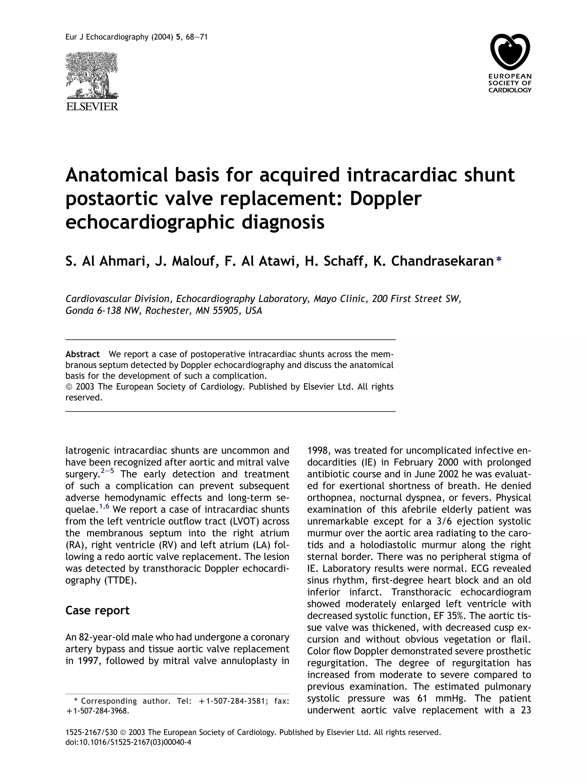
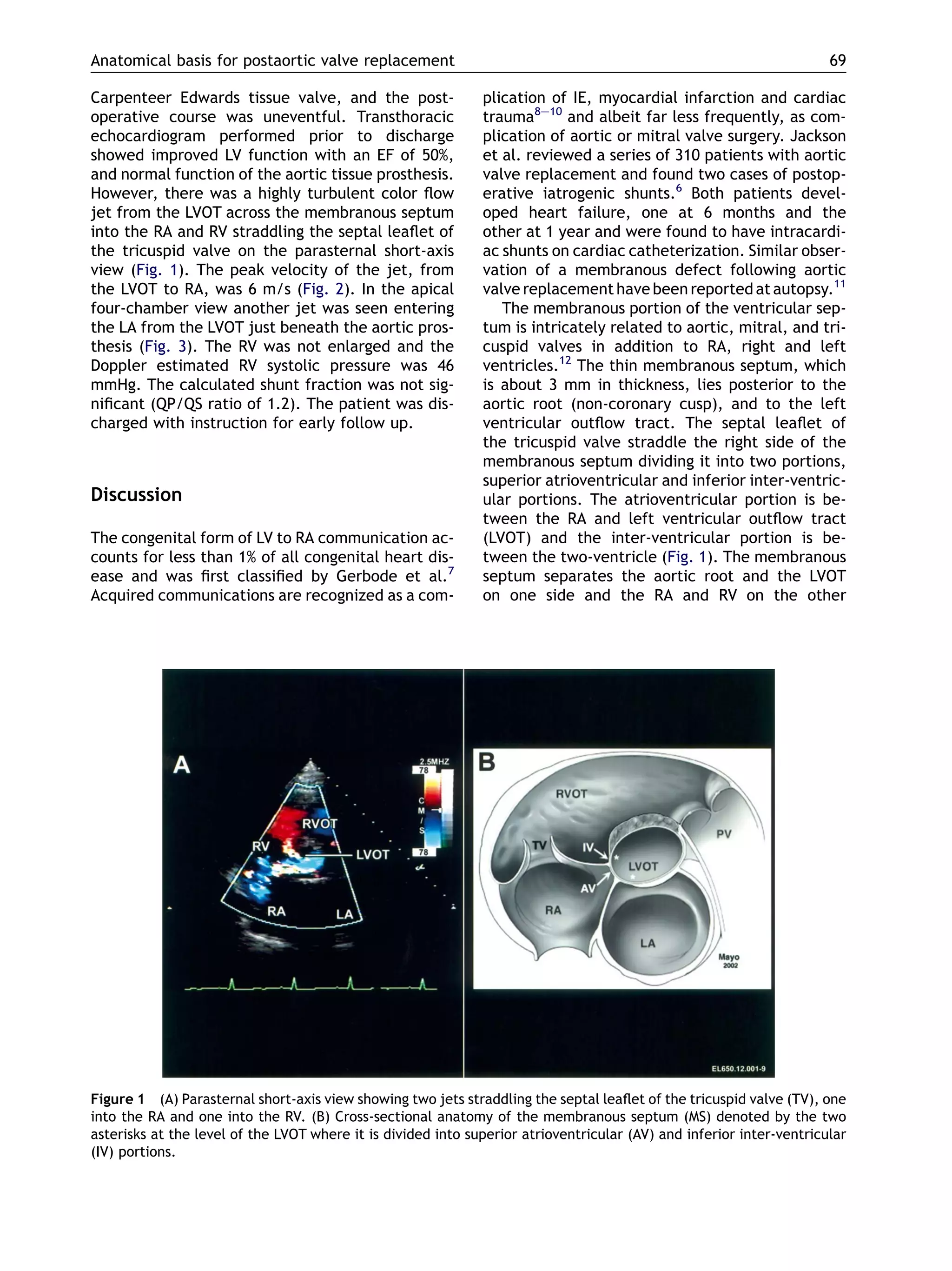
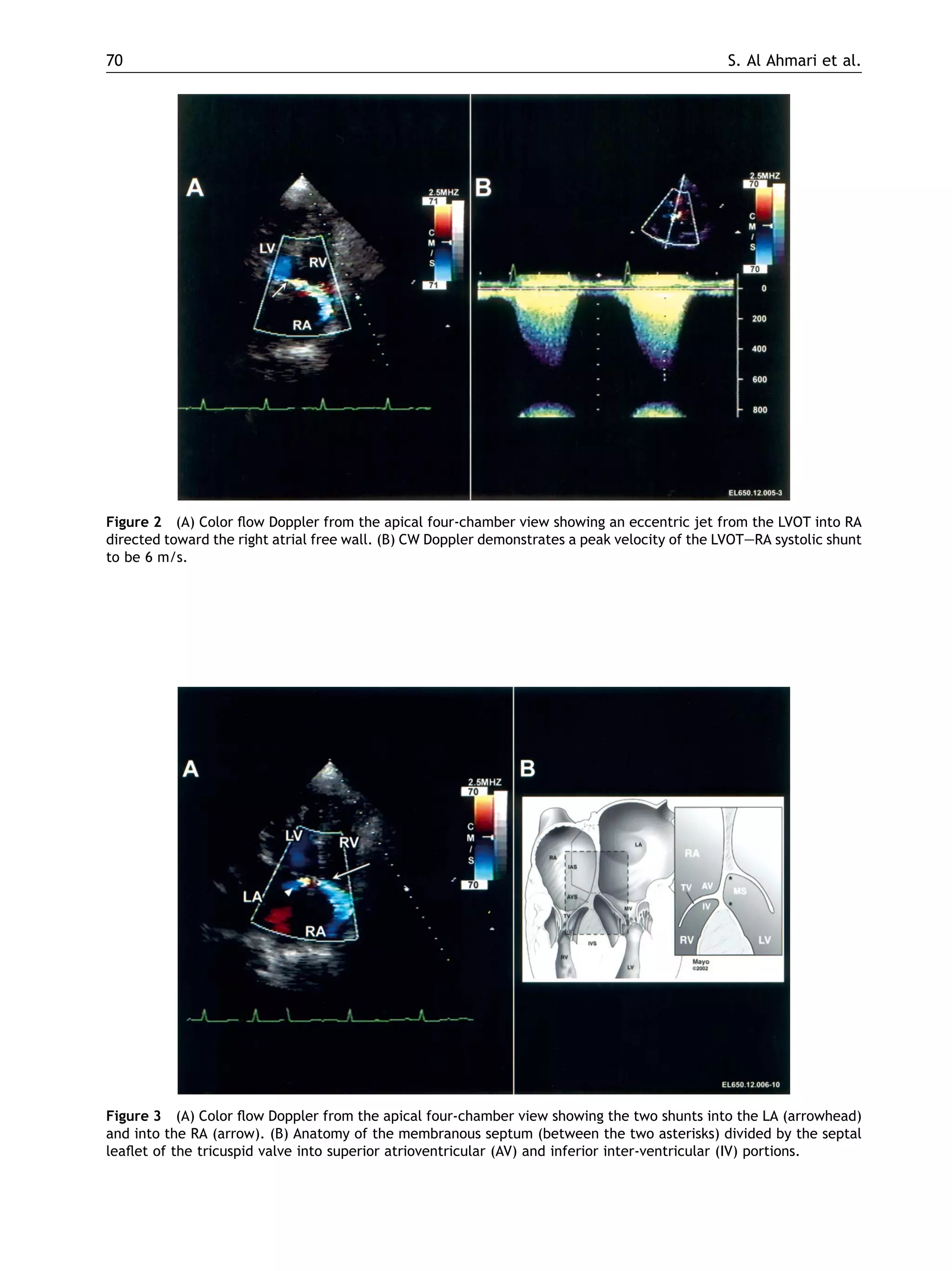
Anatomical basis for acquired intracardiac shunt
postaortic valve replacement: Doppler
echocardiographic diagnosis
S. Al Ahmari, J. Malouf, F. Al Atawi, H. Schaff, K. Chandrasekaran)
Cardiovascular Division, Echocardiography Laboratory, Mayo Clinic, 200 First Street SW,
Gonda 6-138 NW, Rochester, MN 55905, USA
Abstract We report a case of postoperative intracardiac shunts across the mem-
branous septum detected by Doppler echocardiography and discuss the anatomical
basis for the development of such a complication.
ª 2003 The European Society of Cardiology. Published by Elsevier Ltd. All rights
reserved.
Iatrogenic intracardiac shunts are uncommon and
have been recognized after aortic and mitral valve
surgery.2e5
The early detection and treatment
of such a complication can prevent subsequent
adverse hemodynamic effects and long-term se-
quelae.1,6
We report a case of intracardiac shunts
from the left ventricle outflow tract (LVOT) across
the membranous septum into the right atrium
(RA), right ventricle (RV) and left atrium (LA) fol-
lowing a redo aortic valve replacement. The lesion
was detected by transthoracic Doppler echocardi-
ography (TTDE).
Case report
An 82-year-old male who had undergone a coronary
artery bypass and tissue aortic valve replacement
in 1997, followed by mitral valve annuloplasty in
1998, was treated for uncomplicated infective en-
docardities (IE) in February 2000 with prolonged
antibiotic course and in June 2002 he was evaluat-
ed for exertional shortness of breath. He denied
orthopnea, nocturnal dyspnea, or fevers. Physical
examination of this afebrile elderly patient was
unremarkable except for a 3/6 ejection systolic
murmur over the aortic area radiating to the caro-
tids and a holodiastolic murmur along the right
sternal border. There was no peripheral stigma of
IE. Laboratory results were normal. ECG revealed
sinus rhythm, first-degree heart block and an old
inferior infarct. Transthoracic echocardiogram
showed moderately enlarged left ventricle with
decreased systolic function, EF 35%. The aortic tis-
sue valve was thickened, with decreased cusp ex-
cursion and without obvious vegetation or flail.
Color flow Doppler demonstrated severe prosthetic
regurgitation. The degree of regurgitation has
increased from moderate to severe compared to
previous examination. The estimated pulmonary
systolic pressure was 61 mmHg. The patient
underwent aortic valve replacement with a 23
Eur J Echocardiography (2004) 5, 68e71
) Corresponding author. Tel: D1-507-284-3581; fax:
D1-507-284-3968.
1525-2167/$30 ª 2003 The European Society of Cardiology. Published by Elsevier Ltd. All rights reserved.
doi:10.1016/S1525-2167(03)00040-4
Carpenteer Edwards tissue valve, and the post-
operative course was uneventful. Transthoracic
echocardiogram performed prior to discharge
showed improved LV function with an EF of 50%,
and normal function of the aortic tissue prosthesis.
However, there was a highly turbulent color flow
jet from the LVOT across the membranous septum
into the RA and RV straddling the septal leaflet of
the tricuspid valve on the parasternal short-axis
view (Fig. 1). The peak velocity of the jet, from
the LVOT to RA, was 6 m/s (Fig. 2). In the apical
four-chamber view another jet was seen entering
the LA from the LVOT just beneath the aortic pros-
thesis (Fig. 3). The RV was not enlarged and the
Doppler estimated RV systolic pressure was 46
mmHg. The calculated shunt fraction was not sig-
nificant (QP/QS ratio of 1.2). The patient was dis-
charged with instruction for early follow up.
Discussion
The congenital form of LV to RA communication ac-
counts for less than 1% of all congenital heart dis-
ease and was first classified by Gerbode et al.7
Acquired communications are recognized as a com-
plication of IE, myocardial infarction and cardiac
trauma8e10
and albeit far less frequently, as com-
plication of aortic or mitral valve surgery. Jackson
et al. reviewed a series of 310 patients with aortic
valve replacement and found two cases of postop-
erative iatrogenic shunts.6
Both patients devel-
oped heart failure, one at 6 months and the
other at 1 year and were found to have intracardi-
ac shunts on cardiac catheterization. Similar obser-
vation of a membranous defect following aortic
valve replacement have beenreported at autopsy.11
The membranous portion of the ventricular sep-
tum is intricately related to aortic, mitral, and tri-
cuspid valves in addition to RA, right and left
ventricles.12
The thin membranous septum, which
is about 3 mm in thickness, lies posterior to the
aortic root (non-coronary cusp), and to the left
ventricular outflow tract. The septal leaflet of
the tricuspid valve straddle the right side of the
membranous septum dividing it into two portions,
superior atrioventricular and inferior inter-ventric-
ular portions. The atrioventricular portion is be-
tween the RA and left ventricular outflow tract
(LVOT) and the inter-ventricular portion is be-
tween the two-ventricle (Fig. 1). The membranous
septum separates the aortic root and the LVOT
on one side and the RA and RV on the other
Figure 1 (A) Parasternal short-axis view showing two jets straddling the septal leaflet of the tricuspid valve (TV), one
into the RA and one into the RV. (B) Cross-sectional anatomy of the membranous septum (MS) denoted by the two
asterisks at the level of the LVOT where it is divided into superior atrioventricular (AV) and inferior inter-ventricular
(IV) portions.
Anatomical basis for postaortic valve replacement 69
Figure 2 (A) Color flow Doppler from the apical four-chamber view showing an eccentric jet from the LVOT into RA
directed toward the right atrial free wall. (B) CW Doppler demonstrates a peak velocity of the LVOTeRA systolic shunt
to be 6 m/s.
Figure 3 (A) Color flow Doppler from the apical four-chamber view showing the two shunts into the LA (arrowhead)
and into the RA (arrow). (B) Anatomy of the membranous septum (between the two asterisks) divided by the septal
leaflet of the tricuspid valve into superior atrioventricular (AV) and inferior inter-ventricular (IV) portions.
70 S. Al Ahmari et al.
side. This anatomical arrangement explains the
developmental course of fistula communication
from the LVOT to either RV or RA or from the aortic
root to the RV. Additionally, the membranous part
of the ventricular septum lies posterior to the
posteromedial commissure of the mitral valve ad-
joining the LA. During aortic or mitral valve sur-
gery, aggressive debridement of calcific aortic or
mitral valve annuli, and in some cases, inclusion
of the septum in the suture line, may result in in-
jury causing ischemic necrosis and gradual weak-
ening of the septum and formation of fistulae.13,14
The clinical presentation may vary based on the
magnitude of the shunt. Large shunts are associat-
ed with failure of hemodynamic improvement im-
mediately after aortic or mitral valve surgery.1
Small shunts, as in our case, are initially asympto-
tic and the associated murmur may be the only
clue. Progressive enlargement of the defect with
the resultant increase in shunt may lead to heart
failure weeks or months after surgery.6
With the
wide application of intraoperative echocardiogra-
phy these defects can be diagnosed promptly.
While the communication into RA or LA can be de-
tected easily, the communication into anteriorly
located RV can be at times difficult to image due
to the intervening aortic prosthesis. The two-di-
mensional echocardiogram may show the fistulous
tract if it is large, whereas color Doppler will in-
variably show the left to right shunt and the spec-
tral Doppler can estimate the systolic gradient.
The color Doppler jet from the LVOT to RA should
be differentiated from an eccentric tricuspid re-
gurgitation jet. The tricuspid regurgitation usually
has a lower peak velocity unless there is severe
pulmonary hypertension, whereas the shunt from
LVOT to RA will usually be higher and directed to-
ward the right atrial free wall. The hemodynamic
consequences of significant shunts are readily de-
termined by the surface echocardiography and in-
clude the assessment of RV size and function.
Determination of shunt fraction and pulmonary ar-
tery pressure will quantify the hemodynamic bur-
den on the pulmonary circulation. Obtaining this
information from an echo obviates the need for
cardiac catheterization indicated in early case
reports.2,6,13
Surgical trauma to membranous septal tissue,
already weakened from previous valve operations
and previous IE, rendered our patient susceptible
for developing this complication. Since the patient
had no symptoms and the shunt was not signifi-
cant, conservative management with bacterial en-
docarditis prophylaxis and frequent clinical and
echocardiographic monitoring was decided.
References
1. Galbraith JO, Murphy M, Read R, et al. Left ventricleeright
atrial shunt: an unusual cause of hemodynamic deteriora-
tion following aortic valve surgery. J Thorac Cardiovasc Surg
71:383e5.
2. Silverman NA, Seti GK, Scott SM. Acquired left ventricu-
lareright atrial fistula following aortic valve replacement.
Ann Thorac Surg 1980;30:482e6.
3. Benisty J, Roller M, Sahar G, et al. Iatrogenic left
ventriculareright atrial fistula following mitral valve re-
placement and tricuspid annuloplasty: diagnosis by trans-
thoracic and transesophageal echocardiography. J Heart
Valves 2000;9:732e5.
4. Seabra R, Ross DN, Lavin GL. Iatrogenic left ventricleeright
atrial fistula following mitral valve replacement. Thorax
1973;28:235e41.
5. Weinrich MW, Graeter T, Langer F, Schafers H. Left
ventriculareright atrial fistula complicating redo mitral
valve replacement. Ann Thorac Surg 2001;71:343e5.
6. Jackson DH, Murphy GW, Stewart S, et al. Delayed
appearance of left to right shunt following aortic valvular
replacement. Report of two cases. Chest 1979;75:
184e186.
7. Gerbode F, Hultgren H, Melrose D, et al. Syndrome of left
ventricleeright atrial shunt: successful surgical repair of
defect in five cases with observation of bradycardia on
closure. Ann Surg 1958;148:433e6.
8. Sadiq M, Sreeram N, Giovanni J, et al. Endocarditis with
multiple intracardiac shunts: identification and repair. Ann
Thorac Surg 1995;59:753e5.
9. Hole T, Wiseth R, Leaving O. Post-infarction left ventricle to
right atrial fistula diagnosed by transthoracic Doppler
echocardiography. Eur Heart J 1995;16:866e8.
10. Dunseth W, Ferguson TB. Acquired cardiac septal defect due
to thoracic trauma. J Trauma 1965;5:142e9.
11. Roberts WC, Bulkley BH, Morrow AG. Pathologic anatomy of
cardiac valve replacement: a study of 224 necropsies
patients. Prog Cardiovasc Dis 1973;15:539.
12. Edwards JE, Burchell HB. The pathological anatomy of
a deficiency between the aortic root and the heart,
including aortic sinus aneurysms. Thorax 1957;12:125.
13. Lorenz J, Reddy CV, Khan R, et al. Aortico-right ventricular
shunt following aortic valve replacement. Chest 1983;
83:922e5.
14. Timmis CC, Gordon S, Rarvi SA. Iatrogenic atrial septal
defect: an unusual complication of valvular heart surgery.
Am Heart J 1972;83:804e9.
Anatomical basis for postaortic valve replacement 71

Faisal al atawi study2

  • 1.
    Anatomical basis foracquired intracardiac shunt postaortic valve replacement: Doppler echocardiographic diagnosis S. Al Ahmari, J. Malouf, F. Al Atawi, H. Schaff, K. Chandrasekaran) Cardiovascular Division, Echocardiography Laboratory, Mayo Clinic, 200 First Street SW, Gonda 6-138 NW, Rochester, MN 55905, USA Abstract We report a case of postoperative intracardiac shunts across the mem- branous septum detected by Doppler echocardiography and discuss the anatomical basis for the development of such a complication. ª 2003 The European Society of Cardiology. Published by Elsevier Ltd. All rights reserved. Iatrogenic intracardiac shunts are uncommon and have been recognized after aortic and mitral valve surgery.2e5 The early detection and treatment of such a complication can prevent subsequent adverse hemodynamic effects and long-term se- quelae.1,6 We report a case of intracardiac shunts from the left ventricle outflow tract (LVOT) across the membranous septum into the right atrium (RA), right ventricle (RV) and left atrium (LA) fol- lowing a redo aortic valve replacement. The lesion was detected by transthoracic Doppler echocardi- ography (TTDE). Case report An 82-year-old male who had undergone a coronary artery bypass and tissue aortic valve replacement in 1997, followed by mitral valve annuloplasty in 1998, was treated for uncomplicated infective en- docardities (IE) in February 2000 with prolonged antibiotic course and in June 2002 he was evaluat- ed for exertional shortness of breath. He denied orthopnea, nocturnal dyspnea, or fevers. Physical examination of this afebrile elderly patient was unremarkable except for a 3/6 ejection systolic murmur over the aortic area radiating to the caro- tids and a holodiastolic murmur along the right sternal border. There was no peripheral stigma of IE. Laboratory results were normal. ECG revealed sinus rhythm, first-degree heart block and an old inferior infarct. Transthoracic echocardiogram showed moderately enlarged left ventricle with decreased systolic function, EF 35%. The aortic tis- sue valve was thickened, with decreased cusp ex- cursion and without obvious vegetation or flail. Color flow Doppler demonstrated severe prosthetic regurgitation. The degree of regurgitation has increased from moderate to severe compared to previous examination. The estimated pulmonary systolic pressure was 61 mmHg. The patient underwent aortic valve replacement with a 23 Eur J Echocardiography (2004) 5, 68e71 ) Corresponding author. Tel: D1-507-284-3581; fax: D1-507-284-3968. 1525-2167/$30 ª 2003 The European Society of Cardiology. Published by Elsevier Ltd. All rights reserved. doi:10.1016/S1525-2167(03)00040-4
  • 2.
    Carpenteer Edwards tissuevalve, and the post- operative course was uneventful. Transthoracic echocardiogram performed prior to discharge showed improved LV function with an EF of 50%, and normal function of the aortic tissue prosthesis. However, there was a highly turbulent color flow jet from the LVOT across the membranous septum into the RA and RV straddling the septal leaflet of the tricuspid valve on the parasternal short-axis view (Fig. 1). The peak velocity of the jet, from the LVOT to RA, was 6 m/s (Fig. 2). In the apical four-chamber view another jet was seen entering the LA from the LVOT just beneath the aortic pros- thesis (Fig. 3). The RV was not enlarged and the Doppler estimated RV systolic pressure was 46 mmHg. The calculated shunt fraction was not sig- nificant (QP/QS ratio of 1.2). The patient was dis- charged with instruction for early follow up. Discussion The congenital form of LV to RA communication ac- counts for less than 1% of all congenital heart dis- ease and was first classified by Gerbode et al.7 Acquired communications are recognized as a com- plication of IE, myocardial infarction and cardiac trauma8e10 and albeit far less frequently, as com- plication of aortic or mitral valve surgery. Jackson et al. reviewed a series of 310 patients with aortic valve replacement and found two cases of postop- erative iatrogenic shunts.6 Both patients devel- oped heart failure, one at 6 months and the other at 1 year and were found to have intracardi- ac shunts on cardiac catheterization. Similar obser- vation of a membranous defect following aortic valve replacement have beenreported at autopsy.11 The membranous portion of the ventricular sep- tum is intricately related to aortic, mitral, and tri- cuspid valves in addition to RA, right and left ventricles.12 The thin membranous septum, which is about 3 mm in thickness, lies posterior to the aortic root (non-coronary cusp), and to the left ventricular outflow tract. The septal leaflet of the tricuspid valve straddle the right side of the membranous septum dividing it into two portions, superior atrioventricular and inferior inter-ventric- ular portions. The atrioventricular portion is be- tween the RA and left ventricular outflow tract (LVOT) and the inter-ventricular portion is be- tween the two-ventricle (Fig. 1). The membranous septum separates the aortic root and the LVOT on one side and the RA and RV on the other Figure 1 (A) Parasternal short-axis view showing two jets straddling the septal leaflet of the tricuspid valve (TV), one into the RA and one into the RV. (B) Cross-sectional anatomy of the membranous septum (MS) denoted by the two asterisks at the level of the LVOT where it is divided into superior atrioventricular (AV) and inferior inter-ventricular (IV) portions. Anatomical basis for postaortic valve replacement 69
  • 3.
    Figure 2 (A)Color flow Doppler from the apical four-chamber view showing an eccentric jet from the LVOT into RA directed toward the right atrial free wall. (B) CW Doppler demonstrates a peak velocity of the LVOTeRA systolic shunt to be 6 m/s. Figure 3 (A) Color flow Doppler from the apical four-chamber view showing the two shunts into the LA (arrowhead) and into the RA (arrow). (B) Anatomy of the membranous septum (between the two asterisks) divided by the septal leaflet of the tricuspid valve into superior atrioventricular (AV) and inferior inter-ventricular (IV) portions. 70 S. Al Ahmari et al.
  • 4.
    side. This anatomicalarrangement explains the developmental course of fistula communication from the LVOT to either RV or RA or from the aortic root to the RV. Additionally, the membranous part of the ventricular septum lies posterior to the posteromedial commissure of the mitral valve ad- joining the LA. During aortic or mitral valve sur- gery, aggressive debridement of calcific aortic or mitral valve annuli, and in some cases, inclusion of the septum in the suture line, may result in in- jury causing ischemic necrosis and gradual weak- ening of the septum and formation of fistulae.13,14 The clinical presentation may vary based on the magnitude of the shunt. Large shunts are associat- ed with failure of hemodynamic improvement im- mediately after aortic or mitral valve surgery.1 Small shunts, as in our case, are initially asympto- tic and the associated murmur may be the only clue. Progressive enlargement of the defect with the resultant increase in shunt may lead to heart failure weeks or months after surgery.6 With the wide application of intraoperative echocardiogra- phy these defects can be diagnosed promptly. While the communication into RA or LA can be de- tected easily, the communication into anteriorly located RV can be at times difficult to image due to the intervening aortic prosthesis. The two-di- mensional echocardiogram may show the fistulous tract if it is large, whereas color Doppler will in- variably show the left to right shunt and the spec- tral Doppler can estimate the systolic gradient. The color Doppler jet from the LVOT to RA should be differentiated from an eccentric tricuspid re- gurgitation jet. The tricuspid regurgitation usually has a lower peak velocity unless there is severe pulmonary hypertension, whereas the shunt from LVOT to RA will usually be higher and directed to- ward the right atrial free wall. The hemodynamic consequences of significant shunts are readily de- termined by the surface echocardiography and in- clude the assessment of RV size and function. Determination of shunt fraction and pulmonary ar- tery pressure will quantify the hemodynamic bur- den on the pulmonary circulation. Obtaining this information from an echo obviates the need for cardiac catheterization indicated in early case reports.2,6,13 Surgical trauma to membranous septal tissue, already weakened from previous valve operations and previous IE, rendered our patient susceptible for developing this complication. Since the patient had no symptoms and the shunt was not signifi- cant, conservative management with bacterial en- docarditis prophylaxis and frequent clinical and echocardiographic monitoring was decided. References 1. Galbraith JO, Murphy M, Read R, et al. Left ventricleeright atrial shunt: an unusual cause of hemodynamic deteriora- tion following aortic valve surgery. J Thorac Cardiovasc Surg 71:383e5. 2. Silverman NA, Seti GK, Scott SM. Acquired left ventricu- lareright atrial fistula following aortic valve replacement. Ann Thorac Surg 1980;30:482e6. 3. Benisty J, Roller M, Sahar G, et al. Iatrogenic left ventriculareright atrial fistula following mitral valve re- placement and tricuspid annuloplasty: diagnosis by trans- thoracic and transesophageal echocardiography. J Heart Valves 2000;9:732e5. 4. Seabra R, Ross DN, Lavin GL. Iatrogenic left ventricleeright atrial fistula following mitral valve replacement. Thorax 1973;28:235e41. 5. Weinrich MW, Graeter T, Langer F, Schafers H. Left ventriculareright atrial fistula complicating redo mitral valve replacement. Ann Thorac Surg 2001;71:343e5. 6. Jackson DH, Murphy GW, Stewart S, et al. Delayed appearance of left to right shunt following aortic valvular replacement. Report of two cases. Chest 1979;75: 184e186. 7. Gerbode F, Hultgren H, Melrose D, et al. Syndrome of left ventricleeright atrial shunt: successful surgical repair of defect in five cases with observation of bradycardia on closure. Ann Surg 1958;148:433e6. 8. Sadiq M, Sreeram N, Giovanni J, et al. Endocarditis with multiple intracardiac shunts: identification and repair. Ann Thorac Surg 1995;59:753e5. 9. Hole T, Wiseth R, Leaving O. Post-infarction left ventricle to right atrial fistula diagnosed by transthoracic Doppler echocardiography. Eur Heart J 1995;16:866e8. 10. Dunseth W, Ferguson TB. Acquired cardiac septal defect due to thoracic trauma. J Trauma 1965;5:142e9. 11. Roberts WC, Bulkley BH, Morrow AG. Pathologic anatomy of cardiac valve replacement: a study of 224 necropsies patients. Prog Cardiovasc Dis 1973;15:539. 12. Edwards JE, Burchell HB. The pathological anatomy of a deficiency between the aortic root and the heart, including aortic sinus aneurysms. Thorax 1957;12:125. 13. Lorenz J, Reddy CV, Khan R, et al. Aortico-right ventricular shunt following aortic valve replacement. Chest 1983; 83:922e5. 14. Timmis CC, Gordon S, Rarvi SA. Iatrogenic atrial septal defect: an unusual complication of valvular heart surgery. Am Heart J 1972;83:804e9. Anatomical basis for postaortic valve replacement 71