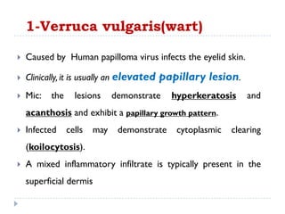
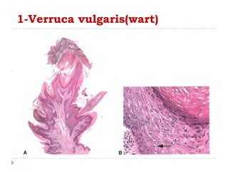
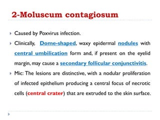
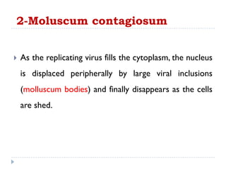
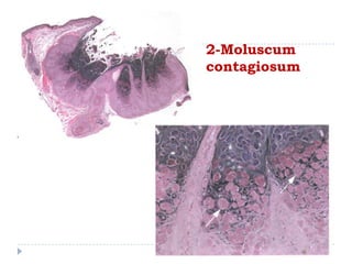
1. Viral infections like verruca vulgaris (wart) caused by human papillomavirus and moluscum contagiosum caused by poxvirus can infect the eyelid. Warts clinically appear as elevated papillary lesions and moluscum contagiosum appear as dome-shaped waxy nodules.
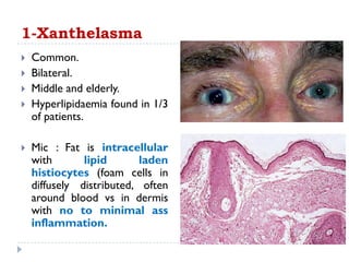
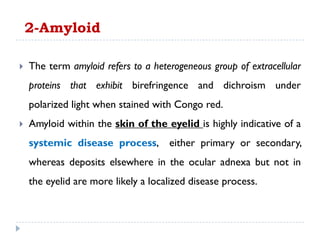
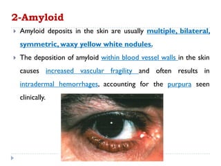
2. Degenerative diseases like xanthelasma and amyloidosis can affect the eyelid. Xanthelasma clinically appears as bilateral yellow plaques on the eyelids and amyloid deposits appear as multiple waxy nodules.
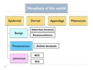
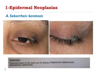
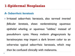
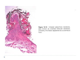
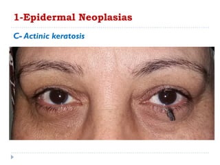
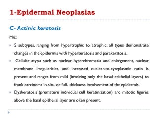
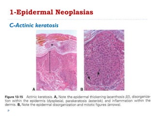
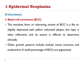
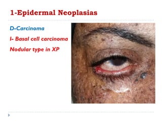
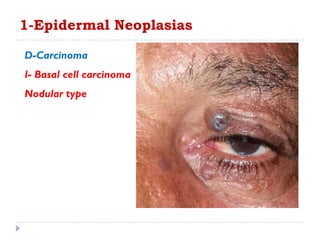
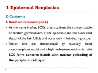
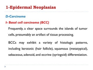
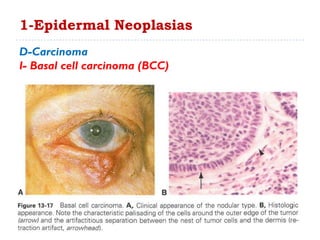
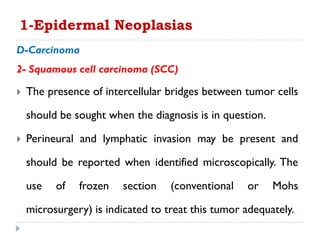
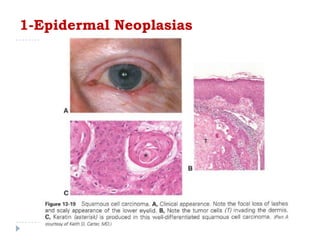
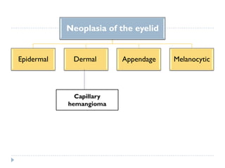
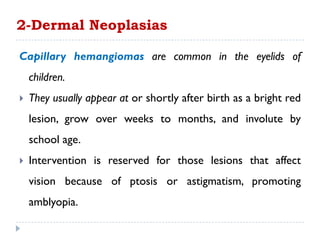
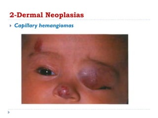
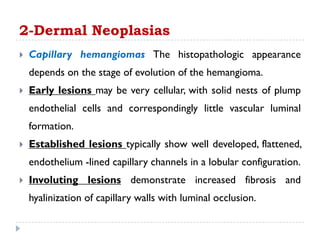
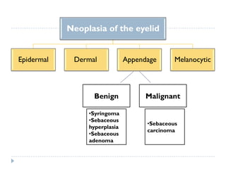
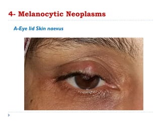
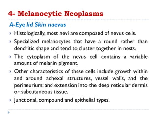
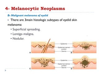
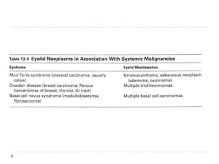
3. Various neoplasms can occur in the eyelid including seborrheic keratosis, basal cell carcinoma, squamous
Eyelid Tumours: A swelling of a part of eyelid generally without inflammation caused by an abnormal growth of tissue.
Types:
Benign &
Malignant
Benign tumors:
Epithelial tumors
Melanocytic tumors
Adnexal cystic lesions
Sweat gland origin
Hair follicle origin
Miscellaneous lesions
Vascular Tumours
Neural Tumours
Malignant tumors:
presentation on intraolcular tumors including detailed explaination on their pathology diagnosis and treatment including details of retinoblastoma. enucleation
Ocular involvement in HIV could be caused by opportunistic infections, vascular abnormalities, neoplasms, neuro-ophthalmic conditions, and adverse effects of medications.
Ocular involvement in HIV infection occurs most commonly due to opportunistic infections and neoplasms. But also can be due to drug related and direct infections.
Opportunistic infections like CMV retinitis occur with a significantly reduced CD4 T-cell count and are one of the common causes of blindness in HIV patients.
Unlike other diseases, ocular infection in these immunosuppressed patients is associated with minimal inflammatory signs.
HIV has been isolated from tears, cornea, vitreous, and chorioretinal tissue in affected persons.
The ocular structures affected by HIV include the adnexa, anterior segment, posterior segment, and orbit.
Neuro ophthalmological manifestations also may be seen.
The institution of highly active antiretroviral therapy (HAART) has caused a dramatic improvement in the immune status of HIV-infected individuals and a change in the clinical presentation and course of opportunistic infections.
Squamous cell carcinoma is the second-most common
cancer of the skin (after basal cell carcinoma but more
common than melanoma). It usually occurs in areas exposed to the sun. Sunlight exposure and immunosuppression are risk factors for SCC of the skin, with chronic sun exposure being the strongest environmental risk factor
Basal Cell Carcinoma (BCC)
BCC is the most common cancer in humans.
Caused by UVR; PTCH gene mutation in most cases.
Clinically different types: nodular, ulcerating, pigmented, sclerosing , and superficial.
BCC is locally invasive, aggressive, and destructive but slow growing, and there is very limited (literally no) tendency to metastasize.
Skin Lesions: There are five clinical types:
1- Nodular
2- Ulcerating
3- Sclerosing (Cicatricial),
4- Superficial,
5- Pigmented.
Invasive Squamous Cell Carcinoma (SCC)
SCC of the skin is a malignant tumor of keratinocytes, arising in the epidermis.
SCC usually arises in epidermal precancerous lesions and, depending on etiology and level of differentiation, varies in its aggressiveness.
The lesion is a plaque or a nodule with varying degrees of keratinization in the nodule and/or on the surface.
Thumb rule:
Undifferentiated SCC: is soft and has no hyperkeratosis;
Differentiated SCC: is hard on palpation and has hyperkeratosis.
Exposure:
Sunlight. Phototherapy, PUVA (oral psoralen + UVA). Excessive photochemotherapy can lead to promotion of SCC, particularly in patients with skin phototypes I and II or in patients with history of previous exposure to ionizing radiation or methotrexate treatment for psoriasis.
Lesions :
Indurated papule, plaque, or nodule ; adherent thick keratotic scale or hyperkeratosis ; when eroded or ulcerated, the lesion may have a crust in the center and a firm, hyperkeratotic, elevated margin
Clark levels
level I, intra-epidermal;
level II, invades papillary dermis;
level III fills papillary dermis;
level IV, invades reticular dermis;
level V, invades subcutaneous fat.
Clarion and Crystal-Clear Cell Acanthoma Reviewed_ Crimson PublishersCrimsonpublishersTTEH
Clarion and Crystal-Clear Cell Acanthoma Reviewed by Anubha Bajaj* in Crimson Publishers: Telemedicine and eHealth Journal
Clear cell acanthoma or Degos’ acanthoma or pale cell acanthoma is an exceptional, asymptomatic, cutaneous benign tumefaction of obscure etiology, emerging from epidermal keratinocytes. Solitary or multiple dome shaped lesions or well delineated nodules or plaques are frequently cogitated on distal extremities. Typically, clear cell acanthoma exhibits a “stuck on” appearance akin to seborrheic keratosis, “vascular countenance” of pyogenic granuloma, “scaling and exudation” elucidated in eczematous reactions and a “progressive margin “associated with an epithelioma. Clear cell acanthoma depicts uniform, pale keratinocytes or pale epithelial cells with abundant cytoplasm composed of excessive glycogen, centric nuclei and distinct foci of transformation. Clinical segregation is required from dermatofibroma, pyogenic granuloma, irritated seborrheic keratosis, keratoacanthoma, actinic keratosis, plaque psoriasis, eccrine poroma, viral warts or malignant cutaneous tumors such as basal cell carcinoma, squamous cell carcinoma, malignant melanoma and metastatic cancer. Dermatoscopy demonstrates a variegated reddish or purple lesion demonstrating a serpiginous pattern akin to a “string of pearls”. Comprehensive surgical eradication of the lesion is the recommended therapeutic option.
https://crimsonpublishers.com/tteh/fulltext/TTEH.000520.php
For more Open access journals in Crimson Publishers
Please click on: https://crimsonpublishers.com/
For more Articles on Telemedicine and eHealth Journal
please click on link: https://crimsonpublishers.com/tteh/index.php
Tom Selleck Health: A Comprehensive Look at the Iconic Actor’s Wellness Journeygreendigital
Tom Selleck, an enduring figure in Hollywood. has captivated audiences for decades with his rugged charm, iconic moustache. and memorable roles in television and film. From his breakout role as Thomas Magnum in Magnum P.I. to his current portrayal of Frank Reagan in Blue Bloods. Selleck's career has spanned over 50 years. But beyond his professional achievements. fans have often been curious about Tom Selleck Health. especially as he has aged in the public eye.
Follow us on: Pinterest
Introduction
Many have been interested in Tom Selleck health. not only because of his enduring presence on screen but also because of the challenges. and lifestyle choices he has faced and made over the years. This article delves into the various aspects of Tom Selleck health. exploring his fitness regimen, diet, mental health. and the challenges he has encountered as he ages. We'll look at how he maintains his well-being. the health issues he has faced, and his approach to ageing .
Early Life and Career
Childhood and Athletic Beginnings
Tom Selleck was born on January 29, 1945, in Detroit, Michigan, and grew up in Sherman Oaks, California. From an early age, he was involved in sports, particularly basketball. which played a significant role in his physical development. His athletic pursuits continued into college. where he attended the University of Southern California (USC) on a basketball scholarship. This early involvement in sports laid a strong foundation for his physical health and disciplined lifestyle.
Transition to Acting
Selleck's transition from an athlete to an actor came with its physical demands. His first significant role in "Magnum P.I." required him to perform various stunts and maintain a fit appearance. This role, which he played from 1980 to 1988. necessitated a rigorous fitness routine to meet the show's demands. setting the stage for his long-term commitment to health and wellness.
Fitness Regimen
Workout Routine
Tom Selleck health and fitness regimen has evolved. adapting to his changing roles and age. During his "Magnum, P.I." days. Selleck's workouts were intense and focused on building and maintaining muscle mass. His routine included weightlifting, cardiovascular exercises. and specific training for the stunts he performed on the show.
Selleck adjusted his fitness routine as he aged to suit his body's needs. Today, his workouts focus on maintaining flexibility, strength, and cardiovascular health. He incorporates low-impact exercises such as swimming, walking, and light weightlifting. This balanced approach helps him stay fit without putting undue strain on his joints and muscles.
Importance of Flexibility and Mobility
In recent years, Selleck has emphasized the importance of flexibility and mobility in his fitness regimen. Understanding the natural decline in muscle mass and joint flexibility with age. he includes stretching and yoga in his routine. These practices help prevent injuries, improve posture, and maintain mobilit
Title: Sense of Smell
Presenter: Dr. Faiza, Assistant Professor of Physiology
Qualifications:
MBBS (Best Graduate, AIMC Lahore)
FCPS Physiology
ICMT, CHPE, DHPE (STMU)
MPH (GC University, Faisalabad)
MBA (Virtual University of Pakistan)
Learning Objectives:
Describe the primary categories of smells and the concept of odor blindness.
Explain the structure and location of the olfactory membrane and mucosa, including the types and roles of cells involved in olfaction.
Describe the pathway and mechanisms of olfactory signal transmission from the olfactory receptors to the brain.
Illustrate the biochemical cascade triggered by odorant binding to olfactory receptors, including the role of G-proteins and second messengers in generating an action potential.
Identify different types of olfactory disorders such as anosmia, hyposmia, hyperosmia, and dysosmia, including their potential causes.
Key Topics:
Olfactory Genes:
3% of the human genome accounts for olfactory genes.
400 genes for odorant receptors.
Olfactory Membrane:
Located in the superior part of the nasal cavity.
Medially: Folds downward along the superior septum.
Laterally: Folds over the superior turbinate and upper surface of the middle turbinate.
Total surface area: 5-10 square centimeters.
Olfactory Mucosa:
Olfactory Cells: Bipolar nerve cells derived from the CNS (100 million), with 4-25 olfactory cilia per cell.
Sustentacular Cells: Produce mucus and maintain ionic and molecular environment.
Basal Cells: Replace worn-out olfactory cells with an average lifespan of 1-2 months.
Bowman’s Gland: Secretes mucus.
Stimulation of Olfactory Cells:
Odorant dissolves in mucus and attaches to receptors on olfactory cilia.
Involves a cascade effect through G-proteins and second messengers, leading to depolarization and action potential generation in the olfactory nerve.
Quality of a Good Odorant:
Small (3-20 Carbon atoms), volatile, water-soluble, and lipid-soluble.
Facilitated by odorant-binding proteins in mucus.
Membrane Potential and Action Potential:
Resting membrane potential: -55mV.
Action potential frequency in the olfactory nerve increases with odorant strength.
Adaptation Towards the Sense of Smell:
Rapid adaptation within the first second, with further slow adaptation.
Psychological adaptation greater than receptor adaptation, involving feedback inhibition from the central nervous system.
Primary Sensations of Smell:
Camphoraceous, Musky, Floral, Pepperminty, Ethereal, Pungent, Putrid.
Odor Detection Threshold:
Examples: Hydrogen sulfide (0.0005 ppm), Methyl-mercaptan (0.002 ppm).
Some toxic substances are odorless at lethal concentrations.
Characteristics of Smell:
Odor blindness for single substances due to lack of appropriate receptor protein.
Behavioral and emotional influences of smell.
Transmission of Olfactory Signals:
From olfactory cells to glomeruli in the olfactory bulb, involving lateral inhibition.
Primitive, less old, and new olfactory systems with different path
Local Advanced Lung Cancer: Artificial Intelligence, Synergetics, Complex Sys...Oleg Kshivets
Overall life span (LS) was 1671.7±1721.6 days and cumulative 5YS reached 62.4%, 10 years – 50.4%, 20 years – 44.6%. 94 LCP lived more than 5 years without cancer (LS=2958.6±1723.6 days), 22 – more than 10 years (LS=5571±1841.8 days). 67 LCP died because of LC (LS=471.9±344 days). AT significantly improved 5YS (68% vs. 53.7%) (P=0.028 by log-rank test). Cox modeling displayed that 5YS of LCP significantly depended on: N0-N12, T3-4, blood cell circuit, cell ratio factors (ratio between cancer cells-CC and blood cells subpopulations), LC cell dynamics, recalcification time, heparin tolerance, prothrombin index, protein, AT, procedure type (P=0.000-0.031). Neural networks, genetic algorithm selection and bootstrap simulation revealed relationships between 5YS and N0-12 (rank=1), thrombocytes/CC (rank=2), segmented neutrophils/CC (3), eosinophils/CC (4), erythrocytes/CC (5), healthy cells/CC (6), lymphocytes/CC (7), stick neutrophils/CC (8), leucocytes/CC (9), monocytes/CC (10). Correct prediction of 5YS was 100% by neural networks computing (error=0.000; area under ROC curve=1.0).
micro teaching on communication m.sc nursing.pdfAnurag Sharma
Microteaching is a unique model of practice teaching. It is a viable instrument for the. desired change in the teaching behavior or the behavior potential which, in specified types of real. classroom situations, tends to facilitate the achievement of specified types of objectives.
Rasamanikya is a excellent preparation in the field of Rasashastra, it is used in various Kushtha Roga, Shwasa, Vicharchika, Bhagandara, Vatarakta, and Phiranga Roga. In this article Preparation& Comparative analytical profile for both Formulationon i.e Rasamanikya prepared by Kushmanda swarasa & Churnodhaka Shodita Haratala. The study aims to provide insights into the comparative efficacy and analytical aspects of these formulations for enhanced therapeutic outcomes.
Title: Sense of Taste
Presenter: Dr. Faiza, Assistant Professor of Physiology
Qualifications:
MBBS (Best Graduate, AIMC Lahore)
FCPS Physiology
ICMT, CHPE, DHPE (STMU)
MPH (GC University, Faisalabad)
MBA (Virtual University of Pakistan)
Learning Objectives:
Describe the structure and function of taste buds.
Describe the relationship between the taste threshold and taste index of common substances.
Explain the chemical basis and signal transduction of taste perception for each type of primary taste sensation.
Recognize different abnormalities of taste perception and their causes.
Key Topics:
Significance of Taste Sensation:
Differentiation between pleasant and harmful food
Influence on behavior
Selection of food based on metabolic needs
Receptors of Taste:
Taste buds on the tongue
Influence of sense of smell, texture of food, and pain stimulation (e.g., by pepper)
Primary and Secondary Taste Sensations:
Primary taste sensations: Sweet, Sour, Salty, Bitter, Umami
Chemical basis and signal transduction mechanisms for each taste
Taste Threshold and Index:
Taste threshold values for Sweet (sucrose), Salty (NaCl), Sour (HCl), and Bitter (Quinine)
Taste index relationship: Inversely proportional to taste threshold
Taste Blindness:
Inability to taste certain substances, particularly thiourea compounds
Example: Phenylthiocarbamide
Structure and Function of Taste Buds:
Composition: Epithelial cells, Sustentacular/Supporting cells, Taste cells, Basal cells
Features: Taste pores, Taste hairs/microvilli, and Taste nerve fibers
Location of Taste Buds:
Found in papillae of the tongue (Fungiform, Circumvallate, Foliate)
Also present on the palate, tonsillar pillars, epiglottis, and proximal esophagus
Mechanism of Taste Stimulation:
Interaction of taste substances with receptors on microvilli
Signal transduction pathways for Umami, Sweet, Bitter, Sour, and Salty tastes
Taste Sensitivity and Adaptation:
Decrease in sensitivity with age
Rapid adaptation of taste sensation
Role of Saliva in Taste:
Dissolution of tastants to reach receptors
Washing away the stimulus
Taste Preferences and Aversions:
Mechanisms behind taste preference and aversion
Influence of receptors and neural pathways
Impact of Sensory Nerve Damage:
Degeneration of taste buds if the sensory nerve fiber is cut
Abnormalities of Taste Detection:
Conditions: Ageusia, Hypogeusia, Dysgeusia (parageusia)
Causes: Nerve damage, neurological disorders, infections, poor oral hygiene, adverse drug effects, deficiencies, aging, tobacco use, altered neurotransmitter levels
Neurotransmitters and Taste Threshold:
Effects of serotonin (5-HT) and norepinephrine (NE) on taste sensitivity
Supertasters:
25% of the population with heightened sensitivity to taste, especially bitterness
Increased number of fungiform papillae
Integrating Ayurveda into Parkinson’s Management: A Holistic ApproachAyurveda ForAll
Explore the benefits of combining Ayurveda with conventional Parkinson's treatments. Learn how a holistic approach can manage symptoms, enhance well-being, and balance body energies. Discover the steps to safely integrate Ayurvedic practices into your Parkinson’s care plan, including expert guidance on diet, herbal remedies, and lifestyle modifications.
Muktapishti is a traditional Ayurvedic preparation made from Shoditha Mukta (Purified Pearl), is believed to help regulate thyroid function and reduce symptoms of hyperthyroidism due to its cooling and balancing properties. Clinical evidence on its efficacy remains limited, necessitating further research to validate its therapeutic benefits.
ABDOMINAL TRAUMA in pediatrics part one.drhasanrajab
Abdominal trauma in pediatrics refers to injuries or damage to the abdominal organs in children. It can occur due to various causes such as falls, motor vehicle accidents, sports-related injuries, and physical abuse. Children are more vulnerable to abdominal trauma due to their unique anatomical and physiological characteristics. Signs and symptoms include abdominal pain, tenderness, distension, vomiting, and signs of shock. Diagnosis involves physical examination, imaging studies, and laboratory tests. Management depends on the severity and may involve conservative treatment or surgical intervention. Prevention is crucial in reducing the incidence of abdominal trauma in children.
3. 1-Verruca vulgaris(wart)
Caused by Human papilloma virus infects the eyelid skin.
Clinically, it is usually an elevated papillary lesion.
Mic: the lesions demonstrate hyperkeratosis and
acanthosis and exhibit a papillary growth pattern.
Infected cells may demonstrate cytoplasmic clearing
(koilocytosis).
A mixed inflammatory infiltrate is typically present in the
superficial dermis
5. Caused by Poxvirus infection.
Clinically, Dome-shaped, waxy epidermal nodules with
central umbilication form and, if present on the eyelid
margin, may cause a secondary follicular conjunctivitis.
Mic: The lesions are distinctive, with a nodular proliferation
of infected epithelium producing a central focus of necrotic
cells (central crater) that are extruded to the skin surface.
2-Moluscum contagiosum
6. As the replicating virus fills the cytoplasm, the nucleus
is displaced peripherally by large viral inclusions
(molluscum bodies) and finally disappears as the cells
are shed.
2-Moluscum contagiosum
9. 1-Xanthelasma
Common.
Bilateral.
Middle and elderly.
Hyperlipidaemia found in 1/3
of patients.
Mic : Fat is intracellular
with lipid laden
histiocytes (foam cells in
diffusely distributed, often
around blood vs in dermis
with no to minimal ass
inflammation.
10. The term amyloid refers to a heterogeneous group of extracellular
proteins that exhibit birefringence and dichroism under
polarized light when stained with Congo red.
Amyloid within the skin of the eyelid is highly indicative of a
systemic disease process, either primary or secondary,
whereas deposits elsewhere in the ocular adnexa but not in
the eyelid are more likely a localized disease process.
2-Amyloid
11. Amyloid deposits in the skin are usually multiple, bilateral,
symmetric, waxy yellow white nodules.
The deposition of amyloid within blood vessel walls in the skin
causes increased vascular fragility and often results in
intradermal hemorrhages, accounting for the purpura seen
clinically.
2-Amyloid
12. Mic: amyloid appears as an amorphous, eosinophilic
extracellular deposit, usually within vessel walls but also in
soft tissue and around peripheral nerves and sweat glands.
Stains useful in demonstrating amyloid deposits include
Congo red, crystal violet, and thioflavinT.
Electron microscopy reveals the deposits to be composed of
randomly oriented extracellular fibrils measuring7-10
nm in diameter.
2-Amyloid
14. Neoplasia of the eyelid
Epidermal
Benign
Precancerous
cancerous
Dermal Appendage Melanocytic
Seborrheic keratosis
Keratoacanthoma
Actinic keratosis
BCC
SCC
15. 1-Epidermal Neoplasias
A. Seborrheic keratosis
A common benign epithelial proliferation.
Occurs in middle age.
Clinically, it is a well-circumscribed, oval, dome-shaped
to verrucoid "stuck-on" papule, varying from pink to
brown in color.
18. A- Seborrheic keratosis
Mic: several architectural patterns are possible,
although all demonstrate hyperkeratosis, acanthosis,
and some degree of papillomatosis.
The acanthosis is a result of the proliferation of
either polygonal or basaloid squamous cells without
dysplasia.
1-Epidermal Neoplasias
19. A- Seborrheic keratosis
A characteristic finding in most types of seborrheic
keratoses is the formation of pseudohorn cysts,
which are concentrically laminated collections of
surface keratin within the acanthotic epithelium.
1-Epidermal Neoplasias
21. A- Seborrheic keratosis
Irritated seborrheic keratosis, also termed inverted
follicular keratosis, shows nonkeratinizing squamous
epithelial whorling, or squamous "eddies;' instead of
pseudohorn cysts. Heavy melanin phagocytosis by
keratinocytes may impart a dark brown color to an
otherwise typical seborrheic keratosis, which may
then be confused clinically with melanoma.
1-Epidermal Neoplasias
22.
23. A- Seborrheic keratosis
Sudden onset of multiple seborrheic keratoses is
known as the Leser- Trelat sign and is associated with a
malignancy, usually a gastrointestinal adenocarcinoma;
1-Epidermal Neoplasias
24. B- Keratoacanthoma
A rapidly growing epithelial proliferation with a potential for
spontaneous involution.
There is strong evidence supporting the idea that
keratoacanthomas are a variant of a well-differentiated
squamous cell carcinoma.
These studies are based on expression of proliferation
markers (cyclins and cyclin-dependent kinases) and
oncoproteins (mutated p53) that are expressed similarly by
both entities.
1-Epidermal Neoplasias
25. 1-Epidermal Neoplasias
B- Keratoacanthoma
Dome-shaped nodules with a keratin-filled central crater
may attain a considerable size, up to 2.5 cm in diameter,
within a matter of weeks to months .
The natural history is typically spontaneous involution
over several months, resulting in a slightly depressed scar.
26. B- Keratoacanthoma
Mic:
keratoacanthomas show a cup-shaped invagination of well-
differentiated squamous cells forming irregularly configured
nests and strands and inciting a chronic inflammatory host
response.
The proliferating epithelial cells undermine the adjacent
normal epidermis.
At the deep aspect of the proliferating nodules, mitotic activity
and nuclear atypia may occur, making the distinction between
keratoacanthoma and invasive squamous cell carcinoma
problematic.
1-Epidermal Neoplasias
28. C- Actinic keratosis
Actinic keratoses are precancerous squamous lesions
that appear, clinically, as erythematous, scaly macules or
papules in middle age on sun-exposed skin, particularly on
the face and the dorsal surfaces of the hands.
Actinic keratoses range from a few millimeters up to 1
cm in greatest dimension..
1-Epidermal Neoplasias
29. C- Actinic keratosis
Hyperkeratotic types may form a cutaneous horn, and
hyperpigmented types may clinically simulate lentigo maligna.
Squamous cell carcinoma may develop from preexisting actinic
keratosis; thus, biopsy of suspicious lesions and long term
follow-up are necessary in patients with this condition.
However, when squamous cell carcinoma arises in actinic
keratosis, the risk of subsequent metastatic dissemination is
very low (0.5%-3.0%).
1-Epidermal Neoplasias
31. C- Actinic keratosis
Mic:
5 subtypes, ranging from hypertrophic to atrophic; all types demonstrate
changes in the epidermis with hyperkeratosis and parakeratosis.
Cellular atypia such as nuclear hyperchromasia and enlargement, nuclear
membrane irregularities, and increased nuclear-to-cytoplasmic ratio is
present and ranges from mild (involving only the basal epithelial layers) to
frank carcinoma in situ, or full- thickness involvement of the epidermis.
Dyskeratosis (premature individual cell keratinization) and mitotic figures
above the basal epithelial layer are often present.
1-Epidermal Neoplasias
33. C-Actinic keratosis
The underlying dermis shows solar elastosis (elastotic
degeneration of collagen) , which manifests as fragmentation,
clumping, and loss of eosinophilia of dermal collagen.
A chronic inflammatory cell infiltrate is usually present in the
superficial dermis.
The base of the lesion must be examined histologically to
determine whether invasive squamous cell carcinoma is
present; for this reason, shave biopsy not including the base of
the lesion is contraindicated.
1-Epidermal Neoplasias
35. D-Carcinoma
I- Basal cell carcinoma (BCC) is the most common malignant
neoplasm of the eyelids, accounting for > than 90% of all eyelid
malignancies.
Exposure to sunlight is the main risk factor, although genetic
factors can play a role in familial syndromes.
The lower eyelid is more commonly involved than the upper
eyelid, with the medial canthus being the second most
common site of involvement.
1-Epidermal Neoplasias
36. D-Carcinoma
I- Basal cell carcinoma (BCC)
Tumors in the medial canthal area are more likely to be deeply
invasive and to involve the orbit.
Clinically, BCC is a slowly enlarging, slightly elevated lesion with
ulceration and pearly, raised, rolled edges.
1-Epidermal Neoplasias
37.
38. D-Carcinoma
I- Basal cell carcinoma (BCC)
The morphea form, or sclerosing, variant of BCC is a flat or
slightly depressed pale yellow indurated plaque; this type is
often infiltrative, and its extent is difficult to determine
clinically.
Other growth patterns include nodular (most common; and
multicentric.A small percentage of BCCs are pigmented.
1-Epidermal Neoplasias
42. D-Carcinoma
I- Basal cell carcinoma (BCC)
As the name implies, BCCs originate from the stratum basale,
or stratum germinativum, of the epidermis and the outer root
sheath of the hair follicle and occur only in hair-bearing tissue.
Tumor cells are characterized by relatively bland,
monomorphous nuclei and a high nuclear-to-cytoplasmic ratio.
BCC forms cohesive islands with nuclear palisading of
the peripheral cell layer.
1-Epidermal Neoplasias
43. D-Carcinoma
I- Basal cell carcinoma (BCC)
Frequently, a clear space surrounds the islands of tumor
cells, presumably an artifact of tissue processing.
BCCs may exhibit a variety of histologic patterns,
including keratotic (hair follicle), squamous (metatypical),
sebaceous, adenoid, and eccrine (syringoid) differentiation.
1-Epidermal Neoplasias
45. D-Carcinoma
I- Basal cell
carcinoma (BCC)
1-Epidermal Neoplasias
The morphea (sclerosing) variant shows thin cords and
strands of tumor cells set in a fibrotic stroma.
46. D-Carcinoma
I- Basal cell carcinoma (BCC)
Complete excision is the treatment of choice, and surgical
margin control is required.
Typically, margin control is achieved with frozen sections
or Mohs micrographic excision.
Morbidity in BCCs is almost always the result of local
spread; metastasis is extremely unusual.
1-Epidermal Neoplasias
47. D-Carcinoma
2- Squamous cell carcinoma (SCC)
Although squamous cell carcinoma may occur in the eyelids, it is at
least 10 - 40 times less common than BCC.
Because most SCCs arise in solar damaged skin, the lower
eyelid is more frequently involved than the upper. However, the
proportion of SCCs occurring in the upper eyelid is larger
than the proportion of BCCs occurring in the upper eyelid.
1-Epidermal Neoplasias
48. D-Carcinoma
2- Squamous cell carcinoma (SCC)
The clinical appearance of SCC is diverse, ranging from
ulcers to plaques to fungating or nodular growths.
Accordingly, the clinical differential diagnosis is a long list,
and pathologic examination of excised tissue is necessary
for accurate diagnosis.
1-Epidermal Neoplasias
49. D-Carcinoma
I- Squamous cell carcinoma (SCC)
Histologic examination shows atypical squamous cells forming
nests and strands, extending beyond the epidermal basement
membrane, infiltrating the dermis, and in citing a fibrotic tissue
reaction. Tumor cells may be well differentiated (forming keratin
and easily recognizable as squamous), moderately differentiated,
or poorly differentiated (requiring ancillary studies to confirm the
nature of the neoplasm).
1-Epidermal Neoplasias
50. D-Carcinoma
2- Squamous cell carcinoma (SCC)
The presence of intercellular bridges between tumor cells
should be sought when the diagnosis is in question.
Perineural and lymphatic invasion may be present and
should be reported when identified microscopically. The
use of frozen section (conventional or Mohs
microsurgery) is indicated to treat this tumor adequately.
1-Epidermal Neoplasias
51. D-Carcinoma
2- Squamous cell carcinoma (SCC)
Regional lymph node metastasis is reported to occur in
up to 20% of patients with SCC of the eyelid.
1-Epidermal Neoplasias
53. Neoplasia of the eyelid
Epidermal Dermal Appendage Melanocytic
Capillary
hemangioma
54. 2-Dermal Neoplasias
Capillary hemangiomas are common in the eyelids of
children.
They usually appear at or shortly after birth as a bright red
lesion, grow over weeks to months, and involute by
school age.
Intervention is reserved for those lesions that affect
vision because of ptosis or astigmatism, promoting
amblyopia.
56. Capillary hemangiomas The histopathologic appearance
depends on the stage of evolution of the hemangioma.
Early lesions may be very cellular, with solid nests of plump
endothelial cells and correspondingly little vascular luminal
formation.
Established lesions typically show well developed, flattened,
endothelium -lined capillary channels in a lobular configuration.
Involuting lesions demonstrate increased fibrosis and
hyalinization of capillary walls with luminal occlusion.
2-Dermal Neoplasias
58. Neoplasia of the eyelid
Epidermal Dermal Appendage Melanocytic
Benign Malignant
•Syringoma
•Sebaceous
hyperplasia
•Sebaceous
adenoma
•Sebaceous
carcinoma
59. 3- Appendage Neoplasms
A- Syringoma
Syringoma is a common benign lesion of the lower eyelid
and typically manifests as multiple tiny papules.
Syringomas result from a malformation of the eccrine sweat
gland ducts.
Histologically, syringomas consist of multiple, comma-shaped
or round ductules lined with a double layer of epithelium
and containing a central lumen, often with secretory material.
61. B-Sebaceous hyperplasia
Sebaceous hyperplasia is an uncommon benign lesion of
the eyelid and face.
Clinically, it appears as a small, yellow papule.
Histologically, it is typically a single enlarged sebaceous
gland with numerous glandular lobules attached to a
single central duct.
3- Appendage Neoplasms
63. C-Sebaceous adenoma
Sebaceous adenoma is a rare benign lesion of the eyelid
that typically manifests as a yellow, circumscribed
nodule.
Histologically, it is composed of multiple sebaceous
lobules that are irregularly shaped and incompletely
differentiated.
Muir-Torre syndrome should be considered when
sebaceous adenoma is diagnosed.
3- Appendage Neoplasms
65. D-Sebaceous carcinoma
A sebaceous carcinoma most commonly involves the upper
eyelid of elderly persons.
It may originate in the meibomian glands of the tarsus, the
glands of Zeis in the skin of the eyelid, or the sebaceous glands
of the caruncle.
Clinical diagnosis is often missed or delayed because of this
lesion's propensity to mimic a chalazion or chronic
blepharoconjunctivitis
3- Appendage Neoplasms
67. Histologically, well-differentiated sebaceous carcinomas are
readily identified by the microvesicular foamy nature of the
tumor cell cytoplasm.
Moderately differentiated tumors may exhibit some degree of
sebaceous differentiation.
Poorly differentiated tumors, however, may be difficult to
distinguish from the other, more common epithelial
malignancies. The demonstration of lipid within the cytoplasm
of tumor cells by special stains, such as oil red 0 or Sudan
black, is diagnostic, but it must be performed on tissue prior
to processing and paraffin embedding.
3- Appendage Neoplasms
D-Sebaceous carcinoma
68. Sebaceous carcinoma, histology. A, Tumor cells often have
hyperchromatic, atypical nuclei. The cytoplasm frequently has a
foamy or vacuolated appearance. Note mitotic figure (arrow)
3- Appendage Neoplasms
69. When sebaceous carcinoma is suspected clinically, the
pathologist should be alerted so that frozen section slides
can be generated for lipid stains.
Another feature, characteristic of but not pathognomonic
for sebaceous cell carcinoma, is the dissemination of
individual tumor cells and clusters of tumor cells within
the epidermis or conjunctival epithelium, known as
pagetoid spread
Another pattern in the conjunctiva is that of complete
replacement of conjunctival epithelium by tumor cells, or
sebaceous carcinoma in situ.
3- Appendage Neoplasms
70. . B, Pagetoid invasion
of epidermis by
individual tumor cells
and small clusters
of tumor cells (arrows).
C, Sebaceous
carcinoma in situ with
complete replacement
of normal conjunctival
epithelium by tumor
cells (between arrows).
3- Appendage Neoplasms
71. Neoplasia of the eyelid
Epidermal Dermal Appendage Melanocytic
Benign Malignant
•Nevus Malignant
melanoma
72. A- Eye lid Skin nevi
It is benign neoplastic proliferations of melanocytic cells
(hamartomatous lesions).
At birth or acquired.
Giant congenital melanocytic nevi.
Congenital nevi of the eyelid may develop in utero before
the separation of the upper and lower eyelids and result
in a "kissing" nevus.
4- Melanocytic Neoplasms
75. Histologically, most nevi are composed of nevus cells.
Specialized melanocytes that have a round rather than
dendritic shape and tend to cluster together in nests.
The cytoplasm of the nevus cell contains a variable
amount of melanin pigment.
Other characteristics of these cells include growth within
and around adnexal structures, vessel walls, and the
perineurium; and extension into the deep reticular dermis
or subcutaneous tissue.
Junctional, compound and epithelial types.
4- Melanocytic Neoplasms
A-Eye lid Skin naevus
76. B- Malignant melanoma of eyelid
There are 3main histologic subtypes of eyelid skin
melanoma:
• Superficial spreading.
• Lentigo maligna.
• Nodular.
4- Melanocytic Neoplasms
77. Pagetoid intraepidermal spread of atypical melanocytic nests.
Band-like lymphocytic host response along the base of the
mass is more common in melanoma than in benign
proliferations.
Prognosis is correlated with depth of invasion
Metastases, when they occur, typically involve regional lymph
nodes first.
4- Melanocytic Neoplasms
A-Eye lid Skin MM