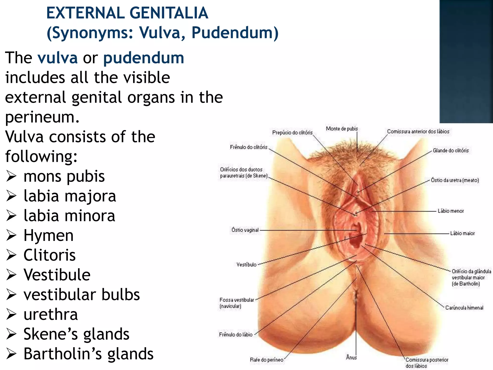
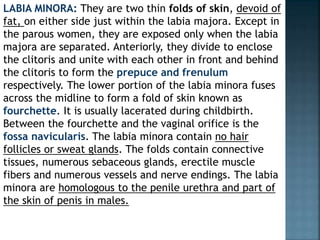
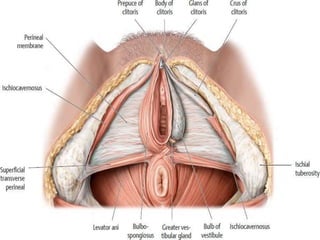
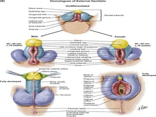
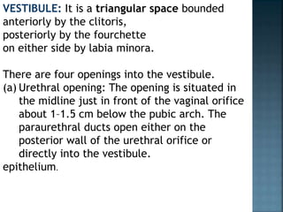
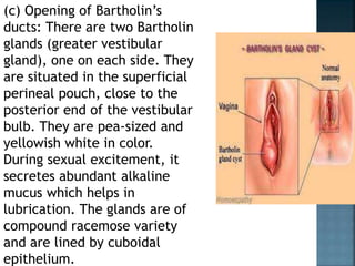
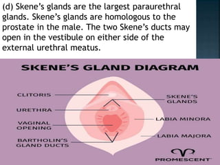
SlideShare a Scribd company logo
EXTERNAL GENITALIA
(Synonyms: Vulva, Pudendum)
The vulva or pudendum
includes all the visible
external genital organs in the
perineum.
Vulva consists of the
following:
 mons pubis
 labia majora
 labia minora
 Hymen
 Clitoris
 Vestibule
 vestibular bulbs
 urethra
 Skene’s glands
 Bartholin’s glands
MONS VENERIS (MONS
PUBIS): It is the pad of
subcutaneous adipose
connective tissue lying in
front of the pubis and in
the adult female is
covered by hair. The hair
pattern (escutcheon) of
most women is
triangular with the base
directed upwards.
LABIA MAJORA: The vulva is
bounded on each side by the
elevation of skin and
subcutaneous tissue which
form the labia majora. They
are continuous where they
join medially to form the
posterior commissure in
front of the anus.
The skin on the outer convex
surface is pigmented and
covered with hair follicle.
The thin skin on the inner
surface has sebaceous glands
but no hair follicle.
The labia majora are covered with
squamous epithelium and contain sweat
glands. Beneath the skin, there is dense
connective tissue and adipose tissue. The
adipose tissue is richly supplied with
venous plexus which may produce
hematoma, if injured during childbirth.
The labia majora are homologous to the
scrotum in the male. The round ligament
terminates at its upper border.
LABIA MINORA: They are two thin folds of skin, devoid of
fat, on either side just within the labia majora. Except in
the parous women, they are exposed only when the labia
majora are separated. Anteriorly, they divide to enclose
the clitoris and unite with each other in front and behind
the clitoris to form the prepuce and frenulum
respectively. The lower portion of the labia minora fuses
across the midline to form a fold of skin known as
fourchette. It is usually lacerated during childbirth.
Between the fourchette and the vaginal orifice is the
fossa navicularis. The labia minora contain no hair
follicles or sweat glands. The folds contain connective
tissues, numerous sebaceous glands, erectile muscle
fibers and numerous vessels and nerve endings. The labia
minora are homologous to the penile urethra and part of
the skin of penis in males.
CLITORIS: It is a small cylindrical erectile body,
measuring about 1.5–2 cm situated in the most
anterior part of the vulva.
It consists of a
glans,
body
two crura.
The clitoris consists of two cylindrical corpora
cavernosa (erectile tissue). The glans is covered
by squamous epithelium and is richly supplied
with nerves. The vessels of the clitoris are
connected with the vestibular bulb and are
liable to be injured during childbirth.
Clitoris is homologous to the penis in the
male but it differs in being entirely
separate from the urethra. It is attached
to the under surface of the symphysis pubis
by the suspensory ligament.
VESTIBULE: It is a triangular space bounded
anteriorly by the clitoris,
posteriorly by the fourchette
on either side by labia minora.
There are four openings into the vestibule.
(a) Urethral opening: The opening is situated in
the midline just in front of the vaginal orifice
about 1–1.5 cm below the pubic arch. The
paraurethral ducts open either on the
posterior wall of the urethral orifice or
directly into the vestibule.
epithelium.
(b) Vaginal orifice and hymen: The vaginal orifice
lies in the posterior end of the vestibule and is of
varying size and shape. In virgins and nulliparae,
the opening is closed by the labia minora, but in
parous, it may be exposed. It is incompletely
closed by a septum of mucous membrane, called
hymen. The membrane varies in shape but is
usually circular or crescentic in virgins. The
hymen is usually ruptured at the consummation
of marriage. During childbirth, the hymen is
extremely lacerated and is later represented by
cicatrized nodules of varying size, called the
carunculae myrtiformes. On both sides it is lined
by stratified squamous
(c) Opening of Bartholin’s
ducts: There are two Bartholin
glands (greater vestibular
gland), one on each side. They
are situated in the superficial
perineal pouch, close to the
posterior end of the vestibular
bulb. They are pea-sized and
yellowish white in color.
During sexual excitement, it
secretes abundant alkaline
mucus which helps in
lubrication. The glands are of
compound racemose variety
and are lined by cuboidal
epithelium.
Each gland has got a duct which measures
about 2 cm and opens into the vestibule
outside the hymen at the junction of the
anterior two-third and posterior one-third
in the groove between the hymen and the
labium minus. The duct is lined by
columnar epithelium but near its opening
by stratified squamous epithelium.
Bartholin’s glands are homologous to the
bulb of the penis in male.
(d) Skene’s glands are the largest paraurethral
glands. Skene’s glands are homologous to the
prostate in the male. The two Skene’s ducts may
open in the vestibule on either side of the
external urethral meatus.
VESTIBULAR BULB: These are bilateral elongated masses
of erectile tissues situated beneath the mucous
membrane of the vestibule. Each bulb lies on either side
of the vaginal orifice in front of the Bartholin’s gland and
is incorporated with the bulbocavernosus muscle. They
are homologous to the bulb of the penis and corpus
spongiosum in the male. They are likely to be injured
during childbirth with brisk hemorrhage (Fig. 1.2)
PERINEUM: The details of its anatomy are described later
in the chapter.
BLOOD SUPPLY:
Arteries—(a) Branches of internal pudendal artery—the
chief being labial, transverse perineal, artery to the
vestibular bulb and deep and dorsal arteries to the
clitoris.
(b) Branches of femoral
artery—superficial and
deep external pudendal.
Veins—The veins form
plexuses and drain into:
(a) Internal pudendal
vein, (b) vesical or
vaginal venous plexus
and (c) Long saphenous
vein. Varicosities during
pregnancy are not
uncommon and may
rupture spontaneously
causing visible bleeding
or hematoma formation
NERVE SUPPLY: The supply is through bilateral
spinal somatic nerves— (a) anterosuperior part is
supplied by the cutaneous branches from the
ilioinguinal and genital branch of genitofemoral
nerve(L1 and L2 ) and the posteroinferior part by
the pudendal branches from the posterior
cutaneous nerve of thigh (S1.2.3). Between
these two groups, the vulva is supplied by the
labial and perineal branches of the pudendal
nerve (S2.3.4).
LYMPHATICS: Vulval lymphatics have bilateral drainage.
Lymphatics drain into—(a) superficial inguinal nodes, (b)
intermediate groups of inguinal lymph nodes—gland of
Cloquet and (c) external and internal iliac lymph nodes.
DEVELOPMENT: External genitalia is developed in the
region of the cranial aspect of ectodermal cloacal fossa;
clitoris from the genital tubercle; labia minora from the
genital folds; labia majora from the labioscrotal swelling
and the vestibule from the urogenital sinus

More Related Content

What's hot

Anatomy of Ureter
Anatomy of UreterAnatomy of Ureter
Anatomy of Ureter
Dr. Vibhash Kumar Vaidya
 
UTERUS.pptx
UTERUS.pptxUTERUS.pptx
UTERUS.pptx
Dr. sana yaseen
 
Anterior abdominal wall
Anterior abdominal wallAnterior abdominal wall
The umbilical cord
The umbilical cordThe umbilical cord
The umbilical cord
raj kumar
 
Anatomy of testis
Anatomy of testisAnatomy of testis
Anatomy of testis
Dr. Sravani kommuru
 
Antomy of scrotum and testis
Antomy of scrotum and testisAntomy of scrotum and testis
Antomy of scrotum and testis
priyanka rana
 
Breast anatomy
Breast anatomyBreast anatomy
Breast anatomy
Animesh Agrawal
 
Anatomyof female genital tract
Anatomyof female genital tractAnatomyof female genital tract
Anatomyof female genital tract
Ayman Shehata
 
The placenta and fetal membranes
The placenta and fetal membranesThe placenta and fetal membranes
The placenta and fetal membranes
Snigdha Gupta
 
instruments ostetrics and gynaecology ppt
instruments ostetrics and gynaecology pptinstruments ostetrics and gynaecology ppt
instruments ostetrics and gynaecology ppt
TONY SCARIA
 
Anatomy Of Urinary Bladder
Anatomy Of Urinary BladderAnatomy Of Urinary Bladder
Anatomy Of Urinary Bladder
Umair Majeed
 
Umbilical Cord (General Embryology)
Umbilical Cord (General Embryology)Umbilical Cord (General Embryology)
Umbilical Cord (General Embryology)
Dr. Sherif Fahmy
 
Urinary bladder
Urinary bladderUrinary bladder
Urinary bladder
Dr. Vibhash Kumar Vaidya
 
Mal presentation & Mal position
 Mal presentation & Mal position				 Mal presentation & Mal position
Mal presentation & Mal position
golden4host
 
Penile anatomy
Penile anatomyPenile anatomy
Penile anatomy
Faheem Andrabi
 
Fallopian tube radiology - Dr. Sumit Sharma
Fallopian tube radiology - Dr. Sumit SharmaFallopian tube radiology - Dr. Sumit Sharma
Fallopian tube radiology - Dr. Sumit Sharma
Sumit Sharma
 
ANATOMY OF FEMALE GENITAL TRACT
ANATOMY OF FEMALE GENITAL TRACTANATOMY OF FEMALE GENITAL TRACT
ANATOMY OF FEMALE GENITAL TRACT
PRANATI PATRA
 
Prostate anatomy
Prostate anatomyProstate anatomy
Prostate anatomy
Sambit Tripathy
 
Bony pelvis
Bony pelvisBony pelvis
Bony pelvis
Lawrence James
 
Anatomy of female reproductive organs
Anatomy of female reproductive organsAnatomy of female reproductive organs
Anatomy of female reproductive organs
amrit kaur
 

What's hot (20)

Anatomy of Ureter
Anatomy of UreterAnatomy of Ureter
Anatomy of Ureter
 
UTERUS.pptx
UTERUS.pptxUTERUS.pptx
UTERUS.pptx
 
Anterior abdominal wall
Anterior abdominal wallAnterior abdominal wall
Anterior abdominal wall
 
The umbilical cord
The umbilical cordThe umbilical cord
The umbilical cord
 
Anatomy of testis
Anatomy of testisAnatomy of testis
Anatomy of testis
 
Antomy of scrotum and testis
Antomy of scrotum and testisAntomy of scrotum and testis
Antomy of scrotum and testis
 
Breast anatomy
Breast anatomyBreast anatomy
Breast anatomy
 
Anatomyof female genital tract
Anatomyof female genital tractAnatomyof female genital tract
Anatomyof female genital tract
 
The placenta and fetal membranes
The placenta and fetal membranesThe placenta and fetal membranes
The placenta and fetal membranes
 
instruments ostetrics and gynaecology ppt
instruments ostetrics and gynaecology pptinstruments ostetrics and gynaecology ppt
instruments ostetrics and gynaecology ppt
 
Anatomy Of Urinary Bladder
Anatomy Of Urinary BladderAnatomy Of Urinary Bladder
Anatomy Of Urinary Bladder
 
Umbilical Cord (General Embryology)
Umbilical Cord (General Embryology)Umbilical Cord (General Embryology)
Umbilical Cord (General Embryology)
 
Urinary bladder
Urinary bladderUrinary bladder
Urinary bladder
 
Mal presentation & Mal position
 Mal presentation & Mal position				 Mal presentation & Mal position
Mal presentation & Mal position
 
Penile anatomy
Penile anatomyPenile anatomy
Penile anatomy
 
Fallopian tube radiology - Dr. Sumit Sharma
Fallopian tube radiology - Dr. Sumit SharmaFallopian tube radiology - Dr. Sumit Sharma
Fallopian tube radiology - Dr. Sumit Sharma
 
ANATOMY OF FEMALE GENITAL TRACT
ANATOMY OF FEMALE GENITAL TRACTANATOMY OF FEMALE GENITAL TRACT
ANATOMY OF FEMALE GENITAL TRACT
 
Prostate anatomy
Prostate anatomyProstate anatomy
Prostate anatomy
 
Bony pelvis
Bony pelvisBony pelvis
Bony pelvis
 
Anatomy of female reproductive organs
Anatomy of female reproductive organsAnatomy of female reproductive organs
Anatomy of female reproductive organs
 

Similar to EXTERNAL GENITALIA (1).pptx

Anatomy of female genital tract
Anatomy of female genital tractAnatomy of female genital tract
Anatomy of female genital tract
PRIYAPRAJEESH
 
Anatomy & physiology of female reproductive system
Anatomy & physiology of female reproductive systemAnatomy & physiology of female reproductive system
Anatomy & physiology of female reproductive system
Dr Ndayisaba Corneille
 
Breast
BreastBreast
Breast
Anmol Bansal
 
0_ANATOMY OF FEMALE REPRODUCTIVE SYSTEM.pptx
0_ANATOMY OF FEMALE REPRODUCTIVE SYSTEM.pptx0_ANATOMY OF FEMALE REPRODUCTIVE SYSTEM.pptx
0_ANATOMY OF FEMALE REPRODUCTIVE SYSTEM.pptx
nehal791634
 
MATERNAL ANATOMY.pptx
MATERNAL ANATOMY.pptxMATERNAL ANATOMY.pptx
MATERNAL ANATOMY.pptx
AJAY MANDAL
 
HAP 8 semester 2 PCI FIRST YEAR BPHARMACY
HAP 8 semester 2 PCI FIRST YEAR BPHARMACYHAP 8 semester 2 PCI FIRST YEAR BPHARMACY
HAP 8 semester 2 PCI FIRST YEAR BPHARMACY
christinajohn24
 
anatomy of female genital tract.pptx
anatomy of female genital tract.pptxanatomy of female genital tract.pptx
anatomy of female genital tract.pptx
kajalgupta681731
 
anatomyandphysiologyoffemalereproductivesystem-160229181400.pdf
anatomyandphysiologyoffemalereproductivesystem-160229181400.pdfanatomyandphysiologyoffemalereproductivesystem-160229181400.pdf
anatomyandphysiologyoffemalereproductivesystem-160229181400.pdf
AnkitaSamuel2
 
anatomyandphysiologyoffemalereproductivesystem-160229181400.pdf
anatomyandphysiologyoffemalereproductivesystem-160229181400.pdfanatomyandphysiologyoffemalereproductivesystem-160229181400.pdf
anatomyandphysiologyoffemalereproductivesystem-160229181400.pdf
AnkitaSamuel2
 
Anatomy and physiology of female reproductive system
Anatomy and physiology of female reproductive systemAnatomy and physiology of female reproductive system
Anatomy and physiology of female reproductive system
Shalie Antony
 
7. Male and Female reproductive system.pptx
7. Male and Female reproductive system.pptx7. Male and Female reproductive system.pptx
7. Male and Female reproductive system.pptx
AbhiDabra
 
1.Anatomy2009.2.22
1.Anatomy2009.2.221.Anatomy2009.2.22
1.Anatomy2009.2.22
Deep Deep
 
Anatomy2009
Anatomy2009Anatomy2009
Anatomy2009
Deep Deep
 
13th female and male genital tract.pptx
13th female and male genital tract.pptx13th female and male genital tract.pptx
13th female and male genital tract.pptx
ChangezKhan33
 
Organs Of The Reproductive System.ppt
Organs Of The Reproductive System.pptOrgans Of The Reproductive System.ppt
Organs Of The Reproductive System.ppt
Shama
 
Organs of the Reproductive System.ppt
Organs of the Reproductive System.pptOrgans of the Reproductive System.ppt
Organs of the Reproductive System.ppt
Shama
 
Breast and it's benign diseases
Breast and it's benign diseasesBreast and it's benign diseases
Breast and it's benign diseases
DrPoojaPandey4
 
lecture 1 Clinical anatomy of the reproductive tract ,vulvovaginitis (1).pptx
lecture 1 Clinical anatomy of the reproductive tract  ,vulvovaginitis (1).pptxlecture 1 Clinical anatomy of the reproductive tract  ,vulvovaginitis (1).pptx
lecture 1 Clinical anatomy of the reproductive tract ,vulvovaginitis (1).pptx
tengizbaindurishvili
 
lecture 1 Clinical anatomy of the reproductive tract ,vulvovaginitis.pptx
lecture 1 Clinical anatomy of the reproductive tract  ,vulvovaginitis.pptxlecture 1 Clinical anatomy of the reproductive tract  ,vulvovaginitis.pptx
lecture 1 Clinical anatomy of the reproductive tract ,vulvovaginitis.pptx
giadunkin
 
Anatomy of female external genital tract, urethra, urinary bladder
Anatomy of female external genital tract, urethra, urinary bladderAnatomy of female external genital tract, urethra, urinary bladder
Anatomy of female external genital tract, urethra, urinary bladder
eshna gupta
 

Similar to EXTERNAL GENITALIA (1).pptx (20)

Anatomy of female genital tract
Anatomy of female genital tractAnatomy of female genital tract
Anatomy of female genital tract
 
Anatomy & physiology of female reproductive system
Anatomy & physiology of female reproductive systemAnatomy & physiology of female reproductive system
Anatomy & physiology of female reproductive system
 
Breast
BreastBreast
Breast
 
0_ANATOMY OF FEMALE REPRODUCTIVE SYSTEM.pptx
0_ANATOMY OF FEMALE REPRODUCTIVE SYSTEM.pptx0_ANATOMY OF FEMALE REPRODUCTIVE SYSTEM.pptx
0_ANATOMY OF FEMALE REPRODUCTIVE SYSTEM.pptx
 
MATERNAL ANATOMY.pptx
MATERNAL ANATOMY.pptxMATERNAL ANATOMY.pptx
MATERNAL ANATOMY.pptx
 
HAP 8 semester 2 PCI FIRST YEAR BPHARMACY
HAP 8 semester 2 PCI FIRST YEAR BPHARMACYHAP 8 semester 2 PCI FIRST YEAR BPHARMACY
HAP 8 semester 2 PCI FIRST YEAR BPHARMACY
 
anatomy of female genital tract.pptx
anatomy of female genital tract.pptxanatomy of female genital tract.pptx
anatomy of female genital tract.pptx
 
anatomyandphysiologyoffemalereproductivesystem-160229181400.pdf
anatomyandphysiologyoffemalereproductivesystem-160229181400.pdfanatomyandphysiologyoffemalereproductivesystem-160229181400.pdf
anatomyandphysiologyoffemalereproductivesystem-160229181400.pdf
 
anatomyandphysiologyoffemalereproductivesystem-160229181400.pdf
anatomyandphysiologyoffemalereproductivesystem-160229181400.pdfanatomyandphysiologyoffemalereproductivesystem-160229181400.pdf
anatomyandphysiologyoffemalereproductivesystem-160229181400.pdf
 
Anatomy and physiology of female reproductive system
Anatomy and physiology of female reproductive systemAnatomy and physiology of female reproductive system
Anatomy and physiology of female reproductive system
 
7. Male and Female reproductive system.pptx
7. Male and Female reproductive system.pptx7. Male and Female reproductive system.pptx
7. Male and Female reproductive system.pptx
 
1.Anatomy2009.2.22
1.Anatomy2009.2.221.Anatomy2009.2.22
1.Anatomy2009.2.22
 
Anatomy2009
Anatomy2009Anatomy2009
Anatomy2009
 
13th female and male genital tract.pptx
13th female and male genital tract.pptx13th female and male genital tract.pptx
13th female and male genital tract.pptx
 
Organs Of The Reproductive System.ppt
Organs Of The Reproductive System.pptOrgans Of The Reproductive System.ppt
Organs Of The Reproductive System.ppt
 
Organs of the Reproductive System.ppt
Organs of the Reproductive System.pptOrgans of the Reproductive System.ppt
Organs of the Reproductive System.ppt
 
Breast and it's benign diseases
Breast and it's benign diseasesBreast and it's benign diseases
Breast and it's benign diseases
 
lecture 1 Clinical anatomy of the reproductive tract ,vulvovaginitis (1).pptx
lecture 1 Clinical anatomy of the reproductive tract  ,vulvovaginitis (1).pptxlecture 1 Clinical anatomy of the reproductive tract  ,vulvovaginitis (1).pptx
lecture 1 Clinical anatomy of the reproductive tract ,vulvovaginitis (1).pptx
 
lecture 1 Clinical anatomy of the reproductive tract ,vulvovaginitis.pptx
lecture 1 Clinical anatomy of the reproductive tract  ,vulvovaginitis.pptxlecture 1 Clinical anatomy of the reproductive tract  ,vulvovaginitis.pptx
lecture 1 Clinical anatomy of the reproductive tract ,vulvovaginitis.pptx
 
Anatomy of female external genital tract, urethra, urinary bladder
Anatomy of female external genital tract, urethra, urinary bladderAnatomy of female external genital tract, urethra, urinary bladder
Anatomy of female external genital tract, urethra, urinary bladder
 

Recently uploaded

Complementary feeding in infant IAP PROTOCOLS
Complementary feeding in infant IAP PROTOCOLSComplementary feeding in infant IAP PROTOCOLS
Complementary feeding in infant IAP PROTOCOLS
chiranthgowda16
 
Ear and its clinical correlations By Dr. Rabia Inam Gandapore.pptx
Ear and its clinical correlations By Dr. Rabia Inam Gandapore.pptxEar and its clinical correlations By Dr. Rabia Inam Gandapore.pptx
Ear and its clinical correlations By Dr. Rabia Inam Gandapore.pptx
Dr. Rabia Inam Gandapore
 
Journal Article Review on Rasamanikya
Journal Article Review on RasamanikyaJournal Article Review on Rasamanikya
Journal Article Review on Rasamanikya
Dr. Jyothirmai Paindla
 
Aortic Association CBL Pilot April 19 – 20 Bern
Aortic Association CBL Pilot April 19 – 20 BernAortic Association CBL Pilot April 19 – 20 Bern
Aortic Association CBL Pilot April 19 – 20 Bern
suvadeepdas911
 
Adhd Medication Shortage Uk - trinexpharmacy.com
Adhd Medication Shortage Uk - trinexpharmacy.comAdhd Medication Shortage Uk - trinexpharmacy.com
Adhd Medication Shortage Uk - trinexpharmacy.com
reignlana06
 
TEST BANK For Community Health Nursing A Canadian Perspective, 5th Edition by...
TEST BANK For Community Health Nursing A Canadian Perspective, 5th Edition by...TEST BANK For Community Health Nursing A Canadian Perspective, 5th Edition by...
TEST BANK For Community Health Nursing A Canadian Perspective, 5th Edition by...
Donc Test
 
Muscles of Mastication by Dr. Rabia Inam Gandapore.pptx
Muscles of Mastication by Dr. Rabia Inam Gandapore.pptxMuscles of Mastication by Dr. Rabia Inam Gandapore.pptx
Muscles of Mastication by Dr. Rabia Inam Gandapore.pptx
Dr. Rabia Inam Gandapore
 
Cell Therapy Expansion and Challenges in Autoimmune Disease
Cell Therapy Expansion and Challenges in Autoimmune DiseaseCell Therapy Expansion and Challenges in Autoimmune Disease
Cell Therapy Expansion and Challenges in Autoimmune Disease
Health Advances
 
Osteoporosis - Definition , Evaluation and Management .pdf
Osteoporosis - Definition , Evaluation and Management .pdfOsteoporosis - Definition , Evaluation and Management .pdf
Osteoporosis - Definition , Evaluation and Management .pdf
Jim Jacob Roy
 
Chapter 11 Nutrition and Chronic Diseases.pptx
Chapter 11 Nutrition and Chronic Diseases.pptxChapter 11 Nutrition and Chronic Diseases.pptx
Chapter 11 Nutrition and Chronic Diseases.pptx
Earlene McNair
 
Role of Mukta Pishti in the Management of Hyperthyroidism
Role of Mukta Pishti in the Management of HyperthyroidismRole of Mukta Pishti in the Management of Hyperthyroidism
Role of Mukta Pishti in the Management of Hyperthyroidism
Dr. Jyothirmai Paindla
 
Abortion PG Seminar Power point presentation
Abortion PG Seminar Power point presentationAbortion PG Seminar Power point presentation
Abortion PG Seminar Power point presentation
AksshayaRajanbabu
 
Diabetic nephropathy diagnosis treatment
Diabetic nephropathy diagnosis treatmentDiabetic nephropathy diagnosis treatment
Diabetic nephropathy diagnosis treatment
arahmanzai5
 
THERAPEUTIC ANTISENSE MOLECULES .pptx
THERAPEUTIC ANTISENSE MOLECULES    .pptxTHERAPEUTIC ANTISENSE MOLECULES    .pptx
THERAPEUTIC ANTISENSE MOLECULES .pptx
70KRISHPATEL
 
Vestibulocochlear Nerve by Dr. Rabia Inam Gandapore.pptx
Vestibulocochlear Nerve by Dr. Rabia Inam Gandapore.pptxVestibulocochlear Nerve by Dr. Rabia Inam Gandapore.pptx
Vestibulocochlear Nerve by Dr. Rabia Inam Gandapore.pptx
Dr. Rabia Inam Gandapore
 
A Classical Text Review on Basavarajeeyam
A Classical Text Review on BasavarajeeyamA Classical Text Review on Basavarajeeyam
A Classical Text Review on Basavarajeeyam
Dr. Jyothirmai Paindla
 
OCT Training Course for clinical practice Part 1
OCT Training Course for clinical practice Part 1OCT Training Course for clinical practice Part 1
OCT Training Course for clinical practice Part 1
KafrELShiekh University
 
Efficacy of Avartana Sneha in Ayurveda
Efficacy of Avartana Sneha in AyurvedaEfficacy of Avartana Sneha in Ayurveda
Efficacy of Avartana Sneha in Ayurveda
Dr. Jyothirmai Paindla
 
TEST BANK For An Introduction to Brain and Behavior, 7th Edition by Bryan Kol...
TEST BANK For An Introduction to Brain and Behavior, 7th Edition by Bryan Kol...TEST BANK For An Introduction to Brain and Behavior, 7th Edition by Bryan Kol...
TEST BANK For An Introduction to Brain and Behavior, 7th Edition by Bryan Kol...
rightmanforbloodline
 
Top 10 Best Ayurvedic Kidney Stone Syrups in India
Top 10 Best Ayurvedic Kidney Stone Syrups in IndiaTop 10 Best Ayurvedic Kidney Stone Syrups in India
Top 10 Best Ayurvedic Kidney Stone Syrups in India
Swastik Ayurveda
 

Recently uploaded (20)

Complementary feeding in infant IAP PROTOCOLS
Complementary feeding in infant IAP PROTOCOLSComplementary feeding in infant IAP PROTOCOLS
Complementary feeding in infant IAP PROTOCOLS
 
Ear and its clinical correlations By Dr. Rabia Inam Gandapore.pptx
Ear and its clinical correlations By Dr. Rabia Inam Gandapore.pptxEar and its clinical correlations By Dr. Rabia Inam Gandapore.pptx
Ear and its clinical correlations By Dr. Rabia Inam Gandapore.pptx
 
Journal Article Review on Rasamanikya
Journal Article Review on RasamanikyaJournal Article Review on Rasamanikya
Journal Article Review on Rasamanikya
 
Aortic Association CBL Pilot April 19 – 20 Bern
Aortic Association CBL Pilot April 19 – 20 BernAortic Association CBL Pilot April 19 – 20 Bern
Aortic Association CBL Pilot April 19 – 20 Bern
 
Adhd Medication Shortage Uk - trinexpharmacy.com
Adhd Medication Shortage Uk - trinexpharmacy.comAdhd Medication Shortage Uk - trinexpharmacy.com
Adhd Medication Shortage Uk - trinexpharmacy.com
 
TEST BANK For Community Health Nursing A Canadian Perspective, 5th Edition by...
TEST BANK For Community Health Nursing A Canadian Perspective, 5th Edition by...TEST BANK For Community Health Nursing A Canadian Perspective, 5th Edition by...
TEST BANK For Community Health Nursing A Canadian Perspective, 5th Edition by...
 
Muscles of Mastication by Dr. Rabia Inam Gandapore.pptx
Muscles of Mastication by Dr. Rabia Inam Gandapore.pptxMuscles of Mastication by Dr. Rabia Inam Gandapore.pptx
Muscles of Mastication by Dr. Rabia Inam Gandapore.pptx
 
Cell Therapy Expansion and Challenges in Autoimmune Disease
Cell Therapy Expansion and Challenges in Autoimmune DiseaseCell Therapy Expansion and Challenges in Autoimmune Disease
Cell Therapy Expansion and Challenges in Autoimmune Disease
 
Osteoporosis - Definition , Evaluation and Management .pdf
Osteoporosis - Definition , Evaluation and Management .pdfOsteoporosis - Definition , Evaluation and Management .pdf
Osteoporosis - Definition , Evaluation and Management .pdf
 
Chapter 11 Nutrition and Chronic Diseases.pptx
Chapter 11 Nutrition and Chronic Diseases.pptxChapter 11 Nutrition and Chronic Diseases.pptx
Chapter 11 Nutrition and Chronic Diseases.pptx
 
Role of Mukta Pishti in the Management of Hyperthyroidism
Role of Mukta Pishti in the Management of HyperthyroidismRole of Mukta Pishti in the Management of Hyperthyroidism
Role of Mukta Pishti in the Management of Hyperthyroidism
 
Abortion PG Seminar Power point presentation
Abortion PG Seminar Power point presentationAbortion PG Seminar Power point presentation
Abortion PG Seminar Power point presentation
 
Diabetic nephropathy diagnosis treatment
Diabetic nephropathy diagnosis treatmentDiabetic nephropathy diagnosis treatment
Diabetic nephropathy diagnosis treatment
 
THERAPEUTIC ANTISENSE MOLECULES .pptx
THERAPEUTIC ANTISENSE MOLECULES    .pptxTHERAPEUTIC ANTISENSE MOLECULES    .pptx
THERAPEUTIC ANTISENSE MOLECULES .pptx
 
Vestibulocochlear Nerve by Dr. Rabia Inam Gandapore.pptx
Vestibulocochlear Nerve by Dr. Rabia Inam Gandapore.pptxVestibulocochlear Nerve by Dr. Rabia Inam Gandapore.pptx
Vestibulocochlear Nerve by Dr. Rabia Inam Gandapore.pptx
 
A Classical Text Review on Basavarajeeyam
A Classical Text Review on BasavarajeeyamA Classical Text Review on Basavarajeeyam
A Classical Text Review on Basavarajeeyam
 
OCT Training Course for clinical practice Part 1
OCT Training Course for clinical practice Part 1OCT Training Course for clinical practice Part 1
OCT Training Course for clinical practice Part 1
 
Efficacy of Avartana Sneha in Ayurveda
Efficacy of Avartana Sneha in AyurvedaEfficacy of Avartana Sneha in Ayurveda
Efficacy of Avartana Sneha in Ayurveda
 
TEST BANK For An Introduction to Brain and Behavior, 7th Edition by Bryan Kol...
TEST BANK For An Introduction to Brain and Behavior, 7th Edition by Bryan Kol...TEST BANK For An Introduction to Brain and Behavior, 7th Edition by Bryan Kol...
TEST BANK For An Introduction to Brain and Behavior, 7th Edition by Bryan Kol...
 
Top 10 Best Ayurvedic Kidney Stone Syrups in India
Top 10 Best Ayurvedic Kidney Stone Syrups in IndiaTop 10 Best Ayurvedic Kidney Stone Syrups in India
Top 10 Best Ayurvedic Kidney Stone Syrups in India
 

EXTERNAL GENITALIA (1).pptx

  • 1. EXTERNAL GENITALIA (Synonyms: Vulva, Pudendum) The vulva or pudendum includes all the visible external genital organs in the perineum. Vulva consists of the following:  mons pubis  labia majora  labia minora  Hymen  Clitoris  Vestibule  vestibular bulbs  urethra  Skene’s glands  Bartholin’s glands
  • 2. MONS VENERIS (MONS PUBIS): It is the pad of subcutaneous adipose connective tissue lying in front of the pubis and in the adult female is covered by hair. The hair pattern (escutcheon) of most women is triangular with the base directed upwards.
  • 3. LABIA MAJORA: The vulva is bounded on each side by the elevation of skin and subcutaneous tissue which form the labia majora. They are continuous where they join medially to form the posterior commissure in front of the anus. The skin on the outer convex surface is pigmented and covered with hair follicle. The thin skin on the inner surface has sebaceous glands but no hair follicle.
  • 4. The labia majora are covered with squamous epithelium and contain sweat glands. Beneath the skin, there is dense connective tissue and adipose tissue. The adipose tissue is richly supplied with venous plexus which may produce hematoma, if injured during childbirth. The labia majora are homologous to the scrotum in the male. The round ligament terminates at its upper border.
  • 5. LABIA MINORA: They are two thin folds of skin, devoid of fat, on either side just within the labia majora. Except in the parous women, they are exposed only when the labia majora are separated. Anteriorly, they divide to enclose the clitoris and unite with each other in front and behind the clitoris to form the prepuce and frenulum respectively. The lower portion of the labia minora fuses across the midline to form a fold of skin known as fourchette. It is usually lacerated during childbirth. Between the fourchette and the vaginal orifice is the fossa navicularis. The labia minora contain no hair follicles or sweat glands. The folds contain connective tissues, numerous sebaceous glands, erectile muscle fibers and numerous vessels and nerve endings. The labia minora are homologous to the penile urethra and part of the skin of penis in males.
  • 6.
  • 7. CLITORIS: It is a small cylindrical erectile body, measuring about 1.5–2 cm situated in the most anterior part of the vulva. It consists of a glans, body two crura. The clitoris consists of two cylindrical corpora cavernosa (erectile tissue). The glans is covered by squamous epithelium and is richly supplied with nerves. The vessels of the clitoris are connected with the vestibular bulb and are liable to be injured during childbirth.
  • 8. Clitoris is homologous to the penis in the male but it differs in being entirely separate from the urethra. It is attached to the under surface of the symphysis pubis by the suspensory ligament.
  • 9.
  • 10. VESTIBULE: It is a triangular space bounded anteriorly by the clitoris, posteriorly by the fourchette on either side by labia minora. There are four openings into the vestibule. (a) Urethral opening: The opening is situated in the midline just in front of the vaginal orifice about 1–1.5 cm below the pubic arch. The paraurethral ducts open either on the posterior wall of the urethral orifice or directly into the vestibule. epithelium.
  • 11. (b) Vaginal orifice and hymen: The vaginal orifice lies in the posterior end of the vestibule and is of varying size and shape. In virgins and nulliparae, the opening is closed by the labia minora, but in parous, it may be exposed. It is incompletely closed by a septum of mucous membrane, called hymen. The membrane varies in shape but is usually circular or crescentic in virgins. The hymen is usually ruptured at the consummation of marriage. During childbirth, the hymen is extremely lacerated and is later represented by cicatrized nodules of varying size, called the carunculae myrtiformes. On both sides it is lined by stratified squamous
  • 12. (c) Opening of Bartholin’s ducts: There are two Bartholin glands (greater vestibular gland), one on each side. They are situated in the superficial perineal pouch, close to the posterior end of the vestibular bulb. They are pea-sized and yellowish white in color. During sexual excitement, it secretes abundant alkaline mucus which helps in lubrication. The glands are of compound racemose variety and are lined by cuboidal epithelium.
  • 13. Each gland has got a duct which measures about 2 cm and opens into the vestibule outside the hymen at the junction of the anterior two-third and posterior one-third in the groove between the hymen and the labium minus. The duct is lined by columnar epithelium but near its opening by stratified squamous epithelium. Bartholin’s glands are homologous to the bulb of the penis in male.
  • 14. (d) Skene’s glands are the largest paraurethral glands. Skene’s glands are homologous to the prostate in the male. The two Skene’s ducts may open in the vestibule on either side of the external urethral meatus.
  • 15. VESTIBULAR BULB: These are bilateral elongated masses of erectile tissues situated beneath the mucous membrane of the vestibule. Each bulb lies on either side of the vaginal orifice in front of the Bartholin’s gland and is incorporated with the bulbocavernosus muscle. They are homologous to the bulb of the penis and corpus spongiosum in the male. They are likely to be injured during childbirth with brisk hemorrhage (Fig. 1.2)
  • 16. PERINEUM: The details of its anatomy are described later in the chapter. BLOOD SUPPLY: Arteries—(a) Branches of internal pudendal artery—the chief being labial, transverse perineal, artery to the vestibular bulb and deep and dorsal arteries to the clitoris.
  • 17. (b) Branches of femoral artery—superficial and deep external pudendal.
  • 18. Veins—The veins form plexuses and drain into: (a) Internal pudendal vein, (b) vesical or vaginal venous plexus and (c) Long saphenous vein. Varicosities during pregnancy are not uncommon and may rupture spontaneously causing visible bleeding or hematoma formation
  • 19. NERVE SUPPLY: The supply is through bilateral spinal somatic nerves— (a) anterosuperior part is supplied by the cutaneous branches from the ilioinguinal and genital branch of genitofemoral nerve(L1 and L2 ) and the posteroinferior part by the pudendal branches from the posterior cutaneous nerve of thigh (S1.2.3). Between these two groups, the vulva is supplied by the labial and perineal branches of the pudendal nerve (S2.3.4).
  • 20. LYMPHATICS: Vulval lymphatics have bilateral drainage. Lymphatics drain into—(a) superficial inguinal nodes, (b) intermediate groups of inguinal lymph nodes—gland of Cloquet and (c) external and internal iliac lymph nodes. DEVELOPMENT: External genitalia is developed in the region of the cranial aspect of ectodermal cloacal fossa; clitoris from the genital tubercle; labia minora from the genital folds; labia majora from the labioscrotal swelling and the vestibule from the urogenital sinus