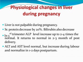
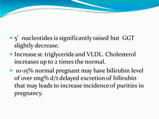
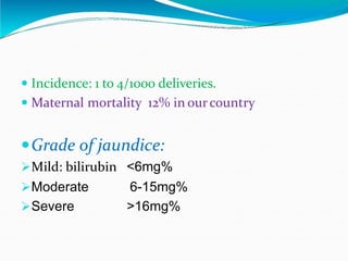
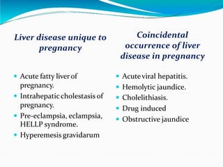
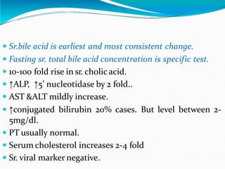
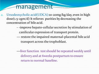
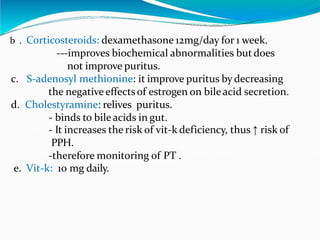
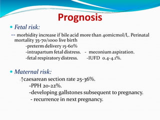
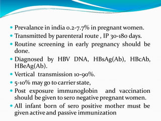
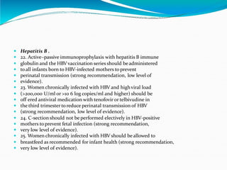
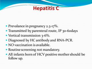
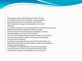
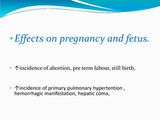
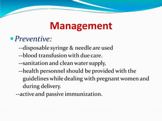
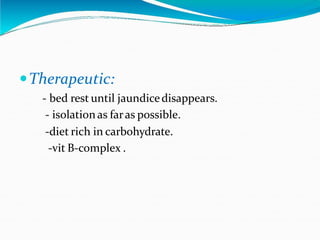
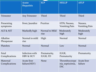
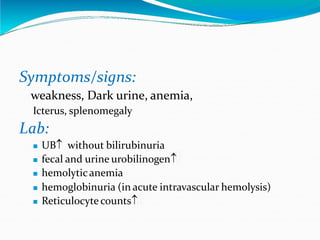
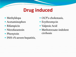
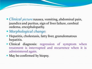
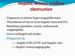
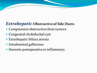
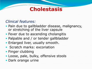
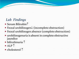
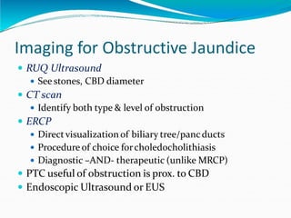
JAUNDICE IN PREGNANCY
DR. S.N. BERA & MITALI DASH
M K C G MEDICAL COLLEGE
 Yellow discoloration
of skin, conjunctiva,
sclera and mucosa
associated with rise
in serum bilirubin
above 2mg/dl
( normal 0.2-01mg/dl)
Latent jaundice:1-2mg/dl
Mechanisms:
 ↑ production of bilirubin
 ↓ hepatocyte transport orconjugation
 Impaired excretion of bilirubin
 Impaired delivery of bilirubin into intestine
--“surgically relevant jaundice” or obstructive
jaundice
“Cholestasis” refers to the latter two, impaired
excretion and obstructive jaundice.
Physiological changes in liver
during pregnancy
 Liver is not palpable during pregnancy.
 Sr. protein decrease by 20%. Bilirubin also decrease
 In 3
rd trimester ALP level increase up to 2-4 times the
normal. It returns to normal in 2-3 month of post
delivery.
 ALT and AST level normal, but increase during labour
and normalize in 1-2 days postpartum.
 5’ nucleotides is significantly raised but GGT
slightly decrease.
 Increase sr. triglycerideand VLDL. Cholesterol
increases up to 2 times the normal.
 10-15% normal pregnant may have bilirubin level
of over 1mg% d/t delayed excretion of bilirubin
that may leads to increase incidenceof purities in
pregnancy.
 Incidence: 1 to 4/1000 deliveries.
 Maternal mortality 12% in ourcountry
Grade of jaundice:
Mild: bilirubin <6mg%
Moderate
Severe
6-15mg%
>16mg%
Cause of jaundice during
pregnancy
Coincidental
occurrence of liver
disease in pregnancy
Liver disease unique to
pregnancy
 Acute fatty liverof
pregnancy.
 Intrahepaticcholestasis of
pregnancy.
 Pre-eclampsia, eclampsia,
 Acuteviral hepatitis.
 Hemolytic jaundice.
 Cholelithiasis.
 Drug induced
 Obstructive jaundice
HELLP syndrome.
 Hyperemesis gravidarum
Jaundice develop during pregnancy
on chronic liver disease:
A. Cirrhosis of liver.
B. Chronic hepatitis
Liver disease probably related to
pregnancy:
. Portal HTN.
. Cholecystitis.
. Pancreatitis.
. Wilsons ds.
. Gilbirts syndrome.
. Budd chiari syndrome.
Acute fatty liver of pregnancy
 Acute hepatic failure in absence of viral hepatitis, IHC,
etc.
 Manifest usually in 3
rd trimester sometimes present
afterdelivery.
 Incidence: 1 in 7000-15000 pregnancies.
 More common in primipara and associated with pre-
eclampsia, multiple pregnancy, having male fetus.
Etiology:
 may be mitochondrial dysfunction.--- >LCHAD
def.
--->defect in oxidation pathway of fatty acid.
. ↑estrogenin3
rd trimester leads to ↓ mitochondrial
oxidation.
 Table 4 . Swansea criteria for diagnosis of acute fatty liverof
 pregnancy
 Six or more criteria required in the absence of another cause
 Vomiting
 Abdominal pain
 Polydipsia/polyuria
 Encephalopathy
 Elevated bilirubin >14 μ mol/l
 Hypoglycaemia <4 mmol/l
 Elevated urea >340 μ mol/l
 Leucocytosis >11×10 6 cells/l
 Ascites or bright liveron ultrasound scan
 Elevated transaminases (AST or ALT) >42 IU/l
 Elevated ammonia >47 μ mol/l
 Renal impairment; creatinine >150 μ mol/l
 Coagulopathy; prothrombin time >14 s or APPT>34 s
 Microvesicular steatosis on liver biopsy
Clinical features and diagnosis
Clinical features: Anorexia, nausea, vomiting,
headache, fatigue, altered mental status,
polydypsia with orwith out polyuria.
 Jaundice 90%,
 Right upperquadrant pain.
 Puritus and ascites 50% cases.
 Hepatic encephalopathy seen in latter.
 Clinical improvementoccurs 1-4 weeksafter
delivery..
 Conjugated hyperbilirubinimia up to 5-15 mg/dl.
 ↑ALT but <1000iu/ml.
 ↑ALP 3-4times. Prolonged PT.
 ↑BUN and amonia level..
 ↓sr. fibrinogen,hypoglycemia. Hyperurecemia,
 leukocytosis,
 USG, CT, MRI demonstrate fatty infiltration.
 liver biopsy is gold standered.
--microvesicular fatty infiltration
--portal inflamation with cholestasis.
Prognosis
 Maternal mortality:10-15% d/t
 Hepatic encephalopathy, DIC, ARDS, Renal failure, Acute
pancreatitis, PPH.
 Fetal mortality: 15-20% d/t
 IUD , prematurity.
Full clinical and laboratory remission occurs over 1-4
weeks post delivery..
Prenatal diagnosis in next can be done by CVS or
amniocentesis
Recurrence in future pregnancy 15-25% in those have
LCHAD deficiency.
Management
 Supportive and directed towards management of
liver insufficiencyand complication.
 Optimal management is prompt delivery.. Transfer
to high dependency unit.
 FFP, platelets, blood transfusion to correct
coagulopathy.
 ivglucose to maintain normoglycemia.
 monitor LFT with PT.
 In fulminant hepatic failure liver transplantation
is only treatment.
Intrahepatic cholestasis of
pregnancy
 Typically occurs in late pregnancy. Affecting 1.5-2% of
pregnancies.
 Disappears spontaneously, recurs in subsequent
pregnancy.
 ETIOLOGY:
i. Hereditary hepatic ↑ sensitivity of pregnancy
hormone → affects gallbladder function →slow or
stop the bile flow →build upof bileacid in liver.
ii. Genetic mutation in the hepatocellular phospholipid
transporter MDR3 in 15% case.
iii. Familial.
iv. Selenium deficiency in diet.
v. Previous liverdamage.
Intrahepatic cholestasis of
pregnancy
. Typically manifest in 3rd trimester but can occure as
early as 10 weeks.
. Puritus in 2nd half of pregnancy which is otherwise
unexplained is most charecteristic manifestation 70%
cases.
. Purituson palm and sole, intensity more in night.
. Severe puritus leads to insomnia, fatigue, depression
,mental disturbance.
. Dark coloururine.
. Jaundice seen in 10% cases usually mild ,develop 1-4
wks afterpuritus.
 Jaundice usuallydisappears with in weeks following
delivery.
 Puritus begins todecline a few hours afterdelivery and
disappears with in few days. Puritiesas a rule persist
longer than jaundice.
 Biochemical abnormalities normalize in few weeks
following delivery.
 If persist more than three month afterdelivery,
investigate to ruleoutchronic liverdisease.
 Sr.bile acid is earliest and most consistent change.
 Fasting sr. total bile acid concentration is specific test.
 10-100 fold rise in sr. cholicacid.
 ↑ALP, ↑5’ nucleotidase by 2 fold..
 AST &ALT mildly increase.
 ↑conjugated bilirubin 20% cases. But level between 2-
5mg/dl.
 PT usually normal.
 Serum cholesterol increases 2-4 fold
 Sr. viral marker negative.
management
a. Ursodeoxycholic acid(UDCA):10-20mg/kg/day, even in high
dose(1.5-2gm/d) It relieves purities bydecreasing the
concentration of bile acid.
- -improve hepato-cellularsecretion by stimulation of
canilicularexpression of transport protein.
--- restore the impaired maternal-placental bile acid
transport across the trophoblast.
---liver function test should be repeated weekly until
deliveryand at 6weeks postpartum to ensure
return to normal baseline.
b . Corticosteroids: dexamethasone 12mg/day for 1 week.
---improves biochemical abnormalities but does
not improve puritus.
c. S-adenosyl methionine: it improve puritus by decreasing
the negative effectsof estrogen on bileacid secretion.
d. Cholestyramine: relives puritus.
- binds to bile acids in gut.
- It increases the risk of vit-k deficiency, thus ↑ risk of
PPH.
-therefore monitoring of PT .
e. Vit-k: 10 mg daily.
Prognosis
 Fetal risk:
-- morbidity increase if bile acid more than 40micmol/l,. Perinatal
mortality 35-70/1000 live birth
-preterm delivery 15-60%
-intrapartum fetal distress. - meconium aspiration.
-fetal respiratorydistress. -IUFD 0.4-4.1%.
 Maternal risk:
-↑caesarean section rate 25-36%.
-PPH 20-22%.
-developing gallstones subsequent to pregnancy.
- recurrence in next pregnancy.
HELLP syndrome
 Coined by Dr.Louis weinstein in 1982.
 0.5-0.9% of all pregnancies, and 10-20% case with
preeclampsia.
 characterized by haemolysis, elevated liver enzyme ,low
platelet.
 More common in multiparous and older pregnant
women.
 Develops during antepartum periods in 70% cases with
frequency between 27th and 37th wks GA.
 Majority with HELLP syndrome have HTN and
proteinuria, but it may absent in10-20% cases.
Pathogenesis: as in preeclampsia activation of
complementand coagulation cascade→increse
vascular tone,platlet aggregation , alteration in
TXA2 and prostacycline ratio. Leade to
--micro angiopathic hemolytic anemia.
--elevated liverenzyme(d/t periportal
hepatic necrosis)
--thrombocytopenia.
. Clincal picture: nausea, vomiting, malaise,
headache,weight gain, upperabdominal pain,
HTN,
DIAGNOSIS
Peripheral smear: features of hemolysis i.e
schizocytes, Burrcells,↑reticulocytes, ↑LDH.
↑Uncojugated bilirubin
-- thrombocytopenia.
Elevated liverenzyme--↑ALT and AST.
Diagnostic criteria: Mississippi classification.
CT or USG if subcapsular hematoma is suspected.
Complication:
Maternal:--eclampsia.
--DIC
—abruptio placentae,
-- ARF
--pulmonaryoedema
--Cerebral oedema.
: ---IUGR ---PREMATURITY
--perinatal asphyxia .– still birth
Fetal
Management
 Stabilizes maternal condition.
 Control HTN.
 Antiseizure prophylaxis with mgso4.
 Correct coagulopathy. Assessmentof fetal wellbeing.
 Definitive therapy is delivery irrespective of
gestation.
 Modeof delivery: vaginal delivery prefered.
 If CS has to be done 10 units of platlets should be
arranged if count <40000/cmm,
 Intensive monitoring for 48 hrs,
 Dexamethasone therapy to continue untill
postpartum resolution of disease occurs.
Prognosis
. Maternal mortality 2-25%.
. Perinatal mortality 33% .
. Recurrence in subsequent pregnancy 25%..
. Most patient have rapid, early resolution of
HELLP syndrome afterdelivery, with
normalization of platlets 5-7 postpartum day.
Hyper emesis gravidarum
 Extremeof spectrum of morning sickness.
 c/b intractable nausea, vomiting ,dehydration, metalic
acidosis or alkalosis, electrolyte imbalance, weight
loss.
 Incidence less than 1 in 1000 pregnancy.
 Limited in 1st trimester, more in 1st pregnancy , recur in
next pregnancy, more in multiple pregnancy with
hydatidiform mole.
 Etiology:
a) Hormonal: hCG, estrogen, progesterone.
b) Psychological(neurosis)
Pathology:
 Liver: centri lobular hepatic infiltration with out
necrosis.
 Heart: small heart , occasonal sub endocardial
hemorrhage.
 Brain: small hemorrhage in hypothalamic region
giving the manifestation of wernick’s
encephalopathy
Complication:
1. Neurological: wernick’s encephalopathy,
korsakoff’s psychosis, pontine myelinolysis,
peripheral neuritis.
2. Stress ulcer in stomach,
3. Mallory weiss syndrome,
4. Convulsion, coma.
5. jaundice
Management
 Hospitalization in severe case,
 Restriction of oral fluid, and iv fluid given to
correctdehydration.
 Anti emetics and vitamins.
 Hydrocortisone use in severe case.
 Nutritional support with vit b1, vitb6, vit b12, vitc.
Acute viral hepatitis
 MC cause of jaundice in pregnancy.
 Caused by hepatiti A,B,C,D and E VIRUS.
Hepatitis A:
 Occurs in area of crowding orpoorsanitation
 Transmitted by fecooral route, with IP 28-30 days.
 c/b initial period of fever, anorexia , nausea, vomiting,
jaundice, subsides 2-4 weeks
 In acutestage IgM, and IgG promotes immunity.
 Self limited, no carrier state, novertical transmission,
 Not teratogenic, but ↑ risk of preterm birth.
 Post exposure immunoglobin and vaccine may be
given toseronagative.
 Infant born of seropositive mother may be given active
and passive immunization.
Hepatitis B
 Prevalance in india 0.2-7.7% in pregnant women.
 Transmitted by parenteral route , IP 30-180 days.
 Routine screening in early pregnancy should be
done.
 Diagnosed by HBV DNA, HBsAg(Ab), HBcAb,
HBeAg(Ab).
 Vertical transmission 10-90%.
 5-10% may go to carrierstate,
 Post exposure immunoglobin and vaccination
should be given to sero negative pregnant women.
 All infant born of sero positive mother must be
given activeand passive immunization
 Hepatitis B .
 22. Active–passive immunoprophylaxis with hepatitis B immune
 globulin and the HBV vaccination series should be administered
 to all infants born to HBV-infected mothers to prevent
 perinatal transmission (strong recommendation, low level of
 evidence).
 23. Women chronically infected with HBV and highviral load
 (>200,000 U/ml or >10 6 log copies/ml and higher) should be
 off ered antiviral medication with tenofovir or telbivudine in
 the third trimester to reduce perinatal transmission of HBV
 (strong recommendation, low level of evidence).
 24. C-section should not be performed electively in HBV-positive
 mothers to prevent fetal infection (strong recommendation,
 very low level of evidence).
 25. Women chronically infected with HBV should be allowed to
 breastfeed as recommended for infant health (strong recommendation,
 very low level of evidence).
Hepatitis C
 Prevalance in pregnancy 2.3-17%.
 Transmitted by parenteral route, IP 30-60days
 Vertical transmission 3-6%.
 Diagnosed by HC antibody and RNA-PCR.
 NO vaccination is available.
 Routine screening not mandatory.
 All infants born of HCV positive mothershould be
follow up.
 All pregnant women with risk factors for HCV should
 be screened with anti-HCV antibody. Screening should
 not be performed in women without risk factors for
 HCV acquisition (strong recommendation, low level of
 evidence).
 27. Invasive procedures (e.g., amniocentesis, invasive fetal monitoring)
 should be minimized in infected mothers and their
 fetus to prevent vertical transmission of hepatitis C (strong
 recommendation, very low level of evidence).
 28. C-section should not be performed electively in HCV-positive
 mothers to prevent fetal infection (strong recommendation,
 very low level of evidence).
 29. Women chronically infected with HCVshould be allowed to
 breastfeed as indicated for infant health (strong recommendation,
 very low level of evidence).
 30. Hepatitis C therapy should not beoff ered to pregnant
 women to either treat HCVordecrease the risk forvertical
 transmission (strong recommendation, very low level of























Autoimmune hepatitis
31. Pregnant women with autoimmune hepatitis (AIH) should
becontinued on their treatment with corticosteroids and/or
azathioprine (AZA) (strong recommendation, very low level
of evidence).
32. Pregnant women with primary biliary cirrhosis (PBC) should
becontinued on their treatment with UDCA (strong recommendation,
very low level of evidence).
33. Pregnant women with Wilson’s disease (WD) should be
continued, with dose reduction if possible, on their treatment
with penicillamine, trientine, or zinc (strong recommendation,
very low level of evidence).
34. Pregnant women with suspected portal hypertension should
undergo screening with upperendoscopy foresophageal
varices in the second trimester (strong recommendation, low
level of evidence).
35. Pregnant womenwho are found to have large esophageal
varices should be treated with beta-blockers and/or band
ligation (conditional recommendation, very low level of
evidence).
36. Pregnant women with a history of liver transplantation
should continue their immunosuppression except for
mycophenolic acid (strong recommendation, moderate level
Hepatitis D
 Deltavirus; defective RNA virus.
 Mustco-infectwith HBV.
 Transmission similarto HBV.
 Chronic co-infection with HBV and HDV is more
severe.
 Neonatal transmission is unusual.
Hepatitis E
 Transmitted by feco-oral route.
 Fulminantvarity is most common among all types.
 HEV specific IgM and IgG can be detected.
 Chance of vertical transmission.
 Chronic infection is uncommon but may occurs.
 Hepatitis E vaccine(Hecolin) .
 Maternal mortality 15-20%.
Clinical features of viral hepatitis
 Prodromal phase: nausea, vomiting, pyrexia, lossof
appetite, upperabdominal pain, myalgia, artthralgia,
dark urine, claycolourstool. Palpable liver.
 Icteric phase: appears 1-2 weeks aftersymptom.
 Recovery phase: jaundice recedes, liverenlargement
regress, it takes 6-12 weeks forcompleterecovery.
 Investigation:
---LFT
---liver biopsy.
•Effects on pregnancy and fetus.
• ↑incidence of abortion, pre term labour, still birth,
• ↑incidence of primary pulmonary hypertention ,
hemorrhagic manifestation, hepatic coma,
Management
Preventive:
--disposablesyringe & needle are used
--blood transfusionwith duecare.
--sanitation and clean watersupply,
--health personnel should be provided with the
guidelines whiledealing with pregnant women and
during delivery.
--activeand passive immunization.
Therapeutic:
- bed rest until jaundicedisappears.
- isolationas faras possible.
-diet rich in carbohydrate.
-vit B-complex .
Acute ICP HELLP AFLP
Hepatitis
Trimester Any Trimester Third Third Third
Presenting
symptoms
Fever, Jaundice Pruritus HTN, Nausea,
Vomiting,Pain Vomiting,Pain
Nausea,
ALT & AST Markedly high Normal to Mild Moderately Moderately
high
rise high
Alkaline
Phospatase
Normal to mild High
rise
Normal Normal
Platelets Normal Normal Low Normal
Fetal
Complications
Infection with
HBV & HCV
Prematurity,
IUGR, FD
IUGR,
Prematurity
Prematurity
Maternal
Complications
Acute liver
failure(HEV)
None Thrombocytope Acute liver
nia, septicemia, failure
DIC
Hemolytic jaundice
 Hereditary
----thalassemia, sickle cell anemia.
----spherocytosis.
 Acquired
---- malaria.
---- mismatched blood transfusion.
---- leptospira,
----cl. Wiilchi
All causes unconjugated hyperbilirubinemia,
Sicklecell anaemia with pregnancy :
 Pregnancy is high risk in sickle cell disease (mostly SS
 Haemolyticcrisis causes rapidlydeveloping anemia with
jaundice.
 Vaso-occlusive crisis causes osteonecrosis, infarction of
kidney, lung. Hepato-splenomegaly, heart failureare
common.
 Increased incidence of maternal pre-eclampsia, PPH,
infection. Increased morbidity and mortalitydue to CVA,
pulm infarction, CHF, embolism etc.
 Increased risk of abortion, prematurity, IUGR, fetal loss,
perinatal mortality.
Management includes :
• Pre-conceptional councelling
• During pregnancy: RegularANC, Prophylactic folic
acid 1 mg daily, Iron supplementationetc. Infection or
appearance of unusual symptoms needs
hospitalization
• During labour: Oxygen inhalation, Adequate
hydration, Antibiotics . Epidural anesthesia preferred.
Caesarian section should be performed forobstetric
indication only.
• Contraception: Sterilization even with low parity. OC
pills, IUCD contraindicated. Barrier method is ideal.
Malaria
 Malaria is imp. causeof jaundice in ourcountry.
 Effectson mother: Anemia, Hypoglycemia, Metabolic
acidosis, Hepatic dysfunctionwith jaundice, Renal
failure, Pulmonaryedema, Convulsions, comaetc.
 Effects on the fetus: Abortion, Preterm labour, Pre-
maturity, IUGR, IUFD etc.
 Congenital malaria is rare (<5%) unless placenta is
damaged (with Plasmodium falciparum in 2nd half of
pregnancy ).
 Effects of pregnancy on malaria: Risk and severityof
infection increases, Complicationsare high.
Management:
Management:
• Prevention from mosquito bite.
• Chemoprophylaxis: Cloroquine ( 300mg baseweekly, 2
weeks before travel, covering the period of exposureand 4
weeks after leaving theendemic zone). Mefloquine
250mg/week in cloroquine resistant cases.
• Treatment:
i. Cloroquine- 10mg base/kg followed by 10mg/kg base at
24 hrand 5mg/kg at 48 hr.
ii. Cloroquine resistant cases: Quinine- 10mg salt/kg p.o
,every 8 hr for 7 days.
iii. Complicated malaria: Artesunate IV 2.4mg/kg at 0, 12, 24
hr, then daily for 5 days. Oral therapy ( 2mg/kg) when
the pt is stable.
Symptoms/signs:
weakness, Dark urine, anemia,
Icterus, splenomegaly
Lab:
 UB without bilirubinuria
 fecal and urine urobilinogen
 hemolyticanemia
 hemoglobinuria (in acute intravascular hemolysis)
 Reticulocyte counts
Drug induced
 Methyldopa
 Acetaminophen
 Rifampicin
 OCP’s-cholestasis,
 Erythromycin
 Valproic Acid
 Nitrofurantoin
 Phenytoin
 Methotrexate-indolent
cirrhosis
 INH-1% severe hepatitis,
 Clinical picture:nausea, vomiting, abdominal pain,
jaundiceand puritus, sign of liver failure, cerebral
oedema, encephalopathy.
 Morphological change:
 Hepatitis, cholestasis, fatty liver, granulomatous
hepatitis.
 Clinical diagnosis: regression of symptom when
treatment is interrupted and recurrence when it is
administered again.
 May be confirmed by biopsy.
Hepatic venous outflow
obstruction
o Pregnancy is relative hypercoagulable state.
o Thrombosis of oneor more hepaticveinsand IVC.
o Resulting injaundice, ascitis, oedemaand
coagulopathy.
o Liver is enlarged and tender.
oDiagnosis by
---- doppler USG of IVC and hepaticvein.
----hepatic venous angiography.
Pregnancy and chronic liver
disease
 CLD has mainly two types of manifestation
Firstly ,those related to portal HTN such as
oesophageal and gastric varices. Thevarices may
be low or high grade
secondly ,features of liver failure such as
ascites, jaundice, coagulopathy, hepatorenal
syndrome, hepatic encephalopathy.
CLD with well preserved liver function
--cirrhosis is not contraindication to pregnancy.
--pregnancy may be uneventful.
--all pregnant patient should screen for varices in second
trimester.
--low grade varices should be observed, high risk varices should
receives primary prophylasix either beta blockers or
endoscopic variceal ligation.
--vasopressin is contraindicated in pregnancy as it may causes
placental ischemia, necrosis and amputation of fetal digit.
--coagulation should be corrected before induction orcs.
--vaginal delivery prefered, should be assited and second stage
shortened.
--early termination of pregnancy should be consider when
hepaticdecompensation is present.
CLD with decompensation of liver function
 Most women are amenorrhoeic and unable to
conceive because of associated hypothalamic-
pituitary dysfunction.
 It is high risk pregnancy with maternal and fetal
complication 50% with increase fetal loss .
Obstructive jaundice
Intrahepatic-Livercell Damage/Blockage of Bile
Canaliculi
 Drugs orchemical toxins
 Dubin-Johnson syndrome
 Estrogens or Pregnancy
 Infiltrative tumors
 Intrahepatic biliary hypoplasiaoratresia
 Primary biliary cirrhosis
Extrahepatic-Obstructiveof bile Ducts
 Compression obstruction from tumors
 Congenital choledochal cyst
 Extrahepatic biliary atresia
 Intraluminal gallstones
 Stenosis-postoperativeor inflammary
Cholestasis
Clinical features:
 Pain due to gallbladder disease, malignancy,
or stretching of the liver capsule
 Fever due to ascending cholangitis
 Palpable and / or tender gallbladder
 Enlarged liver, usually smooth.
 Scratch marks: excoriation
 Finger clubbing
 Loose, pale, bulky, offensive stools
 Dark orange urine
Lab Findings
 Serum Bilirubin
 Feceal urobilinogen (incompleteobstruction)
 Feceal urobilinogen absence (completeobstruction)
 urobilinogenuria is absent in completeobstructive
jaundice
 bilirubinuria 
 ALP 
 cholesterol 
Imaging for Obstructive Jaundice
 RUQ Ultrasound
 See stones, CBD diameter
 CT scan
 Identify both type & level of obstruction
 ERCP
 Directvisualization of biliary tree/pancducts
 Procedure of choice forcholedocholithiasis
 Diagnostic –AND- therapeutic (unlike MRCP)
 PTC useful of obstruction is prox. to CBD
 Endoscopic Ultrasound or EUS
Treatment
 If Medical, then treat theetiology
 If Obstructive Jaundice:
 Should r/oascending cholangitis,
 Forcholangitis: IVF, IV Antibiotics, Decompression
 Stones (remove stones vs stent vs drainage)
 Donevia ERCP or PTC oropen (surgery)
 Benign stricture (stent vs drainage catheter)
 Cancer (Stentvs drainage +/- resect the CA)
 The key principle is decompression, either
externally(drainage) or internally(stenting) the duct
open toallow betterdrainage
Cholelithiasis
 Pregnancy alters bilecomposition and gall bladder
emptying slows in the second trimester increasing the risk
of gall stones.
 Risk factors are multiparityand previousgallbladder
disease.
 Choledocholithiasis accounts forapproximately 7% of
patients with jaundice in pregnancy but jaundice occurs in
only 5% of cholelithiasis.
 Treatment involves laparoscopic cholecystectomy.
 Uncomplicated cholecystectomy is safer in first and second
trimesters with fetal loss about 5%
 But with common bileduct exploration and if with
pancreatitis then 15 % maternal and 60% fetal mortality
Jaundice in pregnancy should be evaluated thoroughly
with proper history, clinical examination, laboratory
investigation to identify the underlying cause.
Proper management may improve maternal and
fetal outcome.
THANK YOU

jaundiceself-160811104352.pptx

  • 1.
    JAUNDICE IN PREGNANCY DR.S.N. BERA & MITALI DASH M K C G MEDICAL COLLEGE
  • 2.
     Yellow discoloration ofskin, conjunctiva, sclera and mucosa associated with rise in serum bilirubin above 2mg/dl ( normal 0.2-01mg/dl) Latent jaundice:1-2mg/dl
  • 4.
    Mechanisms:  ↑ productionof bilirubin  ↓ hepatocyte transport orconjugation  Impaired excretion of bilirubin  Impaired delivery of bilirubin into intestine --“surgically relevant jaundice” or obstructive jaundice “Cholestasis” refers to the latter two, impaired excretion and obstructive jaundice.
  • 5.
    Physiological changes inliver during pregnancy  Liver is not palpable during pregnancy.  Sr. protein decrease by 20%. Bilirubin also decrease  In 3 rd trimester ALP level increase up to 2-4 times the normal. It returns to normal in 2-3 month of post delivery.  ALT and AST level normal, but increase during labour and normalize in 1-2 days postpartum.
  • 6.
     5’ nucleotidesis significantly raised but GGT slightly decrease.  Increase sr. triglycerideand VLDL. Cholesterol increases up to 2 times the normal.  10-15% normal pregnant may have bilirubin level of over 1mg% d/t delayed excretion of bilirubin that may leads to increase incidenceof purities in pregnancy.
  • 7.
     Incidence: 1to 4/1000 deliveries.  Maternal mortality 12% in ourcountry Grade of jaundice: Mild: bilirubin <6mg% Moderate Severe 6-15mg% >16mg%
  • 8.
    Cause of jaundiceduring pregnancy Coincidental occurrence of liver disease in pregnancy Liver disease unique to pregnancy  Acute fatty liverof pregnancy.  Intrahepaticcholestasis of pregnancy.  Pre-eclampsia, eclampsia,  Acuteviral hepatitis.  Hemolytic jaundice.  Cholelithiasis.  Drug induced  Obstructive jaundice HELLP syndrome.  Hyperemesis gravidarum
  • 9.
    Jaundice develop duringpregnancy on chronic liver disease: A. Cirrhosis of liver. B. Chronic hepatitis
  • 10.
    Liver disease probablyrelated to pregnancy: . Portal HTN. . Cholecystitis. . Pancreatitis. . Wilsons ds. . Gilbirts syndrome. . Budd chiari syndrome.
  • 11.
    Acute fatty liverof pregnancy  Acute hepatic failure in absence of viral hepatitis, IHC, etc.  Manifest usually in 3 rd trimester sometimes present afterdelivery.  Incidence: 1 in 7000-15000 pregnancies.  More common in primipara and associated with pre- eclampsia, multiple pregnancy, having male fetus.
  • 12.
    Etiology:  may bemitochondrial dysfunction.--- >LCHAD def. --->defect in oxidation pathway of fatty acid. . ↑estrogenin3 rd trimester leads to ↓ mitochondrial oxidation.
  • 13.
     Table 4. Swansea criteria for diagnosis of acute fatty liverof  pregnancy  Six or more criteria required in the absence of another cause  Vomiting  Abdominal pain  Polydipsia/polyuria  Encephalopathy  Elevated bilirubin >14 μ mol/l  Hypoglycaemia <4 mmol/l  Elevated urea >340 μ mol/l  Leucocytosis >11×10 6 cells/l  Ascites or bright liveron ultrasound scan  Elevated transaminases (AST or ALT) >42 IU/l  Elevated ammonia >47 μ mol/l  Renal impairment; creatinine >150 μ mol/l  Coagulopathy; prothrombin time >14 s or APPT>34 s  Microvesicular steatosis on liver biopsy
  • 14.
    Clinical features anddiagnosis Clinical features: Anorexia, nausea, vomiting, headache, fatigue, altered mental status, polydypsia with orwith out polyuria.  Jaundice 90%,  Right upperquadrant pain.  Puritus and ascites 50% cases.  Hepatic encephalopathy seen in latter.  Clinical improvementoccurs 1-4 weeksafter delivery..
  • 15.
     Conjugated hyperbilirubinimiaup to 5-15 mg/dl.  ↑ALT but <1000iu/ml.  ↑ALP 3-4times. Prolonged PT.  ↑BUN and amonia level..  ↓sr. fibrinogen,hypoglycemia. Hyperurecemia,  leukocytosis,  USG, CT, MRI demonstrate fatty infiltration.  liver biopsy is gold standered. --microvesicular fatty infiltration --portal inflamation with cholestasis.
  • 16.
    Prognosis  Maternal mortality:10-15%d/t  Hepatic encephalopathy, DIC, ARDS, Renal failure, Acute pancreatitis, PPH.  Fetal mortality: 15-20% d/t  IUD , prematurity. Full clinical and laboratory remission occurs over 1-4 weeks post delivery.. Prenatal diagnosis in next can be done by CVS or amniocentesis Recurrence in future pregnancy 15-25% in those have LCHAD deficiency.
  • 17.
    Management  Supportive anddirected towards management of liver insufficiencyand complication.  Optimal management is prompt delivery.. Transfer to high dependency unit.  FFP, platelets, blood transfusion to correct coagulopathy.  ivglucose to maintain normoglycemia.  monitor LFT with PT.  In fulminant hepatic failure liver transplantation is only treatment.
  • 18.
    Intrahepatic cholestasis of pregnancy Typically occurs in late pregnancy. Affecting 1.5-2% of pregnancies.  Disappears spontaneously, recurs in subsequent pregnancy.  ETIOLOGY: i. Hereditary hepatic ↑ sensitivity of pregnancy hormone → affects gallbladder function →slow or stop the bile flow →build upof bileacid in liver. ii. Genetic mutation in the hepatocellular phospholipid transporter MDR3 in 15% case. iii. Familial. iv. Selenium deficiency in diet. v. Previous liverdamage.
  • 19.
    Intrahepatic cholestasis of pregnancy .Typically manifest in 3rd trimester but can occure as early as 10 weeks. . Puritus in 2nd half of pregnancy which is otherwise unexplained is most charecteristic manifestation 70% cases. . Purituson palm and sole, intensity more in night. . Severe puritus leads to insomnia, fatigue, depression ,mental disturbance. . Dark coloururine. . Jaundice seen in 10% cases usually mild ,develop 1-4 wks afterpuritus.
  • 20.
     Jaundice usuallydisappearswith in weeks following delivery.  Puritus begins todecline a few hours afterdelivery and disappears with in few days. Puritiesas a rule persist longer than jaundice.  Biochemical abnormalities normalize in few weeks following delivery.  If persist more than three month afterdelivery, investigate to ruleoutchronic liverdisease.
  • 21.
     Sr.bile acidis earliest and most consistent change.  Fasting sr. total bile acid concentration is specific test.  10-100 fold rise in sr. cholicacid.  ↑ALP, ↑5’ nucleotidase by 2 fold..  AST &ALT mildly increase.  ↑conjugated bilirubin 20% cases. But level between 2- 5mg/dl.  PT usually normal.  Serum cholesterol increases 2-4 fold  Sr. viral marker negative.
  • 22.
    management a. Ursodeoxycholic acid(UDCA):10-20mg/kg/day,even in high dose(1.5-2gm/d) It relieves purities bydecreasing the concentration of bile acid. - -improve hepato-cellularsecretion by stimulation of canilicularexpression of transport protein. --- restore the impaired maternal-placental bile acid transport across the trophoblast. ---liver function test should be repeated weekly until deliveryand at 6weeks postpartum to ensure return to normal baseline.
  • 23.
    b . Corticosteroids:dexamethasone 12mg/day for 1 week. ---improves biochemical abnormalities but does not improve puritus. c. S-adenosyl methionine: it improve puritus by decreasing the negative effectsof estrogen on bileacid secretion. d. Cholestyramine: relives puritus. - binds to bile acids in gut. - It increases the risk of vit-k deficiency, thus ↑ risk of PPH. -therefore monitoring of PT . e. Vit-k: 10 mg daily.
  • 24.
    Prognosis  Fetal risk: --morbidity increase if bile acid more than 40micmol/l,. Perinatal mortality 35-70/1000 live birth -preterm delivery 15-60% -intrapartum fetal distress. - meconium aspiration. -fetal respiratorydistress. -IUFD 0.4-4.1%.  Maternal risk: -↑caesarean section rate 25-36%. -PPH 20-22%. -developing gallstones subsequent to pregnancy. - recurrence in next pregnancy.
  • 25.
    HELLP syndrome  Coinedby Dr.Louis weinstein in 1982.  0.5-0.9% of all pregnancies, and 10-20% case with preeclampsia.  characterized by haemolysis, elevated liver enzyme ,low platelet.  More common in multiparous and older pregnant women.  Develops during antepartum periods in 70% cases with frequency between 27th and 37th wks GA.  Majority with HELLP syndrome have HTN and proteinuria, but it may absent in10-20% cases.
  • 26.
    Pathogenesis: as inpreeclampsia activation of complementand coagulation cascade→increse vascular tone,platlet aggregation , alteration in TXA2 and prostacycline ratio. Leade to --micro angiopathic hemolytic anemia. --elevated liverenzyme(d/t periportal hepatic necrosis) --thrombocytopenia. . Clincal picture: nausea, vomiting, malaise, headache,weight gain, upperabdominal pain, HTN,
  • 27.
    DIAGNOSIS Peripheral smear: featuresof hemolysis i.e schizocytes, Burrcells,↑reticulocytes, ↑LDH. ↑Uncojugated bilirubin -- thrombocytopenia. Elevated liverenzyme--↑ALT and AST. Diagnostic criteria: Mississippi classification. CT or USG if subcapsular hematoma is suspected.
  • 28.
    Complication: Maternal:--eclampsia. --DIC —abruptio placentae, -- ARF --pulmonaryoedema --Cerebraloedema. : ---IUGR ---PREMATURITY --perinatal asphyxia .– still birth Fetal
  • 29.
    Management  Stabilizes maternalcondition.  Control HTN.  Antiseizure prophylaxis with mgso4.  Correct coagulopathy. Assessmentof fetal wellbeing.  Definitive therapy is delivery irrespective of gestation.  Modeof delivery: vaginal delivery prefered.  If CS has to be done 10 units of platlets should be arranged if count <40000/cmm,  Intensive monitoring for 48 hrs,  Dexamethasone therapy to continue untill postpartum resolution of disease occurs.
  • 30.
    Prognosis . Maternal mortality2-25%. . Perinatal mortality 33% . . Recurrence in subsequent pregnancy 25%.. . Most patient have rapid, early resolution of HELLP syndrome afterdelivery, with normalization of platlets 5-7 postpartum day.
  • 31.
    Hyper emesis gravidarum Extremeof spectrum of morning sickness.  c/b intractable nausea, vomiting ,dehydration, metalic acidosis or alkalosis, electrolyte imbalance, weight loss.  Incidence less than 1 in 1000 pregnancy.  Limited in 1st trimester, more in 1st pregnancy , recur in next pregnancy, more in multiple pregnancy with hydatidiform mole.  Etiology: a) Hormonal: hCG, estrogen, progesterone. b) Psychological(neurosis)
  • 32.
    Pathology:  Liver: centrilobular hepatic infiltration with out necrosis.  Heart: small heart , occasonal sub endocardial hemorrhage.  Brain: small hemorrhage in hypothalamic region giving the manifestation of wernick’s encephalopathy
  • 33.
    Complication: 1. Neurological: wernick’sencephalopathy, korsakoff’s psychosis, pontine myelinolysis, peripheral neuritis. 2. Stress ulcer in stomach, 3. Mallory weiss syndrome, 4. Convulsion, coma. 5. jaundice
  • 34.
    Management  Hospitalization insevere case,  Restriction of oral fluid, and iv fluid given to correctdehydration.  Anti emetics and vitamins.  Hydrocortisone use in severe case.  Nutritional support with vit b1, vitb6, vit b12, vitc.
  • 35.
    Acute viral hepatitis MC cause of jaundice in pregnancy.  Caused by hepatiti A,B,C,D and E VIRUS.
  • 36.
    Hepatitis A:  Occursin area of crowding orpoorsanitation  Transmitted by fecooral route, with IP 28-30 days.  c/b initial period of fever, anorexia , nausea, vomiting, jaundice, subsides 2-4 weeks  In acutestage IgM, and IgG promotes immunity.  Self limited, no carrier state, novertical transmission,  Not teratogenic, but ↑ risk of preterm birth.  Post exposure immunoglobin and vaccine may be given toseronagative.  Infant born of seropositive mother may be given active and passive immunization.
  • 38.
    Hepatitis B  Prevalancein india 0.2-7.7% in pregnant women.  Transmitted by parenteral route , IP 30-180 days.  Routine screening in early pregnancy should be done.  Diagnosed by HBV DNA, HBsAg(Ab), HBcAb, HBeAg(Ab).  Vertical transmission 10-90%.  5-10% may go to carrierstate,  Post exposure immunoglobin and vaccination should be given to sero negative pregnant women.  All infant born of sero positive mother must be given activeand passive immunization
  • 39.
     Hepatitis B.  22. Active–passive immunoprophylaxis with hepatitis B immune  globulin and the HBV vaccination series should be administered  to all infants born to HBV-infected mothers to prevent  perinatal transmission (strong recommendation, low level of  evidence).  23. Women chronically infected with HBV and highviral load  (>200,000 U/ml or >10 6 log copies/ml and higher) should be  off ered antiviral medication with tenofovir or telbivudine in  the third trimester to reduce perinatal transmission of HBV  (strong recommendation, low level of evidence).  24. C-section should not be performed electively in HBV-positive  mothers to prevent fetal infection (strong recommendation,  very low level of evidence).  25. Women chronically infected with HBV should be allowed to  breastfeed as recommended for infant health (strong recommendation,  very low level of evidence).
  • 40.
    Hepatitis C  Prevalancein pregnancy 2.3-17%.  Transmitted by parenteral route, IP 30-60days  Vertical transmission 3-6%.  Diagnosed by HC antibody and RNA-PCR.  NO vaccination is available.  Routine screening not mandatory.  All infants born of HCV positive mothershould be follow up.
  • 41.
     All pregnantwomen with risk factors for HCV should  be screened with anti-HCV antibody. Screening should  not be performed in women without risk factors for  HCV acquisition (strong recommendation, low level of  evidence).  27. Invasive procedures (e.g., amniocentesis, invasive fetal monitoring)  should be minimized in infected mothers and their  fetus to prevent vertical transmission of hepatitis C (strong  recommendation, very low level of evidence).  28. C-section should not be performed electively in HCV-positive  mothers to prevent fetal infection (strong recommendation,  very low level of evidence).  29. Women chronically infected with HCVshould be allowed to  breastfeed as indicated for infant health (strong recommendation,  very low level of evidence).  30. Hepatitis C therapy should not beoff ered to pregnant  women to either treat HCVordecrease the risk forvertical  transmission (strong recommendation, very low level of
  • 42.
                           Autoimmune hepatitis 31. Pregnantwomen with autoimmune hepatitis (AIH) should becontinued on their treatment with corticosteroids and/or azathioprine (AZA) (strong recommendation, very low level of evidence). 32. Pregnant women with primary biliary cirrhosis (PBC) should becontinued on their treatment with UDCA (strong recommendation, very low level of evidence). 33. Pregnant women with Wilson’s disease (WD) should be continued, with dose reduction if possible, on their treatment with penicillamine, trientine, or zinc (strong recommendation, very low level of evidence). 34. Pregnant women with suspected portal hypertension should undergo screening with upperendoscopy foresophageal varices in the second trimester (strong recommendation, low level of evidence). 35. Pregnant womenwho are found to have large esophageal varices should be treated with beta-blockers and/or band ligation (conditional recommendation, very low level of evidence). 36. Pregnant women with a history of liver transplantation should continue their immunosuppression except for mycophenolic acid (strong recommendation, moderate level
  • 43.
    Hepatitis D  Deltavirus;defective RNA virus.  Mustco-infectwith HBV.  Transmission similarto HBV.  Chronic co-infection with HBV and HDV is more severe.  Neonatal transmission is unusual.
  • 44.
    Hepatitis E  Transmittedby feco-oral route.  Fulminantvarity is most common among all types.  HEV specific IgM and IgG can be detected.  Chance of vertical transmission.  Chronic infection is uncommon but may occurs.  Hepatitis E vaccine(Hecolin) .  Maternal mortality 15-20%.
  • 45.
    Clinical features ofviral hepatitis  Prodromal phase: nausea, vomiting, pyrexia, lossof appetite, upperabdominal pain, myalgia, artthralgia, dark urine, claycolourstool. Palpable liver.  Icteric phase: appears 1-2 weeks aftersymptom.  Recovery phase: jaundice recedes, liverenlargement regress, it takes 6-12 weeks forcompleterecovery.  Investigation: ---LFT ---liver biopsy.
  • 46.
    •Effects on pregnancyand fetus. • ↑incidence of abortion, pre term labour, still birth, • ↑incidence of primary pulmonary hypertention , hemorrhagic manifestation, hepatic coma,
  • 47.
    Management Preventive: --disposablesyringe & needleare used --blood transfusionwith duecare. --sanitation and clean watersupply, --health personnel should be provided with the guidelines whiledealing with pregnant women and during delivery. --activeand passive immunization.
  • 48.
    Therapeutic: - bed restuntil jaundicedisappears. - isolationas faras possible. -diet rich in carbohydrate. -vit B-complex .
  • 49.
    Acute ICP HELLPAFLP Hepatitis Trimester Any Trimester Third Third Third Presenting symptoms Fever, Jaundice Pruritus HTN, Nausea, Vomiting,Pain Vomiting,Pain Nausea, ALT & AST Markedly high Normal to Mild Moderately Moderately high rise high Alkaline Phospatase Normal to mild High rise Normal Normal Platelets Normal Normal Low Normal Fetal Complications Infection with HBV & HCV Prematurity, IUGR, FD IUGR, Prematurity Prematurity Maternal Complications Acute liver failure(HEV) None Thrombocytope Acute liver nia, septicemia, failure DIC
  • 50.
    Hemolytic jaundice  Hereditary ----thalassemia,sickle cell anemia. ----spherocytosis.  Acquired ---- malaria. ---- mismatched blood transfusion. ---- leptospira, ----cl. Wiilchi All causes unconjugated hyperbilirubinemia,
  • 51.
    Sicklecell anaemia withpregnancy :  Pregnancy is high risk in sickle cell disease (mostly SS  Haemolyticcrisis causes rapidlydeveloping anemia with jaundice.  Vaso-occlusive crisis causes osteonecrosis, infarction of kidney, lung. Hepato-splenomegaly, heart failureare common.  Increased incidence of maternal pre-eclampsia, PPH, infection. Increased morbidity and mortalitydue to CVA, pulm infarction, CHF, embolism etc.  Increased risk of abortion, prematurity, IUGR, fetal loss, perinatal mortality.
  • 52.
    Management includes : •Pre-conceptional councelling • During pregnancy: RegularANC, Prophylactic folic acid 1 mg daily, Iron supplementationetc. Infection or appearance of unusual symptoms needs hospitalization • During labour: Oxygen inhalation, Adequate hydration, Antibiotics . Epidural anesthesia preferred. Caesarian section should be performed forobstetric indication only. • Contraception: Sterilization even with low parity. OC pills, IUCD contraindicated. Barrier method is ideal.
  • 53.
    Malaria  Malaria isimp. causeof jaundice in ourcountry.  Effectson mother: Anemia, Hypoglycemia, Metabolic acidosis, Hepatic dysfunctionwith jaundice, Renal failure, Pulmonaryedema, Convulsions, comaetc.  Effects on the fetus: Abortion, Preterm labour, Pre- maturity, IUGR, IUFD etc.  Congenital malaria is rare (<5%) unless placenta is damaged (with Plasmodium falciparum in 2nd half of pregnancy ).  Effects of pregnancy on malaria: Risk and severityof infection increases, Complicationsare high.
  • 54.
    Management: Management: • Prevention frommosquito bite. • Chemoprophylaxis: Cloroquine ( 300mg baseweekly, 2 weeks before travel, covering the period of exposureand 4 weeks after leaving theendemic zone). Mefloquine 250mg/week in cloroquine resistant cases. • Treatment: i. Cloroquine- 10mg base/kg followed by 10mg/kg base at 24 hrand 5mg/kg at 48 hr. ii. Cloroquine resistant cases: Quinine- 10mg salt/kg p.o ,every 8 hr for 7 days. iii. Complicated malaria: Artesunate IV 2.4mg/kg at 0, 12, 24 hr, then daily for 5 days. Oral therapy ( 2mg/kg) when the pt is stable.
  • 55.
    Symptoms/signs: weakness, Dark urine,anemia, Icterus, splenomegaly Lab:  UB without bilirubinuria  fecal and urine urobilinogen  hemolyticanemia  hemoglobinuria (in acute intravascular hemolysis)  Reticulocyte counts
  • 56.
    Drug induced  Methyldopa Acetaminophen  Rifampicin  OCP’s-cholestasis,  Erythromycin  Valproic Acid  Nitrofurantoin  Phenytoin  Methotrexate-indolent cirrhosis  INH-1% severe hepatitis,
  • 57.
     Clinical picture:nausea,vomiting, abdominal pain, jaundiceand puritus, sign of liver failure, cerebral oedema, encephalopathy.  Morphological change:  Hepatitis, cholestasis, fatty liver, granulomatous hepatitis.  Clinical diagnosis: regression of symptom when treatment is interrupted and recurrence when it is administered again.  May be confirmed by biopsy.
  • 58.
    Hepatic venous outflow obstruction oPregnancy is relative hypercoagulable state. o Thrombosis of oneor more hepaticveinsand IVC. o Resulting injaundice, ascitis, oedemaand coagulopathy. o Liver is enlarged and tender. oDiagnosis by ---- doppler USG of IVC and hepaticvein. ----hepatic venous angiography.
  • 59.
    Pregnancy and chronicliver disease  CLD has mainly two types of manifestation Firstly ,those related to portal HTN such as oesophageal and gastric varices. Thevarices may be low or high grade secondly ,features of liver failure such as ascites, jaundice, coagulopathy, hepatorenal syndrome, hepatic encephalopathy.
  • 60.
    CLD with wellpreserved liver function --cirrhosis is not contraindication to pregnancy. --pregnancy may be uneventful. --all pregnant patient should screen for varices in second trimester. --low grade varices should be observed, high risk varices should receives primary prophylasix either beta blockers or endoscopic variceal ligation. --vasopressin is contraindicated in pregnancy as it may causes placental ischemia, necrosis and amputation of fetal digit. --coagulation should be corrected before induction orcs. --vaginal delivery prefered, should be assited and second stage shortened. --early termination of pregnancy should be consider when hepaticdecompensation is present.
  • 61.
    CLD with decompensationof liver function  Most women are amenorrhoeic and unable to conceive because of associated hypothalamic- pituitary dysfunction.  It is high risk pregnancy with maternal and fetal complication 50% with increase fetal loss .
  • 62.
    Obstructive jaundice Intrahepatic-Livercell Damage/Blockageof Bile Canaliculi  Drugs orchemical toxins  Dubin-Johnson syndrome  Estrogens or Pregnancy  Infiltrative tumors  Intrahepatic biliary hypoplasiaoratresia  Primary biliary cirrhosis
  • 63.
    Extrahepatic-Obstructiveof bile Ducts Compression obstruction from tumors  Congenital choledochal cyst  Extrahepatic biliary atresia  Intraluminal gallstones  Stenosis-postoperativeor inflammary
  • 64.
    Cholestasis Clinical features:  Paindue to gallbladder disease, malignancy, or stretching of the liver capsule  Fever due to ascending cholangitis  Palpable and / or tender gallbladder  Enlarged liver, usually smooth.  Scratch marks: excoriation  Finger clubbing  Loose, pale, bulky, offensive stools  Dark orange urine
  • 65.
    Lab Findings  SerumBilirubin  Feceal urobilinogen (incompleteobstruction)  Feceal urobilinogen absence (completeobstruction)  urobilinogenuria is absent in completeobstructive jaundice  bilirubinuria   ALP   cholesterol 
  • 66.
    Imaging for ObstructiveJaundice  RUQ Ultrasound  See stones, CBD diameter  CT scan  Identify both type & level of obstruction  ERCP  Directvisualization of biliary tree/pancducts  Procedure of choice forcholedocholithiasis  Diagnostic –AND- therapeutic (unlike MRCP)  PTC useful of obstruction is prox. to CBD  Endoscopic Ultrasound or EUS
  • 67.
    Treatment  If Medical,then treat theetiology  If Obstructive Jaundice:  Should r/oascending cholangitis,  Forcholangitis: IVF, IV Antibiotics, Decompression  Stones (remove stones vs stent vs drainage)  Donevia ERCP or PTC oropen (surgery)  Benign stricture (stent vs drainage catheter)  Cancer (Stentvs drainage +/- resect the CA)  The key principle is decompression, either externally(drainage) or internally(stenting) the duct open toallow betterdrainage
  • 68.
    Cholelithiasis  Pregnancy altersbilecomposition and gall bladder emptying slows in the second trimester increasing the risk of gall stones.  Risk factors are multiparityand previousgallbladder disease.  Choledocholithiasis accounts forapproximately 7% of patients with jaundice in pregnancy but jaundice occurs in only 5% of cholelithiasis.  Treatment involves laparoscopic cholecystectomy.  Uncomplicated cholecystectomy is safer in first and second trimesters with fetal loss about 5%  But with common bileduct exploration and if with pancreatitis then 15 % maternal and 60% fetal mortality
  • 69.
    Jaundice in pregnancyshould be evaluated thoroughly with proper history, clinical examination, laboratory investigation to identify the underlying cause. Proper management may improve maternal and fetal outcome.
  • 70.