SlideShare a Scribd company logo
Computer


                  Analysis of Lung Function
                                                                                              21
Spirometry is a valuable tool for analyzing the flow rate of air passing into and out of the lungs.
Flow rates vary over the course of a respiratory cycle (a single inspiration followed by a single
expiration) and are dependent upon a variety of factors. Maximal inspiration results from
contraction of the diaphragm downward and the movement of the ribs upward and outward, both
of which expand the chest cavity. Forced expiration is the result of the rapid contraction of chest
and abdominal muscles, as well as the relaxation of the diaphragm.

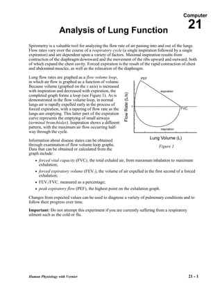
Lung flow rates are graphed as a flow volume loop,
in which air flow is graphed as a function of volume.
Because volume (graphed on the x axis) is increased
with inspiration and decreased with expiration, the
completed graph forms a loop (see Figure 1). As is
demonstrated in the flow volume loop, in normal
lungs air is rapidly expelled early in the process of
forced expiration, with a tapering of flow rate as the
lungs are emptying. This latter part of the expiration
curve represents the emptying of small airways
(terminal bronchioles). Inspiration shows a different
pattern, with the maximum air flow occurring half-
way through the cycle.

Information about disease states can be obtained
through examination of flow volume loop graphs.                             Figure 1
Data that can be obtained or calculated from the
graph include:
   • forced vital capacity (FVC), the total exhaled air, from maximum inhalation to maximum
     exhalation;
   • forced expiratory volume (FEV1), the volume of air expelled in the first second of a forced
     exhalation;
   • FEV1/FVC, measured as a percentage;
   • peak expiratory flow (PEF), the highest point on the exhalation graph.

Changes from expected values can be used to diagnose a variety of pulmonary conditions and to
follow their progress over time.
Important: Do not attempt this experiment if you are currently suffering from a respiratory
ailment such as the cold or flu.


Human Physiology with Vernier                                                                 21 - 1
Computer 21

OBJECTIVES
In this experiment, you will
   •     Obtain graphical representation of a flow volume loop.
   •     Find the forced expiratory volume at 1 s (FEV1) and the forced vital capacity (FVC).
   •     Calculate FEV1/FVC.
   •     Find the peak expiratory flow rate (PEF).
   •     Create flow volume loops for several clinical scenarios.


MATERIALS
       computer                                            disposable mouthpiece
       Vernier computer interface                          disposable bacterial filter
       Vernier Spirometer                                  nose clip
       Logger Pro


PROCEDURE
Select one person from your lab group to be the subject.

1. Connect the Spirometer to the Vernier computer interface. Open the file
   “21 Analyze Lung Function” from the Human Physiology with Vernier folder.
2. Attach the larger diameter side of a white bacterial filter to the side of the Spirometer head
   marked “Inlet.” Attach a disposable mouthpiece to the other end of the bacterial filter (see
   Figure 2).


                                               Figure 2
3. Hold the Spirometer in one or both hands. Brace your arm(s) against a solid surface, such as a
   table, and click      . Note: The Spirometer must be held straight up and down (see
   Figure 2) and not moved during data collection.
4. Collect exhalation and inhalation data.
   a. Put on the nose clip and put your mouth against the mouthpiece.
   b. Click         to begin data collection.
   c. Take the deepest breath possible and exhale forcefully as quickly as possible through the
      Spirometer mouthpiece. Important: Force ALL of the air out of your lungs. Your
      exhalation will trigger data collection. Without removing your mouth from the mouthpiece,
      inhale as deeply as possible.


21 - 2                                                                  Human Physiology with Vernier
Analysis of Lung Function

5. If your data does not approximate Figure 1, repeat Step 4. It is helpful to watch the screen
   while performing this test. Important: It is essential that maximum effort be expended when
   performing tests of air flow.
6. Find FEV1 by scrolling down the data table to 1.00 s. Scroll to the right to see the volume.
   Record this value to the nearest 0.01 L in the data table.

7. Click the Examine button, . Move the examine line to the rightmost point of the flow
   volume loop. This is the FVC. Record the volume value to the nearest 0.01 L in the data
   table. Close the Statistics box by clicking the X in the corner of the box.

8. Find FEV1/FVC, multiply by 100, and enter the calculated value in the data table.

9. Click the Statistics button, . Enter the maximum flow rate value to the nearest 0.01 L/s as
   peak expiratory flow rate (PEF) in the data table.

DATA
           FEV1                     FVC                    FEV1/FVC                    PEF
            (L)                      (L)                      (%)                      (L/s)


DATA ANALYSIS
1. Using the flow volume loops pictured below as guides, superimpose loops for the following
   scenarios. In your answer consider whether the problem affects inspiration, expiration, or
   both; whether it is likely to affect the first half of the curve, the second half of the curve, or
   both. (Hint: It may be helpful to begin by estimating the maximum expected total lung
   capacity for each situation and drawing this point on the graph first.)

    a. There is a narrowing of the trachea, causing blockage in this large airway.


Human Physiology with Vernier                                                                    21 - 3
Computer 21

b. A small grape is lodged in the right mainstem bronchus, completely blocking off the right
      lung.


   c. Emphysema reduces all flow rates, but has a greater effect on small airways, many of
      which are lost to the disease. (Acute asthma may show similar changes because of mucous
      obstruction of these same small airways.)


   d. Severe osteoporosis may result in kyphoscoliosis, in which there is a curvature of the
      upper spine that limits the normal ability of the ribs to expand with inspiration.


21 - 4                                                               Human Physiology with Vernier
Analysis of Lung Function

2. Lung disease is often divided into two broad categories: obstructive disease and restrictive
   disease. Examples of obstructive disease are emphysema, chronic bronchitis, and asthma.
   Examples of restrictive disease are abnormalities of the spine and chest and diseases within the
   lungs that make them less elastic (“stiffer”), such as pulmonary fibrosis. Calculate the
   FEV1/FVC in the table below for obstructive and restrictive disease and compare to your
   values. How might these values be helpful diagnostically?

        Volume (L)              Normal (your data)   Obstructive disease    Restrictive disease

         FVC (L)                                             4                      4

         FEV1 (L)                                           1.8                    3.5

      FEV1/FVC (%)


Human Physiology with Vernier                                                                21 - 5

More Related Content

What's hot

Lung volumes and_capacities
Lung volumes and_capacitiesLung volumes and_capacities
Lung volumes and_capacitiesnc479bn
 
Anat and phys
Anat and physAnat and phys
Anat and physks0477bn
 
Lung volumes and_capacities
Lung volumes and_capacitiesLung volumes and_capacities
Lung volumes and_capacities
ashleeeeyg
 
Lung volumes and_capacities
Lung volumes and_capacitiesLung volumes and_capacities
Lung volumes and_capacitiesXxDeadmanx698xX
 
Lung volumes-and-capacities
Lung volumes-and-capacitiesLung volumes-and-capacities
Lung volumes-and-capacitieskierra_johnson13
 
Lungs volumes and capacities
Lungs volumes and capacitiesLungs volumes and capacities
Lungs volumes and capacitieszs4033bn
 
Experiment 4 and 5
Experiment 4 and 5Experiment 4 and 5
Experiment 4 and 5hb0809
 
Experiment 5
Experiment 5Experiment 5
Experiment 5ab0046bn
 
Lung volume and capacities
Lung volume and capacitiesLung volume and capacities
Lung volume and capacitiesYogesh Ramasamy
 
Lung volumes
Lung volumesLung volumes
Lung volumes
Vamshi Keshavaram
 
Spirometry : To record static and dynamic lung volumes and capacities
Spirometry : To record static and dynamic lung volumes and capacitiesSpirometry : To record static and dynamic lung volumes and capacities
Spirometry : To record static and dynamic lung volumes and capacities
Dr.Ravi Dutt
 
Lung volumes in COPD
Lung volumes in COPDLung volumes in COPD
Lung volumes in COPD
Ashraf ElAdawy
 
Lung volumes & capacities and Dead Space
Lung volumes & capacities and Dead SpaceLung volumes & capacities and Dead Space
Lung volumes & capacities and Dead Space
vinai0710
 
Volumes capacities 2
Volumes  capacities 2Volumes  capacities 2
Volumes capacities 2Michael Wrock
 
Lungs volume and capacites
Lungs volume and capacites Lungs volume and capacites
Lungs volume and capacites
Dr. Prashant Kaushik
 
Pressure changes during Respiration
Pressure changes during RespirationPressure changes during Respiration
Pressure changes during Respiration
SRILATHA BASHETTI
 
20 respiratory response1
20 respiratory response120 respiratory response1
20 respiratory response1ag1430bn
 
3) measurement of lung function
3) measurement  of lung function3) measurement  of lung function
3) measurement of lung function
Ayub Abdi
 

What's hot (20)

Lung volumes and_capacities
Lung volumes and_capacitiesLung volumes and_capacities
Lung volumes and_capacities
 
Experiment 4
Experiment 4Experiment 4
Experiment 4
 
Anat and phys
Anat and physAnat and phys
Anat and phys
 
Lung volumes and_capacities
Lung volumes and_capacitiesLung volumes and_capacities
Lung volumes and_capacities
 
Lab 4
Lab 4Lab 4
Lab 4
 
Lung volumes and_capacities
Lung volumes and_capacitiesLung volumes and_capacities
Lung volumes and_capacities
 
Lung volumes-and-capacities
Lung volumes-and-capacitiesLung volumes-and-capacities
Lung volumes-and-capacities
 
Lungs volumes and capacities
Lungs volumes and capacitiesLungs volumes and capacities
Lungs volumes and capacities
 
Experiment 4 and 5
Experiment 4 and 5Experiment 4 and 5
Experiment 4 and 5
 
Experiment 5
Experiment 5Experiment 5
Experiment 5
 
Lung volume and capacities
Lung volume and capacitiesLung volume and capacities
Lung volume and capacities
 
Lung volumes
Lung volumesLung volumes
Lung volumes
 
Spirometry : To record static and dynamic lung volumes and capacities
Spirometry : To record static and dynamic lung volumes and capacitiesSpirometry : To record static and dynamic lung volumes and capacities
Spirometry : To record static and dynamic lung volumes and capacities
 
Lung volumes in COPD
Lung volumes in COPDLung volumes in COPD
Lung volumes in COPD
 
Lung volumes & capacities and Dead Space
Lung volumes & capacities and Dead SpaceLung volumes & capacities and Dead Space
Lung volumes & capacities and Dead Space
 
Volumes capacities 2
Volumes  capacities 2Volumes  capacities 2
Volumes capacities 2
 
Lungs volume and capacites
Lungs volume and capacites Lungs volume and capacites
Lungs volume and capacites
 
Pressure changes during Respiration
Pressure changes during RespirationPressure changes during Respiration
Pressure changes during Respiration
 
20 respiratory response1
20 respiratory response120 respiratory response1
20 respiratory response1
 
3) measurement of lung function
3) measurement  of lung function3) measurement  of lung function
3) measurement of lung function
 

Similar to Experiment 5

Interpretation of pulmonary function tests.a practical guide
Interpretation of pulmonary function tests.a practical guideInterpretation of pulmonary function tests.a practical guide
Interpretation of pulmonary function tests.a practical guide
SoM
 
Pulmonary Function Testing 2023.pdf
Pulmonary Function Testing 2023.pdfPulmonary Function Testing 2023.pdf
Pulmonary Function Testing 2023.pdf
aljamhori teaching hospital
 
Pulmonary Function Test.ppt
Pulmonary Function Test.pptPulmonary Function Test.ppt
Pulmonary Function Test.pptShama
 
PULMONARY FUNCTION TEST.pdf
PULMONARY FUNCTION TEST.pdfPULMONARY FUNCTION TEST.pdf
PULMONARY FUNCTION TEST.pdf
Shilpasree Saha
 
Discussion #11. What physical findings might be indicative of a .docx
Discussion #11. What physical findings might be indicative of a .docxDiscussion #11. What physical findings might be indicative of a .docx
Discussion #11. What physical findings might be indicative of a .docx
mecklenburgstrelitzh
 
Pulmonary Function Tests
Pulmonary Function TestsPulmonary Function Tests
Pulmonary Function Testsguest2379201
 
Ultrasonography evaluation during the weaning process
Ultrasonography evaluation during the  weaning processUltrasonography evaluation during the  weaning process
Ultrasonography evaluation during the weaning process
Fadel Omar
 
Anesthesia For Patients Requiring Advanced Ventilatory Support
Anesthesia For Patients Requiring Advanced Ventilatory SupportAnesthesia For Patients Requiring Advanced Ventilatory Support
Anesthesia For Patients Requiring Advanced Ventilatory Supportsxbenavides
 
dynamic spirometry............................
dynamic spirometry............................dynamic spirometry............................
dynamic spirometry............................
DrManjushaShinde
 
Exercise 7: Respiratory System Mechanics: Activity 1: Measuring Respiratory V...
Exercise 7: Respiratory System Mechanics: Activity 1: Measuring Respiratory V...Exercise 7: Respiratory System Mechanics: Activity 1: Measuring Respiratory V...
Exercise 7: Respiratory System Mechanics: Activity 1: Measuring Respiratory V...
ViscKand
 
Respiratory Muscle Assessment
Respiratory Muscle AssessmentRespiratory Muscle Assessment
Respiratory Muscle Assessment
Dr Riham Hazem Raafat
 
Pulmonary function testing
Pulmonary function testingPulmonary function testing
Pulmonary function testing
aljamhori teaching hospital
 
a detailed study on pulmonary function test
a detailed study on pulmonary function testa detailed study on pulmonary function test
a detailed study on pulmonary function test
martinshaji
 
My presentation
My presentationMy presentation
My presentation
Ghada Bashandy
 
Pulmonary function tests
Pulmonary function testsPulmonary function tests
Pulmonary function tests
richamistry3
 
Pulmonary Function Testing
Pulmonary Function TestingPulmonary Function Testing
Pulmonary Function Testing
Dr Riham Hazem Raafat
 
Noninvasive Tests for Asthma Diagnosis
Noninvasive Tests for Asthma DiagnosisNoninvasive Tests for Asthma Diagnosis
Noninvasive Tests for Asthma DiagnosisAli Taki
 
ASSESSMENT, EVALUATION AND TREATMENT OF RESPIRATORY CONDITIONS
ASSESSMENT, EVALUATION AND TREATMENT  OF RESPIRATORY CONDITIONSASSESSMENT, EVALUATION AND TREATMENT  OF RESPIRATORY CONDITIONS
ASSESSMENT, EVALUATION AND TREATMENT OF RESPIRATORY CONDITIONSPeace Samuel
 
Double lung ventilation
Double lung ventilation Double lung ventilation
Double lung ventilation
Rawan Herz
 
Resp Physio and PFTmade by me welcome to
Resp Physio and PFTmade by me welcome toResp Physio and PFTmade by me welcome to
Resp Physio and PFTmade by me welcome to
kaqib1234789
 

Similar to Experiment 5 (20)

Interpretation of pulmonary function tests.a practical guide
Interpretation of pulmonary function tests.a practical guideInterpretation of pulmonary function tests.a practical guide
Interpretation of pulmonary function tests.a practical guide
 
Pulmonary Function Testing 2023.pdf
Pulmonary Function Testing 2023.pdfPulmonary Function Testing 2023.pdf
Pulmonary Function Testing 2023.pdf
 
Pulmonary Function Test.ppt
Pulmonary Function Test.pptPulmonary Function Test.ppt
Pulmonary Function Test.ppt
 
PULMONARY FUNCTION TEST.pdf
PULMONARY FUNCTION TEST.pdfPULMONARY FUNCTION TEST.pdf
PULMONARY FUNCTION TEST.pdf
 
Discussion #11. What physical findings might be indicative of a .docx
Discussion #11. What physical findings might be indicative of a .docxDiscussion #11. What physical findings might be indicative of a .docx
Discussion #11. What physical findings might be indicative of a .docx
 
Pulmonary Function Tests
Pulmonary Function TestsPulmonary Function Tests
Pulmonary Function Tests
 
Ultrasonography evaluation during the weaning process
Ultrasonography evaluation during the  weaning processUltrasonography evaluation during the  weaning process
Ultrasonography evaluation during the weaning process
 
Anesthesia For Patients Requiring Advanced Ventilatory Support
Anesthesia For Patients Requiring Advanced Ventilatory SupportAnesthesia For Patients Requiring Advanced Ventilatory Support
Anesthesia For Patients Requiring Advanced Ventilatory Support
 
dynamic spirometry............................
dynamic spirometry............................dynamic spirometry............................
dynamic spirometry............................
 
Exercise 7: Respiratory System Mechanics: Activity 1: Measuring Respiratory V...
Exercise 7: Respiratory System Mechanics: Activity 1: Measuring Respiratory V...Exercise 7: Respiratory System Mechanics: Activity 1: Measuring Respiratory V...
Exercise 7: Respiratory System Mechanics: Activity 1: Measuring Respiratory V...
 
Respiratory Muscle Assessment
Respiratory Muscle AssessmentRespiratory Muscle Assessment
Respiratory Muscle Assessment
 
Pulmonary function testing
Pulmonary function testingPulmonary function testing
Pulmonary function testing
 
a detailed study on pulmonary function test
a detailed study on pulmonary function testa detailed study on pulmonary function test
a detailed study on pulmonary function test
 
My presentation
My presentationMy presentation
My presentation
 
Pulmonary function tests
Pulmonary function testsPulmonary function tests
Pulmonary function tests
 
Pulmonary Function Testing
Pulmonary Function TestingPulmonary Function Testing
Pulmonary Function Testing
 
Noninvasive Tests for Asthma Diagnosis
Noninvasive Tests for Asthma DiagnosisNoninvasive Tests for Asthma Diagnosis
Noninvasive Tests for Asthma Diagnosis
 
ASSESSMENT, EVALUATION AND TREATMENT OF RESPIRATORY CONDITIONS
ASSESSMENT, EVALUATION AND TREATMENT  OF RESPIRATORY CONDITIONSASSESSMENT, EVALUATION AND TREATMENT  OF RESPIRATORY CONDITIONS
ASSESSMENT, EVALUATION AND TREATMENT OF RESPIRATORY CONDITIONS
 
Double lung ventilation
Double lung ventilation Double lung ventilation
Double lung ventilation
 
Resp Physio and PFTmade by me welcome to
Resp Physio and PFTmade by me welcome toResp Physio and PFTmade by me welcome to
Resp Physio and PFTmade by me welcome to
 

Experiment 5

  • 1. Computer Analysis of Lung Function 21 Spirometry is a valuable tool for analyzing the flow rate of air passing into and out of the lungs. Flow rates vary over the course of a respiratory cycle (a single inspiration followed by a single expiration) and are dependent upon a variety of factors. Maximal inspiration results from contraction of the diaphragm downward and the movement of the ribs upward and outward, both of which expand the chest cavity. Forced expiration is the result of the rapid contraction of chest and abdominal muscles, as well as the relaxation of the diaphragm. Lung flow rates are graphed as a flow volume loop, in which air flow is graphed as a function of volume. Because volume (graphed on the x axis) is increased with inspiration and decreased with expiration, the completed graph forms a loop (see Figure 1). As is demonstrated in the flow volume loop, in normal lungs air is rapidly expelled early in the process of forced expiration, with a tapering of flow rate as the lungs are emptying. This latter part of the expiration curve represents the emptying of small airways (terminal bronchioles). Inspiration shows a different pattern, with the maximum air flow occurring half- way through the cycle. Information about disease states can be obtained through examination of flow volume loop graphs. Figure 1 Data that can be obtained or calculated from the graph include: • forced vital capacity (FVC), the total exhaled air, from maximum inhalation to maximum exhalation; • forced expiratory volume (FEV1), the volume of air expelled in the first second of a forced exhalation; • FEV1/FVC, measured as a percentage; • peak expiratory flow (PEF), the highest point on the exhalation graph. Changes from expected values can be used to diagnose a variety of pulmonary conditions and to follow their progress over time. Important: Do not attempt this experiment if you are currently suffering from a respiratory ailment such as the cold or flu. Human Physiology with Vernier 21 - 1
  • 2. Computer 21 OBJECTIVES In this experiment, you will • Obtain graphical representation of a flow volume loop. • Find the forced expiratory volume at 1 s (FEV1) and the forced vital capacity (FVC). • Calculate FEV1/FVC. • Find the peak expiratory flow rate (PEF). • Create flow volume loops for several clinical scenarios. MATERIALS computer disposable mouthpiece Vernier computer interface disposable bacterial filter Vernier Spirometer nose clip Logger Pro PROCEDURE Select one person from your lab group to be the subject. 1. Connect the Spirometer to the Vernier computer interface. Open the file “21 Analyze Lung Function” from the Human Physiology with Vernier folder. 2. Attach the larger diameter side of a white bacterial filter to the side of the Spirometer head marked “Inlet.” Attach a disposable mouthpiece to the other end of the bacterial filter (see Figure 2). Figure 2 3. Hold the Spirometer in one or both hands. Brace your arm(s) against a solid surface, such as a table, and click . Note: The Spirometer must be held straight up and down (see Figure 2) and not moved during data collection. 4. Collect exhalation and inhalation data. a. Put on the nose clip and put your mouth against the mouthpiece. b. Click to begin data collection. c. Take the deepest breath possible and exhale forcefully as quickly as possible through the Spirometer mouthpiece. Important: Force ALL of the air out of your lungs. Your exhalation will trigger data collection. Without removing your mouth from the mouthpiece, inhale as deeply as possible. 21 - 2 Human Physiology with Vernier
  • 3. Analysis of Lung Function 5. If your data does not approximate Figure 1, repeat Step 4. It is helpful to watch the screen while performing this test. Important: It is essential that maximum effort be expended when performing tests of air flow. 6. Find FEV1 by scrolling down the data table to 1.00 s. Scroll to the right to see the volume. Record this value to the nearest 0.01 L in the data table. 7. Click the Examine button, . Move the examine line to the rightmost point of the flow volume loop. This is the FVC. Record the volume value to the nearest 0.01 L in the data table. Close the Statistics box by clicking the X in the corner of the box. 8. Find FEV1/FVC, multiply by 100, and enter the calculated value in the data table. 9. Click the Statistics button, . Enter the maximum flow rate value to the nearest 0.01 L/s as peak expiratory flow rate (PEF) in the data table. DATA FEV1 FVC FEV1/FVC PEF (L) (L) (%) (L/s) DATA ANALYSIS 1. Using the flow volume loops pictured below as guides, superimpose loops for the following scenarios. In your answer consider whether the problem affects inspiration, expiration, or both; whether it is likely to affect the first half of the curve, the second half of the curve, or both. (Hint: It may be helpful to begin by estimating the maximum expected total lung capacity for each situation and drawing this point on the graph first.) a. There is a narrowing of the trachea, causing blockage in this large airway. Human Physiology with Vernier 21 - 3
  • 4. Computer 21 b. A small grape is lodged in the right mainstem bronchus, completely blocking off the right lung. c. Emphysema reduces all flow rates, but has a greater effect on small airways, many of which are lost to the disease. (Acute asthma may show similar changes because of mucous obstruction of these same small airways.) d. Severe osteoporosis may result in kyphoscoliosis, in which there is a curvature of the upper spine that limits the normal ability of the ribs to expand with inspiration. 21 - 4 Human Physiology with Vernier
  • 5. Analysis of Lung Function 2. Lung disease is often divided into two broad categories: obstructive disease and restrictive disease. Examples of obstructive disease are emphysema, chronic bronchitis, and asthma. Examples of restrictive disease are abnormalities of the spine and chest and diseases within the lungs that make them less elastic (“stiffer”), such as pulmonary fibrosis. Calculate the FEV1/FVC in the table below for obstructive and restrictive disease and compare to your values. How might these values be helpful diagnostically? Volume (L) Normal (your data) Obstructive disease Restrictive disease FVC (L) 4 4 FEV1 (L) 1.8 3.5 FEV1/FVC (%) Human Physiology with Vernier 21 - 5
  • 6. Analysis of Lung Function 2. Lung disease is often divided into two broad categories: obstructive disease and restrictive disease. Examples of obstructive disease are emphysema, chronic bronchitis, and asthma. Examples of restrictive disease are abnormalities of the spine and chest and diseases within the lungs that make them less elastic (“stiffer”), such as pulmonary fibrosis. Calculate the FEV1/FVC in the table below for obstructive and restrictive disease and compare to your values. How might these values be helpful diagnostically? Volume (L) Normal (your data) Obstructive disease Restrictive disease FVC (L) 4 4 FEV1 (L) 1.8 3.5 FEV1/FVC (%) Human Physiology with Vernier 21 - 5
  • 7. Analysis of Lung Function 2. Lung disease is often divided into two broad categories: obstructive disease and restrictive disease. Examples of obstructive disease are emphysema, chronic bronchitis, and asthma. Examples of restrictive disease are abnormalities of the spine and chest and diseases within the lungs that make them less elastic (“stiffer”), such as pulmonary fibrosis. Calculate the FEV1/FVC in the table below for obstructive and restrictive disease and compare to your values. How might these values be helpful diagnostically? Volume (L) Normal (your data) Obstructive disease Restrictive disease FVC (L) 4 4 FEV1 (L) 1.8 3.5 FEV1/FVC (%) Human Physiology with Vernier 21 - 5
  • 8. Analysis of Lung Function 2. Lung disease is often divided into two broad categories: obstructive disease and restrictive disease. Examples of obstructive disease are emphysema, chronic bronchitis, and asthma. Examples of restrictive disease are abnormalities of the spine and chest and diseases within the lungs that make them less elastic (“stiffer”), such as pulmonary fibrosis. Calculate the FEV1/FVC in the table below for obstructive and restrictive disease and compare to your values. How might these values be helpful diagnostically? Volume (L) Normal (your data) Obstructive disease Restrictive disease FVC (L) 4 4 FEV1 (L) 1.8 3.5 FEV1/FVC (%) Human Physiology with Vernier 21 - 5