Oesophageal cancer is the 14th most common malignancy in the UK. There are two major types - squamous cell carcinoma and adenocarcinoma. The main risk factors are smoking, alcohol consumption, and chronic reflux. Symptoms include dysphagia. Diagnosis involves endoscopy with biopsies. Treatment depends on staging and may include surgery, chemotherapy, radiotherapy, or palliative care. Prognosis is poor with a 5-year survival of around 16% but depends on stage, with early-stage disease having a better prognosis if treated.
This Presentation gives summarized overview of Gall Bladder Carcinoma especially the management as per latest National Comprehensive Cancer Network(NCCN) Guidelines version 2.2013
The stomach J-shaped. It has two surfaces (the anterior & posterior), two curvatures (the greater & lesser), two orifices (the cardia & pylorus). It has fundus, body and pyloric antrum.
Blood supply
The left gastric artery
Right gastric artery
Right gastro-epiploic artery
Left gastro-epiploic artery
Short gastric arteries
Stomach cancer begins when cancer cells form in the inner lining of your stomach. These cells can grow into a tumor. Also called gastric cancer, the disease usually grows slowly over many years.
It could be:
malignant or benign
primary or secondary
GALLBLADDER CANCER UNDERSTANDING THE DISEASE AND TREATMENT OPTIONS AVAILABLE....Lovina Kapoor
The gallbladder is a pear-shaped organ in the upper right side of the abdomen below the liver. Its prime function is to store and deliver bile (a fluid secreted by liver to digest fats).
TEST BANK for Operations Management, 14th Edition by William J. Stevenson, Ve...kevinkariuki227
TEST BANK for Operations Management, 14th Edition by William J. Stevenson, Verified Chapters 1 - 19, Complete Newest Version.pdf
TEST BANK for Operations Management, 14th Edition by William J. Stevenson, Verified Chapters 1 - 19, Complete Newest Version.pdf
MANAGEMENT OF ATRIOVENTRICULAR CONDUCTION BLOCK.pdfJim Jacob Roy
Cardiac conduction defects can occur due to various causes.
Atrioventricular conduction blocks ( AV blocks ) are classified into 3 types.
This document describes the acute management of AV block.
These simplified slides by Dr. Sidra Arshad present an overview of the non-respiratory functions of the respiratory tract.
Learning objectives:
1. Enlist the non-respiratory functions of the respiratory tract
2. Briefly explain how these functions are carried out
3. Discuss the significance of dead space
4. Differentiate between minute ventilation and alveolar ventilation
5. Describe the cough and sneeze reflexes
Study Resources:
1. Chapter 39, Guyton and Hall Textbook of Medical Physiology, 14th edition
2. Chapter 34, Ganong’s Review of Medical Physiology, 26th edition
3. Chapter 17, Human Physiology by Lauralee Sherwood, 9th edition
4. Non-respiratory functions of the lungs https://academic.oup.com/bjaed/article/13/3/98/278874
Title: Sense of Smell
Presenter: Dr. Faiza, Assistant Professor of Physiology
Qualifications:
MBBS (Best Graduate, AIMC Lahore)
FCPS Physiology
ICMT, CHPE, DHPE (STMU)
MPH (GC University, Faisalabad)
MBA (Virtual University of Pakistan)
Learning Objectives:
Describe the primary categories of smells and the concept of odor blindness.
Explain the structure and location of the olfactory membrane and mucosa, including the types and roles of cells involved in olfaction.
Describe the pathway and mechanisms of olfactory signal transmission from the olfactory receptors to the brain.
Illustrate the biochemical cascade triggered by odorant binding to olfactory receptors, including the role of G-proteins and second messengers in generating an action potential.
Identify different types of olfactory disorders such as anosmia, hyposmia, hyperosmia, and dysosmia, including their potential causes.
Key Topics:
Olfactory Genes:
3% of the human genome accounts for olfactory genes.
400 genes for odorant receptors.
Olfactory Membrane:
Located in the superior part of the nasal cavity.
Medially: Folds downward along the superior septum.
Laterally: Folds over the superior turbinate and upper surface of the middle turbinate.
Total surface area: 5-10 square centimeters.
Olfactory Mucosa:
Olfactory Cells: Bipolar nerve cells derived from the CNS (100 million), with 4-25 olfactory cilia per cell.
Sustentacular Cells: Produce mucus and maintain ionic and molecular environment.
Basal Cells: Replace worn-out olfactory cells with an average lifespan of 1-2 months.
Bowman’s Gland: Secretes mucus.
Stimulation of Olfactory Cells:
Odorant dissolves in mucus and attaches to receptors on olfactory cilia.
Involves a cascade effect through G-proteins and second messengers, leading to depolarization and action potential generation in the olfactory nerve.
Quality of a Good Odorant:
Small (3-20 Carbon atoms), volatile, water-soluble, and lipid-soluble.
Facilitated by odorant-binding proteins in mucus.
Membrane Potential and Action Potential:
Resting membrane potential: -55mV.
Action potential frequency in the olfactory nerve increases with odorant strength.
Adaptation Towards the Sense of Smell:
Rapid adaptation within the first second, with further slow adaptation.
Psychological adaptation greater than receptor adaptation, involving feedback inhibition from the central nervous system.
Primary Sensations of Smell:
Camphoraceous, Musky, Floral, Pepperminty, Ethereal, Pungent, Putrid.
Odor Detection Threshold:
Examples: Hydrogen sulfide (0.0005 ppm), Methyl-mercaptan (0.002 ppm).
Some toxic substances are odorless at lethal concentrations.
Characteristics of Smell:
Odor blindness for single substances due to lack of appropriate receptor protein.
Behavioral and emotional influences of smell.
Transmission of Olfactory Signals:
From olfactory cells to glomeruli in the olfactory bulb, involving lateral inhibition.
Primitive, less old, and new olfactory systems with different path
Ozempic: Preoperative Management of Patients on GLP-1 Receptor Agonists Saeid Safari
Preoperative Management of Patients on GLP-1 Receptor Agonists like Ozempic and Semiglutide
ASA GUIDELINE
NYSORA Guideline
2 Case Reports of Gastric Ultrasound
Explore natural remedies for syphilis treatment in Singapore. Discover alternative therapies, herbal remedies, and lifestyle changes that may complement conventional treatments. Learn about holistic approaches to managing syphilis symptoms and supporting overall health.
- Video recording of this lecture in English language: https://youtu.be/lK81BzxMqdo
- Video recording of this lecture in Arabic language: https://youtu.be/Ve4P0COk9OI
- Link to download the book free: https://nephrotube.blogspot.com/p/nephrotube-nephrology-books.html
- Link to NephroTube website: www.NephroTube.com
- Link to NephroTube social media accounts: https://nephrotube.blogspot.com/p/join-nephrotube-on-social-media.html
New Directions in Targeted Therapeutic Approaches for Older Adults With Mantl...i3 Health
i3 Health is pleased to make the speaker slides from this activity available for use as a non-accredited self-study or teaching resource.
This slide deck presented by Dr. Kami Maddocks, Professor-Clinical in the Division of Hematology and
Associate Division Director for Ambulatory Operations
The Ohio State University Comprehensive Cancer Center, will provide insight into new directions in targeted therapeutic approaches for older adults with mantle cell lymphoma.
STATEMENT OF NEED
Mantle cell lymphoma (MCL) is a rare, aggressive B-cell non-Hodgkin lymphoma (NHL) accounting for 5% to 7% of all lymphomas. Its prognosis ranges from indolent disease that does not require treatment for years to very aggressive disease, which is associated with poor survival (Silkenstedt et al, 2021). Typically, MCL is diagnosed at advanced stage and in older patients who cannot tolerate intensive therapy (NCCN, 2022). Although recent advances have slightly increased remission rates, recurrence and relapse remain very common, leading to a median overall survival between 3 and 6 years (LLS, 2021). Though there are several effective options, progress is still needed towards establishing an accepted frontline approach for MCL (Castellino et al, 2022). Treatment selection and management of MCL are complicated by the heterogeneity of prognosis, advanced age and comorbidities of patients, and lack of an established standard approach for treatment, making it vital that clinicians be familiar with the latest research and advances in this area. In this activity chaired by Michael Wang, MD, Professor in the Department of Lymphoma & Myeloma at MD Anderson Cancer Center, expert faculty will discuss prognostic factors informing treatment, the promising results of recent trials in new therapeutic approaches, and the implications of treatment resistance in therapeutic selection for MCL.
Target Audience
Hematology/oncology fellows, attending faculty, and other health care professionals involved in the treatment of patients with mantle cell lymphoma (MCL).
Learning Objectives
1.) Identify clinical and biological prognostic factors that can guide treatment decision making for older adults with MCL
2.) Evaluate emerging data on targeted therapeutic approaches for treatment-naive and relapsed/refractory MCL and their applicability to older adults
3.) Assess mechanisms of resistance to targeted therapies for MCL and their implications for treatment selection
Lung Cancer: Artificial Intelligence, Synergetics, Complex System Analysis, S...Oleg Kshivets
RESULTS: Overall life span (LS) was 2252.1±1742.5 days and cumulative 5-year survival (5YS) reached 73.2%, 10 years – 64.8%, 20 years – 42.5%. 513 LCP lived more than 5 years (LS=3124.6±1525.6 days), 148 LCP – more than 10 years (LS=5054.4±1504.1 days).199 LCP died because of LC (LS=562.7±374.5 days). 5YS of LCP after bi/lobectomies was significantly superior in comparison with LCP after pneumonectomies (78.1% vs.63.7%, P=0.00001 by log-rank test). AT significantly improved 5YS (66.3% vs. 34.8%) (P=0.00000 by log-rank test) only for LCP with N1-2. Cox modeling displayed that 5YS of LCP significantly depended on: phase transition (PT) early-invasive LC in terms of synergetics, PT N0—N12, cell ratio factors (ratio between cancer cells- CC and blood cells subpopulations), G1-3, histology, glucose, AT, blood cell circuit, prothrombin index, heparin tolerance, recalcification time (P=0.000-0.038). Neural networks, genetic algorithm selection and bootstrap simulation revealed relationships between 5YS and PT early-invasive LC (rank=1), PT N0—N12 (rank=2), thrombocytes/CC (3), erythrocytes/CC (4), eosinophils/CC (5), healthy cells/CC (6), lymphocytes/CC (7), segmented neutrophils/CC (8), stick neutrophils/CC (9), monocytes/CC (10); leucocytes/CC (11). Correct prediction of 5YS was 100% by neural networks computing (area under ROC curve=1.0; error=0.0).
CONCLUSIONS: 5YS of LCP after radical procedures significantly depended on: 1) PT early-invasive cancer; 2) PT N0--N12; 3) cell ratio factors; 4) blood cell circuit; 5) biochemical factors; 6) hemostasis system; 7) AT; 8) LC characteristics; 9) LC cell dynamics; 10) surgery type: lobectomy/pneumonectomy; 11) anthropometric data. Optimal diagnosis and treatment strategies for LC are: 1) screening and early detection of LC; 2) availability of experienced thoracic surgeons because of complexity of radical procedures; 3) aggressive en block surgery and adequate lymph node dissection for completeness; 4) precise prediction; 5) adjuvant chemoimmunoradiotherapy for LCP with unfavorable prognosis.
Flu Vaccine Alert in Bangalore Karnatakaaddon Scans
As flu season approaches, health officials in Bangalore, Karnataka, are urging residents to get their flu vaccinations. The seasonal flu, while common, can lead to severe health complications, particularly for vulnerable populations such as young children, the elderly, and those with underlying health conditions.
Dr. Vidisha Kumari, a leading epidemiologist in Bangalore, emphasizes the importance of getting vaccinated. "The flu vaccine is our best defense against the influenza virus. It not only protects individuals but also helps prevent the spread of the virus in our communities," he says.
This year, the flu season is expected to coincide with a potential increase in other respiratory illnesses. The Karnataka Health Department has launched an awareness campaign highlighting the significance of flu vaccinations. They have set up multiple vaccination centers across Bangalore, making it convenient for residents to receive their shots.
To encourage widespread vaccination, the government is also collaborating with local schools, workplaces, and community centers to facilitate vaccination drives. Special attention is being given to ensuring that the vaccine is accessible to all, including marginalized communities who may have limited access to healthcare.
Residents are reminded that the flu vaccine is safe and effective. Common side effects are mild and may include soreness at the injection site, mild fever, or muscle aches. These side effects are generally short-lived and far less severe than the flu itself.
Healthcare providers are also stressing the importance of continuing COVID-19 precautions. Wearing masks, practicing good hand hygiene, and maintaining social distancing are still crucial, especially in crowded places.
Protect yourself and your loved ones by getting vaccinated. Together, we can help keep Bangalore healthy and safe this flu season. For more information on vaccination centers and schedules, residents can visit the Karnataka Health Department’s official website or follow their social media pages.
Stay informed, stay safe, and get your flu shot today!
Title: Sense of Taste
Presenter: Dr. Faiza, Assistant Professor of Physiology
Qualifications:
MBBS (Best Graduate, AIMC Lahore)
FCPS Physiology
ICMT, CHPE, DHPE (STMU)
MPH (GC University, Faisalabad)
MBA (Virtual University of Pakistan)
Learning Objectives:
Describe the structure and function of taste buds.
Describe the relationship between the taste threshold and taste index of common substances.
Explain the chemical basis and signal transduction of taste perception for each type of primary taste sensation.
Recognize different abnormalities of taste perception and their causes.
Key Topics:
Significance of Taste Sensation:
Differentiation between pleasant and harmful food
Influence on behavior
Selection of food based on metabolic needs
Receptors of Taste:
Taste buds on the tongue
Influence of sense of smell, texture of food, and pain stimulation (e.g., by pepper)
Primary and Secondary Taste Sensations:
Primary taste sensations: Sweet, Sour, Salty, Bitter, Umami
Chemical basis and signal transduction mechanisms for each taste
Taste Threshold and Index:
Taste threshold values for Sweet (sucrose), Salty (NaCl), Sour (HCl), and Bitter (Quinine)
Taste index relationship: Inversely proportional to taste threshold
Taste Blindness:
Inability to taste certain substances, particularly thiourea compounds
Example: Phenylthiocarbamide
Structure and Function of Taste Buds:
Composition: Epithelial cells, Sustentacular/Supporting cells, Taste cells, Basal cells
Features: Taste pores, Taste hairs/microvilli, and Taste nerve fibers
Location of Taste Buds:
Found in papillae of the tongue (Fungiform, Circumvallate, Foliate)
Also present on the palate, tonsillar pillars, epiglottis, and proximal esophagus
Mechanism of Taste Stimulation:
Interaction of taste substances with receptors on microvilli
Signal transduction pathways for Umami, Sweet, Bitter, Sour, and Salty tastes
Taste Sensitivity and Adaptation:
Decrease in sensitivity with age
Rapid adaptation of taste sensation
Role of Saliva in Taste:
Dissolution of tastants to reach receptors
Washing away the stimulus
Taste Preferences and Aversions:
Mechanisms behind taste preference and aversion
Influence of receptors and neural pathways
Impact of Sensory Nerve Damage:
Degeneration of taste buds if the sensory nerve fiber is cut
Abnormalities of Taste Detection:
Conditions: Ageusia, Hypogeusia, Dysgeusia (parageusia)
Causes: Nerve damage, neurological disorders, infections, poor oral hygiene, adverse drug effects, deficiencies, aging, tobacco use, altered neurotransmitter levels
Neurotransmitters and Taste Threshold:
Effects of serotonin (5-HT) and norepinephrine (NE) on taste sensitivity
Supertasters:
25% of the population with heightened sensitivity to taste, especially bitterness
Increased number of fungiform papillae
micro teaching on communication m.sc nursing.pdfAnurag Sharma
Microteaching is a unique model of practice teaching. It is a viable instrument for the. desired change in the teaching behavior or the behavior potential which, in specified types of real. classroom situations, tends to facilitate the achievement of specified types of objectives.
The POPPY STUDY (Preconception to post-partum cardiovascular function in prim...
Esophageal Carcinoma
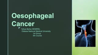
1. z Yahya Berke DEMIREL
Odessa National Medical University
1st Group
5th Course
2. z
Oesophageal cancer is the 14th most common malignancy in adults in the UK.
The oesophagus is a muscular tube that is situated within the thorax and runs from the
pharynx to stomach. It is pivotal in the transfer of food material to the stomach and
broadly divided into upper, middle and lower.
There are two major types of cancer that arise from the oesophagus, depending on the
cell of origin.
• Squamous cell carcinoma (SCC): usually located in the upper or middle oesophagus.
Accounts for >90% of cases worldwide.
• Adenocarcinoma (AC): usually located in the lower oesophagus. Due to chronic reflux
and development of a columnar metaplasia, which is a precursor lesion known
as Barrett’s oesophagus.
Rarer forms of oesophageal cancers include small cell carcinoma, sarcoma, lymphoma,
melanoma and choriocarcinoma.
The hallmark clinical feature of oesophageal cancer is dysphagia, which refers to
difficulty swallowing. This is due to obstruction of the oesophageal lumen.
3. z
Epidemiology
The incidence of oesophageal adenocarcinoma, particularly at
the gastro-oesophageal junction, has increased dramatically.
Oesophageal cancer is rare in young people and more common
as we age. The peak incidence is seen in the 7-8th decades.
Globally, SCC is the most common cause of oesophageal
cancer, but the incidence of AC is increasing, particularly in
Western countries. This is due to an increase in AC risk factors
such as obesity.
AC is more commonly seen in men, whereas the male-to-female
ratio is more equal in SCC.
4. z
Etiology
Smoking and alcohol consumption are the two major risk factors
for the development of SCC.
Oesophageal neoplasia develops due to sequential mutations
that occur in the oesophageal epithelium, which allows cells to
proliferate uncontrollably. Several risk factors for both SCC and
AC increase the likelihood of developing these mutations.
5. z
Squamous cell carcinoma
The biggest risk factors for development of SCC include alcohol
consumption and smoking.
• Smoking
• Alcohol consumption
• Foods containing N-nitroso compounds
• Chewing of areca nuts
• Previous partial gastrectomy
• Atrophic gastritis
• Human papillomavirus (HPV): mainly genotypes 16 and 18
• Tylosis: rare condition leading to hyperkeratosis of hands and feet
Squamous cell carcinoma of the esophagus
6. z
Adenocarcinoma
The majority of AC cases arise from Barrett’s oesophagus, which refers to
columnar metaplasia of the lower oesophagus due to chronic reflux. This a
pre-malignant lesion.
• Chronic reflux
• Barrett’s oesophagus: 30-fold increase risk of AC
• Smoking
• Obesity
• Zollinger-Ellison syndrome: gastrin-secreting tumour leading to excess
hydrochloric acid.
7. z
T1a adenocarcinoma of esophagus with
further subdivision according to the depth pf
invasion.
8. z
Pathophysiolog
y
The majority of oesophageal SCCs arise from the mid-
oesophagus and ACs from near the gastro-oesophageal junction
(GOJ).
Squamous cell carcinoma
The majority of SCCs occur due to chronic alcohol consumption and
smoking, which damage cellular DNA. This leads to development of
genetic mutations that promote abnormal cell growth. Overtime, the
cells proliferate uncontrollably leading to invasive cancer.
SCC may be seen as an infiltrating and ulcerated mass in the middle
oesophagus, especially if advanced. There is early invasion into
surrounding lymph nodes and the tumour may metastasize to liver,
bone and lung.
9. z
Adenocarcinoma
Typically, chronic reflux leads to inflammation and damage of the lower
oesophageal mucosa. This results in columnar metaplasia, which refers
to the transformation of the mature squamous cell type to columnar cell
type.
This metaplastic epithelium may become dysplastic, which refers to
cells with abnormal growth and development. The dysplastic epithelium
acquires further genetic mutation that promotes development of
invasive carcinoma. AC is most commonly located near the GOJ and
there is usually early lymph node involvement.
10. z
The hallmark feature of oesophageal cancer is dysphagia, which refers to difficulty
swallowing.
Symptoms
• Constitutional symptoms: fevers, anorexia, lethargy, weight loss
• Dysphagia: difficulty swallowing
• Weight loss: due to tumour-related anorexia and poor nutrition from swallowing difficulties
• Bleeding: haematemesis and melaena
• Pain: typically retrosternal pain
• Aspiration: cough, shortness of breath, fever
• Hoarseness: if there is extension to involve the recurrent laryngeal nerve
Clinical features
11. z
Signs
• Lymphadenopathy: if local tumour spread
• Cachexia
• Pallor: due to anaemia
• Hepatomegaly: if metastatic spread
12. z
Suspected cancer
referral
The NICE (NG12) guideline outlines recommendations for the referral of suspected
cancer cases including upper gastrointestinal cancers. Below details the
recommended referral for suspected oesophageal cancer.
Urgent (two week wait) referral
This means referring a patient for appropriate investigations (e.g. gastroscopy) for
suspected oesophageal cancer within two weeks. It is usually combined with a clinic
appointment and CT imaging.
• Dysphagia, OR
• > 55 years with weight loss and one of the following:
• Upper abdominal pain
• Reflux
• Dyspepsia
13. z
Non-urgent referral
This means referral for a non-urgent gastroscopy to assess for oesophageal
pathology. Usually performed within 6 weeks.
• Haematemesis, OR
• > 55 years with treatment resistant dyspepsia, OR
• > 55 years with upper abdominal pain and anaemia, OR
• Thrombocytosis with one of the following:
• Nausea/vomiting
• Weight loss
• Reflux
• Dyspepsia
• Upper abdominal pain
14. z
• Nausea/vomiting with one of the following:
• Weight loss
• Reflux
• Dyspepsia
• Upper abdominal pain
NOTE: Upper and lower gastrointestinal (GI) investigations should
also be considered to investigate for GI malignancy (inc.
oesophageal cancer) in all postmenopausal female and male
patients where IDA has been confirmed unless there is a history of
significant overt non-GI blood loss. According to British Society of
Gastroenterology guidelines 2011.
15. z
Diagnosi
s
Oesophageal cancer is diagnosed using upper GI endoscopy
and biopsies of suspected lesions.
The principle test for the diagnosis of oesophageal cancer is an
upper GI endoscopy known as a gastroscopy. This a camera
test that allows direct visualisation of the upper gastrointestinal
tract including oesophagus and gastro-oesophageal junction.
17. z
Investigations
Further investigations allow assessment of distant spread and key-organ
function to help guide management.
Bloods
• Full blood count
• Serum iron, transferrin saturation, total iron binding capacity (TIBC)
• Urea & electrolytes
• Liver function tests
• Bone profile
• Clotting screen
• Renal function
18. z
Imaging
• CT chest/abdomen/pelvis: patients with suspected oesophageal
cancer undergo CT imaging to help stage the cancer. See
staging below.
• Abdominal ultrasound: may be used to assess for liver
metastasis. Usually superseded by CT.
• PET-CT: offered to patients with potentially resectable disease
(i.e. candidates for surgery) to assess for distant disease not
detected by conventional CT.
20. z
Special
• Gastroscopy: principle investigation for diagnosis.
• Endoscopic ultrasound (EUS): can be performed at time of endoscopy.
Sometimes completed to help more accurately stage oesophageal cancer
if it will change management.
• Diagnostic laparoscopy: may be used to more accurately stage
oesophageal cancer if it will alter management.
HER2 testing
Human epidermal growth factor receptor 2 (HER2) testing should be
completed on tumour or biopsy specimens. Targeted therapy against the
HER2 receptor may be offered to patients with HER2 positive metastatic
oesophageal cancer.
22. z
Staging
Tumour
• TX: Primary tumour cannot be assessed
• T0: No evidence of primary tumour
• Tis: Carcinoma in situ/high-grade dysplasia
• T1: Tumour invades lamina propria or submucosa
• T1a: Tumour invades mucosa or lamina propria or muscularis mucosae
• T1b: Tumour invades submucosa
• T2: Tumour invades muscularis propria
• T3: Tumour invades adventitia
• T4: Tumour invades adjacent structures
• T4a: Tumour invades pleura, pericardium, diaphragm or adjacent peritoneum
• T4b: Tumour invades other adjacent structures such as aorta, vertebral body or trachea
23. z
Node
• NX: Regional lymph nodes cannot be assessed
• N0: No regional lymph node metastasis
• N1: Metastasis in 1–2 regional lymph nodes
• N2: Metastasis in 3–6 regional lymph nodes
• N3: Metastasis in 7 or more regional lymph nodes
Metastasis
• MX: Distant metastasis cannot be assessed
• M0: No distant metastasis
• M1: Distant metastasis
NOTE: Non-regional lymph node spread is considered M1a disease.
24. z
Management
principles
Treatment options
There are numerous treatment options for the management of oesophageal cancer:
• Surgery: resection of oesophageal or gastro-oesophageal tumours (e.g. oesophagectomy).
• Endoscopic techniques: mucosal resection or mucosal dissection.
• Radiotherapy: use of high energy rays to destroy cancer cells.
• Chemotherapy: use of anti-cancer medications to destroy cancer cells.
• Targeted cancer drugs: monoclonal antibodies against certain receptors (e.g. HER2).
• Palliative care: use of chemotherapy/radiotherapy or stenting to control disease
and/or symptoms without aiming to cure.
• Best supportive care: focus primarily on symptoms and quality of life without systemic
treatments.
25. z
Determining choice
The choice of treatment depends on whether the cancer is limited,
locally advanced or advanced/metastatic.
• Limited: refers to small tumours without lymph node involvement or
distant spread
• Locally advanced: refers to larger tumours with/without lymph node
involvement but without distant spread
• Advanced/metastatic: refers to metastatic disease with spread to
distant sites
26. z
Limited and locally
advanced
The treatment of choice for limited disease is surgical or
endoscopic resection.
Surgical resection, radiotherapy, chemotherapy, or a
combination, may be used for patients with limited or locally
advanced disease.
• Limited disease (Staging: T1-2, N0, M0)
• Locally advanced disease (Staging: T3-4 or N1-2, M0)
28. z
Surgery
Surgical resection of oesophageal cancer is the treatment of choice if the patient is fit to
undergo an operation and the disease is limited. Surgery may be an option for patients
with locally advanced oesophageal cancer. This can be considered following systemic anti-
cancer therapy (e.g. chemoradiotherapy). The idea is to shrink the tumour, which then
becomes resectable.
The surgical technique of choice is an oesophagectomy, which refers to removal of part of
the oesophagus and subsequent joining of the remaining oesophagus to the stomach. If
the tumour involves the GOJ or proximal stomach then an oesophago-gastrectomy or
extended total gastrectomy may be undertaken. In addition to surgical removal of the
oesophagus, lymph node dissection may be considered at the time of surgery.
Surgical resection is an option for:
• Limited disease
• Locally advanced disease (deemed resectable following neoadjuvant chemoradiotherapy)
29. z
Endoscopic therapy
Patients with limited disease may be suitable for endoscopic
therapy to remove the oesophageal cancer.
Two options include:
• Endoscopic mucosal resection (EMR)
• Endoscopic submucosal dissection (ESD)
These are typically performed in specialist upper gastrointestinal
centres any may require preoperative assessment with endoscopic
ultrasound (EUS) to ensure no localised lymph node spread.
30. z
Systemic anti-cancer therapy
The two main systemic anti-cancer therapies used in both SCC and AC
are chemotherapy and radiotherapy.
• Squamous cell carcinoma: a combination of chemotherapy (e.g.
cisplatin/5-fluorouracil) and radiotherapy may be given as neoadjuvant
therapy to shrink the tumour prior to resection or as definite treatment.
SCC is highly chemo/radio sensitive, which is why it can be used as
radical therapy. Patients undergoing radical chemoradiotheray are
followed-up regularly to assess whether a salvage resection should be
undertaken.
• Adenocarcinoma: either chemotherapy (e.g. cisplatin/5-fluorouracil) alone
or in combination with radiotherapy should be given to patients as
neoadjuvant therapy. If there is a response to treatment and the tumour
shrinks becoming resectable on restaging then surgical intervention should
be considered. Radial chemoradiotherapy (i.e. main treatment to cure
cancer) is not an option in AC.
31. z
Palliative management
Palliative treatment should be considered in patients with locally advanced
disease who are not operative candidates or not fit enough to undergo
radical treatment (e.g. poor performance status, extensive co-morbidities).
Options include:
• Radiotherapy: if tumour lies within a radiotherapy field that allows high-
doses to be applied.
• Chemotherapy: regimens depend on fitness of the patient.
• Local tumour treatment: endoscopic stenting, palliative radiotherapy.
• Best supportive care: focusing on symptom control only.
NOTE: oesophageal stenting involves endoscopically placing a stent within
the oesophagus to keep the lumen open and prevent dysphagia.
32. z
Nutritional support
Patients being considered for radical treatment (e.g. surgical
resection or chemoradiotherapy) need to have their nutrition
optimised. This may mean temporary enteral or parenteral
nutrition.
In patients undergoing chemoradiotherapy, they should be
considered for enteral nutrition with placement of a radiologically
inserted gastrostomy (RIG) tube. This is because
chemoradiotherapy will damage the oesophagus leading to
localised inflammation during treatment that will impair nutritional
intake. A percutaneous endoscopic gastrostomy (PEG) is not
suitable in these cases due to the risk of tumour migration from
endoscopic pull through of the tube.
33. z
Prognosis
The five year survival of oesophageal cancer is poor at ~16%
Survival from oesophageal cancer depends on stage. The five
year survival for stage 1 disease is 52.8% but only 16% for
stage 3 disease. Early diagnosis and treatment is potentially
curative, especially if the disease is resectable. However,
oesophagectomy is a major operation with significant morbidity
and mortality.