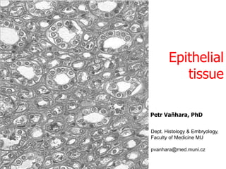
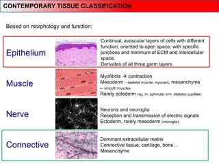
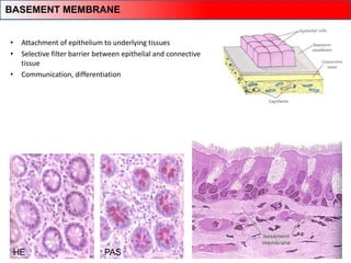
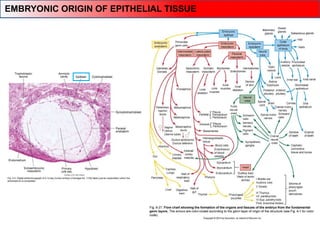
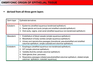
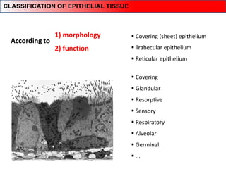
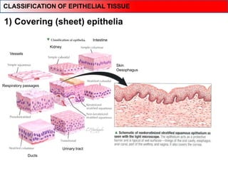
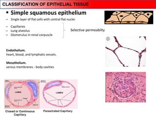
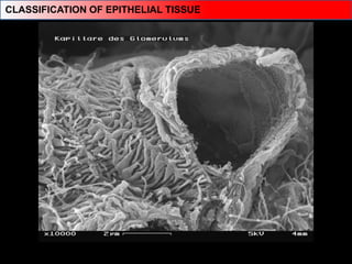
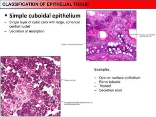
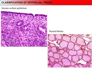
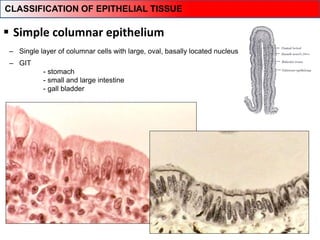
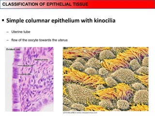
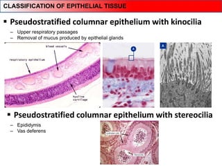
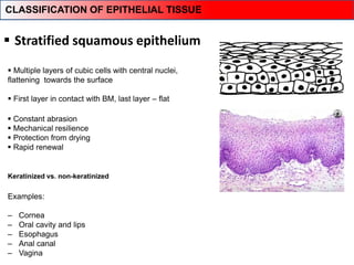
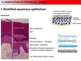
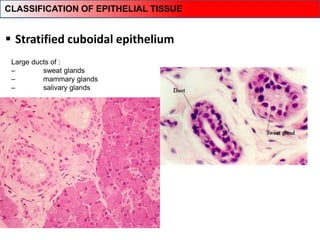
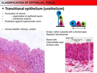
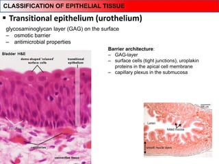
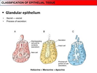
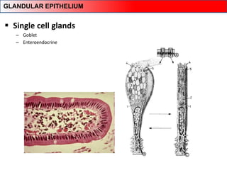
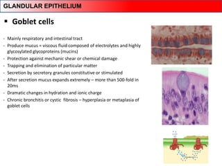
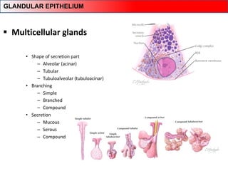
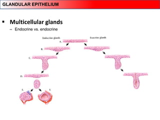
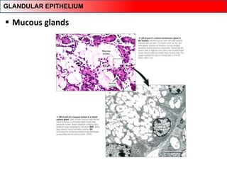
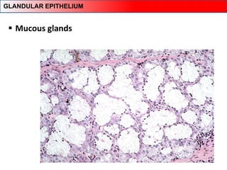
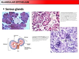
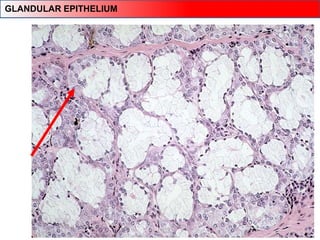
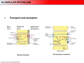
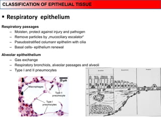
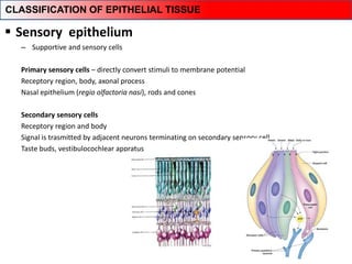
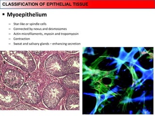
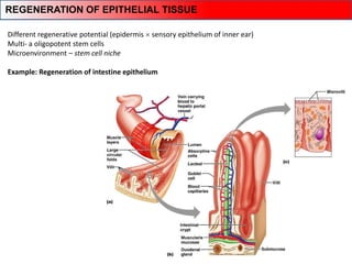
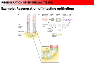
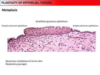
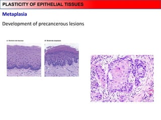
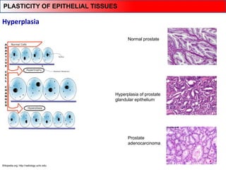
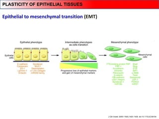
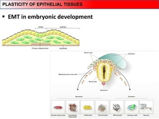
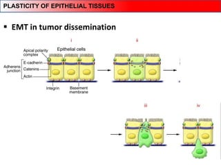
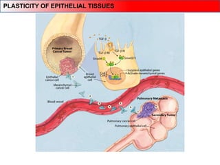
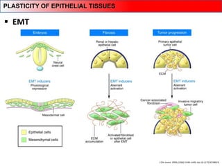
Epithelial
tissue
Dept. Histology & Embryology,
Faculty of Medicine MU
pvanhara@med.muni.cz
Petr Vaňhara, PhD
Epithelium
Muscle
Nerve
Connective
Based on morphology and function:
Myofibrils  contraction
Mesoderm – skeletal muscle, myocard, mesenchyme
– smooth muscles
Rarely ectoderm (eg. m. sphincter a m. dilatator pupillae)
Neurons and neuroglia
Reception and transmission of electric signals
Ectoderm, rarely mesoderm (microglia)
Dominant extracellular matrix
Connective tissue, cartilage, bone…
Mesenchyme
Continual, avascular layers of cells with different
function, oriented to open space, with specific
junctions and minimum of ECM and intercellular
space.
Derivates of all three germ layers
CONTEMPORARY TISSUE CLASSIFICATION
General characteristics
EPITHELIAL TISSUE
‒ Very early event and very novel innovation in Metazoa evolution
‒ From simple colonies of cells to highly specialized tissue structures
‒ Boundaries and interfaces
‒ Dividing of the body into separated compartments → separating individual milieu
‒ Lining of cavities or interfaces of open space
‒ Attachment and adhesion
‒ Basal membrane
What can sea sponges and hydras teach us?
General characteristics – lessons from primitive metazoans
Evolution of epithelia
Complexity of cell-cell junctions
EPITHELIAL VARIABILITY IN HUMANS
• Typical morphology and cell connections
• Avascular (without blood supply) – nutrition by diffusion from a highly vascular and innervated area
of loose connective tissue (lamina propria) just below the basement membrane
• Highly cellular – cohesive sheet or groups of cells with no or little extracellular matrix
GENERAL CHARACTERISTICS OF EPITHELIAL TISSUE
www.webanatomy.net
HALLMARKS OF A TYPICAL EPITHELIAL CELL
• Attachment of epithelium to underlying tissues
• Selective filter barrier between epithelial and connective
tissue
• Communication, differentiation
PAS
HE
BASEMENT MEMBRANE
 50-100nm
 Glycosaminoglycans – heparansulfate
 Laminin, collagen III, IV, VI,
 Nidogen/entactin
 Perlecan
 Proteoglycans
BASAL LAMINA vs. BASEMENT MEMBRANE
Dunsmore SE, Chambers RC, Laurent GJ. 2003. Matrix Proteins. Figure 2.1.2. In: Respiratory Medicine, 3rd ed. London.
Saunders, p. 83; Dunsmore SE, Laurent GJ. 2007. Lung Connective Tissue. Figure 40.1. In: Chronic Obstructive
Pulmonary Disease: A Practical Guide to Management, 1st ed. Oxford. Wiley-Blackwell, p. 467.
 Basement membrane
Kolagen IV
Laminin
Perlecan,
Nidogen/Entactin
BASEMENT MEMBRANE
Lamina
basalis
Lamina
fibroreticularis
Kolagenní
vazivo
BM
ARCHITECTURE OF BASEMENT MEMBRANE
• Two basic layers
– lamina basalis
• lamina densa,
• lamina rara ext. et int.
– lamina fibroreticularis
• Tissue specific modifications
‒ Descemet membrane (cornea)
‒ Glomerular BM (Bowman’s capsule)
‒ Part of Bruch’s membran of retina
…
MODIFICATION OF BASEMENT MEMBRANE
BASEMENT MEMBRANE IN CORPUSCULUM RENIS
• Clinical correlations – Membranous glomerulonefritis
- circulationg Abs bind to BM of capillary wall
- complement (C5b-C9) attacks glomerular endothelial cells
- filtation barrier compromised
- proteinuria, edema, hematouria, renal failure
BASEMENT MEMBRANE IN CORPUSCULUM RENIS
EMBRYONIC ORIGIN OF EPITHELIAL TISSUE
Germ layer Epithelial derivatives
Ectoderm 1. Epidermis (stratified squamous keratinized epithelium)
2. Sweat glands and ducts (simple and stratified cuboidal epithelium)
3. Oral cavity, vagina, anal canal (stratified squamous non-keratinized epithelium)
Mesoderm 1. Endothelium of blood vessels (simple squamous epithelium)
2. Mesothelium of body cavities (simple squamous epithelium)
3. Urinary and reproductive passages (transitional, pseudostratified and stratified
columnar epithelium, simple cuboidal and columnar epithelium)
Endoderm 1. Esophagus (stratified squamous non-keratinized epithelium)
2. GIT (simple columnar epithelium)
3. Výstelka žlučníku (simple columnar epithelium)
4. Solid glands (liver, pancreas)
5. Respiratory passages (ciliated pseudostratified columnar epithelium, ciliated simple
columnar epithelium, cuboidal, squamous)
 derived from all three germ layers
EMBRYONIC ORIGIN OF EPITHELIAL TISSUE
 Covering (sheet) epithelium
 Trabecular epithelium
 Reticular epithelium
1) morphology
2) function
 Covering
 Glandular
 Resorptive
 Sensory
 Respiratory
 Alveolar
 Germinal
 ...
CLASSIFICATION OF EPITHELIAL TISSUE
According to
Morphology
CLASSIFICATION OF EPITHELIAL TISSUE
Vessels
Kidney
Intestine
Respiratory passages
Skin
Oesophagus
Ducts
Urinary tract
1) Covering (sheet) epithelia
CLASSIFICATION OF EPITHELIAL TISSUE
Endothelium.
heart, blood, and lymphatic vessels.
Mesothelium.
serous membranes - body cavities
 Simple squamous epithelium
‒ Capillaries
‒ Lung alveolus
‒ Glomerulus in renal corpuscle
Selective permeabilty
‒ Single layer of flat cells with central flat nuclei
CLASSIFICATION OF EPITHELIAL TISSUE
 Simple cuboidal epithelium
‒ Single layer of cubic cells with large, spherical
central nuclei
Examples:
‒ Ovarian surface epithelium
‒ Renal tubules
‒ Thyroid
‒ Secretion acini
‒ Secretion or resorption
CLASSIFICATION OF EPITHELIAL TISSUE
Ovarian surface epithelium
Thyroid follicles
CLASSIFICATION OF EPITHELIAL TISSUE
 Simple columnar epithelium
‒ GIT
- stomach
- small and large intestine
- gall bladder
Resorption / Secretion
‒ Single layer of columnar cells with large, oval, basally located nucleus
CLASSIFICATION OF EPITHELIAL TISSUE
 Simple columnar epithelium with kinocilia
‒ Uterine tube
‒ flow of the oocyte towards the uterus
CLASSIFICATION OF EPITHELIAL TISSUE
 Pseudostratified columnar epithelium with kinocilia
‒ Upper respiratory passages
‒ Removal of mucus produced by epithelial glands
‒ Epididymis
‒ Vas deferens
CLASSIFICATION OF EPITHELIAL TISSUE
 Pseudostratified columnar epithelium with stereocilia
 Stratified squamous epithelium
Keratinized vs. non-keratinized
 Constant abrasion
 Mechanical resilience
 Protection from drying
 Rapid renewal
Examples:
‒ Cornea
‒ Oral cavity and lips
‒ Esophagus
‒ Anal canal
‒ Vagina
 Multiple layers of cubic cells with central nuclei,
flattening towards the surface
 First layer in contact with BM, last layer – flat
CLASSIFICATION OF EPITHELIAL TISSUE
 Stratified squamous epithelium
Keratinized
Skin (epidermis)
Nail
Keratins
Fibrous proteins, ~ 40 types
Very stable, multimeric
Disorders of keratin expression
– variety of clinical symptoms
e.g. Epidermolysis bullosa simplex
CLASSIFICATION OF EPITHELIAL TISSUE
 Stratified cuboidal epithelium
Large ducts of :
‒ sweat glands
‒ mammary glands
‒ salivary glands
CLASSIFICATION OF EPITHELIAL TISSUE
‒ Fluctuation of volume
‒ - organization of epithelial layers
‒ - membrane reserve
‒ Protection against hyperosmotic urine
‒ Urinary bladder, kidneys, ureters
 Transitional epithelium (urothelium)
Empty: rather cuboidal with a domed apex
Relaxed: flat,stretched
Basal cells
Intermediate layer
Surface cells
CLASSIFICATION OF EPITHELIAL TISSUE
 Transitional epithelium (urothelium)
glycosaminoglycan layer (GAG) on the surface
‒ osmotic barrier
‒ antimicrobial properties
Barrier architecture:
‒ GAG-layer
‒ surface cells (tight junctions), uroplakin
proteins in the apical cell membrane
‒ capillary plexus in the submucosa
CLASSIFICATION OF EPITHELIAL TISSUE
©http://www.cytochemistry.net/microanatomy/epithelia/salivary7.jpg
 Stratified columnar epithelia
‒ several layers of columnar cells
‒ secretion / protection
‒ ocular conjunctiva
‒ pharynx, anus – transitions
‒ uterus, male urethra, vas deferens
‒ intralobular ducts of salivary glands
CLASSIFICATION OF EPITHELIAL TISSUE
2) Trabecular epithelium
Liver parenchyma
CLASSIFICATION OF EPITHELIAL TISSUE
• Cords of hepatocytes
Trabecular epithelium
CLASSIFICATION OF EPITHELIAL TISSUE
Islets of Langerhans
Cords of endocrine active cells
 Endocrine glands
CLASSIFICATION OF EPITHELIAL TISSUE
Adrenal cortex
Cortex of adrenal gland – epithelial cells in cords secreting corticoid
 Endocrine glands
CLASSIFICATION OF EPITHELIAL TISSUE
Adenohypophysis – anterior pituitary
 Endocrine glands
CLASSIFICATION OF EPITHELIAL TISSUE
Reticular epithelium
Thymus
CLASSIFICATION OF EPITHELIAL TISSUE
Function
CLASSIFICATION OF EPITHELIAL TISSUE
 Glandular epithelium
• Secret ↔ excret
• Process of secretion:
Holocrine  Merocrine  Apocrine
CLASSIFICATION OF EPITHELIAL TISSUE
 Single cell glands
– Goblet
– Enteroendocrine
GLANDULAR EPITHELIUM
 Goblet cells
- Mainly respiratory and intestinal tract
- Produce mucus = viscous fluid composed of electrolytes and highly
glycosylated glycoproteins (mucins)
- Protection against mechanic shear or chemical damage
- Trapping and elimination of particular matter
- Secretion by secretory granules constitutive or stimulated
- After secretion mucus expands extremely – more than 500-fold in
20ms
- Dramatic changes in hydration and ionic charge
- Chronic bronchitis or cystic fibrosis – hyperplasia or metaplasia of
goblet cells
GLANDULAR EPITHELIUM
GOBLET CELL
 Multicellular glands
• Shape of secretion part
– Alveolar (acinar)
– Tubular
– Tubuloalveolar (tubuloacinar)
• Branching
– Simple
– Branched
– Compound
• Secretion
– Mucous
– Serous
– Compound
GLANDULAR EPITHELIUM
 Multicellular glands
– Endocrine vs. endocrine
GLANDULAR EPITHELIUM
 Mucous glands
GLANDULAR EPITHELIUM
 Mucous glands
GLANDULAR EPITHELIUM
 Serous glands
GLANDULAR EPITHELIUM
 Compound glands
- both serous and mucous
GLANDULAR EPITHELIUM
 Transport and resorption
Glucose transport HCl secretion in stomach
http://www.ncbi.nlm.nih.gov/books/NBK21502/
GLANDULAR EPITHELIUM
 Respiratory epithelium
Respiratory passages
– Moisten, protect against injury and pathogen
– Remove particles by „mucociliary escalator“
– Pseudostratified columanr epithelim with cilia
– Basal cells- epithelium renewal
Alveolar epitheithelium
– Gas exchange
– Respiratory bronchiols, alveolar passages and alveoli
– Type I and II pneumocytes
CLASSIFICATION OF EPITHELIAL TISSUE
 Sensory epithelium
– Supportive and sensory cells
Primary sensory cells – directly convert stimuli to membrane potential
Receptory region, body, axonal process
Nasal epithelium (regio olfactoria nasi), rods and cones
Secondary sensory cells
Receptory region and body
Signal is trasmitted by adjacent neurons terminating on secondary sensory cell
Taste buds, vestibulocochlear appratus
CLASSIFICATION OF EPITHELIAL TISSUE
 Myoepithelium
– Star-like or spindle cells
– Connected by nexus and desmosomes
– Actin microfilaments, myosin and tropomyosin
– Contraction
– Sweat and salivary glands – enhancing secretion
CLASSIFICATION OF EPITHELIAL TISSUE
Different regenerative potential (epidermis  sensory epithelium of inner ear)
Multi- a oligopotent stem cells
Microenvironment – stem cell niche
Example: Regeneration of intestine epithelium
REGENERATION OF EPITHELIAL TISSUE
Example: Regeneration of intestine epithelium
REGENERATION OF EPITHELIAL TISSUE
Metaplasia
Squamous metaplasia of cervix uteri
Respiratory passages
Simple columnar epithelium
Stratified squamous epithelium
Simple columnar epithelium
PLASTICITY OF EPITHELIAL TISSUES
Metaplasia
Development of precancerous lesions
PLASTICITY OF EPITHELIAL TISSUES
Wikipedia.org; http://radiology.uchc.edu
Hyperplasia
Normal prostate
Hyperplasia of prostate
glandular epithelium
Prostate
adenocarcinoma
PLASTICITY OF EPITHELIAL TISSUES
Epithelial to mesenchymal transition (EMT)
J Clin Invest. 2009;119(6):1420–1428. doi:10.1172/JCI39104.
PLASTICITY OF EPITHELIAL TISSUES
 EMT in embryonic development
PLASTICITY OF EPITHELIAL TISSUES
 EMT in tumor dissemination
PLASTICITY OF EPITHELIAL TISSUES
J Clin Invest. 2009;119(6):1438–1449. doi:10.1172/JCI38019.
 EMT
PLASTICITY OF EPITHELIAL TISSUES
http://www.med.muni.cz/histology
FURTHER STUDY
Thank you for attention
pvanhara@med.muni.cz
http://www.med.muni.cz/histology/education/
http://www.med.muni.cz/histology/petr-vanhara/

EPITHELIAL_TISSUE_ENG_2018.pdf

  • 1.
    Epithelial tissue Dept. Histology &Embryology, Faculty of Medicine MU pvanhara@med.muni.cz Petr Vaňhara, PhD
  • 2.
    Epithelium Muscle Nerve Connective Based on morphologyand function: Myofibrils  contraction Mesoderm – skeletal muscle, myocard, mesenchyme – smooth muscles Rarely ectoderm (eg. m. sphincter a m. dilatator pupillae) Neurons and neuroglia Reception and transmission of electric signals Ectoderm, rarely mesoderm (microglia) Dominant extracellular matrix Connective tissue, cartilage, bone… Mesenchyme Continual, avascular layers of cells with different function, oriented to open space, with specific junctions and minimum of ECM and intercellular space. Derivates of all three germ layers CONTEMPORARY TISSUE CLASSIFICATION
  • 3.
  • 4.
    ‒ Very earlyevent and very novel innovation in Metazoa evolution ‒ From simple colonies of cells to highly specialized tissue structures ‒ Boundaries and interfaces ‒ Dividing of the body into separated compartments → separating individual milieu ‒ Lining of cavities or interfaces of open space ‒ Attachment and adhesion ‒ Basal membrane What can sea sponges and hydras teach us? General characteristics – lessons from primitive metazoans
  • 5.
    Evolution of epithelia Complexityof cell-cell junctions
  • 6.
  • 7.
    • Typical morphologyand cell connections • Avascular (without blood supply) – nutrition by diffusion from a highly vascular and innervated area of loose connective tissue (lamina propria) just below the basement membrane • Highly cellular – cohesive sheet or groups of cells with no or little extracellular matrix GENERAL CHARACTERISTICS OF EPITHELIAL TISSUE
  • 8.
    www.webanatomy.net HALLMARKS OF ATYPICAL EPITHELIAL CELL
  • 9.
    • Attachment ofepithelium to underlying tissues • Selective filter barrier between epithelial and connective tissue • Communication, differentiation PAS HE BASEMENT MEMBRANE
  • 10.
     50-100nm  Glycosaminoglycans– heparansulfate  Laminin, collagen III, IV, VI,  Nidogen/entactin  Perlecan  Proteoglycans BASAL LAMINA vs. BASEMENT MEMBRANE
  • 11.
    Dunsmore SE, ChambersRC, Laurent GJ. 2003. Matrix Proteins. Figure 2.1.2. In: Respiratory Medicine, 3rd ed. London. Saunders, p. 83; Dunsmore SE, Laurent GJ. 2007. Lung Connective Tissue. Figure 40.1. In: Chronic Obstructive Pulmonary Disease: A Practical Guide to Management, 1st ed. Oxford. Wiley-Blackwell, p. 467.  Basement membrane Kolagen IV Laminin Perlecan, Nidogen/Entactin BASEMENT MEMBRANE
  • 12.
  • 13.
    • Two basiclayers – lamina basalis • lamina densa, • lamina rara ext. et int. – lamina fibroreticularis • Tissue specific modifications ‒ Descemet membrane (cornea) ‒ Glomerular BM (Bowman’s capsule) ‒ Part of Bruch’s membran of retina … MODIFICATION OF BASEMENT MEMBRANE
  • 14.
    BASEMENT MEMBRANE INCORPUSCULUM RENIS
  • 15.
    • Clinical correlations– Membranous glomerulonefritis - circulationg Abs bind to BM of capillary wall - complement (C5b-C9) attacks glomerular endothelial cells - filtation barrier compromised - proteinuria, edema, hematouria, renal failure BASEMENT MEMBRANE IN CORPUSCULUM RENIS
  • 16.
    EMBRYONIC ORIGIN OFEPITHELIAL TISSUE
  • 17.
    Germ layer Epithelialderivatives Ectoderm 1. Epidermis (stratified squamous keratinized epithelium) 2. Sweat glands and ducts (simple and stratified cuboidal epithelium) 3. Oral cavity, vagina, anal canal (stratified squamous non-keratinized epithelium) Mesoderm 1. Endothelium of blood vessels (simple squamous epithelium) 2. Mesothelium of body cavities (simple squamous epithelium) 3. Urinary and reproductive passages (transitional, pseudostratified and stratified columnar epithelium, simple cuboidal and columnar epithelium) Endoderm 1. Esophagus (stratified squamous non-keratinized epithelium) 2. GIT (simple columnar epithelium) 3. Výstelka žlučníku (simple columnar epithelium) 4. Solid glands (liver, pancreas) 5. Respiratory passages (ciliated pseudostratified columnar epithelium, ciliated simple columnar epithelium, cuboidal, squamous)  derived from all three germ layers EMBRYONIC ORIGIN OF EPITHELIAL TISSUE
  • 18.
     Covering (sheet)epithelium  Trabecular epithelium  Reticular epithelium 1) morphology 2) function  Covering  Glandular  Resorptive  Sensory  Respiratory  Alveolar  Germinal  ... CLASSIFICATION OF EPITHELIAL TISSUE According to
  • 19.
  • 20.
    Vessels Kidney Intestine Respiratory passages Skin Oesophagus Ducts Urinary tract 1)Covering (sheet) epithelia CLASSIFICATION OF EPITHELIAL TISSUE
  • 21.
    Endothelium. heart, blood, andlymphatic vessels. Mesothelium. serous membranes - body cavities  Simple squamous epithelium ‒ Capillaries ‒ Lung alveolus ‒ Glomerulus in renal corpuscle Selective permeabilty ‒ Single layer of flat cells with central flat nuclei CLASSIFICATION OF EPITHELIAL TISSUE
  • 22.
  • 23.
     Simple cuboidalepithelium ‒ Single layer of cubic cells with large, spherical central nuclei Examples: ‒ Ovarian surface epithelium ‒ Renal tubules ‒ Thyroid ‒ Secretion acini ‒ Secretion or resorption CLASSIFICATION OF EPITHELIAL TISSUE
  • 24.
    Ovarian surface epithelium Thyroidfollicles CLASSIFICATION OF EPITHELIAL TISSUE
  • 25.
     Simple columnarepithelium ‒ GIT - stomach - small and large intestine - gall bladder Resorption / Secretion ‒ Single layer of columnar cells with large, oval, basally located nucleus CLASSIFICATION OF EPITHELIAL TISSUE
  • 26.
     Simple columnarepithelium with kinocilia ‒ Uterine tube ‒ flow of the oocyte towards the uterus CLASSIFICATION OF EPITHELIAL TISSUE
  • 27.
     Pseudostratified columnarepithelium with kinocilia ‒ Upper respiratory passages ‒ Removal of mucus produced by epithelial glands ‒ Epididymis ‒ Vas deferens CLASSIFICATION OF EPITHELIAL TISSUE  Pseudostratified columnar epithelium with stereocilia
  • 28.
     Stratified squamousepithelium Keratinized vs. non-keratinized  Constant abrasion  Mechanical resilience  Protection from drying  Rapid renewal Examples: ‒ Cornea ‒ Oral cavity and lips ‒ Esophagus ‒ Anal canal ‒ Vagina  Multiple layers of cubic cells with central nuclei, flattening towards the surface  First layer in contact with BM, last layer – flat CLASSIFICATION OF EPITHELIAL TISSUE
  • 29.
     Stratified squamousepithelium Keratinized Skin (epidermis) Nail Keratins Fibrous proteins, ~ 40 types Very stable, multimeric Disorders of keratin expression – variety of clinical symptoms e.g. Epidermolysis bullosa simplex CLASSIFICATION OF EPITHELIAL TISSUE
  • 30.
     Stratified cuboidalepithelium Large ducts of : ‒ sweat glands ‒ mammary glands ‒ salivary glands CLASSIFICATION OF EPITHELIAL TISSUE
  • 31.
    ‒ Fluctuation ofvolume ‒ - organization of epithelial layers ‒ - membrane reserve ‒ Protection against hyperosmotic urine ‒ Urinary bladder, kidneys, ureters  Transitional epithelium (urothelium) Empty: rather cuboidal with a domed apex Relaxed: flat,stretched Basal cells Intermediate layer Surface cells CLASSIFICATION OF EPITHELIAL TISSUE
  • 32.
     Transitional epithelium(urothelium) glycosaminoglycan layer (GAG) on the surface ‒ osmotic barrier ‒ antimicrobial properties Barrier architecture: ‒ GAG-layer ‒ surface cells (tight junctions), uroplakin proteins in the apical cell membrane ‒ capillary plexus in the submucosa CLASSIFICATION OF EPITHELIAL TISSUE
  • 33.
    ©http://www.cytochemistry.net/microanatomy/epithelia/salivary7.jpg  Stratified columnarepithelia ‒ several layers of columnar cells ‒ secretion / protection ‒ ocular conjunctiva ‒ pharynx, anus – transitions ‒ uterus, male urethra, vas deferens ‒ intralobular ducts of salivary glands CLASSIFICATION OF EPITHELIAL TISSUE
  • 34.
    2) Trabecular epithelium Liverparenchyma CLASSIFICATION OF EPITHELIAL TISSUE
  • 35.
    • Cords ofhepatocytes Trabecular epithelium CLASSIFICATION OF EPITHELIAL TISSUE
  • 36.
    Islets of Langerhans Cordsof endocrine active cells  Endocrine glands CLASSIFICATION OF EPITHELIAL TISSUE
  • 37.
    Adrenal cortex Cortex ofadrenal gland – epithelial cells in cords secreting corticoid  Endocrine glands CLASSIFICATION OF EPITHELIAL TISSUE
  • 38.
    Adenohypophysis – anteriorpituitary  Endocrine glands CLASSIFICATION OF EPITHELIAL TISSUE
  • 39.
  • 40.
  • 41.
     Glandular epithelium •Secret ↔ excret • Process of secretion: Holocrine  Merocrine  Apocrine CLASSIFICATION OF EPITHELIAL TISSUE
  • 42.
     Single cellglands – Goblet – Enteroendocrine GLANDULAR EPITHELIUM
  • 43.
     Goblet cells -Mainly respiratory and intestinal tract - Produce mucus = viscous fluid composed of electrolytes and highly glycosylated glycoproteins (mucins) - Protection against mechanic shear or chemical damage - Trapping and elimination of particular matter - Secretion by secretory granules constitutive or stimulated - After secretion mucus expands extremely – more than 500-fold in 20ms - Dramatic changes in hydration and ionic charge - Chronic bronchitis or cystic fibrosis – hyperplasia or metaplasia of goblet cells GLANDULAR EPITHELIUM
  • 44.
  • 45.
     Multicellular glands •Shape of secretion part – Alveolar (acinar) – Tubular – Tubuloalveolar (tubuloacinar) • Branching – Simple – Branched – Compound • Secretion – Mucous – Serous – Compound GLANDULAR EPITHELIUM
  • 46.
     Multicellular glands –Endocrine vs. endocrine GLANDULAR EPITHELIUM
  • 47.
  • 48.
  • 49.
  • 50.
     Compound glands -both serous and mucous GLANDULAR EPITHELIUM
  • 51.
  • 52.
     Transport andresorption Glucose transport HCl secretion in stomach http://www.ncbi.nlm.nih.gov/books/NBK21502/ GLANDULAR EPITHELIUM
  • 53.
     Respiratory epithelium Respiratorypassages – Moisten, protect against injury and pathogen – Remove particles by „mucociliary escalator“ – Pseudostratified columanr epithelim with cilia – Basal cells- epithelium renewal Alveolar epitheithelium – Gas exchange – Respiratory bronchiols, alveolar passages and alveoli – Type I and II pneumocytes CLASSIFICATION OF EPITHELIAL TISSUE
  • 54.
     Sensory epithelium –Supportive and sensory cells Primary sensory cells – directly convert stimuli to membrane potential Receptory region, body, axonal process Nasal epithelium (regio olfactoria nasi), rods and cones Secondary sensory cells Receptory region and body Signal is trasmitted by adjacent neurons terminating on secondary sensory cell Taste buds, vestibulocochlear appratus CLASSIFICATION OF EPITHELIAL TISSUE
  • 55.
     Myoepithelium – Star-likeor spindle cells – Connected by nexus and desmosomes – Actin microfilaments, myosin and tropomyosin – Contraction – Sweat and salivary glands – enhancing secretion CLASSIFICATION OF EPITHELIAL TISSUE
  • 56.
    Different regenerative potential(epidermis  sensory epithelium of inner ear) Multi- a oligopotent stem cells Microenvironment – stem cell niche Example: Regeneration of intestine epithelium REGENERATION OF EPITHELIAL TISSUE
  • 57.
    Example: Regeneration ofintestine epithelium REGENERATION OF EPITHELIAL TISSUE
  • 58.
    Metaplasia Squamous metaplasia ofcervix uteri Respiratory passages Simple columnar epithelium Stratified squamous epithelium Simple columnar epithelium PLASTICITY OF EPITHELIAL TISSUES
  • 59.
    Metaplasia Development of precancerouslesions PLASTICITY OF EPITHELIAL TISSUES
  • 60.
    Wikipedia.org; http://radiology.uchc.edu Hyperplasia Normal prostate Hyperplasiaof prostate glandular epithelium Prostate adenocarcinoma PLASTICITY OF EPITHELIAL TISSUES
  • 61.
    Epithelial to mesenchymaltransition (EMT) J Clin Invest. 2009;119(6):1420–1428. doi:10.1172/JCI39104. PLASTICITY OF EPITHELIAL TISSUES
  • 62.
     EMT inembryonic development PLASTICITY OF EPITHELIAL TISSUES
  • 63.
     EMT intumor dissemination PLASTICITY OF EPITHELIAL TISSUES
  • 64.
  • 65.
    J Clin Invest.2009;119(6):1438–1449. doi:10.1172/JCI38019.  EMT PLASTICITY OF EPITHELIAL TISSUES
  • 66.
  • 67.
    Thank you forattention pvanhara@med.muni.cz http://www.med.muni.cz/histology/education/ http://www.med.muni.cz/histology/petr-vanhara/