
Recommended
More Related Content
What's hot
What's hot (20)
Similar to INTRAVENOUS REGIONAL ANAESTHESIA.pptx
Similar to INTRAVENOUS REGIONAL ANAESTHESIA.pptx (20)
Recently uploaded
Recently uploaded (20)
INTRAVENOUS REGIONAL ANAESTHESIA.pptx
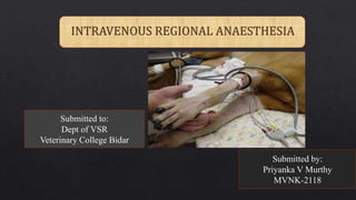
- 1. Submitted to: Dept of VSR Veterinary College Bidar Submitted by: Priyanka V Murthy MVNK-2118
- 3. INDICATIONS
- 5. PROS??
- 6. CONS??
- 7. Using a standard technique, catheterize a vein in the distal limb using a small‐gauge catheter Secure the catheter to prevent its dislodgement when the bandage exsanguinates the limb. The distal extremity must be exsanguinated using a flexible bandage (Esmarch, flexible self‐adhering bandaging tape). This prevents the injected local anesthetic solution from being diluted by blood, making it more effective for analgesia. The tourniquet should then be applied to the limb. There are two main types of tourniquets: non‐ pneumatic tourniquets that are made of rubber or elasticized cloth, and pneumatic cuff tourniquets inflated with air. TECHNIQUE
- 8. If a pneumatic tourniquet is used, the cuff should be inflated to a pressure 50–100 mmHg A less expensive alternative for use in small animal patients is to use a sphygmomanometer and a blood pressure cuff (after the cuff is first tested for leaks). The sphygmomanometer is used to inflate the blood pressure cuff and to monitor and adjust cuff pressure over time. In the case of a non‐pneumatic rubber tourniquet, the band should be placed above the elastic wrap that was used for exsanguination and secured tightly to prevent inadvertent release. Document the time of tourniquet application in the anesthetic chart. The remaining procedures (e.g., completion of IVRA, preparation for surgery, surgery) should be completed in less than 90 min to avoid ischemia of tissues under the tourniquet.
- 9. Once the tourniquet is in place, the elastic bandage should be carefully removed. Confirm that the previously identified peripheral pulse is now absent. Never proceed with the block if an arterial pulse is detected. If arterial blood is allowed to enter the distal limb while venous blood is being occluded, the limb will become edematous over the course of the procedure. The local anesthetic solution should be slowly injected over 2–3 min as distally as possible, avoiding high injection pressures that might increase venous pressure and cause leakage of the local anesthetic under the tourniquet. Document the time of local anesthetic injection and continually observe the patient for signs of systemic toxicity thereafter. Following injection, the catheter can be removed. At the end of the procedure, the tourniquet is removed slowly while the surgical site is closely evaluated for hemorrhage during initial re- perfusion
- 11. IVRA OF TEAT
- 12. IVRA OF CATTLE LIMB
- 13. POST SURGERY
- 15. INDICATIONS
- 16. painless and not induce fear or apprehension reliable rapid safe and simple to operate irreversible inexpensive as far as possible be aesthetic be safe for predators/consumers CRITERIA FOR EVALUATING EUTHANASIA
- 17. PHYSICAL METHODS Stunning Gun shot Captive bolt Electrocution Cervical dislocation Decapitation CHEMICAL METHODS Drug injections Gas or vapor inhalation METHODS OF EUTHANASIA
- 18. BASIC MECHANISMS OF TERMINATING LIFE Direct depression of neurons vital for life function Physical disruption of brain activity &destruction of neurons necessary for life
- 25. INHALANT AGENTS
- 26. Carbon dioxide gasflow of 70% CO2 for atleast 1minute • Advantages o Rapid depressant, analgesic and anesthetic effect of CO2 o Readily available and can be purchased in compressed gas cylinders are well established o Inexpensive, noninflammable, nonexplosive and poses minimal hazard to personnel o No accumulation of tissue residues in food producing animals
- 28. Carbon monoxide o Carbon monoxide is noninflammable and nonexplosive unless concentration exceeds 10 % o It combines with hemoglobin to form carboxyhemoglobin and blocks uptake of O2 by erythrocytes, leading to fatal hypoxemia o Death occurs rapidly if concentrations of 4 to 6% are used
- 29. INJECTABLE AGENTS • Most rapid and reliable method • Most desirable method • Intravenous administration most effective in well restrained animals • Intraperitoneal administration is acceptable if IV administration is impractical • Intracardiac injection is acceptable only in heavily sedated or anesthetized animals
- 30. Barbituric acid derivatives- Na Pentobarbital & Thiopental Na (100mg/kg) o IV injection is preferred in dogs, cats, other small animals, and horses o Intraperitoneal injection may be used in situations when an intravenous injection is distressful or even dangerous o Intracardiac injection must only be used if the animal is heavily sedated, unconscious, or anesthetized o • Depress the central nervous system o • With an overdose, depression of neurons responsible for vital functions leading to depression of respiratory center, which is followed by cardiac arrest. o induce euthanasia smoothly, with minimal discomfort to the animal
- 32. Tricaine methane sulfonate (MS 222, TMS) • Euthanasia choice for amphibians and fish • Concentration ≥ 250 mg/L concentration used for euthanasia o Fish should be left in this solution for at least 10 minutes following cessation of opercular movement
- 33. Potassium chloride in conjunction with prior general anesthesia • Unacceptable and condemned when used in conscious animals • The use of fully saturated solution in anesthetized animals is an acceptable method to produce cardiac arrest and death • 1 to 2 mmol/kg IV
- 36. POST EUTHANASIA INDICATORS OF DEATH
- 38. Epidural space is that compartment between the duramater and the bony and ligamentous wall of the spinal canal This space is filled with extradural fat, internal vertebral plexus of veins and the spinal nerves Injection of local anaesthetics will desensitize the spinal nerves at their origin in vertebral canal EPIDURAL SPACE
- 39. desensitize tail,vagina,rectum,vulva,anus,caudal prepuce,scrotum and urethra in low dose-1ml/100kg it is routinely used in cows, sheep, and goats. Indications • obstetric manipulations • caudal surgical procedures • adjunct treatment for rectal tenesmus. Needle placement is either at the sacrococcygeal (S5- Col) or more commonly at the first coccygeal (Col-Co2) interspace. Caudal epidural/low epidural
- 40. The Col-Co 2 interspace is larger and more easily penetrated than the S5-Col site. The S5-Col interspace may be ossified in older cows and is not so easily detectable in fat cows 3.75- to 5-cm, 18-gauge needle is used Locate Sacro coccygeal joint by moving the tail up and down, this joint moves very little and located anterior to the anal folds The first intercoccygeal joint is easily located by its movement, much wider and posterior to anal folds Insert the needle exactly at middle of the first intercoccygeal space at a right angle (aprox.10 degrees to vertical) to the skin surface
- 41. Push the needle ventrally through the interarcuate ligament to the floor of the neural canal, which is aprox. 2 to 4cm Withdraw the needle slightly (about 0.5cm) into epidural space Test by injecting 1 ml of air, no resistance should be felt
- 42. ANAESTHETIC EXTENT the area of anesthesia extends cranially to the middle of the sacrum and ventrally over the perineum to the inner aspect of the thigh Proper techniques should desensitize the pelvic viscera and genitalia, and paralyze the tail and abolish abdominal contractions. locomotor function of the hind legs and uterine motility remain unaffected. Maximal anesthesia may require 10 to 20 min and can be expected to last 30 min to 2.5 h
- 43. In sheep and goats useful for tail docking in lambs and for intravaginal obstetric procedures Site: Between S4 and Co1 , Co1 and CO 2 and lumbosacral space (b/w L6 and S1)
- 44. Needle used: A 2.5- to 3.75-cm, 18-gauge needle Anesthetic dose: no more than 1 mL of a 2% lidocaine hydrochloride solution per 50 kg of body weight is injected.
- 45. This is indicated in cattle and sheep with prolapse of the vagina and/or rectum, which can provoke severe continuous straining. The technique in cattle is simply performed by inserting a fine catheter into the epidural space through a 7.5-cm, 16- or 17-gauge needle. the spinal needle with stylet in place and bevel directed cranially is advanced for 5 to 8 cm while being directed at approximately 45° to vertical until resistance to needle passage is abruptly reduced. The stylet from the needle is removed, and a test dose of 2 to 3 mL of anesthetic is injected with almost no resistance, assuring proper placement of the needle in the epidural space
- 46. A30-cm medical-grade vinyl epidural catheter (0.036-cm outside diameter) with gradual markings is introduced into the canal through the needle and advanced cranially 3 to 4 cm beyond the tip of the needle The needle is then withdrawn, leaving the catheter in position. The catheter adapter or 23-gauge needle and three-way stopcock are placed on the free end of the catheter The catheter can be sutured to the skin puncture site. The sterility of the free end of the catheter is maintained by using protective sterile gauze 3 to 5 mL of a 2% lidocaine hydrochloride solution is injected into the catheter at 4- to 6-h intervals or whenever the animal shows signs of straining
- 47. It is advocated for all procedures caudal to the diaphragm. The anesthetic solution, dosed at 1 mL/4.5 kg b.wt, Site: lumbosacral (L6-S1) junction for immature cattle ,sheeps and goats provide analgesia of the the entire inguinal region, the flanks, and the abdominal wall up to the umbilicus Increasing the dose (volume X concentration) increases the area desensitized
- 48. Used in surgical conditions for cesarean section; intra-abdominal, pelvic, or hind-limb surgery; and udder surgery in small ruminants. Site: Lumbosacral intervertebral space Needle used: a 6- to 7-cm, 20-gauge spinal needle with a fitted stylet Dose:1 mL of a 2% lidocaine hydrochloride solution per 4.5 kg of body weight
- 49. include loss of consciousness, convulsions, respiratory paralysis, hypotension, and hypothermia after overdose Animals with severe hypotension show signs of distress, collapse, tachycardia or bradycardia, weak pulse, and shallow, rapid respiration
- 50. In horses, the spinal cord and its meninges end in the midsacral region, and only the coccygeal nerves and the thin phylum terminale remain in the spinal canal. Produce regional anesthesia of the anus, perineum, rectum, vulva, vagina, urethra, and bladder in horses. The injection site is the epidural space between the first and second coccygeal vertebrae. The first coccygeal interspace (ColCo2) is identified as the first obvious midline depression caudal to the sacrum.
- 51. In the standard technique, a 5- to 7.5-cm, 18- gauge spinal needle with fitted stylet is inserted through the disinfected skin in the center of the Col-Co 2 joint space needle is directed at almost right angles to the general contour of the croup A popping sensation is often detected as the interarcuate ligament is penetrated A mature mare (450 kg) may require a total of 6 to 8 mL of a 2% lidocaine HC1 solution (0.26 to 0.35 mg/kg) to anesthetize the anus, perineum, rectum, vulva, vagina, urethra, and bladder.
- 52. Continuous caudal epidural anesthesia can be used in horses for extended surgery in the anal and perineal region, for obstetric procedures, and for relief of tenesmus A10.2-cm, 18- gauge thin-walled Tuohy needle with stylet is aseptically inserted on the midline into the Col-Co2 interspace Acommercially available 91.8- cm, 20-gauge Teflon epidural catheter with graduated markings and stylet can be introduced into the needle and advanced cranially 2 to 4 cm beyond the tip of the needle The needle can then be removed from the catheter while the catheter is left in position A threeway stopcock and 2.5-cm, 23-gauge needle is placed on the free end of the catheter for an injection port.
- 53. Also known asArthur’s block Area blocked: The skin area caudal to the T13 or L1 spinous process and flank on both sides Nerves blocked: T13 and anterior lumbar nerves Site: Epidural space between L1 and L2 vertebrae
- 55. The site is aseptically prepared 2 to 4 mL of a 2% lidocaine hydrochloride solution is injected subcutaneously and adjacent to the interspinous (T13-L1 or L1-L2) ligaments to minimize pain during the puncture procedure. A11.25-cm, 18-gauge spinal needle with stylet is inserted at that site. The spinal needle is inserted for a distance of 8 to 12 cm while being directed ventrally and medially at an angle of 10° to 15° with the vertical, at which point the needle has reached the epidural space
- 56. The needle is properly placed into the epidural space if no blood or CSF flows from the needle hub or is obtained upon aspiration. Asucking sound is heard as air enters the needle immediately after penetration of the interarcuate ligament. Most often, the epidural space is identified by the loss-of resistance method An alternative method is hanging-drop method Anaesthetic: 8ml of 2% lidocaine in a 500 kg cow No more than 1ml/50 kg of lidocaine in sheep and goats
- 57. This amount of anesthetic is sufficient to desensitize the T13, LI , and L2 dermatomes within 7 to 20 min with a duration of 45 min to 2 h The epidural space at the thoracolumbar (T13-L1) or first lumbar (L1- L2) intervertebral space in cattle cannot be reached by the spinal needle if the interarcuate ligament is ossified because of old age (>8 years) satisfactory anesthesia and relaxation of the abdominal wall and flank for operations such as rumenotomy and cesarean section, with the animal standing
- 58. The advantages of segmental dorsolumbar epidural anesthesia as compared with proximal and distal paravertebral anesthesia include: the use of a single injection of a small quantity of anesthetic uniform analgesia and relaxation of the skin, musculature, and parietal peritoneum The disadvantages include: difficulty in performing the technique, potential for trauma to the spinal cord or venous sinuses and loss of motor control of the pelvic limbs;
- 59. This can be achieved by aseptically placing a catheter into the epidural space . A10.2-cm, 18-gauge thin-walled Tuohy needle with stylet is inserted into the epidural space at the thoracolumbar (T13-L1) interspace commercially available 91.8-cm, 20-gauge Teflon epidural catheter with graduated markings and stylet is introduced into the needle and advanced caudally 3 to 5 cm beyond the tip of the needle. The needle is removed from the catheter while the catheter is left in position and sutured to the skin at the site of emergence from the skin
- 60. Injection of apx. 6 mL of a 2% lidocaine hydrochloride solution at the anterior portion of the lumbar (LI to L2) Analgesia lasts 1 h to 1 h 40 min Advantages providing a route for repeated small fractional maintenance doses of local anesthetic drug,
- 64. DOG CAT RUMINANTS HORSE Lignocaine 1ml/5kg(2%) 1ml/4kg(2%) 1ml/100kg(ant.) 1ml/5kg(post.) 8ml/450kg(2%) Bupivacaine 1ml/5kg(0.5%) 1ml/4kg(0.25%) 0.125mg/kg 0.06mg/kg(0.5% ) Ropivacaine 5mg/kg(0.5%) 1ml/4kg(0.2%) 0.11mg/kg 8ml/450kg(0.5% ) Mepivacaine 7mg/kg(2%) _ _ 5ml/450kg(2%)
- 65. RUMINANTS HORSE DOG CAT XYLAZINE 0.05mg/kg dil in 5ml NS 0.25mg/kg 0.125mg/kg 0.02-0.2mg/kg DETOMIDINE 40µg/kg 60µg/kg 10µg/kg - MEDETOMIDINE 15µg/kg 15µg/kg 15µg/kg 10µg/kg KETAMINE 0.5-1mg/kg 0.5-1mg/kg in 10ml NS 1-2mg/kg 2.5mg/kg MORPHINE 0.1mg/kg 0.1mg/kg 0.1mg/kg in 4ml 0.1mg/kg OXYMORPHONE - - 0.015mg/kg 0.05-0.1mg/kg BUTARPHANOL - 0.08mg/kg in 20ml NS 0.25mg/kg - TRAMADOL 2mg/kg 1mg/kg in 20ml NS 2mg/kg 2mg/kg
- 66. William J. Tranquilli John C. Thurmon Kurt A. Grimm, Lumb & Jones' Veterinary Anesthesia and Analgesia, Fourth Edition,2007 L.W. Hall, K.W. Clarke , C.M. Trim,Veterinary anaesthesia ,Tenth edition,2001