SlideShare a Scribd company logo
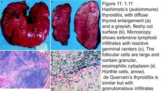
Figure 11: 1.11
Hashimoto’s (autoimmune)
thyroiditis, with diffuse
thyroid enlargement (a)
and a grayish, fleshy cut
surface (b). Microscopy
shows extensive lymphoid
infiltrates with reactive
germinal centers (c). The
follicular cells are large and
contain granular,
eosinophilic cytoplasm (d,
Hürthle cells, arrow).
de Quervain’s thyroiditis is
similar but with
granulomatous infiltrates
Figure 13: 1.13 Follicular adenoma of the thyroid.
Note the sharp demarcation (encapsulation) and
colloid shine of this macrofollicular adenoma (a).
Microscopy (b, c) shows a follicular adenoma of the
thyroid with a well-developed, fibrous capsule
Figure 14: 1.14 (a) This well-circumscribed lesion mimics
adenoma, but a suspicious satellite nodule (arrow)
suggests malignancy. (b) Another view shows a formalin-
fixed (brownish) thyroid with poorly demarcated, diffuse,
and nodular cancerous infiltration (white parts). (c)
Microscopy confirms the diagnosis of papillary carcinoma
Figure 15: 1.15 Carcinomas
of the thyroid have various
histologic appearances,
including papillary
carcinoma with
characteristic ground glass
nuclei (“Orphan Annie eye”)
(a), follicular variant of a
papillary carcinoma (b), and
psammoma bodies of
papillary carcinoma (c)
Figure 16: 1.16 Papillary carcinoma of the thyroid. An
ultrasound image (a) shows a heterogeneous mass with
calcium. Doppler ultrasonography (b) shows
hypervascularity
Figure 17: 1.17 Follicular carcinoma of the thyroid. (a, b)
Note the irregular growth of well-differentiated follicles,
with signs of capsular and vascular invasion
Figure 18: 1.18
Anaplastic thyroid
carcinoma, showing
dense population of
undifferentiated,
pleomorphic cells with
multiple mitotic figures
(arrows)
Figure 19: 1.19
Medullary (C-cell)
carcinoma of the
thyroid.
(a) A gross specimen shows
poorly delimited, slightly
yellowish infiltration of the
gland (arrow). (b) Microscopy
shows small-cell infiltrates
with amyloid deposits (arrow)
in interstitial tissue. (c)
Amyloid shows characteristic
birefringence on polarization
of Congo red-stained sections.
(d) Immunohistochemistry
shows positive staining for
calcitonin
Figure 20: 1.20
Primary malignant non-
Hodgkin’s lymphoma of the
thyroid.
Note the diffuse, whitish enlargement of
the gland (a) caused by pronounced
interstitial infiltration by atypical lymphoid
cell populations (b)
Figure 21: 1.21
Parathyroid glands.
The gross picture (a),
showing the location,
is a view from the
posterior with the
esophagus removed.
The parathyroid
glands are shown on
the posterior surface
of the thyroid gland
(arrows). The normal
location may vary,
however, occasionally
reaching down into the
thymus. Microscopy
shows a normal
parathyroid gland with
abundant fat tissue (b)
and a hyperplastic
gland with extensive
replacement of fat
tissue by parathyroid
endocrine cells (c)
Figure 22: 1.22 Parathyroid adenoma and carcinoma
frequently are distinguishable only by microscopy.
Gross appearance (a) showing several nodular,
tumorous infiltrates of enlarged parathyroid gland
(original magnification ×3). Microscopy shows
parathyroid adenoma (b) and carcinoma with signs of
tumorous invasion of the capsule and adjacent
tissues and cellular polymorphism (c)
Figure 23: 1.23
Hyperparathyroidism—whether
primary (parathyroid adenoma,
carcinoma, or primary
hyperplasia) or secondary (due
to renal disease)—causes
increased bone resorption (such
as the lacunar osteoclastic
resorption shown in this image),
causing dissecting fibro-
osteoclasia and finally osteitis
fibrosa cystica (von
Recklinghausen’s disease).
Hypercalcemia also leads to
calcification of soft tissues and
mucosal surfaces, and to
nephrolithiasis (See also Chaps.
6 and 9 for renal and bone
diseases)
Figure 24: 1.24 Technetium (99mTc) sestamibi parathyroid scan (after 15 min
and 2 h) showing a left inferior parathyroid adenoma (Figure provided by
Bechara Y. Ghorayeb, M.D., Houston, Texas [www. ghorayeb. com])
Figure 25: 1.25 (a) A technetium (99mTc) sestamibi
scan at 15 min (immediate) and after a 3-h delay,
showing a large adenoma. (b, c), surgical pictures of
the same adenoma (Figure provided by Bechara Y.
Ghorayeb, M.D., Houston, Texas [www. ghorayeb.
com])
Figure 26: 1.26 Islet cell tumors of the pancreas
(a, b). These tumors form nodular masses, which
often appear solid and are yellow-tan to white
Figure 27: 1.27 Microscopy of a pancreatic adenoma
producing gastrin (gastrinoma) on hematoxylin-eosin stain
(a) and with immunohistochemistry for gastrin (b brown
cells)
Figure 27: 1.27 Microscopy of a pancreatic adenoma
producing gastrin (gastrinoma) on hematoxylin-eosin stain
(a) and with immunohistochemistry for gastrin (b brown
cells)
Figure 31: 1.31 (a, b) Axial and coronal CT scans of the
pancreas head show prominent nodular alterations
indicative of neuroendocrine tumor
Figure 32: 1.32 Adrenal
hemorrhagic necrosis in
meningococcal septicemia,
causing hemorrhagic shock and
the death of the patient
(Waterhouse–Friderichsen
syndrome; see also Chap. 1).
Note the extensive necrosis and
hemorrhage of the adrenal
cortex (a) and diffuse petechial
and confluent hemorrhages of
the skin (b). The internal organs
show similar hemorrhages
Figure 33: 1.33 Adrenal cortical hypoplasia. (a) The
gross appearance is of a small, brownish, thin, and
soft gland (about 1.8 cm in length). (b), Microscopy
shows hypoplastic adrenal cortex (C) with poorly -
organized layers (arrows). (c), By comparison,
normal cortex has clearly identifiable zones (G
glomerulosa, F fasciculate, R reticularis, M adrenal
medulla)
Figure 2: 1.2 Atrophy of the
pituitary gland (“empty sella
syndrome”). Note the cavitation
of the sella turcica (arrows)
associated with shrinkage of the
pituitary gland
Figure 30: 1.30 This axial CT scan, performed with
contrast, shows a pancreas nodule (presumed to be an
islet cell tumor) in a patient with von Hippel–Lindau
syndrome
Figure 38: 1.38 Left adrenal primary adrenocortical
carcinoma. This postcontrast CT scan shows a large (4.4
cm), irregularly enhancing left adrenal mass. The
differential diagnosis must include metastasis
Figure 39: 1.39 Pheochromocytoma. (a) This axial CT
scan with contrast shows a left adrenal
pheochromocytoma immediately medial and superior to
the left kidney. (b), A coronal CT with contrast in the same
patient
Figure 40: 1.40 Pinealoma (pineal germinoma), shown in
midsagittal (a) and axial (b) postcontrast T1-weighted MR
images (arrows)
Figure 34: 1.34 (a) Diffuse adrenal cortical hyperplasia.
Note the yellowish thickening of poorly demarcated
adrenal cortex (arrows), as shown in this cross section of
the gland. (b) Adrenal cortical adenoma. Note the well-
demarcated yellow nodule (arrows) (F periadrenal fat
tissue, AG adrenal gland). (c) Typical microscopy of the
adenoma, with fairly uniform, pale polygonal cells with
small dark nuclei and no mitoses. Adrenal cortical
adenomas and carcinomas may be associated with
primary hyperaldosteronism (Conn’s syndrome),
Cushing’s syndrome, feminization, or virilization, or may
be nonfunctional
Figure 35: 1.35 Adrenal cortical
carcinomas. (a–c), Note the
irregular structure with yellowish,
partly necrotic, and hemorrhagic
tissues with pseudocystic
degeneration. (d) Microscopy
shows irregular growth of more
polymorphic, partially poorly
differentiated cells, including giant
cells
Figure 36: 1.36 Adrenal medullary
pheochromocytoma: gross
appearance on the surface (a)
and cut surface (b), with soft
yellowish red “medullary” tissue,
focal hemorrhage, and
pseudocystic degeneration.
Microscopy (c) shows a
population of polymorphic
“epithelioid” cells with giant cells
Figure 37: 1.37 Adrenal medullary neuroblastoma. (a)
Gross appearance shows a whitish, nodular infiltrate of
the gland with focal degeneration and hemorrhage. (b)
Microscopy shows a typical small cellular (“lymphoid”)
infiltration with characteristic neurofibrillary rosettes
Figure 41: 1.41 (a) Gross sagittal section from a brain with
a pineal tumor. (b) Microscopy shows the biphasic pattern
of the pinealoma, with areas composed of large primitive
spheroidal cells and stromal areas with a prominent
lymphocytic component. (c) Microscopy of a pineocytoma,
a tumor of pineal parenchymal cells with pineocytomatous
rosettes (large fibrillar zones)
Figure 1: 1.1 Normal pituitary gland: location at the
base of the skull in the sella turcica (a, arrow) with
cross section (b). Enlarged picture of a removed gland
(c) showing the adenohypophysis (A, pink part) and
the neurohypophysis (N, white part)
Figure 4: 1.4 This formalin-fixed specimen (reconstituted in alcohol) shows a large pituitary adenoma (a) expanding
to the optic nerve and the intracerebral part of the internal carotid artery (arrows). Microscopy shows a typical
chromophobe adenoma composed of small cells (b). Chromophobe adenomas generally contain small numbers of
secretory granules (sparsely granulated) but may produce a number of different hormones. A adenoma
Figure 3: 1.3 (a) MRI in sagittal view shows distinct pituitary enlargement representing an adenoma (arrow). (b), A CT
scan of the cranium shows a tumorous mass on the right between the optic chiasm and the base of the third ventricle
(arrow)
Figure 6: 1.6 Pituitary macroadenoma. This coronal
magnetic resonance (MR) image (T1-weighted,
postcontrast) reveals a large, homogenously enhancing
mass that extends above the diaphragma sellae, abuts
and deviates the optic chiasm, and invades the right
cavernous sinus
Figure 7: 1.7 Prolactinoma. This coronal MR image (T1-
weighted, postcontrast) shows the classic findings of a
hypoenhancing mass in the pituitary, deviation of the
infundibulum from the mass, and upward convexity of the
pituitary border
Figure 5: 1.5 Other types of adenoma are composed of
basophilic cells producing corticotropin (clinical, Cushing’s
disease or Nelson’s syndrome). Adenomas composed of
acidophilic cells are typically associated with the
production of growth hormone (somatotropin) or prolactin.
About 25 % of pituitary adenomas are nonfunctional,
without endocrine activity. Newer classifications use
endocrine activities rather than cellular-staining qualities;
shown here, for example, is an FSH-producing adenoma,
with immunohistochemistry showing brown, FSH-positive
cells. This figure shows an immunohistochemical stain for
somatotropin
Figure 8: 1.8 Thyroid hyperplasia
(nodular goiter), showing gross
enlargement with nodular and cystic
(degenerative) structures (a
removed gland, b gland in situ seen
from behind, with larynx and trachea
in the center). Microscopy (c) of a
hyperplastic (adenomatous) nodule
in a patient with a nodular goiter,
demonstrating that the nodule is
partially encapsulated and is
composed of large, colloid-filled
follicles
Figure 9: 1.9 Thyroid gland from a patient with
Graves’ disease (hyperthyroidism, also referred
to as Basedow’s disease). The cut surface
shows diffuse hyperplasia and has a fleshy
appearance (a). Microscopically (b), follicles are
lined by hyperplastic follicular cells with focal
papillary structure and areas of colloid
resorption vacuoles
Figure 10: 1.10 Right thyroid goiter. (a, b) Axial and
coronal postcontrast CT scans, show a large right low-
density cyst consistent with a colloid cyst of goiter
endocrinology

More Related Content

What's hot

48 DAVID SUTTON PICTURES THE SINUSES
48 DAVID SUTTON PICTURES THE SINUSES48 DAVID SUTTON PICTURES THE SINUSES
48 DAVID SUTTON PICTURES THE SINUSES
Dr. Muhammad Bin Zulfiqar
 
18 DAVID SUTTON PICTURES THE SALIVARY GLANDS PHARYNX AND ESOPHAGUS
18 DAVID SUTTON PICTURES THE SALIVARY GLANDS PHARYNX AND ESOPHAGUS18 DAVID SUTTON PICTURES THE SALIVARY GLANDS PHARYNX AND ESOPHAGUS
18 DAVID SUTTON PICTURES THE SALIVARY GLANDS PHARYNX AND ESOPHAGUS
Dr. Muhammad Bin Zulfiqar
 
37 DAVID SUTTON PICTURES AVASCUALR NECROSIS
37 DAVID SUTTON PICTURES AVASCUALR NECROSIS37 DAVID SUTTON PICTURES AVASCUALR NECROSIS
37 DAVID SUTTON PICTURES AVASCUALR NECROSIS
Dr. Muhammad Bin Zulfiqar
 
51 DAVID SUTTON PICTURES THE ORBIT
51 DAVID SUTTON PICTURES THE ORBIT51 DAVID SUTTON PICTURES THE ORBIT
51 DAVID SUTTON PICTURES THE ORBIT
Dr. Muhammad Bin Zulfiqar
 
38 DAVID SUTTON PICTURES DISEASES OF JOINT
38  DAVID SUTTON PICTURES  DISEASES OF JOINT38  DAVID SUTTON PICTURES  DISEASES OF JOINT
38 DAVID SUTTON PICTURES DISEASES OF JOINT
Dr. Muhammad Bin Zulfiqar
 
31 DAVID SUTTON PICTURES THE BLADDER AND PROSTATE
31 DAVID SUTTON PICTURES  THE BLADDER AND PROSTATE31 DAVID SUTTON PICTURES  THE BLADDER AND PROSTATE
31 DAVID SUTTON PICTURES THE BLADDER AND PROSTATE
Dr. Muhammad Bin Zulfiqar
 
34 DAVID SUTTON PICTURES THE GYNAECOLOGICAL IMAGING
34 DAVID SUTTON PICTURES  THE GYNAECOLOGICAL IMAGING34 DAVID SUTTON PICTURES  THE GYNAECOLOGICAL IMAGING
34 DAVID SUTTON PICTURES THE GYNAECOLOGICAL IMAGING
Dr. Muhammad Bin Zulfiqar
 
36 DAVID SUTTON PICTURES PERIOSTEAL REACTION BONE AND JOINT INFECTIONS: SARCOID
36 DAVID SUTTON PICTURES PERIOSTEAL REACTION BONE AND JOINT INFECTIONS: SARCOID36 DAVID SUTTON PICTURES PERIOSTEAL REACTION BONE AND JOINT INFECTIONS: SARCOID
36 DAVID SUTTON PICTURES PERIOSTEAL REACTION BONE AND JOINT INFECTIONS: SARCOID
Dr. Muhammad Bin Zulfiqar
 
40 DAVID SUTTON PICTURES TUMORS AND TUMORS LIKE CONDITIONS OF BONE II
40 DAVID SUTTON PICTURES TUMORS AND TUMORS LIKE CONDITIONS OF BONE II40 DAVID SUTTON PICTURES TUMORS AND TUMORS LIKE CONDITIONS OF BONE II
40 DAVID SUTTON PICTURES TUMORS AND TUMORS LIKE CONDITIONS OF BONE II
Dr. Muhammad Bin Zulfiqar
 
Diagnostic Imaging of Paranasal sinuses and Nose
Diagnostic Imaging of Paranasal sinuses and NoseDiagnostic Imaging of Paranasal sinuses and Nose
Diagnostic Imaging of Paranasal sinuses and Nose
Mohamed M.A. Zaitoun
 
46 DAVID SUTTON PICTURES THE BREAST
46 DAVID SUTTON PICTURES THE BREAST46 DAVID SUTTON PICTURES THE BREAST
46 DAVID SUTTON PICTURES THE BREAST
Dr. Muhammad Bin Zulfiqar
 
33 DAVID SUTTON PICTURES THE OBSTETRIC ULTRASOUND
33 DAVID SUTTON PICTURES  THE OBSTETRIC ULTRASOUND33 DAVID SUTTON PICTURES  THE OBSTETRIC ULTRASOUND
33 DAVID SUTTON PICTURES THE OBSTETRIC ULTRASOUND
Dr. Muhammad Bin Zulfiqar
 
58 DAVID SUTTON PICTURES INTRACRANIAL LESIONS (2)
58 DAVID SUTTON PICTURES INTRACRANIAL LESIONS (2)58 DAVID SUTTON PICTURES INTRACRANIAL LESIONS (2)
58 DAVID SUTTON PICTURES INTRACRANIAL LESIONS (2)
Dr. Muhammad Bin Zulfiqar
 
24 DAVID SUTTON PICTURES THE BILIARY TRACT
24 DAVID SUTTON PICTURES THE BILIARY TRACT24 DAVID SUTTON PICTURES THE BILIARY TRACT
24 DAVID SUTTON PICTURES THE BILIARY TRACT
Dr. Muhammad Bin Zulfiqar
 
22 DAVID SUTTON PICTURES THE ACUTE ABDOMEN
22 DAVID SUTTON PICTURES THE ACUTE ABDOMEN22 DAVID SUTTON PICTURES THE ACUTE ABDOMEN
22 DAVID SUTTON PICTURES THE ACUTE ABDOMEN
Dr. Muhammad Bin Zulfiqar
 
87 splenic lesions on magnetic resonance imaging
87 splenic lesions on magnetic resonance imaging87 splenic lesions on magnetic resonance imaging
87 splenic lesions on magnetic resonance imagingDr. Muhammad Bin Zulfiqar
 
8 Techniques in Thoracic Imaging Dr. Muhammad Bin Zulfiqar Grainger and Allison
8 Techniques in Thoracic Imaging Dr. Muhammad Bin Zulfiqar Grainger and Allison8 Techniques in Thoracic Imaging Dr. Muhammad Bin Zulfiqar Grainger and Allison
8 Techniques in Thoracic Imaging Dr. Muhammad Bin Zulfiqar Grainger and Allison
Dr. Muhammad Bin Zulfiqar
 
47 DAVID SUTTON PICTURES PHARYNX AND LARYNX THE NECK
47 DAVID SUTTON PICTURES PHARYNX AND LARYNX THE NECK47 DAVID SUTTON PICTURES PHARYNX AND LARYNX THE NECK
47 DAVID SUTTON PICTURES PHARYNX AND LARYNX THE NECK
Dr. Muhammad Bin Zulfiqar
 
44 DAVID SUTTON PICTURES SKELETAL TRAUMA REGIONAL
44 DAVID SUTTON PICTURES SKELETAL TRAUMA REGIONAL44 DAVID SUTTON PICTURES SKELETAL TRAUMA REGIONAL
44 DAVID SUTTON PICTURES SKELETAL TRAUMA REGIONAL
Dr. Muhammad Bin Zulfiqar
 
Diagnostic Imaging of Congenital Pulmonary Abnormalities
Diagnostic Imaging of Congenital Pulmonary AbnormalitiesDiagnostic Imaging of Congenital Pulmonary Abnormalities
Diagnostic Imaging of Congenital Pulmonary Abnormalities
Mohamed M.A. Zaitoun
 

What's hot (20)

48 DAVID SUTTON PICTURES THE SINUSES
48 DAVID SUTTON PICTURES THE SINUSES48 DAVID SUTTON PICTURES THE SINUSES
48 DAVID SUTTON PICTURES THE SINUSES
 
18 DAVID SUTTON PICTURES THE SALIVARY GLANDS PHARYNX AND ESOPHAGUS
18 DAVID SUTTON PICTURES THE SALIVARY GLANDS PHARYNX AND ESOPHAGUS18 DAVID SUTTON PICTURES THE SALIVARY GLANDS PHARYNX AND ESOPHAGUS
18 DAVID SUTTON PICTURES THE SALIVARY GLANDS PHARYNX AND ESOPHAGUS
 
37 DAVID SUTTON PICTURES AVASCUALR NECROSIS
37 DAVID SUTTON PICTURES AVASCUALR NECROSIS37 DAVID SUTTON PICTURES AVASCUALR NECROSIS
37 DAVID SUTTON PICTURES AVASCUALR NECROSIS
 
51 DAVID SUTTON PICTURES THE ORBIT
51 DAVID SUTTON PICTURES THE ORBIT51 DAVID SUTTON PICTURES THE ORBIT
51 DAVID SUTTON PICTURES THE ORBIT
 
38 DAVID SUTTON PICTURES DISEASES OF JOINT
38  DAVID SUTTON PICTURES  DISEASES OF JOINT38  DAVID SUTTON PICTURES  DISEASES OF JOINT
38 DAVID SUTTON PICTURES DISEASES OF JOINT
 
31 DAVID SUTTON PICTURES THE BLADDER AND PROSTATE
31 DAVID SUTTON PICTURES  THE BLADDER AND PROSTATE31 DAVID SUTTON PICTURES  THE BLADDER AND PROSTATE
31 DAVID SUTTON PICTURES THE BLADDER AND PROSTATE
 
34 DAVID SUTTON PICTURES THE GYNAECOLOGICAL IMAGING
34 DAVID SUTTON PICTURES  THE GYNAECOLOGICAL IMAGING34 DAVID SUTTON PICTURES  THE GYNAECOLOGICAL IMAGING
34 DAVID SUTTON PICTURES THE GYNAECOLOGICAL IMAGING
 
36 DAVID SUTTON PICTURES PERIOSTEAL REACTION BONE AND JOINT INFECTIONS: SARCOID
36 DAVID SUTTON PICTURES PERIOSTEAL REACTION BONE AND JOINT INFECTIONS: SARCOID36 DAVID SUTTON PICTURES PERIOSTEAL REACTION BONE AND JOINT INFECTIONS: SARCOID
36 DAVID SUTTON PICTURES PERIOSTEAL REACTION BONE AND JOINT INFECTIONS: SARCOID
 
40 DAVID SUTTON PICTURES TUMORS AND TUMORS LIKE CONDITIONS OF BONE II
40 DAVID SUTTON PICTURES TUMORS AND TUMORS LIKE CONDITIONS OF BONE II40 DAVID SUTTON PICTURES TUMORS AND TUMORS LIKE CONDITIONS OF BONE II
40 DAVID SUTTON PICTURES TUMORS AND TUMORS LIKE CONDITIONS OF BONE II
 
Diagnostic Imaging of Paranasal sinuses and Nose
Diagnostic Imaging of Paranasal sinuses and NoseDiagnostic Imaging of Paranasal sinuses and Nose
Diagnostic Imaging of Paranasal sinuses and Nose
 
46 DAVID SUTTON PICTURES THE BREAST
46 DAVID SUTTON PICTURES THE BREAST46 DAVID SUTTON PICTURES THE BREAST
46 DAVID SUTTON PICTURES THE BREAST
 
33 DAVID SUTTON PICTURES THE OBSTETRIC ULTRASOUND
33 DAVID SUTTON PICTURES  THE OBSTETRIC ULTRASOUND33 DAVID SUTTON PICTURES  THE OBSTETRIC ULTRASOUND
33 DAVID SUTTON PICTURES THE OBSTETRIC ULTRASOUND
 
58 DAVID SUTTON PICTURES INTRACRANIAL LESIONS (2)
58 DAVID SUTTON PICTURES INTRACRANIAL LESIONS (2)58 DAVID SUTTON PICTURES INTRACRANIAL LESIONS (2)
58 DAVID SUTTON PICTURES INTRACRANIAL LESIONS (2)
 
24 DAVID SUTTON PICTURES THE BILIARY TRACT
24 DAVID SUTTON PICTURES THE BILIARY TRACT24 DAVID SUTTON PICTURES THE BILIARY TRACT
24 DAVID SUTTON PICTURES THE BILIARY TRACT
 
22 DAVID SUTTON PICTURES THE ACUTE ABDOMEN
22 DAVID SUTTON PICTURES THE ACUTE ABDOMEN22 DAVID SUTTON PICTURES THE ACUTE ABDOMEN
22 DAVID SUTTON PICTURES THE ACUTE ABDOMEN
 
87 splenic lesions on magnetic resonance imaging
87 splenic lesions on magnetic resonance imaging87 splenic lesions on magnetic resonance imaging
87 splenic lesions on magnetic resonance imaging
 
8 Techniques in Thoracic Imaging Dr. Muhammad Bin Zulfiqar Grainger and Allison
8 Techniques in Thoracic Imaging Dr. Muhammad Bin Zulfiqar Grainger and Allison8 Techniques in Thoracic Imaging Dr. Muhammad Bin Zulfiqar Grainger and Allison
8 Techniques in Thoracic Imaging Dr. Muhammad Bin Zulfiqar Grainger and Allison
 
47 DAVID SUTTON PICTURES PHARYNX AND LARYNX THE NECK
47 DAVID SUTTON PICTURES PHARYNX AND LARYNX THE NECK47 DAVID SUTTON PICTURES PHARYNX AND LARYNX THE NECK
47 DAVID SUTTON PICTURES PHARYNX AND LARYNX THE NECK
 
44 DAVID SUTTON PICTURES SKELETAL TRAUMA REGIONAL
44 DAVID SUTTON PICTURES SKELETAL TRAUMA REGIONAL44 DAVID SUTTON PICTURES SKELETAL TRAUMA REGIONAL
44 DAVID SUTTON PICTURES SKELETAL TRAUMA REGIONAL
 
Diagnostic Imaging of Congenital Pulmonary Abnormalities
Diagnostic Imaging of Congenital Pulmonary AbnormalitiesDiagnostic Imaging of Congenital Pulmonary Abnormalities
Diagnostic Imaging of Congenital Pulmonary Abnormalities
 

Similar to endocrinology

Biliary tract
Biliary tractBiliary tract
Biliary tract
Athul Nampoothiri
 
61 chest wall lesions on computed tomography and
61 chest wall lesions on computed tomography and61 chest wall lesions on computed tomography and
61 chest wall lesions on computed tomography and
Dr. Muhammad Bin Zulfiqar
 
Diagnostic Imaging of Cholangiocarcinoma
Diagnostic Imaging of CholangiocarcinomaDiagnostic Imaging of Cholangiocarcinoma
Diagnostic Imaging of Cholangiocarcinoma
Mohamed M.A. Zaitoun
 
28 posterior mediastinal lesions on computed tomography
28 posterior mediastinal lesions on computed tomography28 posterior mediastinal lesions on computed tomography
28 posterior mediastinal lesions on computed tomography
Dr. Muhammad Bin Zulfiqar
 
82 cystic pancreatic masses on ct and mri
82 cystic pancreatic masses on ct and mri82 cystic pancreatic masses on ct and mri
82 cystic pancreatic masses on ct and mri
Dr. Muhammad Bin Zulfiqar
 
RAJ 22.pptx
RAJ 22.pptxRAJ 22.pptx
RAJ 22.pptx
RadiologyReports
 
acute pelvic pain RADIOLOGY LECTURE.pptx
acute pelvic pain RADIOLOGY LECTURE.pptxacute pelvic pain RADIOLOGY LECTURE.pptx
acute pelvic pain RADIOLOGY LECTURE.pptx
SureshChevagoni1
 
10 hypothalamic lesions on magnetic resonance imaging
10 hypothalamic lesions on magnetic resonance imaging10 hypothalamic lesions on magnetic resonance imaging
10 hypothalamic lesions on magnetic resonance imaging
Dr. Muhammad Bin Zulfiqar
 
Renal tuberculosis radiology
Renal tuberculosis radiologyRenal tuberculosis radiology
Renal tuberculosis radiology
docaashishgupt
 
Role of imaging in renal tuberculosis
Role of imaging in renal tuberculosisRole of imaging in renal tuberculosis
Role of imaging in renal tuberculosis
vinothmezoss
 
40 bladder masses on computed tomography and magnetic
40 bladder masses on computed tomography and magnetic40 bladder masses on computed tomography and magnetic
40 bladder masses on computed tomography and magnetic
Dr. Muhammad Bin Zulfiqar
 
83 solid pancreatic masses on computed tomography
83 solid pancreatic masses on computed tomography83 solid pancreatic masses on computed tomography
83 solid pancreatic masses on computed tomography
Dr. Muhammad Bin Zulfiqar
 
Diagnostic Imaging of Perinephric Space
Diagnostic Imaging of Perinephric SpaceDiagnostic Imaging of Perinephric Space
Diagnostic Imaging of Perinephric Space
Mohamed M.A. Zaitoun
 
32 DAVID SUTTON PICTURES THE MALE GENITILIA AND URETHRA
32 DAVID SUTTON PICTURES  THE MALE GENITILIA AND URETHRA32 DAVID SUTTON PICTURES  THE MALE GENITILIA AND URETHRA
32 DAVID SUTTON PICTURES THE MALE GENITILIA AND URETHRA
Dr. Muhammad Bin Zulfiqar
 
Madan_Thoracic_poster_12_BWH
Madan_Thoracic_poster_12_BWHMadan_Thoracic_poster_12_BWH
Madan_Thoracic_poster_12_BWHAnna McCormick
 
50 nodular
50 nodular50 nodular
Presentation1, radiological imaging of popliteal fossa masses.
Presentation1, radiological imaging of popliteal fossa masses.Presentation1, radiological imaging of popliteal fossa masses.
Presentation1, radiological imaging of popliteal fossa masses.
Abdellah Nazeer
 
Diagnostic Imaging of Deep Neck Spaces
Diagnostic Imaging of Deep Neck SpacesDiagnostic Imaging of Deep Neck Spaces
Diagnostic Imaging of Deep Neck Spaces
Mohamed M.A. Zaitoun
 

Similar to endocrinology (20)

Biliary tract
Biliary tractBiliary tract
Biliary tract
 
61 chest wall lesions on computed tomography and
61 chest wall lesions on computed tomography and61 chest wall lesions on computed tomography and
61 chest wall lesions on computed tomography and
 
Diagnostic Imaging of Cholangiocarcinoma
Diagnostic Imaging of CholangiocarcinomaDiagnostic Imaging of Cholangiocarcinoma
Diagnostic Imaging of Cholangiocarcinoma
 
28 posterior mediastinal lesions on computed tomography
28 posterior mediastinal lesions on computed tomography28 posterior mediastinal lesions on computed tomography
28 posterior mediastinal lesions on computed tomography
 
82 cystic pancreatic masses on ct and mri
82 cystic pancreatic masses on ct and mri82 cystic pancreatic masses on ct and mri
82 cystic pancreatic masses on ct and mri
 
RAJ 22.pptx
RAJ 22.pptxRAJ 22.pptx
RAJ 22.pptx
 
acute pelvic pain RADIOLOGY LECTURE.pptx
acute pelvic pain RADIOLOGY LECTURE.pptxacute pelvic pain RADIOLOGY LECTURE.pptx
acute pelvic pain RADIOLOGY LECTURE.pptx
 
10 hypothalamic lesions on magnetic resonance imaging
10 hypothalamic lesions on magnetic resonance imaging10 hypothalamic lesions on magnetic resonance imaging
10 hypothalamic lesions on magnetic resonance imaging
 
Renal tuberculosis radiology
Renal tuberculosis radiologyRenal tuberculosis radiology
Renal tuberculosis radiology
 
Role of imaging in renal tuberculosis
Role of imaging in renal tuberculosisRole of imaging in renal tuberculosis
Role of imaging in renal tuberculosis
 
40 bladder masses on computed tomography and magnetic
40 bladder masses on computed tomography and magnetic40 bladder masses on computed tomography and magnetic
40 bladder masses on computed tomography and magnetic
 
83 solid pancreatic masses on computed tomography
83 solid pancreatic masses on computed tomography83 solid pancreatic masses on computed tomography
83 solid pancreatic masses on computed tomography
 
Diagnostic Imaging of Perinephric Space
Diagnostic Imaging of Perinephric SpaceDiagnostic Imaging of Perinephric Space
Diagnostic Imaging of Perinephric Space
 
32 DAVID SUTTON PICTURES THE MALE GENITILIA AND URETHRA
32 DAVID SUTTON PICTURES  THE MALE GENITILIA AND URETHRA32 DAVID SUTTON PICTURES  THE MALE GENITILIA AND URETHRA
32 DAVID SUTTON PICTURES THE MALE GENITILIA AND URETHRA
 
Madan_Thoracic_poster_12_BWH
Madan_Thoracic_poster_12_BWHMadan_Thoracic_poster_12_BWH
Madan_Thoracic_poster_12_BWH
 
75 focal cystic lesions of the liver
75 focal cystic lesions of the liver75 focal cystic lesions of the liver
75 focal cystic lesions of the liver
 
50 nodular
50 nodular50 nodular
50 nodular
 
79 magnetic resonance imaging of the liver
79 magnetic resonance imaging of the liver79 magnetic resonance imaging of the liver
79 magnetic resonance imaging of the liver
 
Presentation1, radiological imaging of popliteal fossa masses.
Presentation1, radiological imaging of popliteal fossa masses.Presentation1, radiological imaging of popliteal fossa masses.
Presentation1, radiological imaging of popliteal fossa masses.
 
Diagnostic Imaging of Deep Neck Spaces
Diagnostic Imaging of Deep Neck SpacesDiagnostic Imaging of Deep Neck Spaces
Diagnostic Imaging of Deep Neck Spaces
 

Recently uploaded

Superficial & Deep Fascia of the NECK.pptx
Superficial & Deep Fascia of the NECK.pptxSuperficial & Deep Fascia of the NECK.pptx
Superficial & Deep Fascia of the NECK.pptx
Dr. Rabia Inam Gandapore
 
Lung Cancer: Artificial Intelligence, Synergetics, Complex System Analysis, S...
Lung Cancer: Artificial Intelligence, Synergetics, Complex System Analysis, S...Lung Cancer: Artificial Intelligence, Synergetics, Complex System Analysis, S...
Lung Cancer: Artificial Intelligence, Synergetics, Complex System Analysis, S...
Oleg Kshivets
 
Tom Selleck Health: A Comprehensive Look at the Iconic Actor’s Wellness Journey
Tom Selleck Health: A Comprehensive Look at the Iconic Actor’s Wellness JourneyTom Selleck Health: A Comprehensive Look at the Iconic Actor’s Wellness Journey
Tom Selleck Health: A Comprehensive Look at the Iconic Actor’s Wellness Journey
greendigital
 
Pulmonary Thromboembolism - etilogy, types, medical- Surgical and nursing man...
Pulmonary Thromboembolism - etilogy, types, medical- Surgical and nursing man...Pulmonary Thromboembolism - etilogy, types, medical- Surgical and nursing man...
Pulmonary Thromboembolism - etilogy, types, medical- Surgical and nursing man...
VarunMahajani
 
Flu Vaccine Alert in Bangalore Karnataka
Flu Vaccine Alert in Bangalore KarnatakaFlu Vaccine Alert in Bangalore Karnataka
Flu Vaccine Alert in Bangalore Karnataka
addon Scans
 
HOT NEW PRODUCT! BIG SALES FAST SHIPPING NOW FROM CHINA!! EU KU DB BK substit...
HOT NEW PRODUCT! BIG SALES FAST SHIPPING NOW FROM CHINA!! EU KU DB BK substit...HOT NEW PRODUCT! BIG SALES FAST SHIPPING NOW FROM CHINA!! EU KU DB BK substit...
HOT NEW PRODUCT! BIG SALES FAST SHIPPING NOW FROM CHINA!! EU KU DB BK substit...
GL Anaacs
 
Hemodialysis: Chapter 3, Dialysis Water Unit - Dr.Gawad
Hemodialysis: Chapter 3, Dialysis Water Unit - Dr.GawadHemodialysis: Chapter 3, Dialysis Water Unit - Dr.Gawad
Hemodialysis: Chapter 3, Dialysis Water Unit - Dr.Gawad
NephroTube - Dr.Gawad
 
How to Give Better Lectures: Some Tips for Doctors
How to Give Better Lectures: Some Tips for DoctorsHow to Give Better Lectures: Some Tips for Doctors
How to Give Better Lectures: Some Tips for Doctors
LanceCatedral
 
ANATOMY AND PHYSIOLOGY OF URINARY SYSTEM.pptx
ANATOMY AND PHYSIOLOGY OF URINARY SYSTEM.pptxANATOMY AND PHYSIOLOGY OF URINARY SYSTEM.pptx
ANATOMY AND PHYSIOLOGY OF URINARY SYSTEM.pptx
Swetaba Besh
 
Charaka Samhita Sutra sthana Chapter 15 Upakalpaniyaadhyaya
Charaka Samhita Sutra sthana Chapter 15 UpakalpaniyaadhyayaCharaka Samhita Sutra sthana Chapter 15 Upakalpaniyaadhyaya
Charaka Samhita Sutra sthana Chapter 15 Upakalpaniyaadhyaya
Dr KHALID B.M
 
For Better Surat #ℂall #Girl Service ❤85270-49040❤ Surat #ℂall #Girls
For Better Surat #ℂall #Girl Service ❤85270-49040❤ Surat #ℂall #GirlsFor Better Surat #ℂall #Girl Service ❤85270-49040❤ Surat #ℂall #Girls
For Better Surat #ℂall #Girl Service ❤85270-49040❤ Surat #ℂall #Girls
Savita Shen $i11
 
Ozempic: Preoperative Management of Patients on GLP-1 Receptor Agonists
Ozempic: Preoperative Management of Patients on GLP-1 Receptor Agonists  Ozempic: Preoperative Management of Patients on GLP-1 Receptor Agonists
Ozempic: Preoperative Management of Patients on GLP-1 Receptor Agonists
Saeid Safari
 
THOA 2.ppt Human Organ Transplantation Act
THOA 2.ppt Human Organ Transplantation ActTHOA 2.ppt Human Organ Transplantation Act
THOA 2.ppt Human Organ Transplantation Act
DrSathishMS1
 
Phone Us ❤85270-49040❤ #ℂall #gIRLS In Surat By Surat @ℂall @Girls Hotel With...
Phone Us ❤85270-49040❤ #ℂall #gIRLS In Surat By Surat @ℂall @Girls Hotel With...Phone Us ❤85270-49040❤ #ℂall #gIRLS In Surat By Surat @ℂall @Girls Hotel With...
Phone Us ❤85270-49040❤ #ℂall #gIRLS In Surat By Surat @ℂall @Girls Hotel With...
Savita Shen $i11
 
micro teaching on communication m.sc nursing.pdf
micro teaching on communication m.sc nursing.pdfmicro teaching on communication m.sc nursing.pdf
micro teaching on communication m.sc nursing.pdf
Anurag Sharma
 
ARTIFICIAL INTELLIGENCE IN HEALTHCARE.pdf
ARTIFICIAL INTELLIGENCE IN  HEALTHCARE.pdfARTIFICIAL INTELLIGENCE IN  HEALTHCARE.pdf
ARTIFICIAL INTELLIGENCE IN HEALTHCARE.pdf
Anujkumaranit
 
basicmodesofventilation2022-220313203758.pdf
basicmodesofventilation2022-220313203758.pdfbasicmodesofventilation2022-220313203758.pdf
basicmodesofventilation2022-220313203758.pdf
aljamhori teaching hospital
 
Are There Any Natural Remedies To Treat Syphilis.pdf
Are There Any Natural Remedies To Treat Syphilis.pdfAre There Any Natural Remedies To Treat Syphilis.pdf
Are There Any Natural Remedies To Treat Syphilis.pdf
Little Cross Family Clinic
 
The POPPY STUDY (Preconception to post-partum cardiovascular function in prim...
The POPPY STUDY (Preconception to post-partum cardiovascular function in prim...The POPPY STUDY (Preconception to post-partum cardiovascular function in prim...
The POPPY STUDY (Preconception to post-partum cardiovascular function in prim...
Catherine Liao
 
Factory Supply Best Quality Pmk Oil CAS 28578–16–7 PMK Powder in Stock
Factory Supply Best Quality Pmk Oil CAS 28578–16–7 PMK Powder in StockFactory Supply Best Quality Pmk Oil CAS 28578–16–7 PMK Powder in Stock
Factory Supply Best Quality Pmk Oil CAS 28578–16–7 PMK Powder in Stock
rebeccabio
 

Recently uploaded (20)

Superficial & Deep Fascia of the NECK.pptx
Superficial & Deep Fascia of the NECK.pptxSuperficial & Deep Fascia of the NECK.pptx
Superficial & Deep Fascia of the NECK.pptx
 
Lung Cancer: Artificial Intelligence, Synergetics, Complex System Analysis, S...
Lung Cancer: Artificial Intelligence, Synergetics, Complex System Analysis, S...Lung Cancer: Artificial Intelligence, Synergetics, Complex System Analysis, S...
Lung Cancer: Artificial Intelligence, Synergetics, Complex System Analysis, S...
 
Tom Selleck Health: A Comprehensive Look at the Iconic Actor’s Wellness Journey
Tom Selleck Health: A Comprehensive Look at the Iconic Actor’s Wellness JourneyTom Selleck Health: A Comprehensive Look at the Iconic Actor’s Wellness Journey
Tom Selleck Health: A Comprehensive Look at the Iconic Actor’s Wellness Journey
 
Pulmonary Thromboembolism - etilogy, types, medical- Surgical and nursing man...
Pulmonary Thromboembolism - etilogy, types, medical- Surgical and nursing man...Pulmonary Thromboembolism - etilogy, types, medical- Surgical and nursing man...
Pulmonary Thromboembolism - etilogy, types, medical- Surgical and nursing man...
 
Flu Vaccine Alert in Bangalore Karnataka
Flu Vaccine Alert in Bangalore KarnatakaFlu Vaccine Alert in Bangalore Karnataka
Flu Vaccine Alert in Bangalore Karnataka
 
HOT NEW PRODUCT! BIG SALES FAST SHIPPING NOW FROM CHINA!! EU KU DB BK substit...
HOT NEW PRODUCT! BIG SALES FAST SHIPPING NOW FROM CHINA!! EU KU DB BK substit...HOT NEW PRODUCT! BIG SALES FAST SHIPPING NOW FROM CHINA!! EU KU DB BK substit...
HOT NEW PRODUCT! BIG SALES FAST SHIPPING NOW FROM CHINA!! EU KU DB BK substit...
 
Hemodialysis: Chapter 3, Dialysis Water Unit - Dr.Gawad
Hemodialysis: Chapter 3, Dialysis Water Unit - Dr.GawadHemodialysis: Chapter 3, Dialysis Water Unit - Dr.Gawad
Hemodialysis: Chapter 3, Dialysis Water Unit - Dr.Gawad
 
How to Give Better Lectures: Some Tips for Doctors
How to Give Better Lectures: Some Tips for DoctorsHow to Give Better Lectures: Some Tips for Doctors
How to Give Better Lectures: Some Tips for Doctors
 
ANATOMY AND PHYSIOLOGY OF URINARY SYSTEM.pptx
ANATOMY AND PHYSIOLOGY OF URINARY SYSTEM.pptxANATOMY AND PHYSIOLOGY OF URINARY SYSTEM.pptx
ANATOMY AND PHYSIOLOGY OF URINARY SYSTEM.pptx
 
Charaka Samhita Sutra sthana Chapter 15 Upakalpaniyaadhyaya
Charaka Samhita Sutra sthana Chapter 15 UpakalpaniyaadhyayaCharaka Samhita Sutra sthana Chapter 15 Upakalpaniyaadhyaya
Charaka Samhita Sutra sthana Chapter 15 Upakalpaniyaadhyaya
 
For Better Surat #ℂall #Girl Service ❤85270-49040❤ Surat #ℂall #Girls
For Better Surat #ℂall #Girl Service ❤85270-49040❤ Surat #ℂall #GirlsFor Better Surat #ℂall #Girl Service ❤85270-49040❤ Surat #ℂall #Girls
For Better Surat #ℂall #Girl Service ❤85270-49040❤ Surat #ℂall #Girls
 
Ozempic: Preoperative Management of Patients on GLP-1 Receptor Agonists
Ozempic: Preoperative Management of Patients on GLP-1 Receptor Agonists  Ozempic: Preoperative Management of Patients on GLP-1 Receptor Agonists
Ozempic: Preoperative Management of Patients on GLP-1 Receptor Agonists
 
THOA 2.ppt Human Organ Transplantation Act
THOA 2.ppt Human Organ Transplantation ActTHOA 2.ppt Human Organ Transplantation Act
THOA 2.ppt Human Organ Transplantation Act
 
Phone Us ❤85270-49040❤ #ℂall #gIRLS In Surat By Surat @ℂall @Girls Hotel With...
Phone Us ❤85270-49040❤ #ℂall #gIRLS In Surat By Surat @ℂall @Girls Hotel With...Phone Us ❤85270-49040❤ #ℂall #gIRLS In Surat By Surat @ℂall @Girls Hotel With...
Phone Us ❤85270-49040❤ #ℂall #gIRLS In Surat By Surat @ℂall @Girls Hotel With...
 
micro teaching on communication m.sc nursing.pdf
micro teaching on communication m.sc nursing.pdfmicro teaching on communication m.sc nursing.pdf
micro teaching on communication m.sc nursing.pdf
 
ARTIFICIAL INTELLIGENCE IN HEALTHCARE.pdf
ARTIFICIAL INTELLIGENCE IN  HEALTHCARE.pdfARTIFICIAL INTELLIGENCE IN  HEALTHCARE.pdf
ARTIFICIAL INTELLIGENCE IN HEALTHCARE.pdf
 
basicmodesofventilation2022-220313203758.pdf
basicmodesofventilation2022-220313203758.pdfbasicmodesofventilation2022-220313203758.pdf
basicmodesofventilation2022-220313203758.pdf
 
Are There Any Natural Remedies To Treat Syphilis.pdf
Are There Any Natural Remedies To Treat Syphilis.pdfAre There Any Natural Remedies To Treat Syphilis.pdf
Are There Any Natural Remedies To Treat Syphilis.pdf
 
The POPPY STUDY (Preconception to post-partum cardiovascular function in prim...
The POPPY STUDY (Preconception to post-partum cardiovascular function in prim...The POPPY STUDY (Preconception to post-partum cardiovascular function in prim...
The POPPY STUDY (Preconception to post-partum cardiovascular function in prim...
 
Factory Supply Best Quality Pmk Oil CAS 28578–16–7 PMK Powder in Stock
Factory Supply Best Quality Pmk Oil CAS 28578–16–7 PMK Powder in StockFactory Supply Best Quality Pmk Oil CAS 28578–16–7 PMK Powder in Stock
Factory Supply Best Quality Pmk Oil CAS 28578–16–7 PMK Powder in Stock
 

endocrinology

  • 1. Figure 11: 1.11 Hashimoto’s (autoimmune) thyroiditis, with diffuse thyroid enlargement (a) and a grayish, fleshy cut surface (b). Microscopy shows extensive lymphoid infiltrates with reactive germinal centers (c). The follicular cells are large and contain granular, eosinophilic cytoplasm (d, Hürthle cells, arrow). de Quervain’s thyroiditis is similar but with granulomatous infiltrates
  • 2. Figure 13: 1.13 Follicular adenoma of the thyroid. Note the sharp demarcation (encapsulation) and colloid shine of this macrofollicular adenoma (a). Microscopy (b, c) shows a follicular adenoma of the thyroid with a well-developed, fibrous capsule
  • 3. Figure 14: 1.14 (a) This well-circumscribed lesion mimics adenoma, but a suspicious satellite nodule (arrow) suggests malignancy. (b) Another view shows a formalin- fixed (brownish) thyroid with poorly demarcated, diffuse, and nodular cancerous infiltration (white parts). (c) Microscopy confirms the diagnosis of papillary carcinoma
  • 4. Figure 15: 1.15 Carcinomas of the thyroid have various histologic appearances, including papillary carcinoma with characteristic ground glass nuclei (“Orphan Annie eye”) (a), follicular variant of a papillary carcinoma (b), and psammoma bodies of papillary carcinoma (c)
  • 5. Figure 16: 1.16 Papillary carcinoma of the thyroid. An ultrasound image (a) shows a heterogeneous mass with calcium. Doppler ultrasonography (b) shows hypervascularity
  • 6. Figure 17: 1.17 Follicular carcinoma of the thyroid. (a, b) Note the irregular growth of well-differentiated follicles, with signs of capsular and vascular invasion
  • 7. Figure 18: 1.18 Anaplastic thyroid carcinoma, showing dense population of undifferentiated, pleomorphic cells with multiple mitotic figures (arrows)
  • 8. Figure 19: 1.19 Medullary (C-cell) carcinoma of the thyroid. (a) A gross specimen shows poorly delimited, slightly yellowish infiltration of the gland (arrow). (b) Microscopy shows small-cell infiltrates with amyloid deposits (arrow) in interstitial tissue. (c) Amyloid shows characteristic birefringence on polarization of Congo red-stained sections. (d) Immunohistochemistry shows positive staining for calcitonin
  • 9. Figure 20: 1.20 Primary malignant non- Hodgkin’s lymphoma of the thyroid. Note the diffuse, whitish enlargement of the gland (a) caused by pronounced interstitial infiltration by atypical lymphoid cell populations (b)
  • 10. Figure 21: 1.21 Parathyroid glands. The gross picture (a), showing the location, is a view from the posterior with the esophagus removed. The parathyroid glands are shown on the posterior surface of the thyroid gland (arrows). The normal location may vary, however, occasionally reaching down into the thymus. Microscopy shows a normal parathyroid gland with abundant fat tissue (b) and a hyperplastic gland with extensive replacement of fat tissue by parathyroid endocrine cells (c)
  • 11. Figure 22: 1.22 Parathyroid adenoma and carcinoma frequently are distinguishable only by microscopy. Gross appearance (a) showing several nodular, tumorous infiltrates of enlarged parathyroid gland (original magnification ×3). Microscopy shows parathyroid adenoma (b) and carcinoma with signs of tumorous invasion of the capsule and adjacent tissues and cellular polymorphism (c)
  • 12. Figure 23: 1.23 Hyperparathyroidism—whether primary (parathyroid adenoma, carcinoma, or primary hyperplasia) or secondary (due to renal disease)—causes increased bone resorption (such as the lacunar osteoclastic resorption shown in this image), causing dissecting fibro- osteoclasia and finally osteitis fibrosa cystica (von Recklinghausen’s disease). Hypercalcemia also leads to calcification of soft tissues and mucosal surfaces, and to nephrolithiasis (See also Chaps. 6 and 9 for renal and bone diseases)
  • 13. Figure 24: 1.24 Technetium (99mTc) sestamibi parathyroid scan (after 15 min and 2 h) showing a left inferior parathyroid adenoma (Figure provided by Bechara Y. Ghorayeb, M.D., Houston, Texas [www. ghorayeb. com])
  • 14. Figure 25: 1.25 (a) A technetium (99mTc) sestamibi scan at 15 min (immediate) and after a 3-h delay, showing a large adenoma. (b, c), surgical pictures of the same adenoma (Figure provided by Bechara Y. Ghorayeb, M.D., Houston, Texas [www. ghorayeb. com])
  • 15. Figure 26: 1.26 Islet cell tumors of the pancreas (a, b). These tumors form nodular masses, which often appear solid and are yellow-tan to white
  • 16. Figure 27: 1.27 Microscopy of a pancreatic adenoma producing gastrin (gastrinoma) on hematoxylin-eosin stain (a) and with immunohistochemistry for gastrin (b brown cells)
  • 17. Figure 27: 1.27 Microscopy of a pancreatic adenoma producing gastrin (gastrinoma) on hematoxylin-eosin stain (a) and with immunohistochemistry for gastrin (b brown cells)
  • 18. Figure 31: 1.31 (a, b) Axial and coronal CT scans of the pancreas head show prominent nodular alterations indicative of neuroendocrine tumor
  • 19. Figure 32: 1.32 Adrenal hemorrhagic necrosis in meningococcal septicemia, causing hemorrhagic shock and the death of the patient (Waterhouse–Friderichsen syndrome; see also Chap. 1). Note the extensive necrosis and hemorrhage of the adrenal cortex (a) and diffuse petechial and confluent hemorrhages of the skin (b). The internal organs show similar hemorrhages
  • 20. Figure 33: 1.33 Adrenal cortical hypoplasia. (a) The gross appearance is of a small, brownish, thin, and soft gland (about 1.8 cm in length). (b), Microscopy shows hypoplastic adrenal cortex (C) with poorly - organized layers (arrows). (c), By comparison, normal cortex has clearly identifiable zones (G glomerulosa, F fasciculate, R reticularis, M adrenal medulla)
  • 21. Figure 2: 1.2 Atrophy of the pituitary gland (“empty sella syndrome”). Note the cavitation of the sella turcica (arrows) associated with shrinkage of the pituitary gland
  • 22. Figure 30: 1.30 This axial CT scan, performed with contrast, shows a pancreas nodule (presumed to be an islet cell tumor) in a patient with von Hippel–Lindau syndrome
  • 23. Figure 38: 1.38 Left adrenal primary adrenocortical carcinoma. This postcontrast CT scan shows a large (4.4 cm), irregularly enhancing left adrenal mass. The differential diagnosis must include metastasis
  • 24. Figure 39: 1.39 Pheochromocytoma. (a) This axial CT scan with contrast shows a left adrenal pheochromocytoma immediately medial and superior to the left kidney. (b), A coronal CT with contrast in the same patient
  • 25. Figure 40: 1.40 Pinealoma (pineal germinoma), shown in midsagittal (a) and axial (b) postcontrast T1-weighted MR images (arrows)
  • 26. Figure 34: 1.34 (a) Diffuse adrenal cortical hyperplasia. Note the yellowish thickening of poorly demarcated adrenal cortex (arrows), as shown in this cross section of the gland. (b) Adrenal cortical adenoma. Note the well- demarcated yellow nodule (arrows) (F periadrenal fat tissue, AG adrenal gland). (c) Typical microscopy of the adenoma, with fairly uniform, pale polygonal cells with small dark nuclei and no mitoses. Adrenal cortical adenomas and carcinomas may be associated with primary hyperaldosteronism (Conn’s syndrome), Cushing’s syndrome, feminization, or virilization, or may be nonfunctional
  • 27. Figure 35: 1.35 Adrenal cortical carcinomas. (a–c), Note the irregular structure with yellowish, partly necrotic, and hemorrhagic tissues with pseudocystic degeneration. (d) Microscopy shows irregular growth of more polymorphic, partially poorly differentiated cells, including giant cells
  • 28. Figure 36: 1.36 Adrenal medullary pheochromocytoma: gross appearance on the surface (a) and cut surface (b), with soft yellowish red “medullary” tissue, focal hemorrhage, and pseudocystic degeneration. Microscopy (c) shows a population of polymorphic “epithelioid” cells with giant cells
  • 29. Figure 37: 1.37 Adrenal medullary neuroblastoma. (a) Gross appearance shows a whitish, nodular infiltrate of the gland with focal degeneration and hemorrhage. (b) Microscopy shows a typical small cellular (“lymphoid”) infiltration with characteristic neurofibrillary rosettes
  • 30. Figure 41: 1.41 (a) Gross sagittal section from a brain with a pineal tumor. (b) Microscopy shows the biphasic pattern of the pinealoma, with areas composed of large primitive spheroidal cells and stromal areas with a prominent lymphocytic component. (c) Microscopy of a pineocytoma, a tumor of pineal parenchymal cells with pineocytomatous rosettes (large fibrillar zones)
  • 31. Figure 1: 1.1 Normal pituitary gland: location at the base of the skull in the sella turcica (a, arrow) with cross section (b). Enlarged picture of a removed gland (c) showing the adenohypophysis (A, pink part) and the neurohypophysis (N, white part)
  • 32. Figure 4: 1.4 This formalin-fixed specimen (reconstituted in alcohol) shows a large pituitary adenoma (a) expanding to the optic nerve and the intracerebral part of the internal carotid artery (arrows). Microscopy shows a typical chromophobe adenoma composed of small cells (b). Chromophobe adenomas generally contain small numbers of secretory granules (sparsely granulated) but may produce a number of different hormones. A adenoma
  • 33. Figure 3: 1.3 (a) MRI in sagittal view shows distinct pituitary enlargement representing an adenoma (arrow). (b), A CT scan of the cranium shows a tumorous mass on the right between the optic chiasm and the base of the third ventricle (arrow)
  • 34. Figure 6: 1.6 Pituitary macroadenoma. This coronal magnetic resonance (MR) image (T1-weighted, postcontrast) reveals a large, homogenously enhancing mass that extends above the diaphragma sellae, abuts and deviates the optic chiasm, and invades the right cavernous sinus
  • 35. Figure 7: 1.7 Prolactinoma. This coronal MR image (T1- weighted, postcontrast) shows the classic findings of a hypoenhancing mass in the pituitary, deviation of the infundibulum from the mass, and upward convexity of the pituitary border
  • 36. Figure 5: 1.5 Other types of adenoma are composed of basophilic cells producing corticotropin (clinical, Cushing’s disease or Nelson’s syndrome). Adenomas composed of acidophilic cells are typically associated with the production of growth hormone (somatotropin) or prolactin. About 25 % of pituitary adenomas are nonfunctional, without endocrine activity. Newer classifications use endocrine activities rather than cellular-staining qualities; shown here, for example, is an FSH-producing adenoma, with immunohistochemistry showing brown, FSH-positive cells. This figure shows an immunohistochemical stain for somatotropin
  • 37. Figure 8: 1.8 Thyroid hyperplasia (nodular goiter), showing gross enlargement with nodular and cystic (degenerative) structures (a removed gland, b gland in situ seen from behind, with larynx and trachea in the center). Microscopy (c) of a hyperplastic (adenomatous) nodule in a patient with a nodular goiter, demonstrating that the nodule is partially encapsulated and is composed of large, colloid-filled follicles
  • 38. Figure 9: 1.9 Thyroid gland from a patient with Graves’ disease (hyperthyroidism, also referred to as Basedow’s disease). The cut surface shows diffuse hyperplasia and has a fleshy appearance (a). Microscopically (b), follicles are lined by hyperplastic follicular cells with focal papillary structure and areas of colloid resorption vacuoles
  • 39. Figure 10: 1.10 Right thyroid goiter. (a, b) Axial and coronal postcontrast CT scans, show a large right low- density cyst consistent with a colloid cyst of goiter