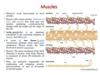
Muscles
• Muscle isa soft tissue found in most
animals.
• Muscle cells contain protein filaments of
actin and myosin that slide past one
another, producing a contraction that
changes both the length and the shape of
the cell.
• Actin, protein that is an important
contributor to the contractile property of
muscle and other cells.
• It exists in two forms: G-actin
(monomeric globular actin) and F-actin
(polymeric fibrous actin), the form
involved in muscle contraction.
• Myosin: The main constituent of the thick
filaments is myosin. Each thick filament
is composed of about 250 molecules of
myosin.
• They are primarily responsible for
maintaining and changing posture,
and locomotion, as well as movement
of internal organs.
3.
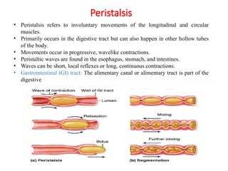
• Peristalsis refersto involuntary movements of the longitudinal and circular
muscles.
• Primarily occurs in the digestive tract but can also happen in other hollow tubes
of the body.
• Movements occur in progressive, wavelike contractions.
• Peristaltic waves are found in the esophagus, stomach, and intestines.
• Waves can be short, local reflexes or long, continuous contractions.
• Gastrointestinal (GI) tract: The alimentary canal or alimentary tract is part of the
digestive
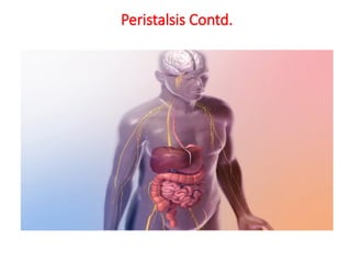
Peristalsis
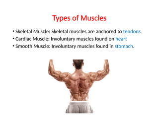
Types of Muscles
•Skeletal Muscle: Skeletal muscles are anchored to tendons
• Cardiac Muscle: Involuntary muscles found on heart
• Smooth Muscle: Involuntary muscles found in stomach.
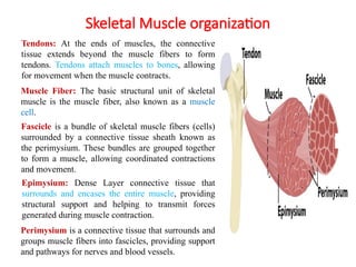
Skeletal Muscle organization
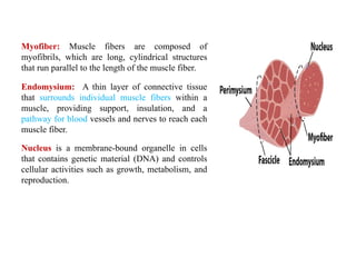
MuscleFiber: The basic structural unit of skeletal
muscle is the muscle fiber, also known as a muscle
cell.
Fascicle is a bundle of skeletal muscle fibers (cells)
surrounded by a connective tissue sheath known as
the perimysium. These bundles are grouped together
to form a muscle, allowing coordinated contractions
and movement.
Epimysium: Dense Layer connective tissue that
surrounds and encases the entire muscle, providing
structural support and helping to transmit forces
generated during muscle contraction.
Tendons: At the ends of muscles, the connective
tissue extends beyond the muscle fibers to form
tendons. Tendons attach muscles to bones, allowing
for movement when the muscle contracts.
Perimysium is a connective tissue that surrounds and
groups muscle fibers into fascicles, providing support
and pathways for nerves and blood vessels.
8.
Endomysium: A thinlayer of connective tissue
that surrounds individual muscle fibers within a
muscle, providing support, insulation, and a
pathway for blood vessels and nerves to reach each
muscle fiber.
Myofiber: Muscle fibers are composed of
myofibrils, which are long, cylindrical structures
that run parallel to the length of the muscle fiber.
Nucleus is a membrane-bound organelle in cells
that contains genetic material (DNA) and controls
cellular activities such as growth, metabolism, and
reproduction.
9.
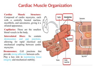
Cardiac Muscle Structure:
Composedof cardio myocytes, each
with a centrally located nucleus,
myofibrils, and sarcomeres, giving it a
striated appearance.
Cardiac Muscle Organization
Capillaries: These are the smallest
blood vessels in the body.
Intercalated Discs: Its contain
desmosomes and gap junctions,
allowing for rapid electrical and
mechanical coupling between cardio
myocytes.
Desmosomes: Cell junctions that
provide strong adhesion between cells.
Play a key role in maintaining tissue
integrity and mechanical stability.
10.
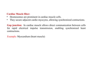
Cardiac Muscle fiber:
•Desmosomes are prominent in cardiac muscle cells.
• They secure adjacent cardio myocytes, allowing synchronized contractions.
Example: Myocardium (heart muscle)
Gap junction: In cardiac muscle allows direct communication between cells
for rapid electrical impulse transmission, enabling synchronized heart
contractions.
11.
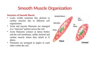
Smooth Muscle Organization
Structureof Smooth Muscle
• Lacks visible striations like skeletal or
cardiac muscles due to different cell
organization.
• Actin and myosin filaments are arranged
in a "staircase" pattern across the cell.
• Actin filaments connect at dense bodies
and the cell membrane, unlike skeletal and
cardiac muscle where they attach to Z
plates.
• Filaments are arranged at angles to each
other within the cell.
12.
Function of SmoothMuscle
• Primary function is contraction, but with notable differences from skeletal
muscle.
• Controlled by the autonomic nervous system, not voluntarily by the somatic
nervous system.
• Contracts persistently, unlike the quick contract-and-release mechanism of
skeletal muscle.
• Calcium levels control the amount of ATP (Adenosine triphosphate)
available for contraction, allowing for sustained tension.
Smooth Muscle Location
• Found in the circulatory system, digestive system, and responsible for
raising body hairs.
• In the circulatory system, smooth muscle regulates blood pressure and
oxygen flow.
• In the digestive system, smooth muscle enables peristalsis, moving food
through the gut.
• Also involved in functions like contracting the iris, raising arm hairs.
13.
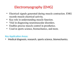
• Electrical signalsgenerated during muscle contraction. EMG
records muscle electrical activity.
• Key role in understanding muscle function.
• Vital in diagnosing neuromuscular disorders.
• Enables precise muscle control in prosthetics.
• Used in sports science, biomechanics, and more.
Key Application Areas:
• Medical diagnosis, research, sports science, biomechanics.
Electromyography (EMG)
14.
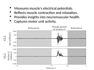
Measures muscle'selectrical potentials.
Reflects muscle contraction and relaxation.
Provides insights into neuromuscular health.
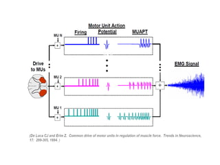
Captures motor unit activity.
15.
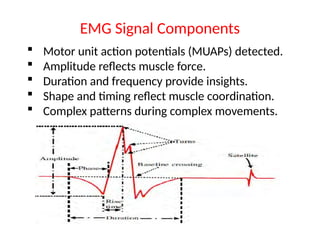
EMG Signal Components
Motor unit action potentials (MUAPs) detected.
Amplitude reflects muscle force.
Duration and frequency provide insights.
Shape and timing reflect muscle coordination.
Complex patterns during complex movements.
16.
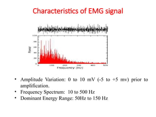
Characteristics of EMGsignal
• Amplitude Variation: 0 to 10 mV (-5 to +5 mv) prior to
amplification.
• Frequency Spectrum: 10 to 500 Hz
• Dominant Energy Range: 50Hz to 150 Hz
17.
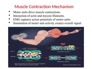
Motor unitsdrive muscle contractions.
Interaction of actin and myosin filaments.
EMG captures action potentials of motor units.
Summation of motor unit activity creates overall signal.
Muscle Contraction Mechanism
18.
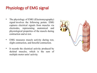
Physiology of EMGsignal
• The physiology of EMG (Electromyography)
signal involves the following points: EMG
captures electrical signals from muscles via
electrodes, representing anatomical and
physiological properties of the muscle during
contraction and at rest.
• EMG measures muscle activity during rest,
slight contraction, and forceful contraction.
• It records the electrical activity produced by
skeletal muscles, which is the sum of
multiple motor units' activity
19.
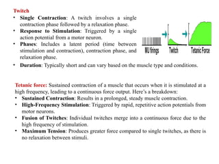
• Single Contraction:A twitch involves a single
contraction phase followed by a relaxation phase.
• Response to Stimulation: Triggered by a single
action potential from a motor neuron.
• Phases: Includes a latent period (time between
stimulation and contraction), contraction phase, and
relaxation phase.
Twitch
Tetanic force: Sustained contraction of a muscle that occurs when it is stimulated at a
high frequency, leading to a continuous force output. Here’s a breakdown:
• Sustained Contraction: Results in a prolonged, steady muscle contraction.
• High-Frequency Stimulation: Triggered by rapid, repetitive action potentials from
motor neurons.
• Fusion of Twitches: Individual twitches merge into a continuous force due to the
high frequency of stimulation.
• Maximum Tension: Produces greater force compared to single twitches, as there is
no relaxation between stimuli.
• Duration: Typically short and can vary based on the muscle type and conditions.
21.
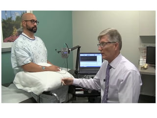
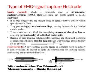
Type of EMG-signalcapture Electrode
Needle electrode, which is commonly used in intramuscular
electromyography (EMG). Here are some key points related to needle
electrodes:
• It inserted directly into the muscle tissue to detect electrical activity within
specific muscle fibers.
• They provide highly localized recordings, making them useful for detailed
muscle studies.
• These electrodes are ideal for identifying neuromuscular disorders or
assessing the functionality of individual motor units.
• Because of their invasive nature, needle electrodes are often used in clinical
or diagnostic settings to monitor deep muscles where surface electrodes may
not be effective.
Microelectrode: A tiny electrode used to record or stimulate electrical activity
in cells or tissues. It's crucial in fields like neuroscience for studying neurons
and creating brain-computer interfaces.
22.
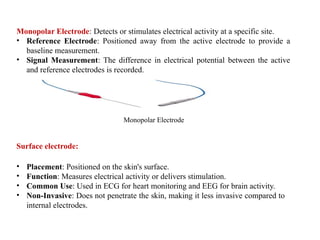
Surface electrode:
Monopolar Electrode:Detects or stimulates electrical activity at a specific site.
• Reference Electrode: Positioned away from the active electrode to provide a
baseline measurement.
• Signal Measurement: The difference in electrical potential between the active
and reference electrodes is recorded.
Monopolar Electrode
• Placement: Positioned on the skin's surface.
• Function: Measures electrical activity or delivers stimulation.
• Common Use: Used in ECG for heart monitoring and EEG for brain activity.
• Non-Invasive: Does not penetrate the skin, making it less invasive compared to
internal electrodes.
23.
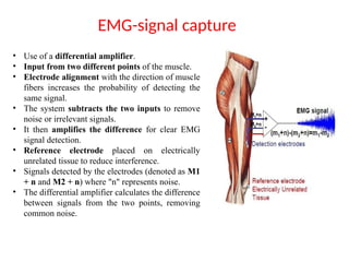
EMG-signal capture
• Useof a differential amplifier.
• Input from two different points of the muscle.
• Electrode alignment with the direction of muscle
fibers increases the probability of detecting the
same signal.
• The system subtracts the two inputs to remove
noise or irrelevant signals.
• It then amplifies the difference for clear EMG
signal detection.
• Reference electrode placed on electrically
unrelated tissue to reduce interference.
• Signals detected by the electrodes (denoted as M1
+ n and M2 + n) where "n" represents noise.
• The differential amplifier calculates the difference
between signals from the two points, removing
common noise.
25.
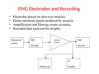
• Electrodes placedon skin over muscles.
• Detect electrical signals produced by muscles.
• Amplification and filtering ensure accuracy.
• Recorded data analyzed for insights.
EMG Electrodes and Recording
26.
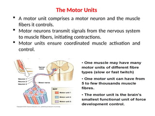
A motorunit comprises a motor neuron and the muscle
fibers it controls.
Motor neurons transmit signals from the nervous system
to muscle fibers, initiating contractions.
Motor units ensure coordinated muscle activation and
control.
The Motor Units
28.
Underlying Physiological Issues
RestingMembrane Potential:
The resting membrane potential (RMP) is the electrical
potential difference across the cell membrane of a non-
excited, resting cell.
Potential difference exists across the sarcomere
Intra-cellular fluid has a high [K+
]
Extra-cellular (interstitial) fluid has a high [Na+
] and [Cl-
]
29.
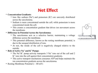
Net Effect
• ConcentrationGradients:
• Ions like sodium (Na ) and potassium (K ) are unevenly distributed
⁺ ⁺
across the sarcolemma.
• Sodium is more concentrated outside the cell, while potassium is more
concentrated inside the cell.
• This creates a concentration gradient that drives ion movement across
the membrane.
• Difference in Potential Across the Sarcolemma:
• The sarcolemma acts as a selective barrier, maintaining a voltage
difference across the membrane.
• This potential difference, known as the resting membrane potential, is
due to the unequal distribution of ions.
• At rest, the inside of the cell is negatively charged relative to the
outside.
• Role of Active Na and K Pumps
⁺ ⁺ :
• The Na /K pump actively transports 3 Na ions out of the cell and 2
⁺ ⁺ ⁺
K ions into the cell against their concentration gradients.
⁺
• This active transport mechanism consumes ATP and helps maintain the
ion concentration gradients across the sarcolemma.
• Resting Membrane Potential (~ -80 mV)
30.
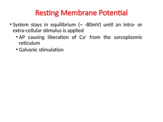
Resting Membrane Potential
•System stays in equilibrium (~ -80mV) until an intra- or
extra-cellular stimulus is applied
• AP causing liberation of Ca+
from the sarcoplasmic
reticulum
• Galvanic stimulation
31.
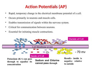
Action Potentials (AP)
•Rapid, temporary change in the electrical membrane potential of a cell.
• Occurs primarily in neurons and muscle cells.
• Enables transmission of signals within the nervous system.
• Critical for communication between neurons.
• Essential for initiating muscle contractions.
Cell Membrane at rest
Na⁺ Cl-
K⁺
Cl-
K⁺ A-
Outside of Cell
Inside of Cell
Potassium (K+) can pass
through to equalize its
concentration
Sodium and Chlorine
cannot pass through
Result:- inside is
negative relative
to outside
- 70 mv
32.
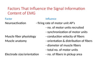
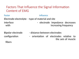
Factors That Influencethe Signal Information
Content of EMG
Factor Influence
Neuroactivation - firing rate of motor unit AP’s
- no. of motor units recruited
- synchronization of motor units
Muscle fiber physiology - conduction velocity of fibers
Muscle anatomy - orientation & distribution of fibers
- diameter of muscle fibers
- total no. of motor units
Electrode size/orientation - no. of fibers in pickup area
33.
Factors That Influencethe Signal Information
Content of EMG
Factor Influence
Electrode-electrolyte - type of material and site
interface - electrode impedance decreases
with increasing frequency
Bipolar electrode - distance between electrodes
configuration - orientation of electrodes relative to
the axis of muscle
fibers
34.
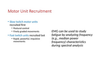
Motor Unit Recruitment
•Slow twitch motor units
recruited first
• Postural control
• Finely graded movements
• Fast twitch units recruited last
• Rapid, powerful, impulsive
movements
EMG can be used to study
fatigue by analyzing frequency
(e.g., median power
frequency) characteristics
during spectral analysis
35.
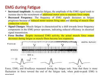
EMG during Fatigue
•Increased Amplitude: As muscles fatigue, the amplitude of the EMG signal tends to
increase due to the recruitment of additional motor units to maintain force output.
• Decreased Frequency: The frequency of EMG signals decreases as fatigue
progresses because of reduced motor neuron firing rates and slowing of muscle fiber
conduction velocity.
• Signal Changes: Muscle fatigue is characterized by a shift towards lower frequency
components in the EMG power spectrum, indicating reduced efficiency in electrical
signal transmission.
• Force Decline: Despite increased EMG activity, the actual muscle force output
decreases during fatigue as muscle fibers become less responsive.
Force, EMG, and H reflexes measured during the fatigue task. Note that there is more
‐
fluctuation in force toward the end of the fatigue task, when peak to peak EMG is
‐ ‐
36.
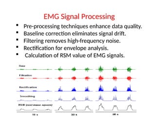
Pre-processing techniquesenhance data quality.
Baseline correction eliminates signal drift.
Filtering removes high-frequency noise.
Rectification for envelope analysis.
Calculation of RSM value of EMG signals.
EMG Signal Processing
37.
Clinical Applications
Diagnosingneuromuscular disorders.
Monitoring progress in rehabilitation.
Assessing muscle activity in patients.
Guiding personalized treatment plans.
Vital tool in neurology and rehabilitation.
38.
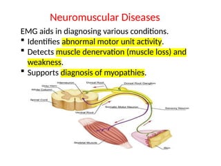
EMG aids indiagnosing various conditions.
Identifies abnormal motor unit activity.
Detects muscle denervation (muscle loss) and
weakness.
Supports diagnosis of myopathies.
Neuromuscular Diseases
39.
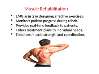
EMG assistsin designing effective exercises.
Monitors patient progress during rehab.
Provides real-time feedback to patients.
Tailors treatment plans to individual needs.
Enhances muscle strength and coordination.
Muscle Rehabilitation
40.
EMG assessesmuscle activation during
movements.
Insights into muscle coordination and function.
Optimizes training and sports performance.
Analyzes muscle fatigue and efficiency.
Guides sports-specific training programs.
Sports Science and Biomechanics
41.
EMG detectsmotor neuron diseases.
Identifies disruptions in motor unit activity.
Supports diagnosis of ALS (Amyotrophic
Lateral Sclerosis).
Monitors disease progression.
Aids in assessing treatment effectiveness.
Motor Neuron Disorders
42.
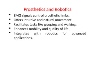
EMG signalscontrol prosthetic limbs.
Offers intuitive and natural movement.
Facilitates tasks like grasping and walking.
Enhances mobility and quality of life.
Integrates with robotics for advanced
applications.
Prosthetics and Robotics
43.
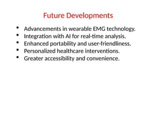
Advancements inwearable EMG technology.
Integration with AI for real-time analysis.
Enhanced portability and user-friendliness.
Personalized healthcare interventions.
Greater accessibility and convenience.
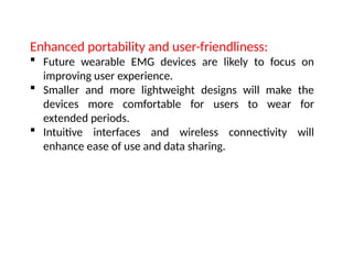
Future Developments
44.
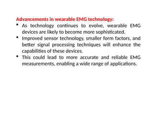
Advancements in wearableEMG technology:
As technology continues to evolve, wearable EMG
devices are likely to become more sophisticated.
Improved sensor technology, smaller form factors, and
better signal processing techniques will enhance the
capabilities of these devices.
This could lead to more accurate and reliable EMG
measurements, enabling a wide range of applications.
45.
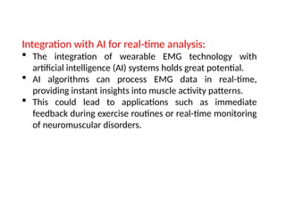
Integration with AIfor real-time analysis:
The integration of wearable EMG technology with
artificial intelligence (AI) systems holds great potential.
AI algorithms can process EMG data in real-time,
providing instant insights into muscle activity patterns.
This could lead to applications such as immediate
feedback during exercise routines or real-time monitoring
of neuromuscular disorders.
46.
Enhanced portability anduser-friendliness:
Future wearable EMG devices are likely to focus on
improving user experience.
Smaller and more lightweight designs will make the
devices more comfortable for users to wear for
extended periods.
Intuitive interfaces and wireless connectivity will
enhance ease of use and data sharing.
47.
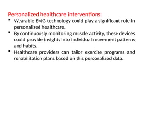
Personalized healthcare interventions:
Wearable EMG technology could play a significant role in
personalized healthcare.
By continuously monitoring muscle activity, these devices
could provide insights into individual movement patterns
and habits.
Healthcare providers can tailor exercise programs and
rehabilitation plans based on this personalized data.
48.
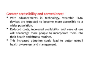
Greater accessibility andconvenience:
With advancements in technology, wearable EMG
devices are expected to become more accessible to a
wider population.
Reduced costs, increased availability, and ease of use
will encourage more people to incorporate them into
their health and fitness routines.
This increased adoption could lead to better overall
health awareness and management.
49.
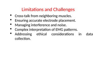
Cross-talk fromneighboring muscles.
Ensuring accurate electrode placement.
Managing interference and noise.
Complex interpretation of EMG patterns.
Addressing ethical considerations in data
collection.
Limitations and Challenges
50.
More Biomedical Signal
Electroneurogram(ENG)
Electrical Signal observed as a stimulus and associated nerve action
potential propagating over the length of the nerve.
Event-related potential (ERP):
It includes ENG or EEG in response to light, sound, electrical potential.
Electrogastrogram (EGG)
Electrical activity of stomach consists of rhythmic waves of
depolarization and repolarization of its stomach muscles. Originates
at mid-corpus of stomach and always present. Not directly associated
with contraction.
(muscle movement i.e. contraction and expansion is such that food
will move in one direction)
51.
More Biomedical Signal
Phonocardiogram(PCG)
Vibration or sound related to contraction of cardio hemic system (the
heart and blood together)
Carotid Pulse (CP)
Pressure signal recorded over carotid artery as it passes near the
surface of body at neck.

![Underlying Physiological Issues
Resting Membrane Potential:
The resting membrane potential (RMP) is the electrical
potential difference across the cell membrane of a non-
excited, resting cell.
Potential difference exists across the sarcomere
Intra-cellular fluid has a high [K+
]
Extra-cellular (interstitial) fluid has a high [Na+
] and [Cl-
]](https://image.slidesharecdn.com/emg-250303110003-bc588e2e/85/EMG_____-___________-________-pptx-28-320.jpg)