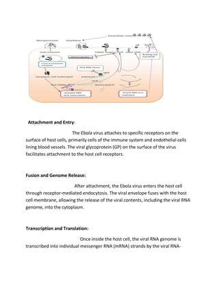
The Ebola virus was first identified in 1976 near the Ebola River. It is transmitted through contact with bodily fluids and causes a sudden onset of fever and other symptoms. While there is no specific treatment, supportive care is important. Outbreaks have primarily occurred in Central and West Africa. Diagnosis involves clinical assessment, blood tests like RT-PCR to detect the virus, and considering exposure history. The virus attaches to host cells and replicates its RNA genome inside the cell before releasing new virus particles. Several commercial tests can rapidly detect the virus.
Combating Ebola- Vaccines and InterferonsStudy Buddy
This ppt tells you about Ebola virus; its transmission methods, how it affects the immune system and evades its action, its major symptoms, epidemiology and how to combat it. Main focus is given on vaccines and use of interferons
The Indian Dental Academy is the Leader in continuing dental education , training dentists in all aspects of dentistry and
offering a wide range of dental certified courses in different formats.for more details please visit
www.indiandentalacademy.com
A detailed description of HIV covering virology, morphology, pathogenesis, clinical stages and manifestations, laboratory diagnosis, and diagnostic strategy, and therapeutic options and prevention.
here i discussed some human oncogenic viruses , their epidemeology, life cycle, treatment, prevention and control. . oncogenic viruses are cancer causing viruses.
There are nearly 100 viruses of the herpes group that infect many different animal species.
Official name of herpesviruses that commonly infect human is Humans herpesvirus (HHV)
herpes simplex virus types 1 (HHV 1)
Herpes simplex virus type 2 (HHV 2)
Varicella-zoster virus (HHV 3)
Epstein-Barr virus, (HHV 4)
Cytomegalovirus (HHV 5)
Human herpesvirus 6 (HHV 6)
Human herpesvirus 7 (HHV 7)
Human herpesvirus 8 (HHV 8) (Kaposi's sarcoma-associated herpesvirus).
Herpes B virus of monkeys can also infect humans
hELMINTHS#corona virus#Aspergillosis#BUGANDO#CUHAS#CUHAS#CUHAS
This presentation intends to explore the application of virus in different biomedical fields and research with special reference to vaccine production and plant viral diseases.
The use of Nauplii and metanauplii artemia in aquaculture (brine shrimp).pptxMAGOTI ERNEST
Although Artemia has been known to man for centuries, its use as a food for the culture of larval organisms apparently began only in the 1930s, when several investigators found that it made an excellent food for newly hatched fish larvae (Litvinenko et al., 2023). As aquaculture developed in the 1960s and ‘70s, the use of Artemia also became more widespread, due both to its convenience and to its nutritional value for larval organisms (Arenas-Pardo et al., 2024). The fact that Artemia dormant cysts can be stored for long periods in cans, and then used as an off-the-shelf food requiring only 24 h of incubation makes them the most convenient, least labor-intensive, live food available for aquaculture (Sorgeloos & Roubach, 2021). The nutritional value of Artemia, especially for marine organisms, is not constant, but varies both geographically and temporally. During the last decade, however, both the causes of Artemia nutritional variability and methods to improve poorquality Artemia have been identified (Loufi et al., 2024).
Brine shrimp (Artemia spp.) are used in marine aquaculture worldwide. Annually, more than 2,000 metric tons of dry cysts are used for cultivation of fish, crustacean, and shellfish larva. Brine shrimp are important to aquaculture because newly hatched brine shrimp nauplii (larvae) provide a food source for many fish fry (Mozanzadeh et al., 2021). Culture and harvesting of brine shrimp eggs represents another aspect of the aquaculture industry. Nauplii and metanauplii of Artemia, commonly known as brine shrimp, play a crucial role in aquaculture due to their nutritional value and suitability as live feed for many aquatic species, particularly in larval stages (Sorgeloos & Roubach, 2021).
ESR spectroscopy in liquid food and beverages.pptxPRIYANKA PATEL
With increasing population, people need to rely on packaged food stuffs. Packaging of food materials requires the preservation of food. There are various methods for the treatment of food to preserve them and irradiation treatment of food is one of them. It is the most common and the most harmless method for the food preservation as it does not alter the necessary micronutrients of food materials. Although irradiated food doesn’t cause any harm to the human health but still the quality assessment of food is required to provide consumers with necessary information about the food. ESR spectroscopy is the most sophisticated way to investigate the quality of the food and the free radicals induced during the processing of the food. ESR spin trapping technique is useful for the detection of highly unstable radicals in the food. The antioxidant capability of liquid food and beverages in mainly performed by spin trapping technique.
Travis Hills' Endeavors in Minnesota: Fostering Environmental and Economic Pr...Travis Hills MN
Travis Hills of Minnesota developed a method to convert waste into high-value dry fertilizer, significantly enriching soil quality. By providing farmers with a valuable resource derived from waste, Travis Hills helps enhance farm profitability while promoting environmental stewardship. Travis Hills' sustainable practices lead to cost savings and increased revenue for farmers by improving resource efficiency and reducing waste.
Nucleophilic Addition of carbonyl compounds.pptxSSR02
Nucleophilic addition is the most important reaction of carbonyls. Not just aldehydes and ketones, but also carboxylic acid derivatives in general.
Carbonyls undergo addition reactions with a large range of nucleophiles.
Comparing the relative basicity of the nucleophile and the product is extremely helpful in determining how reversible the addition reaction is. Reactions with Grignards and hydrides are irreversible. Reactions with weak bases like halides and carboxylates generally don’t happen.
Electronic effects (inductive effects, electron donation) have a large impact on reactivity.
Large groups adjacent to the carbonyl will slow the rate of reaction.
Neutral nucleophiles can also add to carbonyls, although their additions are generally slower and more reversible. Acid catalysis is sometimes employed to increase the rate of addition.
Phenomics assisted breeding in crop improvementIshaGoswami9
As the population is increasing and will reach about 9 billion upto 2050. Also due to climate change, it is difficult to meet the food requirement of such a large population. Facing the challenges presented by resource shortages, climate
change, and increasing global population, crop yield and quality need to be improved in a sustainable way over the coming decades. Genetic improvement by breeding is the best way to increase crop productivity. With the rapid progression of functional
genomics, an increasing number of crop genomes have been sequenced and dozens of genes influencing key agronomic traits have been identified. However, current genome sequence information has not been adequately exploited for understanding
the complex characteristics of multiple gene, owing to a lack of crop phenotypic data. Efficient, automatic, and accurate technologies and platforms that can capture phenotypic data that can
be linked to genomics information for crop improvement at all growth stages have become as important as genotyping. Thus,
high-throughput phenotyping has become the major bottleneck restricting crop breeding. Plant phenomics has been defined as the high-throughput, accurate acquisition and analysis of multi-dimensional phenotypes
during crop growing stages at the organism level, including the cell, tissue, organ, individual plant, plot, and field levels. With the rapid development of novel sensors, imaging technology,
and analysis methods, numerous infrastructure platforms have been developed for phenotyping.
Professional air quality monitoring systems provide immediate, on-site data for analysis, compliance, and decision-making.
Monitor common gases, weather parameters, particulates.
Comparing Evolved Extractive Text Summary Scores of Bidirectional Encoder Rep...University of Maribor
Slides from:
11th International Conference on Electrical, Electronics and Computer Engineering (IcETRAN), Niš, 3-6 June 2024
Track: Artificial Intelligence
https://www.etran.rs/2024/en/home-english/
Toxic effects of heavy metals : Lead and Arsenicsanjana502982
Heavy metals are naturally occuring metallic chemical elements that have relatively high density, and are toxic at even low concentrations. All toxic metals are termed as heavy metals irrespective of their atomic mass and density, eg. arsenic, lead, mercury, cadmium, thallium, chromium, etc.
Observation of Io’s Resurfacing via Plume Deposition Using Ground-based Adapt...Sérgio Sacani
Since volcanic activity was first discovered on Io from Voyager images in 1979, changes
on Io’s surface have been monitored from both spacecraft and ground-based telescopes.
Here, we present the highest spatial resolution images of Io ever obtained from a groundbased telescope. These images, acquired by the SHARK-VIS instrument on the Large
Binocular Telescope, show evidence of a major resurfacing event on Io’s trailing hemisphere. When compared to the most recent spacecraft images, the SHARK-VIS images
show that a plume deposit from a powerful eruption at Pillan Patera has covered part
of the long-lived Pele plume deposit. Although this type of resurfacing event may be common on Io, few have been detected due to the rarity of spacecraft visits and the previously low spatial resolution available from Earth-based telescopes. The SHARK-VIS instrument ushers in a new era of high resolution imaging of Io’s surface using adaptive
optics at visible wavelengths.
Observation of Io’s Resurfacing via Plume Deposition Using Ground-based Adapt...
Ebola Viruse slid show.docx
1. Topic:
Ebola virus
Ebola Virus:
The Ebola virus is a highly infectious and often fatal virus that belongs to the
Filoviridae family. It was first identified in 1976 near the Ebola River in what is
now the Democratic Republic of Congo. Ebola virus disease (EVD), also known as
Ebola hemorrhagic fever, is the illness caused by the Ebola virus.
Ebola virus is transmitted to humans through direct contact with the blood, bodily
fluids, or tissues of infected animals, such as fruit bats, chimpanzees, gorillas,
monkeys, forest antelope, and porcupines. Once the virus enters the human
population, it can spread from person to person through direct contact with the
blood or other bodily fluids of an infected individual. It is important to note that
Ebola is not an airborne virus and does not spread through casual contact or
respiratory droplets like the flu.
SYMPTOMS OF EBOLA VIRUS:
2. The symptoms of Ebola virus disease typically begin with a sudden onset of fever,
fatigue, muscle pain, headache, and sore throat. This is followed by vomiting,
diarrhea, rash, impaired kidney and liver function, and, in some cases, internal
and external bleeding. The severity of the disease can vary, with case fatality rates
ranging from 25% to 90%, depending on the outbreak and the healthcare
resources available.
TREATMENT OF EBOLA VIRUS:
There is currently no specific treatment for Ebola virus disease. Supportive care,
including providing fluids, managing complications, and treating secondary
infections, is crucial. Experimental treatments and vaccines have been developed
and tested, showing promising results in some cases.
OCCURANCE OF EBOLA VIRUS:
Ebola outbreaks have occurred primarily in Central and West African countries,
with occasional outbreaks in other regions. The World Health Organization (WHO)
and other international health organizations work closely with affected countries
to control the spread of the virus, provide medical care, and promote prevention
measures.
It's important to note that information about the Ebola virus is constantly
evolving, and the latest updates should be sought from reputable health
organizations and authorities.
3. The Ebola virus has several structural and non-structural proteins that play
important roles in its lifecycle and pathogenicity. Here are some of the key
proteins associated with the Ebola virus:
PROTEINS OF EBOLA VIRUS:
1-STRUCTURAL PROTEIN:
I. Glycoprotein (GP):
The viral surface glycoprotein (GP) is a critical structural protein
that protrudes from the viral envelope. It plays a crucial role in viral entry into
host cells by binding to specific receptors on the cell surface. The GP protein is
also a major target for host immune responses and is involved in
evading the immune system.
II. Nucleoprotein (NP):
The nucleoprotein forms complexes with the viral
RNA to create the helical nucleocapsid structure
inside the viral envelope. The NP protein is involved
4. in protecting and packaging the viral genetic material
and is crucial for viral replication and transcription.
III. Matrix Protein (VP40):
The matrix protein, VP40, is located beneath the viral
envelope. It interacts with both the viral envelope and the nucleocapsid,
providing structural support to the virus. VP40 is also involved in the assembly
and release of new viral particles.
2-Non-structural Proteins:
I. Viral Protein 24 (VP24):
VP24 is a non-structural protein that plays a role in
counteracting the host's immune response. It
interferes with the production of interferons, which
are important antiviral molecules produced by the
host cells. By inhibiting interferon signaling, VP24
helps the virus evade the immune system.
II. Viral Protein 35 (VP35):
VP35 is another non-structural protein involved in
counteracting the host immune response. It has multiple
functions, including suppressing the production of interferons
and inhibiting the host cell's antiviral defenses. VP35 also plays
a role in viral RNA synthesis and replication.
5. III. Viral Protein 30 (VP30):
VP30 is a non-structural protein that acts as a transcription
factor, regulating viral gene expression and replication. It
interacts with the viral RNA and is essential for initiating
viral transcription.
IV. Viral Protein 40 (VP40):
Although the same name as the structural matrix
protein, the non-structural VP40 is distinct. It plays a role in viral replication
and assembly, specifically in the formation of the viral nucleocapsid.
These are some of the key structural and non-structural proteins associated
with the Ebola virus. Each protein serves specific functions during the virus's
replication cycle and contributes to its pathogenicity. Understanding the roles
of these proteins is crucial in developing targeted therapies and vaccines to
combat Ebola virus infections.
Sample for diagnosis of Ebola virus
6. Diagnosing Ebola virus infection typically involves a combination of clinical
assessment, laboratory testing, and epidemiological information. Here is a general
outline of the diagnostic process for Ebola virus:
7. Clinical Assessment:
Healthcare professionals evaluate the patient's symptoms, medical
history, and potential exposure to the virus. Symptoms of Ebola virus infection
can include fever, headache, muscle pain, weakness, fatigue, vomiting, diarrhea,
and in some cases, bleeding.
Blood Tests:
Blood samples are collected from the patient for laboratory testing. The
samples are handled with extreme caution and analyzed in a specialized
laboratory equipped to handle highly infectious agents like Ebola virus.
a. Reverse Transcription Polymerase Chain Reaction (RT-PCR):
This technique detects
the presence of Ebola virus genetic material (RNA) in the patient's blood. It
involves converting viral RNA into complementary DNA (cDNA) and amplifying
specific regions of the viral genome. RT-PCR provides a sensitive and specific
method for diagnosing Ebola virus.
b. Antigen Detection:
Enzyme-linked immunosorbent assays (ELISAs) or rapid
diagnostic tests (RDTs) can be used to detect viral antigens in the patient's blood.
These tests identify specific viral proteins produced during infection.
Serology:
Serological tests are conducted to detect the presence of antibodies
produced by the patient's immune system in response to Ebola virus infection.
8. These tests are typically performed during the later stages of illness or during the
recovery phase.
a. Enzyme-linked Immunosorbent Assay (ELISA):
ELISA detects the presence of
antibodies, such as immunoglobulin M (IgM) and immunoglobulin G (IgG),
against the Ebola virus in the patient's blood. IgM antibodies are produced
early in the infection, while IgG antibodies are generated later and can
indicate a previous infection or immunity.
Epidemiological Information:
Information regarding the patient's recent travel
history, exposure to infected individuals, or contact with animals associated with
Ebola virus transmission is collected. This helps in assessing the likelihood of Ebola
virus infection and provides important epidemiological data.
It is important to note that the diagnosis of Ebola virus infection requires
specialized laboratory facilities and expertise. Suspected cases should be handled
with strict infection control measures to prevent the spread of the virus.
Healthcare professionals and laboratories following proper protocols and
guidelines ensure accurate and safe diagnosis of Ebola virus infection.
Mechanism of replication in Ebola virus:
There are following mechanism of Replication in Ebola Virus:
9. Attachment and Entry:
The Ebola virus attaches to specific receptors on the
surface of host cells, primarily cells of the immune system and endothelial cells
lining blood vessels. The viral glycoprotein (GP) on the surface of the virus
facilitates attachment to the host cell receptors.
Fusion and Genome Release:
After attachment, the Ebola virus enters the host cell
through receptor-mediated endocytosis. The viral envelope fuses with the host
cell membrane, allowing the release of the viral contents, including the viral RNA
genome, into the cytoplasm.
Transcription and Translation:
Once inside the host cell, the viral RNA genome is
transcribed into individual messenger RNA (mRNA) strands by the viral RNA-
10. dependent RNA polymerase. These viral mRNA molecules are then translated by
the host cell's ribosomes into viral proteins.
Replication:
The Ebola virus replicates its RNA genome in the host cell's cytoplasm.
The viral RNA-dependent RNA polymerase uses the viral mRNA strands as
templates to generate new copies of the viral RNA genome.
Assembly:
Newly synthesized viral proteins, including nucleocapsid proteins and
matrix proteins, are produced in the host cell. These proteins interact with the
replicated viral RNA to form new viral nucleocapsids.
Budding:
The viral nucleocapsids move to the host cell's plasma membrane,
where they acquire a viral envelope derived from the host cell membrane. The
viral envelope contains the viral glycoproteins, including GP. The fully assembled
Ebola viruses bud from the host cell, incorporating the viral glycoproteins into the
envelope.
Release:
The newly formed Ebola viruses are released from the host cell, ready to
infect other cells and continue the replication cycle. This release can occur
through various mechanisms, including cell lysis or budding from the plasma
membrane.
11. The replication mechanism of the Ebola virus is complex and involves intricate
interactions with host cell components. Understanding the details of this process
is crucial for developing strategies to interrupt viral replication and control the
spread of Ebola virus infection.
Commercial test available for diagnosis:
There are following Commercial test available
for diagnosis :
GeneXpert Ebola Assay:
The GeneXpert Ebola Assay is a rapid molecular diagnostic
test developed by Cepheid. It utilizes the GeneXpert platform, which combines
nucleic acid amplification and real-time PCR technology to detect the Ebola virus
RNA in patient samples. The test provides results within a few hours.
ReEBOV Antigen Rapid Test:
The ReEBOV Antigen Rapid Test is a lateral flow
immunoassay developed by Corgenix. It is designed to detect specific Ebola virus
antigens in patient blood samples. The test provides results within 15-30 minutes
and does not require specialized laboratory equipment.
Real Star Filovirus Screen RT-PCR Kit:
The Real Star Filovirus Screen RT-PCR Kit is a
molecular diagnostic test developed by altona Diagnostics. It is designed to detect
and differentiate multiple filoviruses, including Ebola virus, in patient samples
using real-time reverse transcription polymerase chain reaction (RT-PCR)
technology. The test is performed in specialized laboratories equipped for
molecular diagnostics.
12. It's important to note that the availability and specific details of commercial
diagnostic tests may vary over time and across different regions. Additionally, the
use of these tests should be conducted by trained healthcare professionals in
appropriate laboratory settings following established protocols and guidelines.
For the most up-to-date information on commercial tests for Ebola virus
diagnosis, it is recommended to consult with local health authorities or reputable
diagnostic companies.