2. Diffusion Tensor Imaging 535
nates. Diffusion anisotropy in white matter originates
from its specific organization in bundles of more or less
myelinated axonal fibers running in parallel, although
the exact mechanism is still not completely understood
(see below): diffusion in the direction of the fibers is
faster than in the perpendicular direction. It quickly
appeared that this feature could be exploited to map
out the orientation in space of the white matter tracks
in the brain using a color scale, assuming that the
direction of the fastest diffusion would indicate the
overall orientation of the fibers (15).
This pioneering work on diffusion anisotropy really
took off with the introduction of the more rigorous for-
malism of the diffusion tensor by Basser et al. (16 –18).
With diffusion tensor imaging (DTI), diffusion anisot-
ropy effects in diffusion MRI data could be fully ex-
tracted, characterized, and exploited, providing even
more exquisite details of tissue microstructure. Many
studies have been published dealing with the optimiza-
tion of the MRI sequences necessary to gain access to
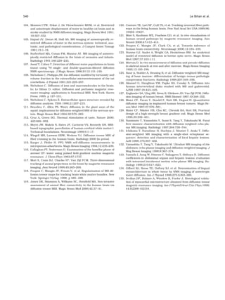
the diffusion tensor, the processing and display of DTI
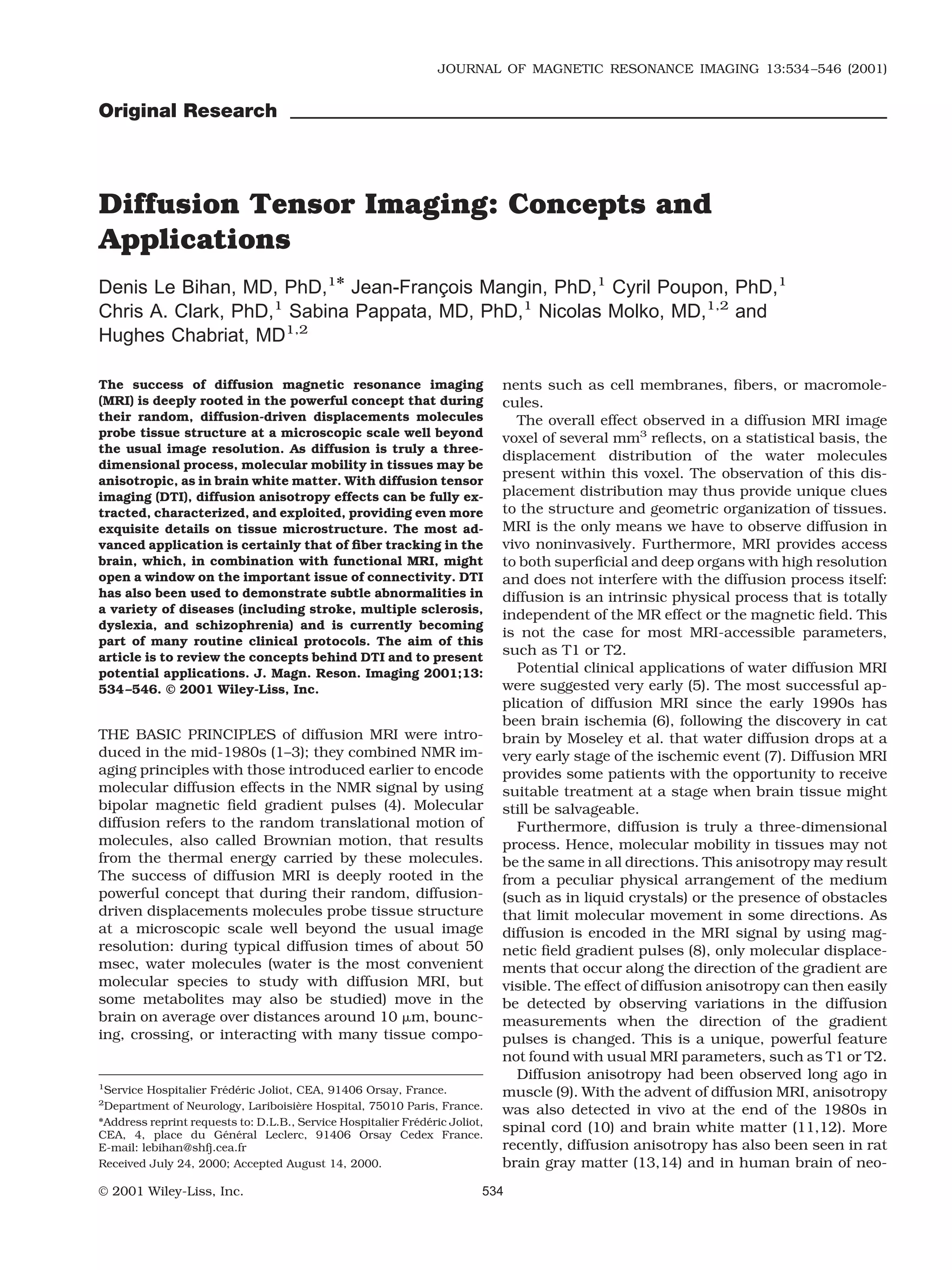
Figure 1. The diffusion tensor, axial slices. Diffusivity along x,
data, and, of course, potential applications. The most
y, and z axes, shown in Dxx (a), Dyy (d), and Dzz (f) images,
advanced application is certainly that of fiber tracking
respectively, is clearly different in white matter, especially in
in the brain, which, in combination with functional the corpus callosum. Nondiagonal images [Dxy (b), Dxz (c), and
MRI, might open a window onto the important issue of Dyz (e)] are not noise images because the x, y, z reference frame
connectivity. of the MRI scanner does not coincide with the diffusion refer-
The aim of this article is to review the concepts behind ence frame of tissues in most voxels.
DTI and to present potential applications.
DIFFUSION TENSOR MRI: DATA ACQUISITION where bii are the elements of the b matrix (which now
AND PROCESSING replaces the b factor) expressed in the coordinates of
The Diffusion Tensor this reference frame.
In practice, unfortunately, measurements are made
With plain diffusion MRI, diffusion is fully described
in the reference frame [ x, y, z] of the MRI scanner
using a single (scalar) parameter, the diffusion coeffi-
gradients, which usually does not coincide with the
cient, D. The effect of diffusion on the MRI signal (most
diffusion frame of the tissue. Therefore, one must also
often a spin-echo signal) is an attenuation, A, which
consider the coupling of the nondiagonal elements, bij,
depends on D and on the “b factor,” which characterizes
of the b matrix with the nondiagonal terms, Dij, (i j),
the the gradient pulses (timing, amplitude, shape) used
of the diffusion tensor (now expressed in the scanner
in the MRI sequence (8):
frame), which reflect correlation between molecular dis-
placements in perpendicular directions (17):
A exp bD . (1)
However, in the presence of anisotropy, diffusion can A exp bijDij (4)
no longer be characterized by a single scalar coefficient, i x,y,z j x,y,z
but requires a tensor, D, which fully describes molecu-
lar mobility along each direction and correlation be- or
tween these directions (4) (Fig. 1):
A exp bxxDxx byyDyy bzzDzz
Dxx Dxy Dxz
D Dyx Dyy Dyz . (2) 2bxyDxy 2bxzDxz 2byzDyz . (5)
Dzx Dzy Dzz
Hence, it is important to note that by using diffusion-
This tensor is symmetric (Dij Dji, with i, j x, y, z). encoding gradient pulses along one direction only, say
In a reference frame [ x , y , z ] that coincides with the x, signal attenuation not only depends on the diffusion
principal or self directions of diffusivity, the off-diagonal effects along this direction but may also include contri-
terms do not exist, and the tensor is reduced only to its bution from other directions, say y and z.
diagonal terms, Dx x , Dy y , Dz z , which represent molec- Calculation of b may quickly become complicated
ular mobility along axes x , y , and z , respectively. The when many gradient pulses are used (20,21), but the
echo attenuation then becomes: full determination of the diffusion tensor is necessary if
one wants to assess properly and fully all anisotropic
A exp bx x Dx x by y Dy y bz z Dz z (3) diffusion effects.
3. 536 Le Bihan et al.
DTI Data Acquisition
To determine the diffusion tensor fully, one must first
collect diffusion-weighted images along several gradi-
ent directions, using diffusion-sensitized MRI pulse se-
quences (22) such as echoplanar imaging (EPI). As the
diffusion tensor is symmetric, measurements along
only six directions are mandatory (instead of nine),
along with an image acquired without diffusion weight-
ing (b 0). A typical set of gradient combinations that
preserves uniform space sampling and similar b values
along each direction is as follows (coefficients for gradi-
ent pulses along the (x, y, z) axes, normalized to a given
amplitude, G):
1/ 2, 0, 1/ 2 ; 1/ 2, 0, 1/ 2 ;
0, 1/ 2, 1/ 2 ; 0, 1/ 2, 1/ 2 ;
1/ 2, 1/ 2, 0 ; 1/ 2, 1/ 2, 0 .
This minimal set of images may be repeated for averag-
ing, as the signal-to-noise ratio (SNR) may be low
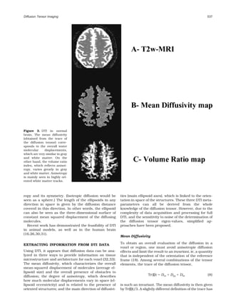
(17,18). Note that it is not necessary (and not efficient in Figure 2. Schemes for directional sampling. Although initial
terms of SNR) to obtain data with different b values for sampling schemes consisted of six (or even four) directions of
each direction. A simple calculation (22) shows that, in measurements, progress is made in scanning space more uni-
the case of scalar diffusion, one gets the best accuracy formly along many directions, especially for fiber orientation
on the diffusion coefficient, D, from two acquisitions mapping.
when using 2 b factors, b1 and b2, differing by about
1/D (23). In the brain, this translates to (b2 b1)
1000–1500 s/mm2. If more than two acquisitions are to
multiple linear regression, using Eq. [5]. From the dif-
be used (for averaging purposes), error propagation the-
fusion tensor components, one may calculate indices
ory (24) shows that it is better to accumulate n1 and n2
that reflect either the average diffusion or the degree of
measurements at each of the low and high b factors, b1
anisotropy in each voxel (see below). Another step is to
and b2, than to use a range of b factors. The accuracy
determine the main direction of diffusivities in each
dD/D is then obtained from the raw image SNR:
voxel and the diffusion values associated with these
1/2 directions. This is equivalent to determining the refer-
dD/D 1/n 1 exp 2D b1 b2 /n2
ence frame [ x , y , z ] where the off-diagonal terms of D
SNR D b1 b2 . (6) are null. This “diagonalization” of the diffusion tensor
provides eigen-vectors and eigen-values, , which cor-
In the anisotropic case, this paradigm would be still respond respectively to the main diffusion directions
useful, but one should bear in mind that D will change and associated diffusivities (17,18). (The eigen-diffu-
for each voxel according to the measurement direction sivities, , are not to be confused with the tortuosity
(25). factors.)
Acquisition performed with linear sets of b value As it is difficult to display tensor data with images
ranges remains, however, extremely useful to assess (multiple images would be necessary), the concept of
the sequence/hardware performance (stability, eddy diffusion ellipsoids has been proposed (17,18). An ellip-
currents, . . . ) (22) or to characterize the diffusion pro- soid is a three-dimensional representation of the diffu-
cess better, in particular in the presence of multiple sion distance covered in space by molecules in a given
diffusion compartments (see below). diffusion time, Td. These ellipsoids, which can be dis-
In the case of axial symmetry, only four directions are played for each voxel of the image, are easily calculated
necessary (tetrahedral encoding) (26), as suggested in from the eigen diffusivities, 1, 2, and 3 (correspond-
the spinal cord (27,28). The acquisition time and the ing to Dx x , Dy y , and Dz z ):
number of images to process are then reduced. In con-
trast, efforts are now being made to collect data along as x 2/ 2 Td y 2/ 2 Td z 2/ 2 Td 1 (7)
1 2 3
many directions in space as possible to avoid sampling
direction biases. This uniform space sampling para- where x , y , and z refer to the frame of the main
digm is particularly interesting for fiber tracking appli- diffusion direction of the tensor. These eigen diffusivi-
cations and provides a gain in SNR (25,29) (Fig. 2 and ties represent the unidimensional diffusion coefficients
see below). in the main directions of diffusivities of the medium.
Therefore, the main axis of the ellipsoid gives the main
DTI Data Processing diffusion direction in the voxel (coinciding with the di-
A second step is to estimate the Dij values from the set rection of the fibers), while the eccentricity of the ellip-
of diffusion/orientation-weighted images, generally by soid provides information about the degree of anisot-
4. Diffusion Tensor Imaging 537
Figure 3. DTI in normal
brain. The mean diffusivity
(obtained from the trace of
the diffusion tensor) corre-
sponds to the overall water
molecular displacements,
which are very similar in gray
and white matter. On the
other hand, the volume ratio
index, which reflects anisot-
ropy, varies greatly in gray
and white matter. Anisotropy
is mainly seen in highly ori-
ented white matter tracks.
ropy and its symmetry. (Isotropic diffusion would be ties (main ellipsoid axes), which is linked to the orien-
seen as a sphere.) The length of the ellipsoids in any tation in space of the structures. These three DTI meta-
direction in space is given by the diffusion distance parameters can all be derived from the whole
covered in this direction. In other words, the ellipsoid knowledge of the diffusion tensor. However, due to the
can also be seen as the three-dimensional surface of complexity of data acquisition and processing for full
constant mean squared displacement of the diffusing DTI, and the sensitivity to noise of the determination of
molecules. the diffusion tensor eigen-values, simplified ap-
Recent work has demonstrated the feasibility of DTI proaches have been proposed.
in animal models, as well as in the human brain
(16,26,30,31).
Mean Diffusivity
EXTRACTING INFORMATION FROM DTI DATA To obtain an overall evaluation of the diffusion in a
voxel or region, one must avoid anisotropic diffusion
Using DTI, it appears that diffusion data can be ana- effects and limit the result to an invariant, ie, a quantity
lyzed in three ways to provide information on tissue that is independent of the orientation of the reference
microstructure and architecture for each voxel (32,33): frame (18). Among several combinations of the tensor
The mean diffusivity, which characterizes the overall elements, the trace of the diffusion tensor,
mean-squared displacement of molecules (average el-
lipsoid size) and the overall presence of obstacles to
diffusion; the degree of anisotropy, which describes Tr D Dxx Dyy Dzz (8)
how much molecular displacements vary in space (el-
lipsoid eccentricity) and is related to the presence of is such an invariant. The mean diffusivity is then given
oriented structures; and the main direction of diffusivi- by Tr(D)/3. A slightly different definition of the trace has
5. 538 Le Bihan et al.
proved useful in assessing the diffusion drop in brain RA, a normalized standard deviation, also represents
ischemia (34) (Fig. 3 and see below). the ratio of the anisotropic part of D to its isotropic part.
Unfortunately, the correct estimation of Tr(D) still FA measures the fraction of the “magnitude” of D that
requires the complete determination of the diffusion can be ascribed to anisotropic diffusion. FA and RA vary
tensor. Generally, one cannot simply add diffusion co- between 0 (isotropic diffusion) and 1( 2 for RA) (infinite
efficients obtained by separately acquiring data with anisotropy). As for VR, which represents the ratio of the
gradient pulses added along x, y, and z axes, as these ellipsoid volume to the volume of a sphere of radius ,
measured coefficients usually do not coincide with Dxx, its range is from 1 (isotropic diffusion) to 0, so that some
Dyy, and Dzz, respectively (see above). The reason is that authors prefer to use (1 VR) (39).
the diffusion attenuation that results, for instance, Once these indices have been defined, it is possible to
from inserting gradients on the x axis (see above) is not evaluate them directly from diffusion-weighted images,
simply A exp[ bxxDxx], unless diffusion is isotropic ie, without the need to calculate the diffusion tensor
(no nondiagonal terms) or the contribution of the gra- (40). For instance, A , which is very similar to RA, has
dient pulses on y and z axes is negligible. (Sequences been proposed as (41):
can be written that way.)
To avoid this problem and to simplify the approach, A Di D 2 2
D xy 2
D xz D2 / 6 D
yz
several groups have designed sequences based on mul- i x,y,z
tiple echoes or acquisitions with tetrahedral gradient (13)
configurations, so as to cancel nondiagonal term con-
tributions to the MRI signal directly (26,35–37). with
Diffusion Anisotropy Indices D D i /3 (14)
i x,y,z
Several scalar indices have been proposed to character-
ize diffusion anisotropy. Initially, simple indices calcu-
Also, images directly sensitive to anisotropy indices, or
lated from diffusion-weighted images (10) or ADCs ob-
anisotropically weighted images, can be obtained (42).
tained in perpendicular directions were used, such as
Finally, the concept of these intravoxel anisotropy indi-
ADCx/ADCy (15) and displayed using a color scale.
ces can be extended to a family of intervoxel or “lattice”
Other groups have devised indices mixing measure-
measures of diffusion anisotropy, which allows neigh-
ments along x, y, and z directions, such as max[ADCx,
boring voxels to be considered together, in a region of
ADCy, ADCz]/min[ADCx, ADCy, ADCz] or the standard
interest, without losing anisotropy effects resulting
deviation of ADCx, ADCy, and ADCz divided by their
from different fiber orientations across voxels (39). Clin-
mean value (34). Unfortunately, none of these indices
ically relevant images of anisotropy indices have been
are really quantitative, as they do not correspond to a
obtained in the human brain (31,43) (Figs. 3, 4; Ta-
single meaningful physical parameter and, more impor-
ble 1).
tantly, are clearly dependent on the choice of directions
made for the measurements. The degree of anisotropy
would then vary according to the respective orientation Fiber Orientation Mapping
of the gradient hardware and the tissue frames of ref- The last family of parameters that can be extracted from
erence and would generally be underestimated. Here the DTI concept relates to the mapping of the orienta-
again, invariant indices must be found to avoid such tion in space of tissue structure. The assumption is that
biases and provide an objective, intrinsic structural the direction of the fibers is colinear with the direction
information (38). of the eigen-vector associated with the largest eigen
Invariant indices are thus made of combinations of diffusivity. This approach opens a completely new way
the terms of the diagonalized diffusion tensor, ie, the to gain direct and in vivo information on the organiza-
eigen-values 1, 2, and 3. The most commonly used tion in space of oriented tissues, such as muscle, myo-
invariant indices are the relative anisotropy (RA), the cardium, and brain or spine white matter, which is of
fractional anisotropy (FA), and the volume ratio (VR) considerable interest, clinically and functionally. Direc-
indices, defined respectively as: tion orientation can be derived from DTI directly from
diffusion/orientation-weighted images or through the
RA 1
2
2
2
3
2
/ 3 calculation of the diffusion tensor. A first issue is to
display fiber orientation on a voxel-by-voxel basis. The
(9) use of color maps has first been suggested (15), followed
where by representation of ellipsoids (18,39), octahedra (44),
or vectors pointing in the fiber direction (45,46) (Figs.
5, 6).
1 2 3 /3. (10)
2 2 2 APPLICATIONS
FA 3 1 2 3 /
2 2 2
It is important to notice that diffusion imaging is a truly
2 1 2 3 . (11) quantitative method. The diffusion coefficient is a phys-
ical parameter that directly reflects the physical prop-
3
VR 1 2 3 / . (12) erties of the tissues, in terms of the random transla-
6. Diffusion Tensor Imaging 539
Figure 4. Comparison of various an-
isotropy indices. a: Simple anisotropy
index given by: ( / 1)/2. b: Rela-
tive anisotropy index (RA). c: Volume
ratio index (VR). d: Fractional anisot-
ropy index (FA).
tional movement of the molecules under study (most Two diffusion compartments have been found in the
often water molecules, but some metabolites have been rat brain (47). Assignment of these compartments to
studied as well; see below). Diffusion coefficients ob- extra- and intracellular water is not, however, straight-
tained at different times in a given patient or in different forward, as the values found for the two compartments
patients or in different hospitals can be compared, in are apparently opposite to the known volume fractions
principle, without any need for normalization. for the intra- and extracellular spaces (82.5% and
However, most diffusion measurements in biologic 17.5%, respectively). This discrepancy may partially re-
tissues refer to an apparent diffusion coefficient, and it sult from the exchange that occurs between compart-
is generally considered that diffusion in the measure- ments. It has been shown that in this case, if the resi-
ment volume (voxel) has a unique diffusion coefficient. dence rate in each compartment is considered, the
In reality, most tissues are made of multiple subcom-
contribution of the fast diffusing component is overes-
partments, at least the intra- and the extracellular com-
timated (48). It is thus not certain whether the two
partments, and the ADC that is measured could depend
compartments that have been observed correspond to
on the range used for the b values, as measurements
the intra- and extracellular compartments.
with low b values would be more sensitive to fast diffu-
sion components. The ideal approach would be to sep- In any case, it is clear that the “apparent” diffusion
arate all subcompartments by fitting the data with a coefficient depends on the range used for the b values
multiexponential decay. Unfortunately, the values for and would not reflect univocally diffusion in the tissue.
Di are often low and not very different from each other, Measurements with low b values (less than 1000
so that very large b values and very high SNRs would be s/mm2) would then be more sensitive to fast diffusion
required. components (possibly as the extracellular compart-
ment). In clinical studies and most animal experiments,
especially when data are fitted to a single exponential,
diffusion patterns observed in tissues have thus to be
Table 1 explained by features of the extracellular space, al-
3
Diffusion Coefficients of Water in Human Brain ( 10 mm2/s)*
though this compartment is physically small. Changes
Anisotropy in the ADC calculated in these conditions should thus
Mean (1-volume be interpreted in terms of changes in the diffusion co-
diffusivity ratio)
efficient of the extracellular space (tortuosity) and in its
Cerebrospinal fluid (CSF) 3.19 0.10 0.02 0.01 fractional volume relative to the intracellular volume.
Gray matter (frontal cortex) 0.83 0.05 0.08 0.05 This explains why diffusion has appeared as a sensitive
Caudate nucleus 0.67 0.02 0.08 0.03 marker of the changes in the extracellular/intracellular
White matter
volume ratio, as observed in brain ischemia, spreading
Pyramidal tract 0.71 0.04 0.93 0.04
Corpus callosum (splenium) 0.69 0.05 0.86 0.05 depression (49 –51), status epilepticus (52), and extra-
Internal capsule 0.64 0.03 0.70 0.08 physiologic manipulations of the cell size through os-
Centrum semiovale 0.65 0.02 0.27 0.03 motic agents (53). Using large b values, it could become
*Measurements were obtained in normal volunteers using diffusion possible to assess separately the intra- and extracellu-
tensor MRI (from ref. 31). lar compartments and their relative volumes, as has
7. 540 Le Bihan et al.
Figure 5. Two-dimensional display of the diffusion tensor. A: Projection of the main eigen-vector on a high-resolution T2-
weighted image. B: Representation of the main eigen-vector direction using a color scale (red x axis, green y axis, blue z
axis).
been suggested in vitro (54), but also in vivo in the rat sults from the energetic failure of the cellular mem-
(47) and human brain (55,56). brane Na/K pumping system. The exact mechanism of
the diffusion decrease is still unclear. Increase of the
Brain Ischemia slow diffusion intracellular volume fraction, changes in
membrane permeability (58), and shrinkage of the ex-
During the acute stage of brain ischemia, water diffu- tracellular space resulting in increased tortuosity for
sion is decreased in the ischemic territory by as much water molecules (59,60) have been suggested. Diffusion
as 50%, as shown in cat brain models (57). This diffu- MRI has been extensively used in animal models to
sion slowdown is linked to cytotoxic edema, which re- establish and test new therapeutic approaches. These
results have been confirmed in human stroke cases,
offering the potential to highlight ischemic regions
within the first hours of the ischemic event, when brain
tissue might still be salvageable (6,61), and well before
conventional MRI becomes abnormal (vasogenic ede-
ma). Combined with perfusion MRI, diffusion MRI is
under clinical evaluation as a tool to help clinicians
optimize their therapeutic approach to individual pa-
tients (62), to monitor patient progress on an objective
basis, and to predict clinical outcome (63– 66). Aniso-
tropic diffusion effects in diffusion-weighted images
may sometimes mimic ischemic regions, especially near
ventricular cavities: if diffusion sensitization is made in
only one direction, which is perpendicular to the white
matter fibers, bright (low diffusion) spots will be visible
on the images. It is, therefore, a good practice to use
images of the mean diffusivity (or the trace of the diffu-
sion tensor) to remove these artifacts (34) (Fig. 7).
Brain White Matter
In the white matter, diffusion MRI has already shown
its potential in diseases such as multiple sclerosis (67).
However, DTI offers more through the separation of
mean diffusivity indices, such as the trace of the diffu-
Figure 6. Three-dimensional display of the diffusion tensor.
Main eigen-vectors are shown as cylinders, the length of which sion tensor, which reflects overall water content, and
is scaled with the degree of anisotropy. Corpus callosum fibers anisotropy indices, which point toward myelin fiber in-
are displayed around ventricles, thalami, putamen, and cau- tegrity. Examples include multiple sclerosis (68 –71),
date nuclei. leukoencephalopathy (72), Wallerian degeneration, Alz-
8. Diffusion Tensor Imaging 541
Figure 7. DTI in acute stroke (7 hours). Acute ischemic regions appear bright in diffusion-weighted images. As normal white
matter regions may also appear bright in some orientations (indicated by the bold arrows) due to anisotropic diffusion effects,
the use of images weighted by the trace of the diffusion tensor is more adequate (the ischemic lesion is indicated by the thin
arrow). (Courtesy of C. Oppenheim, MD. Salpetriere Hospital, Paris)
ˆ `
heimer’s disease (73,74), and CADASIL (Cerebral Auto- anisotropic in white matter (10 –12); b) such anisotropy
somal Dominant Arteriopathy with Subcortical Infarcts has been observed even before fibers are myelinated,
and Leucoenuphalopathy) (75). (Fig. 8). although to a lesser degree (43,43,77,97–102); and c)
It has been shown that the degree of diffusion anisot- the ADCs measured in parallel and perpendicularly to
ropy in white matter increases during the myelination the fibers do not seem to depend on the diffusion time
process (43,76,77), and diffusion MRI could be used to (103,104) at least for diffusion times longer than 20
assess brain maturation in children (78), newborns, or msec (Fig. 8).
premature babies (43,79). Abnormal connectivity in Initial reports suggested that the anisotropic water
white matter based on DTI MRI data has also been diffusion could be explained on the basis that water
reported in frontal regions in schizophrenic patients molecules were restricted in the axons (anisotropically
(80,81) and in left temporo-parietal regions in dyslexic restricted diffusion) due to the presence of the myelin
adults (82). The potential of diffusion MRI has also been sheath (105,106). However, although restricted diffu-
studied in brain tumor grading (83– 85), trauma (86), sion has been seen for intraaxonal metabolites, such as
hypertensive hydrocephalus (87), AIDS (88), eclampsia NAA (N-Acetyl-Aspartate), or for truly intraaxonal water
(89), leukoaraiosis (90,91), migraine (92), and the spi- (107), it now appears that most studies performed with
nal cord in animals (27,66,93,94) and humans (95,96). relatively low b values are mostly sensitive to the extra-
The concepts of restriction, hindrance, tortuosity, cellular, interaxonal space. In this condition, diffusion
and multiple compartments are particularly useful for anisotropy in white matter should be linked to the
understanding diffusion findings in brain white matter anisotropic tortuosity of the interstitial space between
(33). The facts are that: a) water diffusion is highly fibers: diffusion is more impeded perpendicularly to the
9. 542 Le Bihan et al.
Figure 8. DTI in CADASIL, a genetic disease that results in
ischemia of white matter due to abnormalities in microvascu-
lature. White matter lesions, which appear well before clinical
findings, are seen as areas with increased mean diffusivity
(bottom row) in white matter regions that extend well beyond
T2 abnormalities. Decreased anisotropy can also be seen on
VR diffusion images (not shown). Top row, T2-weighted MRI.
Figure 9. Brain fiber tracking.
The most challenging application
of DTI is brain connectivity
through the visualization of white
matter tracks. Using an ad hoc al-
gorithm, it is possible to infer con-
nectivity between neighboring
principal eigen-vector voxels (as
seen in Fig. 6). A: Callosum fibers
around ventricles. B: Occipito-
frontal tracks crossing a “seeding”
region of interest (ROI input).
10. Diffusion Tensor Imaging 543
fibers mainly because of their geometric arrangement Fiber orientation mapping and connectivity studies
(103) (Fig. 7). derived from anisotropic diffusion in white matter will
Considering a bundle where fibers are organized in clearly benefit from a better understanding of the re-
the most compact way, molecules would have to actu- spective contributions of intraaxonal and extraaxonal
ally travel over a distance of d/2, where d is the fiber compartments to anisotropy mechanisms (123).
diameter, to apparently diffuse over a distance equal to
the fiber diameter. Therefore, on average, the ADC mea-
sured perpendicularly to the fibers would then be re- Body
duced, whatever the diffusion time, to: Outside the brain, diffusion has been more difficult to
use successfully, because of the occurrence of strong
2
respiratory motion artifacts in the body and the short
ADC 2/ D0 0.4 D0 (15) T2 values of body tissues, which require shorter TE
than in the brain and thus leave less room for the
where the reference value, D0, is the diffusion value diffusion gradient pulses. These obstacles, however,
measured in parallel to the fibers. This ratio of about can sometimes be overcome with ad hoc MR sequences
0.4 fits reasonably well with literature data (103), al- and hardware. A potential for tissue characterization
though much larger ratios have been reported (39). This has been shown in the extremity muscles (124), the
rough model also implies that the tortuosity factor spine (125,126), the breast (127–129), the kidney, and
would be anisotropic in white matter with a perpendicular/ the liver (130 –133). As for DTI, muscle fiber orientation
parallel ratio of /2( 1.15). Unfortunately, no system- can be approached in organs such as the tongue (134)
atic measurements of have been made yet in white or the heart. Myocardium DTI (135) has the tremen-
matter using ionic extracellular tracers (108,109), al- dous potential of providing data on heart contractility, a
though anisotropy has been seen (110). Another inter- very important parameter, but it remains technically
esting point about this model is that it is compatible very challenging to perform in vivo due to heart motion.
with the fact that no true restricted effects are observed
when the diffusion time is increased, as there are no
actual boundaries to diffusing molecules. Also, the par- CONCLUSIONS
allel organization of the fibers may be sufficient to ex-
plain the presence of anisotropy before myelination. Many tissue features at the microscopic level may
However, as the axonal membranes should be more influence NMR diffusion measurements. Many theo-
permeable to water than the myelin sheaths, the degree retical analyses on the effect of restriction, membrane
of anisotropy should be less pronounced in the absence permeability, hindrance, anisotropy, or tissue
of myelin, as significant exchanges with the axonal inhomo-
spaces should occur. Oriented filaments within the ax- geneity have underlined how much care is necessary
oplasm do not seem do play an important role (111). to interpret diffusion NMR data properly and infer
accurate information on microstructure and micro-
dynamics in vivo in biologic systems. Even at its cur-
Brain Connectivity rent stage, DTI is the only approach available to track
An important potential application of DTI is the visual- brain white matter fibers noninvasively. DTI should
ization of anatomic connections between different parts thus have a tremendous impact on brain function
of the brain on an individual basis. Studies of neuronal studies. DTI has also been used to demonstrate sub-
connectivity are tremendously important for interpret- tle abnormalities in a variety of diseases including
ing functional MRI data and establishing how activated multiple sclerosis and schizophrenia and is currently
foci are linked together through networks (112,113). becoming part of many routine clinical protocols.
This issue is difficult, as one has to infer continuity of With the development of powerful improvements to
fiber orientation from voxel to voxel. This orientation DTI, such as diffusion spectroscopy of metabolites or
may vary due to noisy data, and one has to deal with q-space imaging, one may expect to reach new levels
fiber merging, branching, dividing etc. Also, several fas- and break new ground in the already flourishing field
cicles may cross in a given voxel, which cannot be de- of diffusion imaging.
tected with the diffusion tensor approach in its present
form (114). On the other hand, structures that exhibit
anisotropic diffusion at the molecular level can be iso- REFERENCES
tropically oriented at the microscopic level, resulting in 1. Le Bihan D, Breton E. Imagerie de diffusion in vivo par resonance
´
a “powder average” effect that is difficult to resolve magnetique nucleaire. CR Acad Sci Paris 1985;301:1109 –1112.
´ ´
(115). The plot of the signal attenuation versus b may 2. Merboldt KD, Hanicke W, Frahm J. Self-diffusion NMR imaging
using stimulated echoes. J Magn Reson 1985;64:479 – 486.
not be linear in this case (116). This deviation from
3. Taylor DG, Bushell MC. The spatial mapping of translational dif-
linearity can be ascribed, however, to anisotropy and fusion coefficients by the NMR imaging technique. Phys Med Biol
not to restricted diffusion, because the diffusion mea- 1985;30:345–349.
surements are independent of the diffusion time. Sev- 4. Stejskal EO, Tanner JE. Spin diffusion measurements: spin ech-
eral groups have recently approached the difficult prob- oes in the presence of a time-dependent field gradient. J Chem
Phys 1965;42:288 –292.
lem of inferring connectivity from DTI data, from the 5. Le Bihan D, Breton E, Lallemand D, et al. MR imaging of intravoxel
dead rat brain (117) to the living human brain (118 – incoherent motions: application to diffusion and perfusion in neu-
122) (Fig. 9). rologic disorders. Radiology 1986;161:401– 407.
11. 544 Le Bihan et al.
6. Warach S, Chien D, Li W, Ronthal M, Edelman RR. Fast magnetic ture and function. New York: New York Academy of Sciences,
resonance diffusion-weighted imaging of acute human stroke. 1997. p 123–138.
Neurology 1992;42:1717–1723. 33. Le Bihan D. Molecular diffusion: tissue microdynamics and mi-
7. Moseley ME, Cohen Y, Mintorovitch J. Early detection of regional crostructure. NMR Biomed 1995;8:375–386.
cerebral ischemic injury in cats: evaluation of diffusion and T2- 34. Van Gelderen P, De Vleeschouwer MHM, Des Pres D, et al. Water
weighted MRI and spectroscopy. Magn Reson Med 1990;14:330 – diffusion and acute stroke. Magn Reson Med 1994;31:154 –163.
346. 35. Mori S, Van Zijl PCM. Diffusion weighting by the trace of the
8. Le Bihan D. Molecular diffusion nuclear magnetic resonance im- diffusion tensor within a single scan. Magn Reson Med 1995;33:
aging. Magn Reson Q 1991;7:1–30. 41–52.
9. Cleveland GG, Chang DC, Hazelwood CF. Nuclear magnetic reso- 36. Chun T, Ulug AM, Van Zijl PCM. Single-shot diffusion-weighted
nance measurements of skeletal muscle. Anisotropy of the diffu- trace imaging on a clinical scanner. Magn Reson Med 1998;40:
sion coefficient of the intracellular water. Biophys J 1976;16: 622– 628.
1043–1053. 37. Hsu EW, Mori S. Analytical expressions for the NMR apparent
10. Moseley ME, Cohen Y, Kucharczyk J. Diffusion-weighted MR im- diffusion coefficients in an arisotropic system and a simplified
aging of anisotropic water diffusion in cat central nervous system. method for determining fiber orientation. Magn Reson Med 1995;
Radiology 1990;176:439 – 446.
34:194 –200.
11. Chenevert TL, Brunberg JA, Pipe JG. Anisotropic diffusion within
38. Basser PJ, Pierpaoli C. Microstructural and physiological features
human white matter: demonstration with NMR techniques in vivo.
of tissues elucidated by quantitative-diffusion-tensor MRI. J Magn
Radiology 1990;177:401– 405.
Reson 1996;B111:209 –219.
12. Turner R, Le Bihan D, Maier J, et al. Echo-planar imaging of
39. Pierpaoli C, Basser PJ. Toward a quantitative assessment of dif-
intravoxel incoherent motions. Radiology 1990;177:407– 414.
fusion anisotropy. Magn Reson Med 1996;36:893–906.
13. Lythgoe MF, Busza AL, Calamante F, et al. Effects of diffusion
40. Basser PJ, Pierpaoli C. A simplified method to measure the diffu-
anisotropy on lesion delineation in a rat model of cerebral isch-
sion tensor from seven MR images. Magn Reson Med 1998;39:
emia. Magn Reson Med 1997;38:662– 668.
14. Hoehn Berlage M, Eis M, Schmitz B. Regional and directional 928 –934.
anisotropy of apparent diffusion coefficient in rat brain. NMR 41. Conturo TE, McKinstry RC, Akbudak E, Robinson BH. Encoding
Biomed 1999;12:45–50. of anisotropic diffusion with tetrahedral gradients: a general
15. Douek P, Turner R, Pekar J, Patronas NJ, Le Bihan D. MR color mathematical diffusion formalism and experimental results.
mapping of myelin fiber orientation. J Comput Assist Tomogr Magn Reson Med 1996;35:399 – 412.
1991;15:923–929. 42. Shrager RI, Basser PJ. Anisotropically weighted MRI. Magn Reson
16. Basser PJ, Mattiello J, Turner R, Le Bihan D. Diffusion tensor Med 1998;40:160 –165.
echo-planar imaging of human brain. In: Proceedings of the 43. Neil JJ, Shiran SI, McKinstry RC, et al. Normal brain in human
SMRM, 1993. p 584. newborns: apparent diffusion coefficient and diffusion anisotropy
17. Basser PJ, Mattiello J, Le Bihan D. Estimation of the effective measured by using diffusion tensor MR imaging. Radiology 1998;
self-diffusion tensor from the NMR spin echo. J Magn Reson 1994; 209:57– 66.
103:247–254. 44. Gilbert RJ, Reese TG, Daftary SJ, et al. Determination of lingual
18. Basser PJ, Mattiello J, Le Bihan D. MR diffusion tensor spectros- myoarchitecture in whole tissue by NMR imaging of anisotropic
copy and imaging. Biophys J 1994;66:259 –267. water diffusion. Am J Physiol Gastrointest L 1998;38:G363–G369.
19. Jost W. Diffusion in solids, liquids, gases, 3rd ed. San Diego: 45. Makris N, Worth AJ, Sorensen AG, et al. Morphometry of in vivo
Academic Press, 1960. human white matter association pathways with diffusion-
20. Mattiello J, Basser PJ, Le Bihan D. Analytical expressions for the weighted magnetic resonance imaging. Ann Neurol 1997;42:951–
b matrix in NMR diffusion imaging and Spectroscopy. J Magn 962.
Reson 1994;108:131–141. 46. Werring DJ, Clark CA, Barker GJ, et al. The structural and func-
21. Mattiello J, Basser PJ, Le Bihan D. The b-matrix in diffusion tional mechanisms of motor recovery: complementary use of dif-
tensor echo-planar imaging. Magn Reson Med 1997;37:292–300. fusion tensor and functional magnetic resonance imaging in a
22. Le Bihan D. Diffusion and perfusion magnetic resonance imaging. traumatic injury of the internal capsule. J Neurol Neurosurg Psy-
Applications to functional MRI. New York: Raven Press, 1995. chiatry 1998;65:863– 869.
23. Burdette JH, Elster AD, Ricci PE. Calculation of apparent diffu- 47. Niendorf T, Dijkhuizen RM, Norris DG, Van Lookeren Campagne
sion coefficients (ADCs) in brain using two-point and six-point M, Nicolay K. Biexponential diffusion attenuation in various states
methods. J Comput Assist Tomogr 1998;22:792–794. of brain tissue: implications for diffusion-weighted imaging. Magn
24. Xing D, Papadakis NG, Huang CL, et al. Optimised diffusion- Reson Med 1996;36:847– 857.
weighting for measurement of apparent diffusion coefficient (ADC) 48. Li JG, Stanisz GJ, Henkelman RM. Integrated analysis of diffusion
in human brain. Magn Reson Imaging 1997;15:771–784.
and relaxation of water in blood. Magn Reson Med 1998;40:79 –
25. Jones DK, Horsfield MA, Simmons A. Optimal strategies for mea-
88.
suring diffusion in anisotropic systems by magnetic resonance
49. Latour LL, Hasegawa Y, Formato JE, Fisher M, Sotak CH. Spread-
imaging. Magn Reson Med 1999;42:515–525.
ing waves of decreased diffusion coefficient after cortical stimula-
26. Contouro TE, McKinstry RC, Robinson BH. Encoding of anisotro-
tion in the rat brain. Magn Reson Med 1994;32:189 –198.
pic diffusion with tetrahedral gradients: a general mathematical
50. Hasegawa Y, Latour LL, Formato JE, Sotak CH, Fisher M. Spread-
diffusion formalism and experiments results. Magn Reson Med
ing waves of a reduced diffusion coefficient of water in normal and
1996;35:399 – 412.
ischemic rat brain. J Cereb Blood Flow Metab 1995;15:179 –187.
27. Gulani V, Iwamoto GA, Jiang H, et al. A multiple echo pulse
51. Busch E, Hoehn Berlage M, Eis M, Gyngell ML, Hossmann KA.
sequence for diffusion tensor imaging and its application in ex-
cised rat spinal cords. Magn Reson Med 1997;38:868 – 873. Simultaneous recording of EEG, DC potential and diffusion-
28. Clark CA, Werring DJ, Miller DH. Diffusion imaging of the spinal weighted NMR imaging during potassium induced spreading de-
cord in vivo: estimation of the principal diffusivities and applica- pression in rats. NMR Biomed 1995;8:59 – 64.
tion to multiple sclerosis. Magn Reson Med 2000;43:133–138. 52. Zhong J, Petroff OAC, Prichard JW, Gore JC. Changes in water
29. Papadakis NG, Xing D, Huang CLH, Hall LD, Carpenter TA. A diffusion and relaxation properties of rat cerebrum during status
comparative study of acquisition schemes for diffusion tensor epilepticus. Magn Reson Med 1993;30:241–246.
imaging using MRI. J Magn Reson 1999;137:67– 82. 53. Pfeuffer J, Flogel U, Leibfritz D. Monitoring of cell volume and
30. Davis TL, Wedeen VJ, Weisskoff RM, Rose BR. White matter tract water exchange time in perfused cells by diffusion-weighted H-1
visualization by echo-planar MRI. In: Proceedings of the SMRM, NMR spectroscopy. NMR Biomed 1998;11:11–18.
1993. p 289. 54. Pilatus U, Shim H, Artemov D, et al. Intracellular volume and
31. Pierpaoli C, Jezzard P, Basser PJ, Barnett A, DiChiro G. Diffusion apparent diffusion constants of perfused cancer cell cultures, as
tensor MR imaging of the human brain. Radiology 1996;201:637– measured by NMR. Magn Reson Med 1997;37:825– 832.
648. 55. Mulkern RV, Gudbjartsson H, Westin CF, et al. Multi-component
32. Basser PJ. New histological and physiological stains derived from apparent diffusion coefficients in human brain. NMR Biomed
diffusion-tensor MR images. In: Anonymous imaging: brain struc- 1999;12:51– 62.
12. Diffusion Tensor Imaging 545
56. Clark CA, Le Bihan D. Water diffusion compartmentation and 80. Buchsbaum MS, Tang CY, Peled S, et al. MRI white matter diffu-
anisotropy at high b values in human brain. In: Proceedings of the sion anisotropy and PET metabolic rate in schizophrenia. Neuro-
SMRM, 2000. p 759. report 1998;9:425– 430.
57. Moseley ME, Kucharczyk J, Mintorovitch J, et al. Diffusion- 81. Foong J, Maier M, Clark CA, et al. Neuropathological abnormali-
weighted MR imaging of acute stroke: correlation with T2- ties of the corpus callosum in schizophrenia. J Neurol Neurosurg
weighted and magnetic susceptibility-enhanced MR imaging in Psychiatry 2000;68:242–244.
cats. AJNR 1990;11:423– 429. 82. Klingberg T, Hedehus M, Temple E, et al. Microstructure of tem-
58. Latour LL, Svoboda K, Mitra PP, Sotak CH. Time-dependent dif- poro-parietal white matter as a basis for reading ability: evidence
fusion of water in a biological model system. Proc Natl Acad Sci from diffusion tensor magnetic resonance imaging. Neuron 2000;
USA 1994;91:1229 –1233. 25:493–500.
59. Norris DG, Niendorf T, Leibfritz D. Healthy and infarcted brain 83. Le Bihan D, Douek P, Argyropoulou M, et al. Diffusion and perfu-
tissues studied at short diffusion times: the origins of apparent sion magnetic resonance imaging in brain tumors. Top Magn
restriction and the reduction in apparent diffusion coefficient. Reson Imaging 1993;5:25–31.
NMR Biomed 1994;7:304 –310. 84. Ikezaki K, Takahashi M, Koga H, et al. Apparent diffusion coeffi-
60. Pfeuffer J, Dreher W, Sykova E, Leibfritz D. Water signal attenu- cient (ADC) and magnetization transfer contrast (MTC) mapping of
ation in diffusion-weighted 1H NMR experiments during cerebral experimental brain tumor. Acta Neurochir Suppl (Wien) 1997;70:
ischemia: influence of intracellular restrictions, extracellular tor- 170 –172.
tuosity, and exchange. Magn Reson Imaging 1998;16:1023–1032. 85. Krabbe K, Gideon P, Wagn P, et al. MR diffusion imaging of human
61. Sorensen AG, Buonanno FS, Gonzalez RG, et al. Hyperacute intracranial tumours. Neuroradiology 1997;39:483– 489.
stroke: evaluation with combined multisection diffusion-weighted 86. Barzo P, Marmarou A, Fatouros P, Hayasaki K, Corwin F. Contri-
and hemodynamically weighted echo-planar MR imaging. Radiol- bution of vasogenic and cellular edema to traumatic brain swelling
ogy 1996;199:391– 401. measured by diffusion-weighted imaging. J Neurosurg 1997;87:
62. Warach S, Boska M, Welch KMA. Pitfalls and potential of clinical 900 –907.
diffusion-weighted MR imaging in acute stroke. Stroke 1997;28: 87. Schwartz RB, Mulkern RV, Gudbjartsson H, Jolesz F. Diffusion-
481– 482. weighted MR imaging in hypertensive encephalopathy: clues to
63. Lovblad KO, Baird AE, Schlaug G, et al. Ischemic lesion volumes
¨ pathogenesis. AJNR 1998;19:859 – 862.
in acute stroke by diffusion-weighted magnetic resonance imaging 88. Chang L, Ernst T. MR spectroscopy and diffusion-weighted MR
correlate with clinical outcome. Ann Neurol 1997;42:164 –170. imaging in focal brain lesions in AIDS. Neuroimaging Clin N Am
64. Gonzalez RG, Schaefer PW, Buonanno FS, et al. Diffusion- 1997;7:409 – 426.
weighted MR imaging: diagnostic accuracy in patients imaged 89. Schaefer PW, Buonanno FS, Gonzalez RG, Schwamm LH. Diffu-
within 6 hours of stroke symptom onset. Radiology 1999;210:
sion-weighted imaging discriminates between cytotoxic and vaso-
155–162.
genic edema in a patient with eclampsia. Stroke 1997;28:1082–
65. Warach S, Boska M, Welch KM. Pitfalls and potential of clinical
1085.
diffusion-weighted MR imaging in acute stroke [editorial; com-
90. Okada K, Wu LH, Kobayashi S. Diffusion-weighted MRI in severe
ment]. Stroke 1997;28:481– 482.
leukoaraiosis. Stroke 1999;30:478 – 479.
66. Dreher W, Kahn B, Gyngell ML, et al. Temporal and regional
91. Jones DK, Lythgoe D, Horsfield MA, et al. Characterization of
changes during focal ischemia in rat brain studied by proton
white matter damage in ischemic leukoaraiosis with diffusion
spectroscopic imaging and quantitative diffusion NMR imaging.
tensor MRI. Stroke 1999;30:393–397.
Magn Reson Med 1998;39:878 – 888.
92. Chabriat H, Vahedi K, Clark CA, et al. Decreased hemispheric
67. Ono J, Harada K, Mano T, Sakurai K, Okada S. Differentiation of
water mobility in hemiplegic migraine related to mutation of
dys- and demyelination using diffusional anisotropy. Pediatr Neu-
CACNA1A gene. Neurology 2000;54:510 –512.
rol 1997;16:63– 66.
93. Ford JC, Hackney DB, Lavi E, Phillips M, Patel U. Dependence of
68. Werring DJ, Clark CA, Barker GJ, Thompson AJ, Miller DH. Dif-
apparent diffusion coefficients on axonal spacing, membrane per-
fusion tensor imaging of lesions and normal-appearing white mat-
meability, and diffusion time in spinal cord white matter. J Magn
ter in multiple sclerosis. Neurology 1999;52:1626 –1632.
Reson Imaging 1998;8:775–782.
69. Tievsky AL, Ptak T, Farkas J. Investigation of apparent diffusion
94. Inglis BA, Yang L, Wirth ED, Plant D, Mareci TH. Diffusion anisot-
coefficient and diffusion tensor anisotropy in acute and chronic
multiple sclerosis lesions. AJNR 1999;20:1491–1499. ropy in excised normal rat spinal cord measured by NMR micros-
70. Iwasawa T, Matoba H, Ogi A, et al. Diffusion-weighted imaging of copy. Magn Reson Imaging 1997;15:441– 450.
the human optic nerve: a new approach to evaluate optic neuritis 95. Clark CA, Barker GJ, Tofts PS. Magnetic resonance diffusion im-
in multiple sclerosis. Magn Reson Med 1997;38:484 – 491. aging of the human cervical spinal cord in vivo. Magn Reson Med
71. Horsfield MA, Larsson HB, Jones DK, Gass A. Diffusion magnetic 1999;41:1269 –1273.
resonance imaging in multiple sclerosis. J Neurol Neurosurg Psy- 96. Clark CA, Werring DJ, Miller DH. Diffusion imaging in the spinal
chiatry 1998;64(suppl 1):S80 – 84. cord in vivo: estimation of the principal diffusivities and applica-
72. Ay H, Buonanno FS, Schaefer PW, et al. Posterior leukoencepha- tion to multiple sclerosis. Magn Reson Med 2000;43:133–138.
lopathy without severe hypertension: utility of diffusion-weighted 97. Rutherford MA, Cowan FM, Manzur AY. MR imaging of anisotro-
MRI. Neurology 1998;51:1369 –1376. pically restricted diffusion in the brain neonates and infants.
73. Hanyu H, Shindo H, Kakizaki D, et al. Increased water diffusion in J Comput Assist Tomogr 1991;15:188 –198.
cerebral white matter in Alzheimer’s disease. Gerontology 1997; 98. Toft PB, Leth H, Peitersen B, Lou HC, Thomsen C. The apparent
43:343–351. diffusion coefficient of water in gray and white matter of the infant
74. Hanyu H, Sakurai H, Iwamoto T, et al. Diffusion-weighted MR brain. J Comput Assist Tomogr 1996;20:1006 –1011.
imaging of the hippocampus and temporal white matter in Alzhei- 99. Vorisek I, Sykova E. Evolution of anisotropic diffusion in the de-
mer’s disease. J Neurol Sci 1998;156:195–200. veloping rat corpus callosum. J Neurophysiol 1997;78:912–919.
75. Chabriat H, Papatta S, Poupon C, et al. Clinical severity in CADA- 100. Prayer D, Roberts T, Barkovich AJ, et al. Diffusion-weighted MRI
SIL related to ultrastructural damage in white matter—in vivo of myelination in the rat brain following treatment with gonadal
study with diffusion tensor MRI. Stroke 1999;30:2637–2643. hormones. Neuroradiology 1997;39:320 –325.
76. Li TQ, Chen ZG, Hindmarsh T. Diffusion-weighted MR imaging of 101. Takeda K, Nomura Y, Sakuma H, et al. MR assessment of normal
acute cerebral ischemia. Acta Radiol 1998;39:460 – 473. brain development in neonates and infants: comparative study of
77. Baratti C, Barnett AS, Pierpaoli C. Comparative MR imaging study T1- and diffusion-weighted images. J Comput Assist Tomogr
of brain maturation in kittens with T1, T2, and the trace of the 1997;21:1–7.
diffusion tensor. Radiology 1999;210:133–142. 102. Beaulieu C, Fenrich FR, Allen PS. Multicomponent water proton
78. Zimmerman RA, Haselgrove JC, Wang ZY, et al. Advances in transverse relaxation and T2-discriminated water diffusion in my-
pediatric neuroimaging. Brain Dev 1998;20:275–289. elinated and nonmyelinated nerve. Magn Reson Imaging 1998;16:
79. Huppi PS, Maier SE, Peled S, et al. Microstructural development of 1201–1210.
human newborn cerebral white matter assessed in vivo by diffu- 103. Le Bihan D, Turner R, Douek P. Is water diffusion restricted in
sion tensor magnetic resonance imaging. Pediatr Res 1998;44: human brain white matter? An echo-planar NMR imaging study.
584 –590. Neuroreport 1993;4:887– 890.
13. 546 Le Bihan et al.
104. Moonen CTW, Pekar J, De Vleeschouwer MHM, et al. Restricted 120. Conturo TE, Lori NF, Cull TS, et al. Tracking neuronal fiber path-
and anisotropic displacement of water in healthy cat brain and in ways in the living human brain. Proc Natl Acad Sci USA 1999;96:
stroke studied by NMR diffusion imaging. Magn Reson Med 1991; 10422–10427.
19:327–332. 121. Mori S, Kaufman WE, Pearlson GD, et al. In vivo visualization of
105. Hajnal JV, Doran M, Hall AS. MR imaging of anisotropically re- human neural pathways by magnetic resonance imaging. Ann
stricted diffusion of water in the nervous system: technical, ana- Neurol 2000;47:412– 414.
tomic, and pathological considerations. J Comput Assist Tomogr 122. Poupon C, Mangin JF, Clark CA, et al. Towards inference of
1991;15:1–18. human brain connectivity. Neuroimage 2000;12:184 –195.
106. Rutherford MA, Conan FM, Manzur AY. MR imaging of anisotro- 123. Stanisz GJ, Szafer A, Wright GA, Henkelman RM. An analytical
model of restricted diffusion in bovine optic nerve. Magn Reson
pically restricted diffusion in the brain of neonates and infants.
Med 1997;37:103–111.
Radiology 1991;180:229 –233.
124. Morvan D. In vivo measurement of diffusion and pseudo-diffusion
107. Assaf Y, Cohen Y. Detection of different water populations in brain
in skeletal muscle at rest and after exercise. Magn Reson Imaging
tissue using 2H single- and double-quantum-filtered diffusion
1995;13:193–199.
NMR spectroscopy. J Magn Reson 1996;B112:151–159.
125. Baur A, Stabler A, Bruning R, et al. Diffusion-weighted MR imag-
108. Nicholson C, Philipps JM. Ion diffusion modified by tortuosity and ing of bone marrow: differentiation of benign versus pathologic
volume fraction in the extracellular microenvironment of the rat compression fractures. Radiology 1998;207:349 –356.
cerebellum. J Physiol 1981;321:225–257. 126. Akansel G, Haughton VM, Papke RA, Censky S. Diffusion into
109. Nicholson C. Diffusion of ions and macromolecules in the brain. human intervertebral disks studied with MR and gadoteridol.
In: Le Bihan D, editor. Diffusion and perfusion magnetic reso- AJNR 1997;18:443– 445.
nance imaging: applications to functional MRI. New York: Raven 127. Englander SA, Ulug AM, Brem R, Glickson JD, Van Zijl PCM. Diffu-
Press; 1995. p 127–131. sion imaging of human breast. NMR Biomed 1997;10:348 –352.
110. Nicholson C, Sykova E. Extracellular space structure revealed by 128. Maier CF, Paran Y, Bendel P, Rutt BK, Degani H. Quantitative
diffusion analysis. TINS 1998;21:207–215. diffusion imaging in implanted human breast tumors. Magn Re-
111. Beaulieu C, Allen PS. Water diffusion in the giant axon of the son Med 1997;37:576 –581.
squid: implications for diffusion-weighted MRI of the nervous sys- 129. Maier CF, Nikolov HN, Chu KC, Chronik BA, Rutt BK. Practical
tem. Magn Reson Med 1994;32:579 –583. design of a high-strength breast gradient coil. Magn Reson Med
112. Cruz A, Green BG. Thermal stimulation of taste. Nature 2000; 1998;39:392– 401.
403:889 – 892. 130. Namimoto T, Yamashita Y, Sumi S, Tang Y, Takahashi M. Focal
113. Meyer JW, Makris N, Bates JF, Caviness VS, Kennedy DN. MRI- liver masses: characterization with diffusion-weighted echo-pla-
based topographic parcellation of human cerebral white matter I. nar MR imaging. Radiology 1997;204:739 –744.
Technical foundations. Neuroimage 1999;9:1–17. 131. Ichikawa T, Haradome H, Hachiya J, Nitatori T, Araki T. Diffu-
114. Wiegell MR, Larsson HBW, Wedeen VJ. Diffusion tensor MRI of sion-weighted MR imaging with a single-shot echoplanar se-
quence: detection and characterization of focal hepatic lesions.
fiber crossing in the human brain. Radiology 2000 (in press).
AJR 1998;170:397– 402.
115. Karger J, Pfeifer H. PFG NMR self-diffusion measurements in
132. Yamashita Y, Tang Y, Takahashi M. Ultrafast MR imaging of the
microporous adsorbents. Magn Reson Imaging 1994;12:235–239.
abdomen: echo planar imaging and diffusion-weighted imaging. J
116. Callaghan PT, Soderman O. Examination of the lamellar phase of
Mag Reson Imaging 1998;8:367–374.
aerosol OT: water using pulsed field gradient nuclear magnetic
133. Yamada I, Aung W, Himeno Y, Nakagawa T, Shibuya H. Diffusion
resonance. J Chem Phys 1983;87:1737. coefficients in abdominal organs and hepatic lesions: evaluation
117. Mori S, Crain BJ, Chacko VP, Van Zijl PCM. Three-dimensional with intravoxel incoherent motion echo-planar MR imaging. Ra-
tracking of axonal projections in the brain by magnetic resonance diology 1999;210:617– 623.
imaging. Ann Neurol 1999;45:265–269. 134. Gilbert RJ, Reese TG, Daftary SJ, et al. Determination of lingual
118. Poupon C, Mangin JF, Frouin V, et al. Regularization of MR dif- myoarchitecture in whole tissue by NMR imaging of anisotropic
fusion tensor maps for tracking brain white matter bundles. New water diffusion. Am J Physiol 1998;275:G363–369.
York: Springer-Verlag; 1998. p 489 – 498. 135. Scollan DF, Holmes A, Winslow R, Forder J. Histological valida-
119. Jones DK, Simmons A, Williams SC, Horsfield MA. Non-invasive tion of myocardial microstructure obtained from diffusion tensor
assessment of axonal fiber connectivity in the human brain via magnetic resonance imaging. Am J Physiol Heart Circ Phys 1998;
diffusion tensor MRI. Magn Reson Med 2000;42:37– 41. 44:H2308 –H2318.