Downloaded 65 times


![1
Models for Skin Absorption and
Skin Toxicity Testing
Ulrich F. Schaefer, Steffi Hansen, Marc Schneider, Javiana Luengo
Contreras, and Claus-Michael Lehr
Abstract This chapter includes information about models for skin absorption
testing and skin toxicity testing in a condensed form. At first, the structure and
function of human skin is described discussing the contribution of the different
skin layers and skin appendages to skin absorption. Moreover, various skin
absorption pathways are discussed. Particular attention is paid to the strategies
used to test skin invasion. This chapter includes in vivo, ex vivo as well as in
vitro experimental setups whose advantages and disadvantages are presented.
Finally skin toxicity is addressed paying attention to skin sensitization, skin
irritation and corrosion, and skin phototoxicity testing. Altogether an up-to-
date status on techniques concerning skin invasion testing and skin toxicity
testing is provided.
Keywords: Transdermal drug delivery; Skin toxicity; Microdialysis; In vitro
permeation; In vitro penetration
Abbreviations
ATR-FTIR Attenuated total-reflectance Fourier-transformation infrared
spectroscopy
AUEC Area under the effect curve
EC European Commission
ECETOC European Centre for Ecotoxicology and Toxicology of Chemi-
cals
ECVAM European Centre for the Validation of Alternative Methods
FCA Freund’s complete adjuvant
FDA United States Food and Drug Administration
GPMT Guinea pig maximization test
IPPSF Isolated perfused porcine skin flap
LLNA Local lymph node assay
MPE Mean photo effect
MTT 3-[4,5-dimethylthiazol-2-yl]-2,5-diphenyltetrazolium bromide
NR Neutral red
OECD Organization for Economic Cooperation and Development
pHEMA poly(2-hydroxyethyl methacrylate)
3](https://image.slidesharecdn.com/drugabsorptionstudies-2008-150610022529-lva1-app6892/85/Drug-Absorption-Studies-2008-24-320.jpg)
![4 U. F. Schaefer et al.
PIF Photo irritation factor
SCCNFP Scientific Committee on Cosmetic and Non-Food Products
Intended for Consumers
TER Transcutaneous electrical resistance
TEWL Transepidermal water loss
TG Test guideline
TTS Transdermal therapeutic system
1.1. Introduction
The skin constitutes one of the largest interfaces between the body and the
environment. On the one hand, the function of human skin is to protect our
body against chemical, physical, and microbial injury, loss of water, and other
endogenous substances; on the other hand, it is involved in the thermoregu-
lation of the body and serves as an excretory organ. This bifunctional nature
of the skin depends on its highly differentiated structure, with the main bar-
rier function being located in the outermost skin layer, the stratum corneum.
Understanding of skin absorption processes is needed for several reasons,
such as assessment of the safety aspects of chemicals, other xenobiotics, and
cosmetic formulations and utilizing the opportunity to deliver drug substances
to the skin and further to the systemic circulation. Safety aspects of chemicals
are particularly addressed in the European Union (EU) REACH (Registra-
tion, Evaluation, Authorisation and Restriction of Chemicals) REACH Reg-
ulation (EC) No 1907/2006 and Directive 2006/121/EC amending Directive
67/548/EEC were published in the official journal 30 December 2006. REACH
program, requesting skin absorption data for toxicological examination. More-
over, for substance classification in the context of regulatory purposes, skin
sensitization, skin corrosiveness, and skin phototoxicity data are needed. The
latter ones are also requirements for cosmetically used formulations, addi-
tional to the knowledge of the invasive behavior of active ingredients in these
preparations. In the field of pharmaceutical sciences, drug delivery to the
skin is gaining more and more interest, owing to the high acceptance by
patients. In this regard, two different cases have to be distinguished: local
delivery to selected skin layers (e.g., antimycotics) and systemic delivery (e.g.,
hormones). In the context of bioavailability assessment, knowledge on the
absorption behavior of the active compound is essential. For ethical reasons,
fundamental skin absorption data can normally not be obtained by conducting
in vivo studies. Therefore, other techniques must be used to obtain the desired
information. One option to obtain these data is the use of in vitro penetra-
tion and permeation models. Some basic information on these techniques is
provided in a number of documents, such as the Organization for Economic
Cooperation and Development (OECD) guideline 428 [1] in combination with
OECD guidance 28 [2], the Scientific Committee on Cosmetic and Non-
Food Products Intended for Consumers (SCCNFP) guideline [3], a European
Commission (EC) guide [4], and a United States Food and Drug Admin-
istration (FDA) guidance [5]. More detailed information is provided in this
chapter.](https://image.slidesharecdn.com/drugabsorptionstudies-2008-150610022529-lva1-app6892/85/Drug-Absorption-Studies-2008-25-320.jpg)
![Chapter 1 Models for Skin Absorption and Skin Toxicity Testing 5
1.2. Structure and Function of the Skin
1.2.1. Anatomical Structure of Human Skin
The multitude of different functions of the human skin can only be achieved by
a unique anatomical structure of the different skin layers. These are as follows:
r Epidermis consisting of
r Stratum corneum (outermost layer)
r Viable epidermis
r Dermis
r Subcutis or subcutaneous fatty tissue
1.2.1.1. Epidermis
Because of practical reasons, the human epidermis can be generally divided
into two main layers, the stratum corneum and the viable epidermis.
The stratum corneum consists of separated, nonviable, cornified, almost
nonpermeable corneocytes embedded into a continuous lipid bilayer made
of various classes of lipids, for example, ceramides, cholesterol, cholesterol
esters, free fatty acids, and triglycerides [6]. Structurally, this epidermis layer
is best described by the so-called brick-and-mortar model [7]. The stratum
corneum is crucial for the barrier function of the skin, controlling percutaneous
absorption of dermally applied substances and regulating fluid homeostasis.
The thickness of the stratum corneum is usually 10–25 µm, with exceptions
at the soles of the feet and the palms, and swells several-fold when hydrated.
All components of the stratum corneum originate from the basal layer of the
epidermis, the stratum germinativum.
The viable epidermis is made of several layers starting with the innermost
layer called stratum germinativum (the basal layer), followed by the stratum
spinosum, the stratum granulosum, and the stratum lucidum, which is present
only at the palm of the hand and at the sole of the foot. Over the course of
28 days, cells originating from the stratum germinativium migrate to the skin
surface, undergoing various states of differentiation. The cells in doing so lose
their nuclei, get flattened, discharge lipids into the intercellular space (stratum
granulosum), and are cornified, building up the unique stratum corneum struc-
ture. Furthermore, in the viable epidermis melanocytes, which produce melanin
for light protection, and Langerhans cells, responsible for the immune response
of the skin, are localized. It should be noted that in the viable epidermis no
vascularization exists.
1.2.1.2. Dermis
Depending on the body site, the thickness of the dermis ranges from 3 to 5 mm.
The dermis consists of a matrix of connective tissue composed of collagen,
elastin, and reticulin, and is interspersed by skin appendages, such as sweat
glands, pilosebaceous units, and hair follicles. Furthermore, nerves, lymphatic
and blood vessels are located in this skin layer. Blood vessels are found directly
beneath the stratum germinativum of the viable epidermis, supplying nutrients
and removing metabolites. For systemic drug absorption, both the blood system
and the lymphatic system are responsible, acting as sinks and hence keeping the
drug concentration in the dermis low.](https://image.slidesharecdn.com/drugabsorptionstudies-2008-150610022529-lva1-app6892/85/Drug-Absorption-Studies-2008-26-320.jpg)
![6 U. F. Schaefer et al.
1.2.1.3. Subcutis, Subcutaneous Fatty Tissue
The subcutaneous fat layer acts mainly as a heat insulator and a mechanical
cushion and stores readily available high-energy chemicals.
1.2.2. Biological Activity of the Skin
The human skin is active both biosynthetically and metabolically. For example,
vitamin D and the skin lipids are synthesized in the skin, while various drugs,
for example, testosterone [8] and benzoic acid [9], are metabolized. In gen-
eral, all major enzymes, including aryl hydrocarbon hydroxylase, deethylases,
hydrolases, monooxygenases, esterases, peptidases, and aminopetidases, can
be found in the skin; however, their activity seems somewhat reduced in com-
parison to the liver [10]. Therefore, a reduced first pass effect is often associated
with transdermal drug delivery. To what extent biotransformation is affecting
skin permeability and pharmacological effects is difficult to forecast, since
our knowledge in this field is limited, particularly with regard to the location
of metabolic enzymes. Moreover, concerning in vitro skin experiments, the
activity of some enzymes may be reduced, due to the heating step which is
used to separate dermis and epidermis [11]. A more detailed review on skin
metabolism can be found in reference [12].
1.2.3. Skin Appendages
Skin appendages can be distinguished into hair follicles with their associated
sebaceous glands, eccrine sweat glands, apocrine sweat glands, and nails.
1.2.3.1. Hair Follicles
Hair follicles with their associated sebaceous glands are present all over the
skin surface with the exception of lips, palms, and soles. Furthermore, hair
follicles intersperse down to the subcutis, offering permeation pathways deep
into the skin. The density of hair follicles varies with species and body site; for
example, Bronough [13] reported 11 ± 1 follicles/cm2 for human abdominal
skin and porcine skin from the back. In comparison, 289 ± 21 follicles/cm2
can be found on the back of the rat, 658 ± 38 on the mouse, and 75 ± 6 on
nude mice. These distinctions might explain the differing results across species
obtained in skin permeation studies. In addition, the sebaceous glands produce
the sebum, consisting mainly of squalene, wax esters, triglycerides, cholesterol,
and free fatty acid. Sebum lubricates and protects the skin, and is involved in
the regulation of the pH on the skin surface.
1.2.3.2. Eccrine Glands
Eccrine glands can be found on the entire body surface of humans, except for
the lips, external ear canal, clitoris, and labia minora. These glands play an
important role in thermoregulation, which is necessary for fluid and electrolyte
homeostasis. They secrete a milky or oily odorless liquid which produces the
characteristic body smell after metabolism through surface bacteria of the skin.
1.2.3.3. Apocrine Glands
Apocrine glands produce a viscous secretion that contains compounds related
to communication between individuals of a species, by acting as a sex attractant
or as territorial marker. In humans, these glands are located only in the axillary,
pubic, and perianal region.](https://image.slidesharecdn.com/drugabsorptionstudies-2008-150610022529-lva1-app6892/85/Drug-Absorption-Studies-2008-27-320.jpg)
![Chapter 1 Models for Skin Absorption and Skin Toxicity Testing 7
1.2.3.4. The Nails
The nails are composed of flattened, keratinized cells, fused into a dense and
hard, yet slightly elastic plate. Their thickness varies from 0.5 to 1.0 mm.
In contrast to the stratum corneum (10%), the total lipid content of the nails
lies between 0.1% and 1%, and the keratin domain is harder, due to higher
sulfur content (cystine). Moreover, the water content is only 7% to 12%, in
comparison to 25% in the stratum corneum. The relative water gain may not
exceed 25% at 100% relative humidity, in sharp contrast to 200–300% as found
in the stratum corneum.
1.2.4. Skin Absorption Pathways
Skin absorption pathways can be divided into the transport (a) across the intact
stratum corneum and (b) along using skin appendages. The physicochemical
properties of the compound, as well as the used formulation, are the main
factors influencing the choice of pathway.
1.2.4.1. Transport Across the Intact Stratum Corneum
Generally, the stratum corneum is considered to be the rate limiting layer of
the skin with regard to transdermal drug absorption. However, for the invasion
of very lipophilic compounds, the bottleneck moves from the stratum corneum
down to the viable, very hydrophilic layer of the epidermis, due to substances’
reduced solubility in this rather aqueous layer [14].
Originating from the structure of the stratum corneum, two permeation
pathways are possible: (a) the intercellular route and (b) the transcellular route.
The intercellular route is considered to be the predominantly used path-
way in most cases, especially when steady-state conditions in the stratum
corneum are reached. In case of intercellular absorption, substance transport
occurs in the bilayer-structured, continuous, intercellular lipid domain within
the stratum corneum. Although this pathway is very tortuous and therefore
much longer in distance than the overall thickness of the stratum corneum, the
intercellular route is considered to yield much faster absorption due to the high
diffusion coefficient of most drugs within the lipid bilayer. Resulting from the
bilayer structure, the intercellular pathway provides hydrophilic and lipophilic
regions, allowing more hydrophilic substances to use the hydrophilic and more
lipophilic substances to use the lipophilic route. In addition, it is possible to
influence this pathway by certain excipients in the formulation.
Under normal conditions, the transcellular route is not considered as the
preferred way of dermal invasion, the reason being the very low permeability
through the corneocytes and the obligation to partition several times from the
more hydrophilic corneocytes into the lipid intercellular layers in the stratum
corneum and vice versa. The transcellular pathway can gain in importance
when a penetration enhancer is used, for example, urea, which increases the
permeability of the corneocytes by altering the keratin structure.
1.2.4.2. The Appendages Route
The appendages route consists of the glandular and the follicular pathways,
with the latter one being the more important. However, since appendages
cover only 0.1% of the whole skin surface area, these pathways do not con-
tribute significantly to dermal absorption during steady-state conditions of skin
absorption. In contrast, in the initial stages of a skin absorption process and
in the case of large hydrophilic compounds and ions, invasion through the](https://image.slidesharecdn.com/drugabsorptionstudies-2008-150610022529-lva1-app6892/85/Drug-Absorption-Studies-2008-28-320.jpg)
![8 U. F. Schaefer et al.
appendages may play a considerable role. Recent studies also report that the
appendages route may be involved in the absorption of liposomes, nanoparti-
cles, and cyclodextrin-inclusion complexes [15, 16].
1.3. Strategies for Skin Invasion Testing Classified
According to Their Resemblance of the In Vivo Situation
Several official documents, provided by the European authorities and the FDA,
are at the disposal of researchers in the field of skin research [1, 2, 17–19].
Where ambiguities and freedom of interpretation remain, advice, on how to
practically apply this guidance to protocols in current use, is at hand [20].
In vivo skin absorption measurements are intrinsically rare, due to ethical,
economical, and analytical concerns. Therefore, tremendous focus has been
given to developing and validating alternative in vitro test methods [21–24].
A comprehensive compilation of literature data, comparing the permeability
of chemicals across animal and human skin in vivo, as well as in vitro, has
been published by the European Centre for Ecotoxicology and Toxicology
of Chemicals (ECETOC) [18]. The diversity of existing in vitro and in vivo
techniques shows the difficulties of comparing results between different meth-
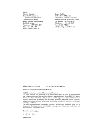
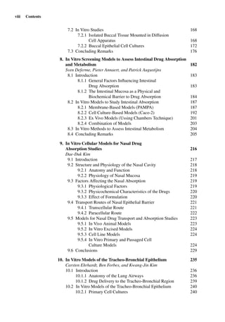
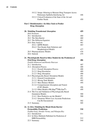
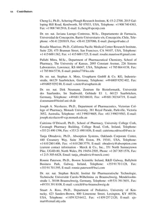
ods, species, ages, as well as healthy and diseased skin. Howes et al. [19]
introduced a hierarchy of frequently applied in vitro methods for measuring
percutaneous absorption according to their resemblance of the in vivo situation
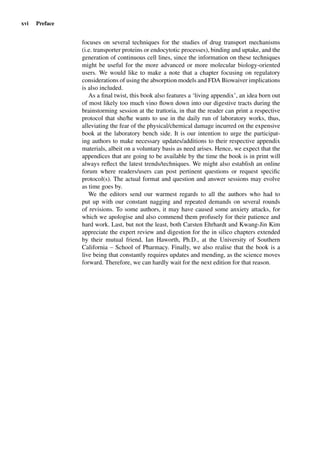
(Figure 1.1). This knowledge is very helpful when comparing different results
of skin absorption studies.
In vivo
Perfused skin
Viable full-thickness skin
Non-viable full thickness skin
Dermatomed skin
Stratum corneum
Reconstituted skin
Mathematical models
Figure 1.1 Hierarchy of frequently applied methods for measuring percutaneous
absorption according to their resemblance of the in vivo situation (adapted from Howes
et al. [19]).](https://image.slidesharecdn.com/drugabsorptionstudies-2008-150610022529-lva1-app6892/85/Drug-Absorption-Studies-2008-29-320.jpg)
![Chapter 1 Models for Skin Absorption and Skin Toxicity Testing 9
After oral administration, drug and metabolite concentrations in blood,
urine, and feces can easily be monitored. In contrast, topical application to
the skin usually aims at a local treatment. Therefore, the main interest lies
in determining the drug level within the skin, in order to evaluate the dermal
bioavailability of compounds or assess the bioequivalence between different
formulations. In the following sections, appropriate analytical techniques will
be presented.
1.3.1. In Vivo Studies Using Pharmacodynamic Response
Only a limited number of chemicals evoke a local quantifiable pharmacody-
namic response, for example, the concentration-dependent vasoconstriction
effect of corticosteroids. In these experiments, resulting skin blanching is
scored visually by one or more qualified investigators, using an ordinal data
scale. The lack of instrumentation has been criticized, because of possible
subjective errors [25]. However, approaches using the Chromameter device,
although recommended by the FDA, have failed to return the desired precision
[26, 27].
In addition to a single time-point measurement, skin blanching may be
followed over a prolonged period of time. In doing so, depot formation and
kinetics of substance removal due to local metabolic or systemic clearance
may be observed. By plotting the magnitude of response against time and
integrating the area under the effect curve (AUEC), different formulations can
be compared. This is the method of choice to determine the bioequivalence
of generic products, as described in the FDA guidance for industry on topical
dermatologic corticosteroids [5]. The ventral forearm is recommended as the
application site. A maximum of eight sites, each of an area of 1 cm2 and
spaced 2.5 cm from centre to centre, may be framed on each arm. If different
formulations or doses are to be tested, these shall be administered randomly
to one arm. On the second arm, the same preparations are applied; however,
the formulations change their positions. It is encouraged to perform a pilot
in vivo study in order to define appropriate parameters for the pivotal study.
This includes defining the linear proportion of the sigmoidal dose–response
curve, the half maximal effective dose ED50, and assessing the lower limits
of sensitivity and the maximum effect. The general study design described
in this guidance document also sets the benchmarks for in vivo studies with
compounds other than corticosteroids.
1.3.2. In Vivo Dermatopharmacokinetic Approach
1.3.2.1. Recovery Studies
Independent of any visible response, the extent of dermal absorption may be
judged from the amount of substance missing within the recovered formulation
after finishing incubation. However, this method keeps the researcher in the
dark about the ultimate fate of the substance. In addition, recovery studies
are not suitable for infinite dosing, as the recovery will always exceed 90%.
Comprehensive mass balance including both drug and metabolite levels in the
remaining formulation, washings, stratum corneum, blood, urine, and feces
can provide a more detailed insight. According to OECD guideline 427 “Skin
absorption: in vivo method” this technique is also to be favored for safety
assessment of chemicals in in vivo animal studies [17].](https://image.slidesharecdn.com/drugabsorptionstudies-2008-150610022529-lva1-app6892/85/Drug-Absorption-Studies-2008-30-320.jpg)
![10 U. F. Schaefer et al.
1.3.2.2. Skin Segmentation Studies – Tape Stripping Method
Sampling from the stratum corneum is usually performed by tape-stripping
single corneocyte layers (see the respective paragraph later in this chap-
ter). Provided the test substance yields a unique infrared signal, distinguish-
able from surrounding tissue and formulation components, attenuated total-
reflectance Fourier-transformation infrared (ATR-FTIR) spectroscopy is a fast,
technically sophisticated, equipment-extensive method of quantification [28].
It has been used to monitor dermal absorption of model substances like 4-
cyanophenol, to investigate depth-dependent changes in barrier properties or
hydration of the stratum corneum, and investigate the effects of formula-
tion excipients and penetration enhancers, such as oleic acid, on the stratum
corneum barrier function [29–32]. It should be noted that with both tape-
stripping and ATR-FTIR, the researcher’s field of vision is limited to the
nonviable part of the skin.
Comparative studies on the bioavailability of three different tretinoin gel
formulations showed that the dimensions of the sampling area may play a
critical role in determining the extent of dermal drug uptake [33, 34]. If, by
lateral spreading, a substance is distributed over an area sufficiently larger than
the sampling area, significant proportions of compound will not be recovered
and hence permeability will be underestimated.
1.3.3. In Vivo Dermal Microdialysis
Originating from the neurosciences, the microdialysis technique has been used
since several years to monitor drug absorption and disposition or the levels
of endogenous substances in the extracellular space of different organs and
fluids, such as bone, lung, liver, brain, and blood. The method has evolved
from its use in different animal species to the human microdialysis during the
late 80s [35].
The technique consists of a microdialysis probe, a thin hollow tube made of
a semi-permeable membrane usually around 200–500 µm in diameter, which
is implanted into the skin and perfused with a receiver solution that recovers
the unbound permeant from the local area. In principle, the driving force
of dialysis is the concentration gradient existing between two compartments
separated by a semi-permeable membrane. For skin under in vivo conditions,
these compartments represent the dermal or subcutaneous extracellular fluid
(depending on the probe position) and an artificial physiological solution inside
the probe [36–38].
Normally, diffusion microdialysis processes are mathematically described
by Fick’s second law relating the diffusion rate of the substance from the
medium to the concentration gradient and the surface area of the membrane.
One of the most important parameters to be considered is the flow rate of the
perfusate (solution flowing into the probe), which is normally in the range of
0.1–5 µl/min, inversely related to the amount of drug recovered in the dialysate
(solution flowing out of the probe). Other factors that strongly influence the
drug recovery from the surrounding medium are the lipophilicity and the extent
of protein binding of the substance, as these will decrease the recovery by
aqueous perfusate media and the molecular size of the compound, which will
limit the pass through the dialysis membrane according to its molecular cutoff.
To improve the recovery of lipophilic drugs, strategies such as addition of](https://image.slidesharecdn.com/drugabsorptionstudies-2008-150610022529-lva1-app6892/85/Drug-Absorption-Studies-2008-31-320.jpg)
![Chapter 1 Models for Skin Absorption and Skin Toxicity Testing 11
solvents (e.g., polyethylene glycol, cyclodextrins, proteins, and lipids) to the
perfusate have been used [36, 38, 39].
The relative recovery (RR) of the probe, essential for data interpretation, is
calculated using the retrodialysis method, which assumes that the net transport
through the microdialysis membrane from the perfusate to the surrounding
tissues equals the net transport from the tissues into the perfusate. The equation
for calculation is represented as follows [39]:
RR =
Cperfusate − Cdialysate
Cperfusate
(1.1)
with Cperfusate and Cdialysate being the compound concentration in the per-
fusate and in the dialysate. Among the techniques used to determine cuta-
neous availability, such as tape-stripping, biopsies, or imaging procedures,
microdialysis has shown to be promising having several advantages for the
assessment of in vivo drug pharmacokinetic profiles. The minimally inva-
sive procedure ensures minor reversible trauma. It allows long-term sampling
under physiological conditions in conscious individuals, and gives indication
of tissue concentration with respect to time. With the other above mentioned
techniques, this would require a large number of volunteers. Due to the
relatively low molecular weight cutoff of the membrane, the obtained sam-
ples are protein free allowing analysis without any further purification steps
and also avoiding an enzymatic degradation of the sample. Nevertheless, the
small sample size is a disadvantage since it requires very sensitive analytical
methods [39–41].
An increasing number of studies are using microdialysis of a wide range
of drugs in animals and humans, supporting the potential of this technique
for bioavailability and bioequivalence studies. Some examples of studies in
vitro, as well as in vivo, involving different delivery systems and species are
iontophoretic drug delivery in rats by Mathy et al. [42]; oral delivery and skin
pharmacokinetics by Bielecka-Grzela and Klimowicz [43]; determination of
salicylic compounds on rat skin by Simonsen et al. [44] and in human skin
by Leveque et al. [45]; and anesthetic extended release products in human
skin by Kopacz et al. [46]. For further details about dermal microdialysis,
refer to the reviews published during the last few years by McCleverty
et al. [35], Kreilgaard [39], Stahl et al. [47], Davies [37], and Elmquist and
Sawchuck [48].
1.3.4. Perfused Skin Models
Perfused skin models are considered the missing link between in vivo and in
vitro methods. Circumventing the potential harm of testing hazardous com-
pounds in living humans or animals, as well as inconveniences connected to
repeated sampling from the venous system or extensive biopsies, perfused skin
models offer the benefits of living tissue with fully active microcirculation
and metabolism [49, 50]. Ears of different animal origin, such as hairless
mice, rabbits, or pigs, have been used. However, the exposed location of the
ears and their involvement in thermoregulation implement a high degree of
vascularization [51–53]. Because of their unusually high permeability, perfused
ear models have been proposed to be predictive models of premature neonate
skin [54].](https://image.slidesharecdn.com/drugabsorptionstudies-2008-150610022529-lva1-app6892/85/Drug-Absorption-Studies-2008-32-320.jpg)
![12 U. F. Schaefer et al.
Further specimens in use are the perfused cow udder, the porcine forelimb,
and the isolated perfused porcine skin flap (IPPSF) [55–57]. While the first
two require the sacrifice of the animal, the last one can be isolated surgically
from the abdomen of the pigs, which afterwards can be returned to their prior
disposition [49].
Basically, these models comprise a surgically prepared portion of animal
skin including a continuous vascular circulation which can be cannulized,
perfused with tissue-culture medium, and sampled for cutaneously applied
chemicals or their metabolites. The easy sampling of substance levels within
the vascular system itself allows analysis of systemically available drugs.
Methods known from in vivo investigations, such as mass balance, including
perfusate levels, surface washings, stratum corneum tape-stripping, and deeper
skin layer biopsies, are easily transferable to the perfused skin. Wester et al.
could show for five compounds with different physicochemical properties that
the dose absorbed in the IPPSF after 8 h (i.e., the cumulative dose recovered
from surface washings, skin, and perfusate) compared favorably to the percuta-
neous absorption after 7 days in man (i.e., urinary levels relative to intravenous
application) [58]. For the perfused cow udder, such relationship still remains
to be shown [59]. Nonetheless, the cow udder proved suitable for investigating
skin irritation. Pittermann et al. tested the effects of a water-soluble coolant
used in metal industry using sodium lauryl sulphate as positive control. The
results showed the benefits of applying special skin protection formulations
before contact with an irritant [60].
In a study using porcine forelimbs, Mahmoud et al. found similar
metabolism rates of 17β-estradiol applied in ethanolic solution, gel, or trans-
dermal therapeutic system (TTS), as found in human plasma of female patients
undergoing transdermal hormone replacement therapy. However, studies by
Wagner et al., analyzing the transdermal availability of estradiol and nitroglyc-
erine from TTS, clearly showed that some work remains to be done before the
porcine forelimb may be used for predicting in vivo percutaneous absorption
in man [56]. Concerns about using animal skin as a surrogate for human risk
assessment and dermal drug absorption are the same as expressed later for in
vitro analyses employing membranes of animal origin.
1.3.5. In Vitro Skin Permeation Studies
Most publications in the field of skin permeation research are carried out using
a large variety of setups and experimental protocols varying from laboratory to
laboratory. This raises questions of standardization and regularization.
Both static and flow-through diffusion cells are approved by the authorities,
and data are available on their relevance for predicting the in vivo situation
[2, 61–64]. Basically, a donor and an acceptor compartment are separated by
a membrane of either native skin or bioengineered materials. These materials
can be of human, animal, or artificial origin. Sampling from the acceptor com-
partment is performed either continuously or at predetermined time intervals.
Dosing is possible in infinite (typically >10 µl/cm2 or 10 mg/cm2) or finite
manner (<10 µl/cm2 or 10 mg/cm2). The donor chamber may either be left
open or be occluded. Nonoccluded conditions permit an exchange with the
environment, such as evaporation of volatile substances and drying of the skin
surface. In contrast, a tight occlusion of the skin surface may lead to excessive](https://image.slidesharecdn.com/drugabsorptionstudies-2008-150610022529-lva1-app6892/85/Drug-Absorption-Studies-2008-33-320.jpg)
![Chapter 1 Models for Skin Absorption and Skin Toxicity Testing 13
hydratization. For the determination of membrane integrity, transepidermal
water loss (TEWL) measurements are recommended in many guidelines.
However, Netzlaff et al. reported certain limitations in applying this method
in vitro [65].
Temperature may be controlled by using a water jacket around each per-
meation cell, an external water bath, or warm air in a drying oven. Usually,
experiments are carried out at 32◦C, that is, the temperature of the skin surface,
or else a temperature gradient may be applied: of 32◦C at the skin surface
to 37◦C in the acceptor compartment, mimicking body temperature. Constant
stirring of the acceptor phase ensures that diffusion is unhampered by the
buildup of high local concentrations and provides sink conditions throughout
the duration of the experiment.
Acceptor solutions preferably comprise buffer solutions of pH 7.4, spiked
with preservatives, such as 0.05% sodium azide, if long-term incubation is
desired. Low aqueous solubility of the compound in question may require the
addition of solubilizers. Although suggested by the original OECD guidance
document 28, ethanol:water (1:1) and BRIJ 98 were both found to inter-
fere with membrane integrity and analytics [20]. To investigate metabolic
processes, the viability of freshly excised skin may be sustained using tissue-
culture medium as acceptor fluid. Further recommendations for experimental
methods are available [2, 20].
1.3.5.1. Membranes
Various membrane types suitable for permeation experiments are listed below.
More details are available in literature [66].
1.3.5.1.a. Membranes of Human Skin Origin: For all in vitro skin experi-
ments, human skin is considered to be the “gold-standard.” Supply is usually
provided from plastic surgery, amputations, or cadavers. While for in vivo
studies the volar forearm is used, in vitro skin sources are mostly abdomi-
nal, back, leg, or breast skin. Much attention has been turned to assessing
the degree and sources of variability [63, 67–70]. Nonetheless, large intra-
and interindividual variations of up to 45% in vivo are documented. These
may be due to differences in lipid composition, skin thickness, or hydration,
which depend on body site and sex or are a consequence of skin diseases
[71–75]. In an in vitro situation even larger variations have been reported,
as excision, storage, and experimental manipulations may cause additional
modifications [76].
After excision, the skin should quickly be freed from subcutaneous fat and
stored deep frozen at −20◦C to −30◦C in tightly sealed plastic bags. It may
then be stored for up to several months without impairing barrier function,
provided that repeated thawing and freezing is avoided [77–79].
Special attention shall be drawn to the preparation of the excised skin prior
to the experiment. Long lag-times encountered with hydrophilic substances,
as well as an unfavorable partition of lipophilic compounds into viable skin
layers, may require the further separation of the skin into its individual layers
[80]. Furthermore, the absence of dermal blood flow in vitro may build up
a significant hindrance to diffusion [81]. Reducing the membrane thickness
will generally reduce experiment times and thus minimize the risk of bacterial
contamination.](https://image.slidesharecdn.com/drugabsorptionstudies-2008-150610022529-lva1-app6892/85/Drug-Absorption-Studies-2008-34-320.jpg)
![14 U. F. Schaefer et al.
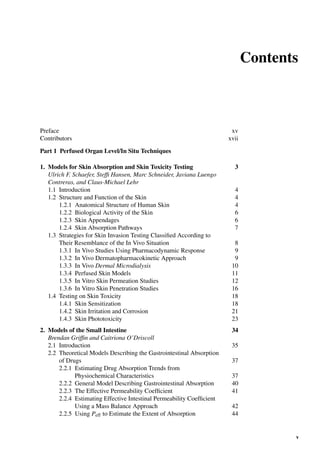
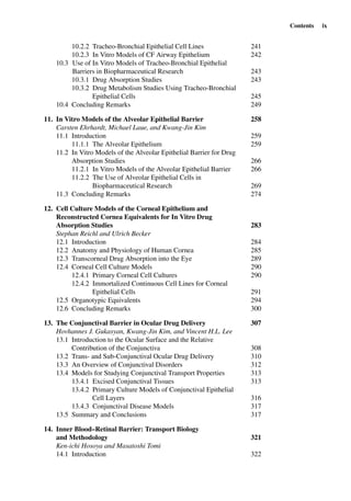
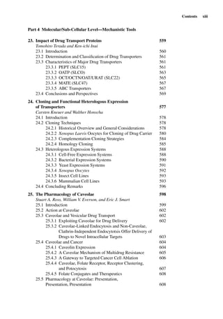
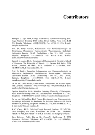
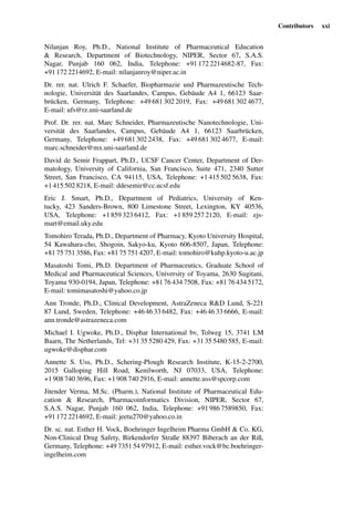
Figure 1.2 Cross sections of (A) full-thickness skin, ×100; (B) dermatomed skin,
×100; (C) heat-separated epidermis, ×400; and (D) trypsin isolated stratum corneum,
×400. (Images courtesy of Leon Muijs, Biopharmaceutics & Pharmaceutical Technol-
ogy, Saarland University, Germany).
Figure 1.2 shows differently prepared skin membranes. The individual mem-
branes were prepared from freshly excised human abdominal skin, originating
from females undergoing reductive surgery. Samples were fixed with xylol,
stained with hematoxylin/eosin, and embedded into paraffin. Cross sections
with a thickness of 4 µm were prepared using a microtome.
Dermatomed Skin or Split Skin The dermatomisation technique may be used
for harvesting skin, as well as for reduction of the dermal thickness. Samples
in the latter case comprise epidermis including stratum corneum, as well as
part of the dermis. Care has to be taken to avoid any damage to the stratum
corneum barrier. This may be achieved by protecting the skin surface with a
piece of plastic [82]. The recommended thickness is 200–400 µm and 400–
700 µm for human and pig skin, respectively [20]. Although the dermatome
will cut through hair follicles, the holes will readily close during incubation in
aqueous media, due to swelling of the tissue.
Epidermis Complete removal of the dermis may be achieved by sev-
eral mechanical, thermal, and chemical techniques. Most commonly, the
epidermal–dermal junction is split by heating the skin to 60 C for 30–120 s
[83, 84]. Pitman et al. [85] could show that such a treatment does not impair the
barrier function. The use of ethylene diamine tetraacetic acid, sodium bromide,
or ammonia fumes has also been reported [80, 83, 86]. It may, however,
be suspected that the use of sufficiently strong acids or bases may change
the buffer capacity of skin, which would especially influence the penetration
behavior of ionizable drugs.
Stratum Corneum The cornified skin layers may be isolated by digestion of
the connective epidermal tissue using trypsin in buffer solutions of pH 7.4.](https://image.slidesharecdn.com/drugabsorptionstudies-2008-150610022529-lva1-app6892/85/Drug-Absorption-Studies-2008-35-320.jpg)
![Chapter 1 Models for Skin Absorption and Skin Toxicity Testing 15
This may be achieved by complete immersion of full-thickness skin in trypsin
solution or by placing the heat-separated epidermis for 24 h at 37◦C on a
filter paper soaked with the enzyme preparation [64, 83]. Other techniques,
such as vacuum or chemically induced blistering, stretching, application of
staphylococcal exfoliatin, or proteolytic digestion of viable cells, are seldom
reported [74, 87–89].
Skin with Impaired Barrier Function (Dermis, Stripped Skin) In the course
of studying the behavior of diseased, injured, or premature skin, it may be
necessary to deliberately impair the skin’s barrier function. As the rate limiting
barrier for most compounds resides within the stratum corneum, removal of
the outermost cornified layer will result in an increased permeability [74, 90].
This has been monitored by measuring the concomitant increase in TEWL and
change in skin impedance [91]. Both tape-stripping and dermatomization seem
to be suitable preparation techniques, since heat separation of epidermis at
60◦C and trypsin-induced removal of the stratum corneum are likely to result
in significant precipitation or digestion of dermal proteins [11].
Reconstructed Human Epidermis Equivalents Because of the limited avail-
ability of human skin, reconstructed human epidermis equivalents are under
investigation to serve as membranes in permeation experiments. A summary
on these replacement tools has been recently published by Netzlaff et al. [92].
First results of a German prevalidation study have shown the suitability of such
bioengineered human epidermis equivalents in permeation studies [93].
1.3.5.1.b. Membranes of Animal Skin Origin: The limited availability of
human skin, together with an increasing need to test the bioavailability of der-
mal products in pharmaceutics and cosmetics, as well as risk assessment of
chemicals, has promoted the search for alternative models. A switch to animal
skin, therefore, seems obvious. Species currently in use are mouse, hairless
rat, hamster (cheek pouch), snake (shed skin), pig (ear, flank, abdomen, or
back), and cow (udder). However, differences in stratum corneum thickness,
number of corneocyte layers, hair density, water content, lipid profile, and
morphology cause animal skin to be more permeable than human skin, leading
to an overemphasis of compound permeability [13]. When comparing results
of several species, human skin finds its closest match in porcine tissue [13, 20,
94]. While animal skin is still mentioned in the OECD guidance document 28,
the explicit use of human skin has later been demanded as the only acceptable
surrogate in human risk assessment [2, 20]. In addition, a general testing, as
well as marketing ban concerning the use of animals for testing cosmetics or
their ingredients, will come into action in the EU in September 2009 [95].
Nonetheless, animal skin may still be suitable for specific, usually veterinary,
applications [20].
1.3.5.1.c. Membranes of Nonskin Origin: The complexity of published setups
ranges from the simple use of dialysis tubing and polymeric membranes, made
up of regenerated cellulose, polycarbonate, polyethylene, or polydimethyl-
siloxane, to amphiphilic multilayer laminates and polymer networks [96–100].
Because of their negligible barrier-forming properties, simple porous mem-
branes do not present a significant hindrance for drug absorption. They may,
however, serve as tools for separation of formulation and acceptor compartment
in drug release studies. Diffusion takes place via aqueous channels along the](https://image.slidesharecdn.com/drugabsorptionstudies-2008-150610022529-lva1-app6892/85/Drug-Absorption-Studies-2008-36-320.jpg)
![16 U. F. Schaefer et al.
tortuous pathway of the pores. Primary fields of application are formulation
optimization, quality control, and comparison of pharmaceutical availability of
different products. Polymeric membranes usually do not provide insight into
the inherently complex mechanisms of skin absorption.
On the other hand, amphiphilic composite membranes made up of silicone
and poly(2-hydroxyethyl methacrylate) (pHEMA) have been reported to form
a significant barrier. Although their absolute permeability is usually higher
than that of skin, the permeability coefficients of several test compounds
correlated reasonably well to values found across rat skin [101–103]. For a set
of compounds, mostly substituted phenols, Geinoz et al. [104] could further
demonstrate similar structure–permeation relationships across silicone mem-
branes and literature data on human epidermis. Lipophilicity and hydrogen-
bond donor acidity are the main determinants for transport across silicone
membranes. Accordingly, another group suggested silicone membranes as a
simple model to study the impact of vehicle components on partition, perme-
ability, and retention of drug molecules [105].
Of particular interest are membranes prepared of an inert porous support
carrying natural or artificial lipids. These coatings may comprise a single
component, such as isopropylmyristate or dodecanol [99, 106], or mixtures
of comparable composition as the stratum corneum intercellular bilayer [107,
108]. Usually, synthetic lipids are used, due to an elaborate isolation procedure
for stratum corneum lipids, with limited yield and the necessity of separa-
tion of triglycerides, originating from subcutaneous fatty tissue or skin care
products [109].
By adjusting the lipid composition, a variety of problems can be addressed.
Mechanistic studies employing wide- and small-angle X-ray diffraction, FT-IR
spectroscopy, and neutron scattering have been used to show the contribution
of the individual classes of skin lipids to barrier formation and the sandwich-
like lipid organization within the intercellular bilayers of the stratum corneum
[110–115]. Moreover, the mode of action of penetration enhancers, such as
oleic acid [116, 117], the structure and hydration of stratum corneum lipids
[118], and the degree of ionization of free fatty acids [119] were investigated.
Lipid-coated membranes have also been applied as surrogates for skin absorp-
tion [120–122]. Jaeckle et al. used an iterative method to examine the influence
of formulation excipients on the diffusion of a model compound through lipid-
coated membrane filter discs [107].
1.3.6. In Vitro Skin Penetration Studies
As described previously in this chapter, efforts have been made to develop
methods for quantification of skin permeability, validation of diffusion cell
setups, and correlation of in vitro data with the in vivo situation. However,
the average drug permeation experiment does not provide insight into the
temporal and local disposition within the tissue, that is, the skin penetration.
The following discussion will give an overview of methods tackling this kind
of problem.
Drug levels within the stratum corneum can be assessed by sampling sin-
gle corneocyte layers with adhesive film. The drug is then extracted from
the tape-strips and quantified by a suitable analytical method. Usually, scin-
tillation counting (for radioactive compounds) or high performance liquid](https://image.slidesharecdn.com/drugabsorptionstudies-2008-150610022529-lva1-app6892/85/Drug-Absorption-Studies-2008-37-320.jpg)
![Chapter 1 Models for Skin Absorption and Skin Toxicity Testing 17
chromatography analytics are employed. Results are reported as the stratum
corneum drug amount absorbed per square cm of skin. In addition, correlat-
ing the local drug concentration with the skin depth will result in a detailed
insight into the concentration gradient across the membrane, that is, the actual
driving force for drug diffusion. Within the deeper skin layers, that is, the
epidermis and dermis, analogous information can be obtained using biopsy
punches. However, the invasive character of the sampling procedure practically
restricts this technique to in vitro studies. After freezing, the punch biopsies
may be segmented parallel to the surface, by means of a cryo-microtome.
After substance extraction, the drug amount is determined using an appropriate
analytical method, for example, high performance liquid chromatography.
The tape-stripping technique has been applied in vivo, as well as in vitro.
The used in vitro incubation devices are the same as described for skin perme-
ation studies. A specialized incubation device developed by Loth and cowork-
ers, the “Saarbrücken penetration model,” allows investigation of skin pen-
etration bypassing the normally occurring nonphysiological hydration of the
dermis [64].
It has been observed that, especially with aqueous donor media, the genera-
tion of concentration-skin depth profiles is limited to a few hours of incubation.
Excessive hydration progressively weakens the coherence of the corneocytes,
which are then torn off by the tapes in large flaps instead of distinct layers. Par-
ticularly, the investigation of hydrophilic or high molecular weight compounds
with long stratum corneum lag-times is affected. Mueller et al. proposed that
incubation can be prolonged up to 48 h if the membrane is allowed to dry at
room conditions for 30 min prior to tape-stripping [123]. Efforts have been
made to correlate short time concentration gradients with steady-state flux.
Pirot et al. correctly predicted the steady-state flux of 4-cyanophenol, by fitting
the concentration gradient measured in vivo after a short time-exposure of 15
min, that is, approximately half the lag-time, to the solution of Fick’s second
law of diffusion [124].
To show the substance distribution in the different skin layers, the allocation
of the different removed skin segments, obtained either by a stripping proce-
dure or by cryo-cutting, is essential.
In the following section, methods of assessing the thickness of stratum
corneum removed per tape are summarized.
1.3.6.1. Determination of the stratum corneum depth by Tape-Strip Number
For untreated skin, repeated stripping may remove a constant amount of stra-
tum corneum, independent of the number of strips already performed [125].
The momentary depth may thus be calculated by dividing the mean thick-
ness of the overall stratum corneum by the total number of tapes necessary
to remove it completely and multiplying with the number of strips already
performed.
However, the validity of a similar assumption has to be questioned, in case
the skin has previously been treated with a topically applied formulation [126].
Opinions differ, whether the distinct curvature of the steady-state stratum
corneum concentration gradient, reported in literature, may be an artifact of
a wrong depth scale, since such a behavior cannot be reasonably explained by
the established diffusion theory.](https://image.slidesharecdn.com/drugabsorptionstudies-2008-150610022529-lva1-app6892/85/Drug-Absorption-Studies-2008-38-320.jpg)
![18 U. F. Schaefer et al.
1.3.6.2. Determination of the stratum corneum depth by Weight
of Removed Tissue
A linearization of the steady-state concentration gradient could be demon-
strated by relating the depth to the weight of the tissue, removed per piece
of adhesive tape. However, large errors, especially, within the first tapes, cast
doubt over these findings [127, 128]. The procedure is time-consuming and
artifacts, due to absorption and desorption of moisture, formulation excipients,
or sebaceous lipids, are likely.
1.3.6.3. Determination of the stratum corneum depth by Amount of Protein
A standard Lowry-based protein assay has been adjusted to the special condi-
tions encountered with skin [126]. Basically, proteins reduce an alkaline solu-
tion of Cu(II)-tartrate to Cu(I) in a concentration-dependent manner. Then, the
formation of a blue complex between Folin-Ciocalteau reagent (a solution of
complex polymeric ions formed from phosphomolybdic and phosphotungstic
heteropoly acids) and Cu(I) can be measured spectrophotometrically at 750 nm.
A calibration curve can be obtained by dissolving known amounts of stratum
corneum in 1 M sodium hydroxide. A piece of tape that has not been in contact
with skin is subjected to an identical procedure and serves as negative control.
The method was recently adapted to a 96-well plate format, notably reducing
analysis times [129].
Assessing the depth by determining the protein amount removed per strip,
Mueller et al. noted a nonlinear steady-state concentration gradient which they
ascribed to an increased permeability of the cornified envelope within the
stratum disjunctum [123].
1.3.6.4. Determination of the stratum corneum depth by Optical
Spectroscopy in the Visible Range
Corneocytes adhered to a piece of adhesive tape will refract light by reflection,
scattering, and diffraction, almost independent of the wavelength [128]. After
stripping, the tape is transferred into a sample holder in a way that the rec-
tangular beam of a specially designed double beam UV/Vis spectrophotometer
penetrates the area carrying corneocytes. The absorbance is then measured at
430 nm, using an empty tape in the reference beam. As most compounds and
excipients absorb light in the ultraviolet spectral range, an overlap of the signal
by formulation components is unlikely.
1.4. Testing on Skin Toxicity
1.4.1. Skin Sensitization
Substances when applied to human skin might exert a sensitizing potential on
the skin and need, therefore, to be evaluated and classified for their possible
toxicity. Every substance that provokes immunologically mediated cutaneous
reactions (i.e., skin sensitization or allergic contact dermatitis) is referred to as
skin sensitizer. Several tests are recommended, but no single method is able
to identify all potential substances capable of inducing sensitization of human
skin. Widely used test methods for the investigation of skin sensitization, the
so-called adjuvant and nonadjuvant tests, are described below.](https://image.slidesharecdn.com/drugabsorptionstudies-2008-150610022529-lva1-app6892/85/Drug-Absorption-Studies-2008-39-320.jpg)
![Chapter 1 Models for Skin Absorption and Skin Toxicity Testing 19
1.4.1.1. Guinea Pig Maximization Test
The guinea pig maximization test (GPMT) is a preferred method for the
detection of skin sensitizers. It belongs to the class of adjuvant-tests, where
the substance will be applied in Freund’s complete adjuvant (FCA). The test
is based on the possible induction of an immune response of the skin during
an induction period (at least 1 week). This pretreatment of the subject will
eventually result in a hypersensitive reaction during a further exposure, the so-
called challenging phase.
The dose used for the induction exposure is chosen in such a way that it is
systemically well tolerated and causes only mild-to-moderate skin irritations.
The dose during the challenging period should be the highest nonirritating
dose. Both doses need to be determined in preliminary tests, in case no other
information on the test substances is available.
The test is started with an intradermal and/or epidermal application of the
test substance, using the induction dose on young adult guinea pigs of either
gender. In the case of female animals, these have to be nulliparous and nonpreg-
nant. For 5 days prior to the test, animals are kept at (20 ± 3)◦C with 30–70%
relative humidity, conventional laboratory diet, and unlimited access to water.
The induction dose is administered to the shoulder region and is followed by
the induction period (10–14 days), in which the animals can rest and a possible
immune response may develop. It should be noted that the location of the
induction dose is kept occluded for the first 48 h after administration.
Thereafter, the animals are exposed for a second time at their flanks, using
the challenging dose for 24 h under occlusive conditions. Twenty-one hours
after removing the challenging dose patches, the area is cleaned and freed
from hair, without abrasion of the skin. After an additional 3-h period (exactly
48 h after the challenging treatment), the skin is investigated in comparison
to that in the control group that received sham treatment during the induction
phase. A second investigation of the incubated area is performed a further 24
h later, and judgments are made according to the Magnusson/Kligman scheme
(Table 1.1) [130].
Along with the testing of the materials of interest, control experiments
with reference substances need to be performed. At least every 6 month, the
control test needs to be carried out using mild-to-moderate skin sensitizers
[recommended substances are α-hexylcinnamaldehyde, benzothiazole-2-thiol
(mercaptobenzothiazole), and benzocaine] to evaluate the testing procedure.
The minimal number of animals to be investigated is 10 in the treatment
group and 5 in the control group. If the findings indicate a nonsensitizing
substance, the number of animals needs to be increased to a total of 20 and
10 for the treatment and the control groups, respectively.
Table 1.1 Magnusson/Kligman grading scale for the
evaluation of challenge patch test reactions [130].
Observed skin reaction Noted value
No visible change 0
Discrete or patchy erythema 1
Moderate or confluent erythema 2
Intense erythema and swelling 3](https://image.slidesharecdn.com/drugabsorptionstudies-2008-150610022529-lva1-app6892/85/Drug-Absorption-Studies-2008-40-320.jpg)
![20 U. F. Schaefer et al.
1.4.1.2. Buehler Test
The Buehler test [131] is the preferred nonadjuvant test method, even though
it might be not as accurate as other tests [132]. Like the GPMT, the Buehler
test consists of two successive phases, the induction treatment followed by the
challenging exposure. The induction dose needs to be chosen in such a way
that it is high enough to cause mild irritation on the skin, while the challenging
dose should equal the highest nonirritating concentration of the investigated
substance. Both doses have to be determined in a pilot study.
Guinea pigs of either sex can be used. If female animals are used, these have
to be nonpregnant and nulliparous. A minimum of 20 animals for testing and
at least 10 as control are recommended.
The test patches are brought in contact with hair-free areas on the flanks
of the guinea pigs under occlusive conditions for 6 h. The control group
(potentially a naive control) will be treated with the vehicle only, which is
applied in a similar manner on hair-free guinea pig flanks. The same treatment
is carried out on days 6–8 and again on days 13–15. On days 27–29 after the
initial incubation, the challenging dose is applied under occlusive conditions to
the, until then, not exposed flank of the guinea pigs (hair-free). These patches
are also held in contact with the skin for 6 h.
Twenty-one hours after the end of the incubation, all hairs are again removed
from the treated flanks (abrasions are to be avoided) and observation of the
skin’s reaction is made after a further 3 h. These are recorded according to
the grades proposed by Magnusson and Kligman (Table 1.1) [130]. After an
additional 24 h, the skin reactions are observed again and recorded following
the same scale.
1.4.1.3. Local Lymph Node Assay
The murine local lymph node assay (LLNA) was developed as an alternative to
the GPMT [133]. Even though it cannot fully replace the GPMT, fewer animals
are necessary to perform the test. The LLNA enables the hazard classification
of substances that induce allergic contact dermatitis, while offering animal
welfare advantages, compared to the GPMT (elimination of pain and reduc-
tion in animal numbers required). Furthermore, the LLNA allows assigning
substances into specific potency categories (classes 1–3) [134]. The LLNA
has recently been accepted by the SCCNFP and is published as OECD test
guideline (TG) 429 updated (2002) [135].
The test method is based on the fact that sensitizers induce a proliferation of
lymphocytes in the lymph node draining the site of substance administration.
The increased proliferation is proportional to the applied dose of the chemical
and the potency of the allergen. Hence, the murine LLNA assesses proliferation
in a dose–response manner, comparing it to the proliferation in a control group.
The ratio of the proliferation after sensitizer application to the control group
defines the Stimulation Index (SI).
SI =
sensitizer stimulated proliferation
sensitizer − free proliferation
An SI ≥ 3 is the minimum requirement to be met, before a chemical has to
undergo further evaluation as a skin sensitizer. Radio labeling is a standard
method to quantitatively assess the proliferation, but other analytical tech-
niques exist. For all investigations, positive controls need to be performed. The](https://image.slidesharecdn.com/drugabsorptionstudies-2008-150610022529-lva1-app6892/85/Drug-Absorption-Studies-2008-41-320.jpg)
![Chapter 1 Models for Skin Absorption and Skin Toxicity Testing 21
positive control should result in an SI > 3, but the substance should be chosen
in such a way that it clearly does not cause excessive reactions (e.g., hexyl cin-
namic aldehyde and mercaptobenzothiazole). Experiments shall be performed
using young adult, nonpregnant, and nulliparous female mice (CBA/Ca or
CBA/J strain). The mice shall be housed at (22 ± 3)◦C with relative humidity
kept between 30% and 70%, at 12-h light–dark alterations, in individual cages
at conventional diet and unlimited water supply. Male animals or other strains
can be used, if enough data are generated to show parity in the LLNA.
The procedure goes over 6 days of which on days 1–3 the verum
or just the vehicle (typical vehicles are acetone/olive oil mixtures, N,N-
dimethylformamide, methyl ethyl ketone, propylene glycol, or dimethyl sulf-
oxide) are applied onto the ears of the animals, whereas the fourth and the fifth
day are without treatment. On the sixth day, a suspension of lymph node cells
is prepared for each mouse.
The LLNA is the preferred method when compared to the GPMT because
(a) it can equivalently predict human contact dermatitis, (b) a dose–response
can be obtained, and (c) it is in line with current animal welfare efforts. Nev-
ertheless, several situations exist where the GPMT is advantageous, depending
predominantly on the choice of test substances. The LLNA is known for
less powerful detection of the sensitization potential of metallic compounds,
high molecular weight proteins, strong irritants, and for substances with low
adhesion to the skin surface (skin wettability is a prerequisite for the successful
application of LLNA) [136–140].
The skin reaction observed after performing the GPMT should also be
recorded analogous to the Magnusson/Kligman grading scale (Table 1.1).
1.4.2. Skin Irritation and Corrosion
Any application of substances onto the skin, for instance, cosmetic products,
must not cause damage to human health when applied under normal conditions.
Therefore, any compound considered for application on human skin needs to
be tested for reversible disadvantageous effects (skin irritation) and irreversible
skin damage (skin corrosion).
To test the irritancy potential of substances, two tests which can reli-
ably distinguish between skin corrosives and noncorrosives are endorsed by
the European Centre for the Validation of Alternative Methods (ECVAM).
The testing procedures are based on the transcutaneous electrical resistance
(TER) measurements of rat skin and on a human skin model. Both test
systems [141–145] will be briefly outlined below. Nevertheless, these tests
are not suited for the group of mild irritants which do not induce an acute
effect on the barrier function. For those substances, new markers need to
be evaluated. First results are available for heat shock protein 27 where
higher levels were observed in skin models after exposure to mildly irritating
chemicals [146, 147].
1.4.2.1. Rat Skin Transcutaneous Electrical Resistance
The rat skin TER assay enables to reliably discriminate between skin corro-
sives and noncorrosive substances [148]. The assay relies on the change in
the bioelectrical properties of the skin in response to the application of test
chemicals. For the measurements, small discs of rat skin are necessary onto
which the substances are applied to the epidermal surface for up to 24 h. In](https://image.slidesharecdn.com/drugabsorptionstudies-2008-150610022529-lva1-app6892/85/Drug-Absorption-Studies-2008-42-320.jpg)
![22 U. F. Schaefer et al.
the case of a corrosive impact on the barrier, the TER value will fall below a
threshold value of 5 k [149]. This effect will only be observed for corrosive
materials, due to the degradation of the barrier integrity, but not for irritant
materials.
For the experiment, the dorsal skin of young rats (Wistar or a comparable
strain) is shaved and washed with an antibiotic solution (containing, e.g.,
streptomycin, penicillin, chloramphenicol, and amphotericin in concentrations
inhibiting bacterial growth). After skin excision, excess fat is peeled off and the
skin is placed over the end of a polytetrafluoroethylene tube with the epidermal
side in touch with the hollow cylinder. The skin is fixed with an O-ring and the
tube interior is sealed. The side of the dermis is then submersed in a magnesium
sulphate solution (154 mM). The samples are applied at 30◦C to the epidermal
side of the skin in such a way that the skin interface is fully covered. After the
incubation time, the substances are removed with prewarmed water; the skin
surface tension is decreased with ethanol which is subsequently replaced with
magnesium sulphate solution (154 mM).
As positive control, 10 M hydrochloric acid (36%) is recommended; distilled
water will work as a negative control.
In the case that TER values below 5 k are measured for surfactants or
neutral organics, an assessment of dye (sulforhodamine B) penetration can be
carried out to determine false-positive results [150]. Preparation of the skin
discs and the washing steps are known to be the critical steps.
The TER assay of rat skin is a fully accepted replacement method of animal
tests for skin corrosion. Hence, in the EU the TER assay is mandatory for the
evaluation of test substances. Investigation of animals to test the corrosivity of
chemicals and/or other substances is not permitted [151].
In 2000, the TER assay has been included into “Annex V. Part B.40 on Skin
Corrosion” of the “Directive 67/548/EEC on the Classification, Packaging and
Labelling of Dangerous Substances” [152]. Moreover, at the 14th Meeting of
the OECD National Coordinators for the TGs Programme, the draft TG 430 for
the TER assay for testing in vitro skin corrosion was granted approval, which
is the basis for a subsequent acceptance at the OECD level [153].
1.4.2.2. Human Skin Model Assay
The human skin model assay involves measuring the effects of corrosives on
viable cells in a reconstituted human skin equivalent. To be accepted as a
valid human skin model, several criteria must be met. The artificial skin must
comprise a functional stratum corneum with an underlying layer of viable cells.
Furthermore, the barrier function of the stratum corneum, as well as the via-
bility of the epidermis, must be verified with appropriate experimental setups.
The chemicals to be tested are applied up to 4 h as a liquid or a wet powder onto
the skin model. Afterwards, careful washing has to be performed, followed by
investigation of the cell viability [e.g., with a (MTT)] reduction assay).
The human skin model assay can provide further data on the degree of
corrosiveness and allows ranking corrosives among each other. It is, therefore,
accepted as a replacement method of animal tests for skin corrosion in the EU.
The following in vitro methods based on reconstructed human skin
models are validated (by the ECVAM) for predicting skin corrosion—
EPISKINR
, EpiDermR
, CorrositexR
—and irritation: EPISKINR
, EpiDermR
,
PREDISKINR
, SIFT [142]. Additionally, the SkinEthicTM was assessed and](https://image.slidesharecdn.com/drugabsorptionstudies-2008-150610022529-lva1-app6892/85/Drug-Absorption-Studies-2008-43-320.jpg)
![Chapter 1 Models for Skin Absorption and Skin Toxicity Testing 23
concordance between the model and the accepted test of OECD TG 430 and
TG 431 was obtained [93, 154, 155].
1.4.3. Skin Phototoxicity
This section will briefly focus on tests used to identify chemicals that lead to
toxic responses after contact with skin and a subsequent exposure to light. This
involves locally as well as systemically administered substances.
1.4.3.1. 3T3-Neutral Red Uptake
This test enables to identify compounds which show a phototoxic effect in vivo
[156]. It allows inter alia assessing the photo irritancy potential of ultraviolet
(UV) filter candidates after topical application and distribution to the skin
[157]. The 3T3-neutral red uptake test, however, is unable to predict other
adverse effects that might result from the combined interaction of chemicals
with light.
The test is based on an in vitro assay of the uptake of the dye, neutral red
(NR), in Balb/c 3T3 fibroblasts. It was developed to detect the phototoxicity
induced by the combined interaction of the test substance and light of the
wavelength range from 315 to 400 nm, the so-called UVA. The cytotoxicity is
evaluated in the presence (+UVA) or absence (−UVA) of UVA light exposure,
after application of a nontoxic dose of the compound. The cytotoxicological
impact is assessed via the inhibition of the fibroblasts to take up the vital dye
NR (NR is a weak cationic dye, penetrating easily into the cell membrane by a
nonionic diffusion and accumulates in the lysosomes) one day after the initial
treatment. Normally, healthy cells may incorporate and bind NR. Alterations
of the cell surface or the lysosomal membranes, however, lead to a decreased
uptake and binding of the dye.
For the in vitro test, the fibroblasts are allowed to form a half-confluent
monolayer within 24 h. Different concentrations of the test chemical are then
incubated for 1 h with two sets of cells in parallel (typically on 96-well plates,
104 cells per well, passage number <100). After the incubation with the test
substances, one set is irradiated with a nontoxic dose of UVA light (5 J/cm2),
while the other set is kept in the dark. Twenty hours after irradiation, cell
viability is evaluated by measuring the uptake of NR for 3 h. After the end
of the absorption process, excess NR is removed and the cells are treated with
an NR desorption solution (ethanol/acetic acid) to extract the dye taken up by
the cells. Subsequently, the optical density of the NR solution is measured at
540 nm. As positive control, a test with chlorpromazine is performed.
For the evaluation of the results, the obtained concentration–response curves
for +UVA and −UVA are compared. The photo irritation factor (PIF) is
calculated, which allows to determine the phototoxic potential of a substance
according to a numerical value. The PIF-value represents the ratio of the
EC50 (effective cytotoxic concentration) (−UVA) and the EC50 (+UVA).
A PIF-value above 5 indicates a phototoxic potential, whereas a PIF-value
below 5 predicts no such potential. To overcome problems in the case of no
phototoxicity up to the highest investigated concentration [no EC50 (−UVA)
and/or no EC50 (+UVA)], a second measure was introduced, the so-called
mean photo effect (MPE) [158], which can be calculated using a freeware
program available at the Humboldt University in Berlin (Germany) from H.G.
Holzhuetter.](https://image.slidesharecdn.com/drugabsorptionstudies-2008-150610022529-lva1-app6892/85/Drug-Absorption-Studies-2008-44-320.jpg)
![24 U. F. Schaefer et al.
1.4.3.2. Reconstructed Human Epidermis in Combination with MTT-Assay
A further option to investigate the phototoxic potential of substances is the
use of reconstructed human skin models. The evaluation of the cell viability
is based on the MTT-assay that is sensitive for the mitochondria activity in
cells. Currently, these in vivo substitutes are still under validation and are not
approved as full standard test methods for the investigation of the phototoxicity
potency of a test chemical. However, several existing models are in use for
prevalidation studies and are described elsewhere in more detail [92].
1.4.3.2.a. SkinEthicR
: The SkinEthicR
model has shown some relevance for
the assessment of phototoxicity by comparing experimental in vitro data to in
vivo data from tolerability studies. The model proved capable of discriminating
between phototoxic and nonphototoxic compounds [159] following a protocol
described by Liebsch et al. [160]
1.4.3.2.b. EpiSkinR
: In studies to examine the suitability of the model for
phototoxicity, the effects of several weak phototoxic substances were compared
to the effect of chlorpromazine, a strongly phototoxic reagent. The substances
were applied topically, and after 1 h of incubation, the EpiSkin R
models
were exposed to a noncytotoxic dose of UVA radiation. After a further 18
h of incubation, an MTT-assay was performed and the IL1α released was
quantified. The phototoxic compounds combined with UVA lead to an increase
in cell mortality and a rise in IL1α release, hence demonstrating the ability of
the model to be used for identification of phototoxic substances [161].
1.4.3.2.c. EpiDermR
: Liebsch et al. adapted a test protocol for phototoxicity
testing to the EpiDermR
model [160]. The test compounds were applied top-
ically at five different concentrations and then the reaction to the irradiation
with UVA/Vis light was evaluated 24 h later by MTT-assay. The EpiDermR
model was able to identify the phototoxic substance [160].
References
1. OECD. Test guideline 428: Skin Absorption: In Vitro Method, OECD, Paris,
2004.
2. OECD. Guidance document for the conduct of skin absorption studies. OECD
Series on Testing and Assessment. No 28, Environment Directorate, Paris, 2004.
3. SCCNFP. Basic criteria for the in-vitro assessment of dermal absorption of cos-
metic ingredients, 2003.
4. EC. Guidance document on dermal absorption, European Comission Health &
Consumer Protection Directorate—General Directorate E—Food Safety: Plant
health, animal welfare, international questions E1—Plant health, 2004.
5. FDA. Guidance for industry: SUPAC-SS In-vitro release testing and in-vivo bioe-
quivalence documentation, Center for Drug Evaluation and Research (CDER),
1997.
6. P. W. Wertz and D. T. Downing. Stratum corneum: Biological and biochemical
considerations. In J. Hadgraft and R. Guy (eds.), Transdermal Drug Delivery,
Marcel Dekker, New York, Basel, 1989, pp. 1–22.
7. P. M. Elias. Epidermal lipids, barrier function, and desquamation. J. Invest. Der-
matol. 80: 44–49 (1983).
8. S. W. Collier, N. M. Sheikh, A. Sakr, J. L. Lichtin, R. F. Stewart, and R. L.
Bronaugh. Maintainance of skin viability during in-vitro percutaneous absorp-
tion/metabolism studies. Toxicol. Appl. Pharmacol. 99: 522–533 (1989).](https://image.slidesharecdn.com/drugabsorptionstudies-2008-150610022529-lva1-app6892/85/Drug-Absorption-Studies-2008-45-320.jpg)
![2
Models of the Small Intestine
Brendan Griffin and Caitriona O’Driscoll
Abstract Predicting the extent of oral drug absorption can be an important
aspect to lead candidate selection during the drug development process. Drug
absorption from the intestine is the culmination of a number of steps, including
drug dissolution in the gastrointestinal tract (GIT), uptake through the intestinal
mucosa, followed by delivery into the systemic circulation. In order to predict
the in vivo performance of a drug after oral administration, it is essential that
the physiochemical and physiological factors affecting drug absorption are
established. The Biopharmaceutical Classification System (BCS) has identified
that drug solubility and intestinal permeability are the key biopharmaceutical
characteristics impacting on drug uptake from the GIT [3]. The current chapter
outlines the theoretical basis for the relationship between intestinal permeabil-
ity estimates (Peff) and the fraction of dose absorbed in humans ( fa), and the
intestinal perfusion models used for determination of intestinal permeability. In
addition, a number of alternative in situ/in vivo intestinal absorption models,
which facilitate a more mechanistic evaluation of the impact of intestinal versus
hepatic first-pass extraction on limiting the oral bioavailability of drugs, are
described.
Keywords: Oral absorption; Bioavailability; Models of small intestine;
Intestinal permeability; Intestinal perfusion techniques; Intestinal versus
hepatic first-pass metabolism
Abbreviations
ABL Aqueous boundary layer
AP Absorption potential
BCS Biopharmaceutics Classification System
CAT Compartmental absorption and transit model
Daq Aqueous diffusion coefficient
GIT Gastrointestinal tract
IVAP Intestinal and vascular access port
log P Octanol/water partition coefficient
Papp Apparent permeability coefficient
Paq ABL permeability
34](https://image.slidesharecdn.com/drugabsorptionstudies-2008-150610022529-lva1-app6892/85/Drug-Absorption-Studies-2008-55-320.jpg)
![Chapter 2 Models of the Small Intestine 35
Peff Effective permeability coefficient
P-gp P-glycoprotein
Pm Membrane permeability
PSA Polar surface area
Ptrans Apparent trans-cellular permeability
2.1. Introduction
Oral drug administration is the most common and convenient route for chronic
drug therapy [1]. Bioavailability is the term used to define both the extent and
the rate at which unchanged drug proceeds from the site of administration to the
site of measurement within the body [2]. Oral bioavailability is directly related
to the kinetic processes whereby drug passes from the gastrointestinal tract
(GIT) through the apical membrane of the epithelial cells (i.e. enterocytes),
through the enterocyte cells into pre-hepatic blood vessels, which collect in the
portal vein prior to passage through the liver, before reaching the systemic
circulation (Figure 2.1). The bioavailability of a drug compound after oral
administration is described by the following general relationship (Eq. 2.1):
F = fa × fg × fh (2.1)
where fa is the fraction of active absorbed from the intestine; fg is the fraction
of intact drug that escapes intestinally mediated metabolism; and fh is the
fraction that escapes hepatically mediated first-pass metabolism. fg and fh
may be further defined in terms of the extraction ratios of the intestinal-based
metabolism (Eg) and hepatic-based metabolism (Eh):
F = fa(1 − Eg)(1 − Eh) (2.2)
Understanding the factors influencing drug absorption and in particular pre-
dicting these values on the basis of pre-clinical in vitro and in situ experiments
remains crucial to bringing safe and effective drugs to the market [3].
The advent of combinatorial synthesis and high throughput screening has
resulted in the rapid identification of potential lead candidates with optimum
pharmacodynamic properties. Coincident with the increasing use of these tech-
nologies, however, has been the greater need for methods that can rapidly and
efficiently assess pharmacokinetic properties of lead candidates. In particular,
in terms of developing an oral dosage form, the ability to screen candidates
for effective bioavailability is paramount. The challenge to balance idealised
pharmacodynamic and pharmacokinetic properties is even further complicated
by the realisation that attributes which tend to provide optimised drug-receptor
binding characteristics tend to result in pharmacokinetic properties that are
far from ideal. For example, high molecular weight lipophilic candidates tend
to display poor solubility, susceptibility to efflux transporters in the gastroin-
testinal membrane, and rapid hepatic extraction. Oral formulation of such
candidate drugs thus presents considerable challenges to the pharmaceutical
scientist. Hence the focus of drug design is not solely on optimising phar-
macodynamic activity but also to ensure adequate pharmacokinetic properties
(i.e. absorption, distribution, metabolism, and elimination) to enable suitable
dosage forms/regimens to be developed.](https://image.slidesharecdn.com/drugabsorptionstudies-2008-150610022529-lva1-app6892/85/Drug-Absorption-Studies-2008-56-320.jpg)
![36 B. Griffin and C. O’Driscoll
Portal vein
Mixed micelle
D
D
D
D
Active transport
T
Active efflux
Lipoprotein
associated
D
D
D
ABL, Mucous, intestinal
mixed micelles milieu
Baso lateralApical
Mesenteric
lymph duct
Mesenteric artery
Passive diffusion
CYP3A
D
+H2
O
MassIN
MassIN
MassIN
MassOUT
MassOUT
MassOUT
fA
fG
fH
to Systemic circulation
Dilution
Figure 2.1 Oral drug absorption process from the gastrointestinal tract (GIT).
Schematic depicting the three major processes ( fa, fg, and fh) affecting absorption
of drug from the site of administration to the systemic circulation, that is oral
bioavailability. fa, fg, and fh can be estimated from the general relationship provided
in Eq. 2.3:
fa, fg, or fh = MassIN −MassOUT
MassIN
(2.3)
(See also color insert.)
Two fundamental reasons for developing predictive oral drug delivery mod-
els are (a) to make reasonable estimates of drug absorption without the need
for human studies and (b) to gain a better understanding of the rate-limiting
processes affecting drug absorption so that oral drug delivery strategies can
be developed [4]. Absorption is a complex kinetic process that is depen-
dent on numerous physiological, physiochemical, and dosage form factors
(Table 2.1). Ho et al. [5] stated that the absorption characteristics of a drug
are dictated by (a) the physiochemical properties of the drug and (b) the
biophysiochemical properties of the gastrointestinal barrier membrane. These
fundamental principles have been extensively explored over the ensuing years,
ultimately leading to the establishment of a Biopharmaceutical Classifica-
tion System (BCS), where drug absorption characteristics are categorised
on the basis of solubility and membrane permeability (Pm) characteristics
[3]. The development of the BCS and its adoption by regulatory authori-
ties in decisions relating to waivers of in vivo bioavailability studies (i.e.
biowaivers) has renewed interest in methods for obtaining estimates of drug
permeability.
There are a variety of methods available for studying intestinal absorption as
will be outlined in this chapter. Given the complexity of the absorption process,
each individual model will differ in terms of merits and inherent assumptions.
The key advantage of in situ/in vivo models will be the intact blood supply and](https://image.slidesharecdn.com/drugabsorptionstudies-2008-150610022529-lva1-app6892/85/Drug-Absorption-Studies-2008-57-320.jpg)
![Chapter 2 Models of the Small Intestine 37
Table 2.1 Factors influencing gastrointestinal absorption of drugs.
Physiochemical factors
of drug substances
Physiological
factors of GIT
Dosage form and
formulation factors
Solubility Stomach emptying rate Dissolution rate
log P Intestinal motility/flow rate Disintegration rate
pKa Membrane surface area Drug release mechanisms
H-bonding potential Intestinal metabolism Excipient effects
Molecular weight/size Transport mechanisms
PSA Native surfactants
Intestinal secretions,
e.g. mucous, enzymes
Intestinal blood/lymph flow
GIT: gastrointestinal tract; PSA: polar surface area; log P: octanol/water partition coefficient.
the ability to perform a mechanistic evaluation of the absorption process under
controlled and semi-conscious conditions.
2.2. Theoretical Models Describing the Gastrointestinal
Absorption of Drugs
2.2.1. Estimating Drug Absorption Trends
from Physiochemical Characteristics
Numerous efforts exist to relate the use of physiochemical descriptors of drug
molecules to the passive diffusion of drugs through biological membranes
and therein predict the extent of absorption from the GIT. The pH partition
hypothesis, originally proposed by Overton [6], was devised to predict the
extent of absorption of an ionisable drug substrate through a lipoidal membrane
on the basis of the pKa and the pH at the absorption site. The basic premise of
this hypothesis is that only the un-ionised form of the drug will diffuse through
a lipophilic membrane. While correlations between absorption rates and pKa
were found to be consistent with this theory, deviations, however, are often
reported, and this is now generally viewed as an over-simplification of the drug
absorption process [7, 8].
Ho et al. [5] presented a theoretical model for gastrointestinal absorption of
drugs (Figure 2.2), which described the biomembrane as a series of lipoidal
and aqueous pores in parallel.
It was postulated that the aqueous pores are available to all molecular
species, both ionic and non-ionic, while the lipoidal pathway is accessible
only to un-ionised species. In addition, Ho and co-workers introduced the
concept of the aqueous boundary layer (ABL) [9, 10]. The ABL is considered
a stagnant water layer adjacent to the apical membrane surface that is created
by incomplete mixing of luminal contents near the intestinal cell surface. The
influence of drug structure on permeability in these domains will be different;
for example ABL permeability (Paq) is inversely related to solute size, whereas
membrane permeability (Pm) is dependent on both size and charge. Using this
model, the apparent permeability coefficient (Papp) through the biomembrane
may therefore be expressed as a function of the resistance of the ABL and](https://image.slidesharecdn.com/drugabsorptionstudies-2008-150610022529-lva1-app6892/85/Drug-Absorption-Studies-2008-58-320.jpg)
![38 B. Griffin and C. O’Driscoll
Aqueous
boundary
layer
Intestinal lumen Blood
sink
Biomembrane
Aqueous pore
Lipoidal
PtransDirection of fluid
flow
Ppara
Paq
Figure 2.2 Schematic diagram
of the physical model for passive
transport of solutes across the
intestinal membrane. The bulk
aqueous solution with an
aqueous boundary layer (ABL)
on the mucosal side is followed
by a heterogeneous membrane
consisting of lipoidal and
aqueous channel pathways and
thereafter by a sink on the
serosal side. (Adapted from Ho
et al. [5]).
membrane resistance in series as expressed in
Papp =
1
1
Paq
+ 1
Pm
(2.4)
where Pm and Paq are the permeability coefficients of the epithelial biomem-
brane and the ABL, respectively. The individual permeability coefficients can
be further defined in terms of the aqueous diffusion coefficient (Daq), thickness
of the ABL (h), fraction of un-ionised drug (Xs), apparent trans-cellular per-
meability (Ptrans), and apparent paracellular permeability (Ppara)(Eqs. 2.5 and
2.6).
Paq =
Daq
h
(2.5)
Pm = Ptrans · Xs + Ppara (2.6)
Because of the lipid nature of biological membranes, drug lipophilicity has
long been considered an important determinant of absorption [11, 12]. The
octanol/water partition coefficient (log P) and/or the octanol/buffer partition
coefficient at a selected pH (log D) are key indicators of lipophilicity, and are
widely used to predict drug permeation through the gastrointestinal membrane.
Higuchi and co-workers demonstrated a sigmoid relationship between apparent
n-octanol/water partition coefficient and fraction absorbed through an intestinal
segment for a group of structurally similar compounds [5]. It was proposed
that if the permeating substance has a high partition coefficient, the membrane
resistance becomes negligible as compared to the ABL resistance resulting in
an ABL-controlled situation. Conversely, for hydrophilic solutes, membrane
resistance tends to be higher than ABL resistance, resulting in a membrane
controlled situation. This relationship led to a model for structure–absorptivity
relationship for passive absorption (Figure 2.3). As the lipophilicity of the
molecule increases, the propensity to partition through a lipoidal membrane
also increases. This increase in permeability will approach a plateau value
beyond which further increases in partition coefficient do not result in increased](https://image.slidesharecdn.com/drugabsorptionstudies-2008-150610022529-lva1-app6892/85/Drug-Absorption-Studies-2008-59-320.jpg)
![Chapter 2 Models of the Small Intestine 39
50
60
70
80
90
100Papp
(expressedas%ofmax)
Reference
drug
Drug lipophilicity (i.e. Log P(o/w)
Plateau = Paq (ABL controlled)
π req
Figure 2.3 Schematic diagram of the method approach in optimising the drug design
for passive absorption by determining the additional lipophilicity required to raise
the absorption rate constant of the lead candidate in dilute solution to >90% of the
maximum (Adapted from Ho et al. [5]).
permeability, that is ABL controlled. They developed a theoretical calculation
for a πreq parameter to define the lipophilicity required to raise the absorp-
tion rate constant of the lead drug to within 10% of the maximum Papp. In
essence, the lipophilicity of the drug can be tailored to improve absorption
by means of synthetic structural analogues or derivatives. However, the rela-
tionship between log P and Papp has generally been demonstrated only for a
group of structurally similar compounds. For compounds that are not passively
absorbed, a simple relationship between Papp and partition coefficient is not
expected.
Dressman et al. [13] developed a dimensionless ‘absorption potential (AP)’
model based on the concept that the fraction of dose absorbed, assuming
negligible luminal instability and first-pass metabolism, is a function of drug
lipophilicity (log Po/w), solubility (Sw), and dose (D), as defined in Eq. 2.7.
AP = log Po/w · Fnon
SwVL
D
(2.7)
where Fnon is the fraction of un-ionised form at gastrointestinal pH (i.e. 6.5)
and VL is the volume of luminal contents (generally assumed to be 250 ml).
Negative values of AP correspond to poor absorption while values greater than
one indicates complete absorption, which implies no limitation on absorption
by equilibrium physiochemical factors. A number of studies have validated this
approach by confirming a strong relationship between the value of AP and the
fraction absorbed for a structurally similar group of compounds [13–16]. In
general, the AP is an empirical correlation that is satisfactory for drugs where
the aqueous to membrane permeability is proportional to octanol/water parti-
tion coefficient (i.e. passive diffusion). However, for drugs which are absorbed
via active transport and/or paracellular transport routes, such correlations are
unlikely to exist.
More recently the H-bonding potential (an estimate of hydrophilicity) and
surface charge characteristics of a molecule have been used as predictors of
passive Pm and intestinal absorption [17, 18]. Theoretical models based on
the determination of polar surface area (PSA) have been utilised to reasonable](https://image.slidesharecdn.com/drugabsorptionstudies-2008-150610022529-lva1-app6892/85/Drug-Absorption-Studies-2008-60-320.jpg)
![40 B. Griffin and C. O’Driscoll
success to predict human intestinal absorption [19]. PSA can be described in
numerous ways, depending on the kind of molecular surface area used and
atoms considered to be polar, but is generally defined as the area occupied by
nitrogen and oxygen atoms, and hydrogen atoms attached to these heteroatoms
[20–22]. A sigmoid relationship was established between the absorbed dose in
humans and the PSA for 20 drugs (r2 = 0.94). However, the prediction for a
larger set of drugs was less impressive [23].
The quasi-equilibrium models, defined by Yu et al. [24] as models based
solely on physiochemical properties and/or molecular descriptors, provide a
basic guideline for understanding drug absorption trends and are useful for the
purpose of early stage library design and lead optimisation. Lipinski’s rule of
five, in particular, has been widely proposed as a qualitative predictive model
for oral absorption trends [25]. It established limits on properties such as log
P, Mw, and number of hydrogen donors and acceptors, beyond which oral
absorption is predicted to be poor. Recently, Yalkowsky et al. [16] proposed
a ‘Rule of unity’ model, based on a modification of the AP concept and
derived from log P, melting point, pKa, and dose, which may in fact be a
more reliable predictor of drug absorption trends. More quantitative structure–
bioavailability relationships [26] and/or quantitative structure–permeability
relationships [27, 28] have been developed to predict the drug absorption on
the basis of equilibrium physiochemical descriptors. However, the difficulty
inherent with all these types of approaches, based solely on physicochemical
descriptors, is that they either predict drug absorption qualitatively or are based
on a limited set of compounds. The complex physiological factors that influ-
ence the rate of drug presentation at the intestinal surface, the trans-membranal
transport process, and finally release into the systemic circulation (e.g. portal
vein) require more than simplified empirical relationships. Techniques for
determining these kinetic rate constants and identification of the rate-limiting
steps within the in vivo physiological conditions of the GIT are essential in
predicting drug absorption from the GIT.
2.2.2. General Model Describing Gastrointestinal Absorption
A basic starting point in the development of predictive absorption models is
to review the mathematical descriptions of rate and extent of drug absorption.
A physical model for simultaneous fluid flow and intestinal absorption that
applies broadly to idealised simulation experiments, animal studies, and in
vivo studies in humans has been described by Ho et al. [30] and is depicted in
Figure 2.4.
The change in blood levels of drug with time will be the result of non-
steady-state drug appearance kinetics within a defined length of intestine and
the rate of elimination from the blood. As drug from the stomach is infused
into a flowing liquid within the small intestine, there occurs a non-steady-
state concentration gradient along the intestinal tract, and the concentration
is a function of both distance and time. The concentration gradient will be
influenced by a number of factors: stomach emptying rate, intestinal fluid
flow, absorption rates, and the extent of spreading of solute along the tract.
Ni et al. [29], using a differential mass balance approach, derived Eq. 2.8 to
describe simultaneous fluid flow and drug absorption at a non-steady state of](https://image.slidesharecdn.com/drugabsorptionstudies-2008-150610022529-lva1-app6892/85/Drug-Absorption-Studies-2008-61-320.jpg)
![Chapter 2 Models of the Small Intestine 41
Blood Compartment
Small intestinal tract
x=0 x=l
Outflow
Cb(t)
Absorption
Elimination
kel
Input
Figure 2.4 Schematic diagram of the physical
model relating non-steady-state intestinal
absorption and blood level kinetics with drug
infusion from the stomach. (Adapted from Ho
et al. [30]).
drug perfusion through the intestine as follows:
dC(x, t)
dt
= α
d2C(x, t)
dx2
− β
dC(x, t)
dx
− kaC(x, t) (2.8)
i.e. [change in concentration with distance and time] = [longitudinal spreading]
− [flow] − [absorption], where C(x, t) is the drug concentration at any dis-
tance along the intestine at time t (mass/cm3), α is the longitudinal spreading
coefficient (cm2/s), β is the linear flow velocity (cm/s) and ka is the apparent
first-order absorption rate constant (per second).
This model, which has also been referred to as the dispersion model, has
been utilised to simulate oral drug absorption [24, 30]. It has also been use-
ful in establishing the anatomical reserve length concept, and predicting the
effect of bile salt sequestrants on human bile salt excretion [31]. Solving this
equation or estimating the extent of absorption in a non-steady-state, exper-
imental setting can be quite complex [24, 29, 32]. However, the model and
equation is a good starting point for understanding some of the key physio-
logical variables affecting the kinetics of oral absorption and lays the foun-
dations for understanding the mass balance approach to estimating intestinal
permeability.
2.2.3. The Effective Permeability Coefficient
The first-order absorption rate constant introduced above may be directly
related to the effective permeability coefficient (Peff) using
ka =
A
V
Peff (2.9)
where Peff is the effective intestinal permeability coefficient through a cylin-
drical intestinal segment of radius r and length L, A represents the surface area
available (2πrL), and V represents the volume (πr2L). The intestinal perme-
ability (Peff) is one of the key biopharmaceutical parameters that determine
the rate (Eq. 2.9) and extent of intestinal drug absorption. Peff is derived from
Fick’s law (Eq. 2.10), which defines the relationship between the net flux (Jwall
or mass per unit area per unit time) through a membrane wall and the drug
concentration drop ( C) across the membrane surface.
Jwall = Peff C (2.10)](https://image.slidesharecdn.com/drugabsorptionstudies-2008-150610022529-lva1-app6892/85/Drug-Absorption-Studies-2008-62-320.jpg)
![42 B. Griffin and C. O’Driscoll
Factors which will affect Peff include physiochemical properties of the drugs
such as lipophilicity, molecular size, hydrogen bonding capacity, PSA, and also
physiological factors in the membrane such as passive versus active transport
and location in the GIT. As drug in solution passes through the intestine, the
concentration of drug in the lumen decreases due to absorption. The transport
rate of drug across the entire intestinal segment (absorbed mass) can be defined
by Fick’s law where net flux is expressed as the mass per unit area per unit time
(Eqs. 2.11 and 2.12):
Jwall =
dM
dt
·
1
A
(2.11)
dM
dt
= A · Peff Cref
lumen − Cref
blood (2.12)
This can be further simplified if it is assumed that there is no accumulation of
drug on the blood side (i.e. Cref
blood << Cref
lumen) or the portal blood is considered
to be under sink conditions. Lennernäs et al. [33] reported that concentrations
in the plasma are generally more than several orders of magnitude below the
intestinal lumen in humans, thus demonstrating the validity of this assumption.
Hence the drug absorption rate is dependent on the area exposed, the perme-
ability coefficient, and the concentration at the membrane surface as follows:
dM
dt
= A · Peff · Cref
lumen (2.13)
More generally, this relationship is used to define the overall rate of absorp-
tion along the entire GIT as per
Rate:
dM
dt
=
A
Peff · Cref
lumendA (2.14)
where the double integral is over the entire absorbing surface and the mass
absorbed at any time (i.e. extent) is given in
Extent: M(t) =
t A
Peff · Cref
lumendA dt (2.15)
where the permeability and concentration are position-dependent and time-
dependent [3].
2.2.4. Estimating Effective Intestinal Permeability Coefficient
Using a Mass Balance Approach
A physical model for simultaneous bulk fluid flow and absorption in the intesti-
nal tract under steady-state conditions is presented in Figure 2.5.
At steady state, the following mass balance can be applied across a perfused
segment of the intestine. The rate of mass entering and exiting the intestinal
segment (i.e. cylindrical tube) is the product of the volumetric flow rate (Q)
and either inlet concentration (Cin) or outlet concentration (Cout), respectively.
Assuming that mass is lost from the tube only by absorption into the blood, the
mass absorbed per unit time is the difference between the rates of mass flow in
and out of the tube as follows:](https://image.slidesharecdn.com/drugabsorptionstudies-2008-150610022529-lva1-app6892/85/Drug-Absorption-Studies-2008-63-320.jpg)
![Chapter 2 Models of the Small Intestine 43
Rate of mass out
(Q.Cout)
Rate of mass in
(Q.Cin)
2prL*Peff*∆CRate of mass
absorbed
L
2r
ref
bloodC
Figure 2.5 Mass balance model for intestinal perfusion experiments at steady state
(Adapted from Johnson and Amidon [129]).
Rate of mass in − Rate of mass out = Rate of mass absorbed
Q · Cin − Q · Cout = A · Peff · Cref
lumen
Peff =
Q
A
·
(Cin − Cout)
Cref
lumen
(2.16)
Estimating the term Cref
lumen (i.e. bulk drug concentration profile in an intesti-
nal segment) is dependent on the flow dynamics in the intestine. Several
mathematical models have been proposed to define this [34, 35]. The two most
frequently models are described here [4]. The simplest model is the mixing tank
or well-stirred model, where complete mixing (i.e. axial and radial) is achieved
within the intestine and as such there is a uniform concentration within the
intestine. The model assumes an instantaneous dilution of the inlet stream
occurring in the intestinal segment. The term Cref
lumen can thus be approximated
with the Cout concentration and Peff calculated as follows:
Peff =
Q
A
·
Cin − Cout
Cout
(2.17)
[Well-stirred model (i.e. mixing tank model)]
An alternative model is the complete radial mixing or parallel tube model
which considers a constant fluid flow down a cylinder. It is also referred to as
an ‘open’ system, where the perfusion solution, containing a drug, flows down
a cylindrical segment and at steady state a constant concentration gradient
is established along the length of the segment. The concentration profile is
considered to be constant radially but not axially. Hence, the concentration
profile will decrease in an exponential fashion, assuming first-order absorption.
The term Cref
lumen can be approximated as the logarithmic mean concentration
(<C >) in the intestine [i.e.<C >=(Cout − Cin)/ ln(Cout/Cin)], substituting
this into Eq. 2.16 yields the following:
Peff = −
Q
A
· ln
Cout
Cin
(2.18)
[Parallel tube model (i.e. complete radial mixing model)]](https://image.slidesharecdn.com/drugabsorptionstudies-2008-150610022529-lva1-app6892/85/Drug-Absorption-Studies-2008-64-320.jpg)
![Chapter 2 Models of the Small Intestine 45
A
B
Figure 2.6 (A) Fraction absorbed in humans versus intestinal rat permeability [36] and
(B) fraction absorbed in humans versus human intestinal permeability [3].
from rat small intestine for nine compounds [36] (Figure 2.6A). The correla-
tion indicated that a dimensionless permeability (P∗
eff) of >1, or ∼25×10−4
cm/min, assuming an average aqueous drug diffusivity of 5×10⁻⁴ cm2/min,
corresponded to a well-absorbed drug. Rather importantly, and unlike much
of the models referred to in Sect. 2.2.1, the relationship was independent of
the drug’s structural class or absorption mechanism. These finding represented
a major paradigm shift away from traditional physiochemical predictors of
absorption (e.g. pKa, log P, and AP) to recognise Peff as the fundamental
absorption parameter of interest. Later a direct comparison of effective perme-
ability values in humans and the extent of absorption was performed [3]. A
key characteristic of the relationship identified (Figure 2.6B) is that the Peff
value of >2 in humans represents complete absorption, whereas <2 represents
incomplete absorption [3].
Further predictions of in vivo drug absorption could be obtained by using
fundamental dimensionless groups such as Do (dose number), Dn (dissolution
number), and An (absorption number). The three dimensionless numbers are
combinations of physiochemical and physiological parameters and represent
the most fundamental view of GIT drug absorption and are the foundation
principles of the BCS [3]. Do is calculated using Eq. 2.26:
Do =
(Mo/Vo)
Cs
(2.26)
where Mo is the highest dose strength, Cs is the saturation solubility, and Vo is
the volume of water taken with the dose. The dissolution number is the ratio of](https://image.slidesharecdn.com/drugabsorptionstudies-2008-150610022529-lva1-app6892/85/Drug-Absorption-Studies-2008-66-320.jpg)
![46 B. Griffin and C. O’Driscoll
the residence time (Tsi) to the dissolution time (Tdiss) and is defined as per
Dn =
Tsi
Tdiss
=
3D
r2
Cs
ρ
Tsi (2.27)
where D is the diffusivity, ρ is the density, and r is the initial particle radius.
An is defined as the ratio of the radial absorption rate (i.e. Peff × πrL) to the
axial convection rate (i.e. Q). Eq. 2.21 can be redefined using this parameter
as illustrated below:
fa = 1 − e−2An
(2.28)
Refinement and expansion of these steady-state mass balance approaches
has led to the development of dynamic models which allow for estimation of
the fraction absorbed as a function of time and can therefore be used to predict
the rate of drug absorption [37]. These compartmental absorption and transit
models (CAT) models have subsequently been used to predict pharmacokinetic
profiles of drugs on the basis of in vitro dissolution and permeability charac-
teristics and drug transit times in the intestine [38].
2.3. In Situ Models
Many variations of intestinal perfusion methods have been used as absorption
models over the years. In situ methods offer advantages over in vitro models.
Although the animal has been anaesthetised and surgically manipulated, neural,
endocrine, lymphatic, and mesenteric blood supplies are intact and therefore all
the transport mechanisms present in a live animal should be functional. As a
result absorption rates from these methods may be more realistic in magnitude
than those determined from in vitro techniques.
2.3.1. Intestinal Perfusion Techniques
The development of stable, vascularly perfused preparations of the small intes-
tine has proved a powerful research tool for the investigation of intestinal drug
transport and metabolism. Since originally proposed by Schanker et al. [39],
this model has gained widespread use and application, owing to its relative
simplicity, relative ease of surgical techniques, and low cost. In this approach,
the abdominal cavity of an anaesthetised animal is exposed by laparotomy.
A number of modifications to the original method have been developed. The
intestinal segment into which drug is introduced can be either ‘closed’ or
‘open’. In closed loop experiments (Sect. 2.3.1.1), described by Doluisio et
al. [40], a drug solution is placed in an isolated segment of the intestine and the
resultant luminal solution is analysed at pre-determined time points. In the open
loop or ‘through and through’ perfusion techniques (Sect. 2.3.1.2), originally
proposed by Higuchi and co-workers, continuous fluid flow is maintained down
the intestine, and intestinal permeability is estimated via the concentration
difference in inlet and outlet perfusate at steady state [41].
An additional consideration when using in situ techniques is the volume
of the luminal drug solution as water absorption and secretion during the
perfusion may introduce errors in the lumenal concentrations and therefore in
the calculated absorption. Various water flux correction methods have been
published [42] including the co-perfusion of a ‘non-absorbed’ marker such](https://image.slidesharecdn.com/drugabsorptionstudies-2008-150610022529-lva1-app6892/85/Drug-Absorption-Studies-2008-67-320.jpg)
![Chapter 2 Models of the Small Intestine 47
as phenol red, inulin, or 14C PEG 4000 (Eq. 2.29). Alternatively, a simpler
gravimetric method may also be applied using Eq. 2.30:
Ccorrected
out = Cout ·
[Concentration of Markerin]
[Concentration of Markerout]
(2.29)
Ccorrected
out = Cout ·
Qout
Qin
(2.30)
where Qin and Qout are the measure flow rates entering and exiting the intesti-
nal segment. Sutton et al. [42] reported that the gravimetric method may in fact
be as accurate as the ‘marker’ methods based on non-absorbable markers, with
the additional advantages of avoiding the risk of interactions between drug and
marker (e.g. analytical) and/or the possible effects of markers on membrane
function.
2.3.1.1. Closed Loop Intestinal Perfusion Technique
In the closed loop experiments, the rats weighing 250–350 g are typically
fasted overnight and allowed access to water ad libitum. It is considered
good practice to allow animals to acclimatise for typically 1 week prior to
experiment. The rats are anaesthetised, placed on a heating pad to maintain a
constant body temperature of 37 ◦C, and a laparotomy is performed to identify
the small intestine. Two L-shaped cannulas are inserted through small slits
at the duodenal and ileal ends. Efforts are made to minimise handling of the
small intestine and to reduce surgery to a minimum in order to maintain an
intact blood supply. The cannulas are secured by ligature. Four-centimetre
segments of polyethylene tubing are attached to the exposed ends of both
cannulas, and a 30-ml hypodermic syringe fitted with a three-way stopcock and
containing perfusion solution, warmed to 37 ◦C, is attached to the duodenal
cannula. The perfusion solution is then passed through the segment to clear
intestinal contents and effluent discarded until running clear. The remaining
perfusion solution is then carefully expelled, by means of air pumped through
the intestine, 10 ml of drug solution is introduced into the isolated intestinal
segment, and a second hypodermic syringe is attached to the ileal cannula.
At pre-determined time intervals (e.g. 5-min intervals for 30 min), the lumen
solution is sampled by pumping it into either the duodenal or the ileal syringe,
removing ∼0.1 ml solution, and returning the lumen solution within 10–15 s.
The method may be modified by the addition of an in-line peristaltic pump
which facilitates constant and gradual mixing of the medium in the lumen,
without the mechanical trauma likely to be exerted upon the intestine each
time the syringe contents are alternated (i.e. recirculation apparatus). Assuming
sink conditions prevail, drug concentration in the ‘closed’ intestinal segment is
assumed to decrease in a first-order fashion with time, t, as defined in
C = C0e−ka· t
(2.31)
where C0 is the initial drug concentration. Hence a plot of drug concentration
remaining versus time may be fitted to a first-order equation, to define ka, the
apparent first-order absorption rate constant. Peff can hence be calculated from
Eq. 2.9.](https://image.slidesharecdn.com/drugabsorptionstudies-2008-150610022529-lva1-app6892/85/Drug-Absorption-Studies-2008-68-320.jpg)
![48 B. Griffin and C. O’Driscoll
2.3.1.2. Single Pass Intestinal Perfusion Technique
For the quantitative estimation of absorption parameters, the Doluisio method
has suffered from the potential drawback that the drug is exposed to the
entire surface area of the intestinal segment throughout the study, which
does not reflect the true in vivo situation (i.e. single-pass intestinal fluid
flow). The single-pass perfusion or ‘through and through’ model, originally
proposed by Higuchi and co-workers, is designed to estimate drug absorp-
tion properties with continuous fluid flow through the intestine. It is gener-
ally considered superior to the Doluisio method giving better control of the
hydrodynamics and increased surface area [30, 43]. In general, Peff values
obtained from both models tend to be similar when normalised for the perfused
volumes and intestinal lengths; however, the single-pass perfusion has been
reported to give more reproducible absorption rate and lower variance within
experiments [44].
The experimental set-up and cannulation of the intestinal segment is similar
to that described in section 2.3.1.1. The drug solution is perfused continuously
(via an infusion pump) down a set length of intestine through the duodenal-
end cannula and perfusate collected from the ileal-end cannula, at flow rates
of between 0.1 and 0.3 ml/min. The samples collected at outflow are assayed
for drug content. In some experiments, to prevent entero-hepatic recycling the
bile duct may be closed before perfusion. Estimation of the effective intestinal
permeability (Peff) is determined by calculating the concentration difference
between inlet and outlet fluids, once steady state has been achieved (i.e. when
the outlet concentrations of the compound are stable over time), using the
parallel tube model (Eq. 2.18). A comparison of both open and closed methods
is presented in Table 2.2.
The choice of flow rates in perfusion experiments is an important considera-
tion as it may affect hydrodynamics [35], ABL thickness [30], intestinal radius
[34], intestinal surface area [45], and time to reach steady-state conditions
[32], all of which can impact on Peff estimates. The intestinal radius has
implications for the estimation of the permeability coefficient. The most widely
used estimate for the rat intestinal radius is 0.18 cm [34]. These authors found
that there was a small change in intra-luminal pressure with an increase in flow
Table 2.2 Comparison of closed loop and single-pass (open loop) intestinal
perfusion models.
Closed loop Open loop
Theoretical basis Determining decrease in drug
concentration for segment
of intestine over time
Determining concentration
drop between inlet and
outlet concentration at
steady state
Fluid volumes 5–10 ml static over ∼30 min 3–100 ml continuous flow
over ∼30 min
Hydrodynamics Well-stirred model, i.e.
uniform concentration
inside intestine
Parallel tube model, i.e.
concentration decrease
exponentially down the
length of the intestine
Intestinal set-up Intestine is removed initially
and may be returned to
intestine during perfusion
Intestine is generally located
outside the abdomen during
perfusion.](https://image.slidesharecdn.com/drugabsorptionstudies-2008-150610022529-lva1-app6892/85/Drug-Absorption-Studies-2008-69-320.jpg)
![Chapter 2 Models of the Small Intestine 49
rate from 0.25 to 0.5 ml/min but that this was without any apparent change in
the distension to the intestinal lumen.
The advantages of the in situ techniques include an intact blood supply;
multiple samples may be taken, thus enabling kinetic studies to be performed.
A fundamental point regarding the in situ intestinal perfusion method is that the
rat model has been demonstrated to correlate with in vivo human data [46–49].
Amidon et al. [36] have demonstrated that it can be used to predict absorp-
tion for both passive and carrier-mediated substrates. However, the intestinal
luminal concentrations used in rat experiments should reflect adequately scaled
and clinically relevant concentrations to ensure appropriate permeability deter-
minations [50]. There are limitations of the in situ rat perfusion models. The
assumption involved in derivation of these models that all drug passes into
portal vein, that is drug disappearance reflects drug absorption, may not be
valid in some circumstances as discussed below.
If the drug under study is metabolised by enterocytes, drug disappearance
from the lumen will not reflect drug appearance into the blood. Conventional
teaching of drug metabolism has tended to focus on hepatic metabolism,
whereas the influence of intestinal bio-transformations has traditionally been
considered a minor factor. However, since Watkins et al. [51] reported that
a major cytochrome P450 enzyme, CYP3A4, is relatively abundant in the
intestinal mucosa, the potential of the intestinal enzyme system to signifi-
cantly reduce oral bioavailability has been a major topic for discussion [52].
Watkins [53] proposed that despite the relatively small amount of P450 in
the intestinal mucosa relative to the liver (i.e. mean microsomal CYP3A4
contents are reported to be ∼50% of the levels found in human liver) the
intestine plays a major role in drug metabolism. The location of the P450,
just below the microvillus border, maximises drug metabolism as it traverses
the intestinal wall. In addition, Watkins [53] suggested that the exposure of
enterocytes to high drug concentrations increase the relative importance of
intestinal metabolism, since CYP3A4 is a low affinity, high capacity enzyme.
The high level of specific CYP P450 becomes of even greater importance when
it is recognised that more than 50% of drugs, for human administration, may be
substrates for this enzyme [54]. Hence the model assumption that metabolism
in intestinal cells is not significant may in many circumstances be untrue.
Similarly, intestinal perfusion models based on disappearance kinetics from
the intestine segment assume drug transport into the enterocyte (i.e. through
the apical membrane) is rate limiting [55]. This view is most likely true in the
case of passively absorbed compounds but exceptions to this would include
drugs which are actively transported through the baso-lateral membrane. For
example, considering the rapid transport of di- and tripeptides through the
oligopeptide carrier, it seems likely that the baso-lateral membrane is the slow-
est step in the overall transport from lumen to portal blood [50, 56]. Similarly
if a drug is transported via the intestinal lymphatics (e.g. lipophilic drugs),
association with intra-cellularly produced lipoproteins appears to be the critical
rate-limiting step for access to the systemic circulation [57, 58].
In general, the intestinal perfusion technique has proved a powerful research
tool, despite these shortcomings. Its primary application has been in the estima-
tion of Peff; the model also lends itself to comparing permeability differences
from one site to another along the intestine, an essential prerequisite for accu-
rately classifying controlled release products [59]. Cross-over experiments tend](https://image.slidesharecdn.com/drugabsorptionstudies-2008-150610022529-lva1-app6892/85/Drug-Absorption-Studies-2008-70-320.jpg)
![50 B. Griffin and C. O’Driscoll
to be impractical, given the animal is anaesthetised, and as such the experimen-
tal numbers tend to be quite high to demonstrate significance outside of inter-
animal variability. In addition, the model can be used to examine concentration-
dependent saturable transport processes, as well as overall contribution of
paracellular transport to drug absorption [60, 61]. It is also relatively robust
when compared to cell culture lines such as Caco 2 cells [49].
There have been some useful modifications of the original method, partic-
ularly when investigating carrier-mediated drug transport in drug absorption.
Carrier-mediated transport processes can be demonstrated by comparing Peff
values at increasing drug concentration in the perfusate, where a reduction
in Peff suggests saturation of carrier-mediated influx [62] and an increasing
Peff values suggests saturation of efflux transporters [63]. Co-administration
of modulators of enterocyte-based metabolism and/or efflux is required to
provide a more specific insight into the type of transport/elimination processes
involved. The metabolite profile in intestinal perfusate may be assessed to
demonstrate intestinally mediated metabolism/efflux and also the impact of
serosal to mucosa efflux following IV administration [64]. Sandström et al.
[65] reported that efflux of R/S-nor-verapamil was high after IV administra-
tion of R/S verapamil, suggesting extensive metabolism of parent drug in the
enterocyte. The efflux of metabolite into the lumen was reduced following
administration of chlorpromazine (a CYP3A4 substrate), suggesting inhibition
of enterocyte metabolism and/or efflux. The closed loop technique may in fact
offer advantages versus the single-pass technique in the investigation of low
permeability (i.e. high efflux) compounds, given that the entire contents of the
solution are in contact with the lumen for the duration of the experiment and
hence disappearance rates (i.e. absorption rates) are higher. It also facilitates
a more precise control of concentrations of drug and/or inhibitor within the
intestinal segment [66].
2.3.2. Intestinal Perfusion with Venous Sampling Models
The intestinal perfusion techniques described in Sect. 2.3.1 facilitate a direct
determination of fraction of dose absorbed on the basis of disappearance kinet-
ics from an intestinal segment. However, the model is limited by the fact that
the drug absorption is predicted by quantifying net drug uptake into enterocyte
cells and not net flux through the cell. By applying a plasma sampling tech-
nique, drug absorption through the enterocytes can be quantified on the basis of
appearance kinetics in pre-hepatic blood (Figure 2.7). The major advantage of
this experimental technique therefore is that it facilitates both a quantification
of steady-state disappearance kinetics from the intestinal lumen and concurrent
appearance kinetics into pre-hepatic blood. Permeability estimates on the basis
of drug appearance in pre-hepatic blood therefore reflect the net effect of (a)
drug absorption into the apical membrane (i.e. fa) and (b) drug flux through
the enterocyte (i.e. fg).
This technique, also referred to as the ‘auto-perfused’ method after experi-
ments by Windmuller and Spaeth [67] involves cannulation and drainage of a
vein from an intestinal segment and donor blood replacement via a sustainable
blood vessel (e.g. jugular vein). The most commonly reported site of cannu-
lation is the mesenteric vein. Cannulation is performed as follows. A midline
incision of ∼4 cm is made and an 8–12-cm segment of the ileum is located to](https://image.slidesharecdn.com/drugabsorptionstudies-2008-150610022529-lva1-app6892/85/Drug-Absorption-Studies-2008-71-320.jpg)
![Chapter 2 Models of the Small Intestine 51
Drug
perfusate in
Drug
perfusate
out
Vena Cava
Hepatic
veinPortal
vein
Mesenteric
vein
Jugular
vein
Donor blood
IV dosing
Systemic
plasma
sampling
Post hepatic
sampling
Pre hepatic
dosing
Pre hepatic
Sampling
Pre hepatic
sampling
Recirculation
Single pass
intestinal
segment
Liver
Figure 2.7 Illustration of the experimental procedures for intestinal perfusion with
possible sites for vascular cannulation.
facilitate an examination of the entire mesenteric blood flow from the intestinal
segment and to facilitate an optimal site for mesenteric cannulation. A segment
of the intestine is cannulated as before. After this, the tissue covering the
mesenteric venules draining blood out of the isolated ileal segment is gently
removed. A 4-0 silk suture is carefully passed below the mesenteric vein at
the site of cannulation. The mesenteric vein is cannulated using a 24-gauge
catheter in which the top 1 cm bevelled tip is cut such that this 1-cm tip
remains in the mesenteric vein, and the 4-0 silk suture passed earlier below the
mesenteric vein is tied around the cannula and mesenteric vein, thus occluding
blood flow to the systemic circulation. The cannula is secured in place using
cyanoacrylate glue and connected to a flexible silastic tubing to facilitate blood
collection. Once blood flow from the mesenteric vein is established, donor
blood is infused at an equal rate via infusion through a jugular vein cannula
(∼0.3–0.5 ml/min) [68]. Any mesenteric venules that do not originate from the
isolated intestinal segment to be cannulated are occluded. In another similar
experimental set-up, portal vein cannulation is accomplished using a 22-gauge](https://image.slidesharecdn.com/drugabsorptionstudies-2008-150610022529-lva1-app6892/85/Drug-Absorption-Studies-2008-72-320.jpg)
![52 B. Griffin and C. O’Driscoll
IV catheter [69]. In this case, periodic samples are taken from the portal vein
(200 µl) and withdrawn blood is replaced with heparinised saline.
The intestinal permeability may be determined from the rate of drug appear-
ance in mesenteric blood (i.e. dM/dt) at steady state, using Eq. 2.12. Estimating
the term Cref
lumen will again depend on the flow dynamics of the model chosen.
The most commonly used experimental procedure is the single-pass perfusion
(i.e. parallel tube model) and the luminal concentration can be estimated using
the logarithmic mean of inlet and outlet concentrations (i.e. <C>).
Singhal et al. [68] compared Peff (disappearance from lumen) to Pblood
eff
(appearance into pre-hepatic blood) for 2 -3 -dideoxyinosine (ddI) and a series
of prodrugs. It was demonstrated that for the parent compound Peff and Pblood
eff
were essentially similar, suggesting minimal intestinal first-pass metabolism,
whereas for a more lipophilic pro-drug form the Pblood
eff was 4.5-fold lower
than the Peff, which indicated extensive metabolism of the pro-drug dur-
ing intestinal transport. The study clearly demonstrates that high Peff values
merely demonstrate high drug uptake into enterocytes (i.e. fa), whereas Pblood
eff
values must be used to assess overall drug flux through the intestinal cells
(i.e. fg).
Cummins et al. [136] assessed the impact of co-administered anti-
metabolites/efflux inhibitors on the degree of intestinal metabolism and/or
efflux of K77 in the in situ rat intestinal perfusion model with pre-hepatic
sampling. K77 is a substrate for intestinal CYP3A and is also actively trans-
ported by the apically polarised efflux protein P-glycoprotein (P-gp). P-gp has
attracted much attention as a possible physiological determinant of the poor
oral absorption; in particular, since the substrates of P-gp and CYP 3A4 are
mutually overlapping. Benet and co-workers have proposed that these proteins
act synergistically to present a barrier to absorption from the small intestine
[54, 70]. It is hypothesised that P-gp has the effect of recycling the substrate,
increasing enterocyte residence time and therein increasing susceptibility to
metabolism by CYP 3A4. The Peff based on lumen disappearance rate was
20-fold higher than Pblood
eff estimated from mesenteric blood, reflecting the
high degree of intestinal metabolism/loss of K77 on passage through the
absorptive cells. The Pblood
eff of K77 was significantly increased following co-
administration of GG918, a selective P-gp inhibitor, confirming the impact of
P-gp on limiting drug transport through intestinal enterocytes. In addition, the
fraction of K77 metabolised after P-gp inhibition was reduced, which supports
the hypothesis that P-gp was increasing the extent of metabolism of drug
when it was active, through repeated cycles of absorption and efflux. The
study therefore demonstrates the impact of P-gp on limiting drug uptake into
intestinal cells (i.e. fa) and also supports a role for P-gp in modulating the
extent of intestinal metabolism (i.e. fg) by controlling drug access to enterocyte
enzymes.
Johnson et al. [71] performed a similar study on verapamil permeability,
absorption, efflux, and metabolism. Verapamil is metabolised by CYP450
enzymes, and in this in situ rat model significant differences in the permeability
estimates on the basis of disappearance or appearance were obtained, confirm-
ing the impact of intestinally mediated metabolism on verapamil. The impact
of co-administering modulators of intestinal membrane function on verapamil
uptake was also assessed. The study clearly demonstrates that inhibition of
intestinal P-gp/CYP did not significantly affect the Peff of verapamil based on](https://image.slidesharecdn.com/drugabsorptionstudies-2008-150610022529-lva1-app6892/85/Drug-Absorption-Studies-2008-73-320.jpg)
![Chapter 2 Models of the Small Intestine 53
0
5
10
15
20
25
30
35
40
45
50
Verapamil
alone
Verapamil +
PSC833
Verapamil +
Midazolam
Verapamil +
Ketoconazole
Effectivepermeabilitycoefficientofverapamil
Peff
Peff blood
*
*
*
Figure 2.8 Effective permeability
coefficients (Peff) (×10−6) of verapamil
calculated from appearance in venous
blood (Pblood
eff ) and disappearance from
lumen (Peff) in the presence and absence
of PSC833, midazolam, or ketoconazole in
the vascularly perfused intestinal.
(Adapted from Johnson et al. [71])
(* Significant difference compared with
control permeability p < 0.025).
lumenal disappearance (Figure 2.8); however, significant increases in Pblood
eff
values based on appearance kinetics were obtained. Blood and intestinal per-
fusate samples were analysed for metabolites of the parent compound. In
addition, a compartmental modelling analysis of the metabolism kinetics was
also performed. As expected the rate of verapamil metabolism was reduced
following co-administration of CYP inhibitors (i.e. midazolam and ketocona-
zole), clearly demonstrating the impact of intestinal CYP on limiting drug flux
through intestinal cells. Similarly, selective inhibition of intestinal P-gp, using
PSC833, resulted in an increased rate of appearance of verapamil in the blood.
Interestingly, despite the increased flux of verapamil through the cell, the rate
of metabolite formation appeared unchanged. The study therefore suggests that
inhibition of P-gp serves predominately to increase the rate of transport of
verapamil through the cell rather than any change in the rate of intra-cellular
metabolism.
2.3.3. The Isolated and Vascularly Perfused Intestinal Models
These techniques involve isolating and cannulation of an intestinal segment,
and, in addition, cannulation of a major supply artery (e.g. mesenteric artery)
and drainage vein (mesenteric vein) of the intestinal segment [72–75]. The
experimental set-up facilities a quantification of intestinal-mediated extraction
( fg) by comparing perfusate profiles following input via either the lumen
versus arterial supply. Generally, the animal is sacrificed after the start of the
perfusion and the success of organ perfusion techniques is very dependent
on maintaining a suitable oxygen/glucose supply to the intestinal segment
[43, 76]. Another type of experimental set-up is the isolated and vascularly
perfused rat intestine-liver technique [77, 78]. Two rats are used for each
experiment, one as the intestinal donor and the other as the liver donor. The
superior mesenteric artery and hepatic artery are cannulated for perfusate
inflow to the intestine and liver, respectively. The pyloric vein is cannulated
for portal sampling and systemic sampling is performed via the hepatic vein.](https://image.slidesharecdn.com/drugabsorptionstudies-2008-150610022529-lva1-app6892/85/Drug-Absorption-Studies-2008-74-320.jpg)
![54 B. Griffin and C. O’Driscoll
The technique allows for single-pass or re-circulating experiments involv-
ing systemic and luminal drug administration. The intestine and liver may
be kept viable for up to 2 h, using an oxygen exchange chamber to re-
oxygenate perfusate (95% O2/5% CO2). Tissue viability can be assessed by
monitoring oxygen and glucose consumption, perfusate pH, active transport
of glucose, and exclusion of macromolecular markers, in addition to histo-
logical or morphological examination. The technique is particularly useful
in examining the relative contribution of intestinal versus hepatic first-pass
metabolism in a controlled experimental setting. However, the major draw-
backs of isolated tissue models will be the impact of humoral/neurogenic
removal on tissue viability and the highly specialised surgical procedures
involved.
2.3.4. Mesenteric Lymph Duct Cannulated Anaesthetised Rat Model
Studies of the uptake and transport of drugs into the body via the intesti-
nal lymphatics have received increasing attention in recent years [79, 80].
Gastrointestinal absorption may occur via two principle routes: portal uptake
or transport through the intestinal lymphatics. The intestinal lymphatics are
responsible for the gastrointestinal uptake of dietary lipid and lipophilic
nutrients and may contribute to the fraction of drug absorbed from the
intestine, particularly for lipophilic drug substances. While the exact mech-
anisms by which a drug is transported by the intestinal lymphatic sys-
tem are not fully elucidated, it is widely reported that lipophilic drugs
with log P > 5 and a lipid solubility >50 mg/ml are potentially good
candidates [81].
Numerous experimental protocols have been reported for assessing intesti-
nal lymphatic drug transport [82]. The most frequent of which involves a
triple cannulation technique, involving cannulation of the carotid artery for
the collection of systemic blood samples, the mesenteric lymph duct for the
collection of the mesenteric lymph, and the duodenum for the administration of
drug. The mesenteric intestinal lymph duct, rather than the thoracic lymphatic
duct, is the preferred site of cannulation as all the lymph draining from the
intestine collects in the mesenteric lymph duct. In contrast, lymph appearing in
the thoracic duct may also be derived following redistribution of lymph flow
throughout the body [83].
Cannulation of the mesenteric lymph duct is performed as follows: The
rat is anaesthetised and a laparotomy is performed. The intestines are gently
pushed to the left side (into the body cavity). A pair of 4-inch pointed forceps
is then placed into the fat pad beneath the right kidney. The forceps, with tips
together, are gently pushed through the fat, under the vena cava, raising the
peritoneal membrane on the other side of the vena cava. Once the peritoneal
membrane had been raised, it is cut to leave the mesenteric artery and the
mesenteric lymph duct exposed. While the forceps are under the vena cava,
the lymphatic cannula can be pulled through. A syringe filled with heparinised
saline (1000 IU/ml) is used to flush the cannula to prevent lymph clotting.
Taking great care, the top of the lymph duct is cut. The polyethylene end of
the cannula is inserted 3–4 mm into the lymph duct. Any auxiliary lymph
ducts to the right of the artery are cut to ensure all the lymph flow is into
the main mesenteric duct. A 5 × 5 mm of muscle tissue which had been](https://image.slidesharecdn.com/drugabsorptionstudies-2008-150610022529-lva1-app6892/85/Drug-Absorption-Studies-2008-75-320.jpg)
![Chapter 2 Models of the Small Intestine 55
cut from the abdominal wall is placed over the cannula and secured with
cynaoacrylate glue to help secure the cannula and to prevent adhesions of the
intestines.
To date much of the research on lymphatic transport has been performed
in anaesthetised rats and the maximum percentage transported in the lymph
for highly lipophilic drugs (log P > 5), administered with lipid-based delivery
systems, tends to be limited to ∼15–20% of the administered dose [84]. How-
ever, a study in a conscious dog model, using the lipophilic drug halofantrine
(Hf), showed much higher levels (54%) of lymphatic transport in the fed state
[85] (Sect. 2.4.2). These studies, in the more physiologically relevant dog
model, have indicated a greater capacity for this route of transport than was
previously thought possible. This, coupled with the increasing trend towards
highly potent, lipophilic drug candidates in drug development programs, has
resulted in renewed interest in quantifying intestinal lymphatic transport [79].
2.3.5. Anaesthetised Large Animal Model
Recently, Petri et al. [86] described a novel pig model involving jejunal single-
pass perfusion, with blood sampling from both portal vein and superior cava
vein, in addition to bile duct collection. The combined perfusion and hepa-
tobiliary sampling method was a useful method to quantitatively compare
intestinal versus hepatic versus hepatobiliary-mediated elimination of the low
permeability compound, fexofenadine. The value for Peff for fexofenadine was
similar to that reported for humans in vivo. However, the Peff in pigs for
two high permeability compounds verapamil and antipyrine were sixfold to
eightfold lower than corresponding values in humans. A plausible explanation
for the lower Peff values seen in the pig model is differences in the effective
absorptive area within the perfused segment induced by surgery and/or the
anaesthesia applied. It may also be explained by species-related differences
in physiology affecting partitioning into the membrane, diffusion coefficient,
and/or diffusion distance [46, 47].
2.4. In Vivo Models
The key advantage of conscious animal models over in situ techniques is
the avoidance of the non-physiological conditions of anaesthesia and surgical
trauma which may affect absorption kinetics. However, as the complexity of the
absorption model increases, an increasing number of factors influencing drug
absorption come into play, which can complicate the interpretation of results. In
addition, establishment of these techniques requires specialised surgical skills
and very intensive animal husbandry, and are therefore generally not suitable
for routine screening purposes.
2.4.1. Cannulated Conscious Rat Models
Poelma and Tukker [130] developed the chronically isolated intestinal loop
method which allows absorption to be studied in the absence of surgical trauma
and anaesthesia. The model also facilitates cross-over experimental schemes. A
major segment of the intestine is identified and surgically isolated under initial
anaesthesia. The loop remains in the peritoneal cavity, with intact blood supply](https://image.slidesharecdn.com/drugabsorptionstudies-2008-150610022529-lva1-app6892/85/Drug-Absorption-Studies-2008-76-320.jpg)
![56 B. Griffin and C. O’Driscoll
C
B IL
P
Figure 2.9 The chronically isolated intestinal loop model. Key: (B) blood catheter; (C)
head-to-tail connection of the intestine; (IL) internal loop with intact blood supply and
innervation; and (P) perfusion tubes (Adapted from Poelma and Tukker [130]).
and innervation but is externalised to the abdominal wall via two permanent
cannulas (6–8 cm) (Figure 2.9). The severed ends of the gut are then rejoined to
allow ‘normal’ digestion and absorption. Animals must be placed in restrain-
ing cages, with food/water access. The loop may be connected, via the two
external cannulas, to a perfusion pump. The rats are allowed 2–4 days to
recover from the surgery before starting the perfusion studies. The single-
pass perfusion is performed at a rate of 1.0 ml/min. Usually, blood sam-
pling is also performed via catheterisation of the inferior vena cava. Hence
a complete mass balance of drug disappearance from the perfusion solution
and appearance rate in the blood can be established. The model also lends
itself to studying site-dependent absorption characterises where different seg-
ments of the intestine were isolated and externalised [87]. In this study the
absorption rates of lipophilic model compounds from the jejunum, ileum, and
colon were comparable; in contrast, the absorption rates for hydrophilic model
compounds were higher in the proximal parts of the intestine, suggesting
preferential absorption in the upper small intestine. The differences in the
absorption characteristics were explained by anatomical differences in the
GIT [88].
Lu et al. (1992) performed a comparison of water and solute uptake in
the in situ single-pass perfusion model and the isolated loops conscious rat
model. Water flux in both experimental set-ups was found to be comparable.
It was found that the solute (i.e. acetaminophen and phenytoin) membrane
permeabilities (Pm) were consistently higher in the chronically isolated loops
compared to the in situ perfusion. It was suggested that this was as a result of
greater luminal fluid mixing in the in vivo system. A key advantage of the
in vivo approach was that each animal can act as its own control for drug
absorption studies.
A number of cannulated conscious rat models involving insertion of a duode-
nal cannula for intra-duodenal dosing, portal vein cannulation for pre-hepatic
dosing/sampling, and systemic vein (e.g. vena cava) cannulation for post-
hepatic sampling or i.v. dosing have been described [89–91]. Cannulation of the
portal vein facilitates determination of the fraction of drug removed by the liver
(Eh) by comparison of dose normalized plasma profiles following either pre-
hepatical dosing (i.e. ... intra-portal vein (i.p.v.)) or post-hepatical dosing (i.e.
intravenous (i.v.)). These models generally do not involve chronically isolating
an intestinal segment, and as such do not facilitate a mechanistic examination
of the drug absorption process, that is cannot be used to estimate fa directly.](https://image.slidesharecdn.com/drugabsorptionstudies-2008-150610022529-lva1-app6892/85/Drug-Absorption-Studies-2008-77-320.jpg)
![Chapter 2 Models of the Small Intestine 57
With this experimental set-up, absolute bioavailability and hepatic extraction
ratio (Eh) can be directly calculated as per Eqs. 2.32 and 2.33:
F =
AUCi.d./Dosei.d.
AUCi.v./Dosei.v.
(2.32)
1 − Eh =
AUCi.p.v./Dosei.d.
AUCi.v./Dosei.v.
(2.33)
where AUCi.d., AUCi.p.v., and AUCi.v. are the systemic AUC values after i.d.,
i.p.v., and i.v. administration. Eg may then be calculated from Eq. 2.2 assuming
fa is known. These models generally do not involve chronically isolating an
intestinal segment, and as such cannot be used to estimate fa.
Using an alternative method, generally referred to as an indirect method, Eg
may also be estimated following i.d. dosing alone and sampling portal blood
and the systemic circulation concurrently. The mass of drug appearing in the
portal vein (Ma) is defined as follows:
Ma = Dosei.d. · fa(1 − Eg) (2.34)
Ma may also be estimated from the absorption flux of drug in the portal vein
as follows:
Ma = (Absorption flux)dt = Qp.v.(Cp.v. − Csys)dt (2.35)
where Qp.v, Cp.v., and Csys are the portal blood flow, blood concentration
in portal vein, and blood concentration in systemic circulation respectively.
Therefore, Eg may be calculated using Eq. 2.36, assuming fa and Qp.v. are
known.
1 − Eg =
Qp.v.(Cp.v. − Csys)dt
Dosei.d. · fa
(2.36)
Direct determination of portal blood flow rate is difficult and would generally
require placement of an electronic flow probe in each animal. However the
technique proposed by Hoffman et al. utilised tritiated water as an absorption
probe (i.e. internal standard) [89]. By dosing and sampling drug/ absorption
probe concurrently, factors such as variable portal blood flow rate are nor-
malised between experiments.
2.4.2. Cannulated Conscious Large Animal Model
Sinko et al. [92] established an Intestinal and Vascular Access Port (IVAP)
model where dogs were fitted with three intestinal catheters for site-specific
administration to various section of the intestine (i.e. duodenum, ileum, and
colon), one vascular catheter for access to the portal vein, and a peripheral
vein (e.g. branchial) for IV access. The animals were allowed to recover for
2 weeks prior to initiation of studies. The extent of intestinal versus hepatic
first-pass metabolism was determined by comparing blood levels following
intra-duodenal (AUCi.d.) versus portal (AUVi.p.v.) versus intravenous (AUCi.v.)
administration. The model also lends itself to a comparison of the impact of
site-specific preferential absorption, and hence a determination of the optimal
site for intestinal delivery.](https://image.slidesharecdn.com/drugabsorptionstudies-2008-150610022529-lva1-app6892/85/Drug-Absorption-Studies-2008-78-320.jpg)
![58 B. Griffin and C. O’Driscoll
The pharmacokinetics of verapamil was studied using this technique [93].
There were no significant differences in bioavailability following i.d. and
i.p.v. administration, confirming that fa and fg are ∼100% in the dog model.
The fraction escaping hepatically mediated first-pass metabolism ( fh) was
estimated to be 21% and was significantly increased by co-administration of
the CYP3A inhibitor, ritonavir. The lack of an appreciable enterocyte-based
metabolism of verapamil in the dog model was unexpected given that vera-
pamil is a substrate for CYP3A and studies in humans indicate a substantial
intestinal first-pass effect (i.e. Eg > 0.50) [94]. This may reflect inter-species
differences in verapamil pharmacokinetics, and indeed a more recent similar
study by this group in an IVAP rabbit model identified substantial intestinally
based metabolism [95]. However, the discrepancy between the dog and humans
data may also reflect alternative approaches to determination of Eg in the differ-
ent models. The current dog study is a direct method, whereby Eg is estimated
using equation 2.2 by firstly determining Eh and F following administration
via i.d. and via i.p.v. (i.e. Eqs. 2.32 and 2.33) and assuming fa for verapamil is
known (∼1). As AUC profiles were similar following i.d. and i.p.v. administra-
tion, Eg were estimated to be ∼0. Using the ‘indirect’ method (eq. 2.36) and
assuming a portal blood flow rate of 30 ml/min/kg, Eg can be estimated to be
0.38. There are numerous other reports in the literature where Eg estimated by
‘direct’ method is substantially lower than that determined using the ‘indirect’
method, for example nifedipine [96] and midazolam [97]. Possible reasons
for these apparent inconsistencies between the different methods have been
reviewed [95, 98] and further studies are required to explore the differing
findings.
Charman and co-workers have developed a triple cannulated conscious
dog model whereby lymphatic drug transport is determined via collection
of thoracic lymph directly, and portal and systemic blood is sampled for
the assessment of non-lymphatic drug absorption and possible enterocyte-
based metabolism [85]. The model was used to delineate the mechanisms
associated with a 12-fold increase in the extent of bioavailability in the fed
versus fasted state for the lipophilic anti-malarial drug, Hf in beagle dogs [99].
Furthermore, administration in the fed state appeared to alter the metabolite
profile in plasma. Hf is predominately metabolised to desbutyl halofantrine
(Hfm), by CYP3A4, which is present in the liver and intestinal epithelium
[52]. The ratio of Hfm:Hf in plasma was significantly reduced in the post-
prandial state, suggesting a reduced rate of metabolite formation. Possible
reasons for the increased bioavailability and reduced metabolite ratio in the
fed state include the multiple effects of food constituents, and in particu-
lar lipid digestion products, on enhancing drug dissolution/solubilisation in
the GIT, increasing drug transport through enterocytes, and/or modulating
enterocyte-based metabolism. In the lymph cannulated dog model, the lym-
phatic transport of Hf increased from 1.3% to 53.9% of the administered
dose in fed versus fasted dogs, respectively, whereas portal concentrations
of Hf were similar in both fed and fasted states. In addition, a comparison
of portal and systemic plasma concentrations confirmed that the proportion
of Hf metabolised to Hfm was unaffected by post-prandial administration.
Hence, it was concluded that the increased bioavailability and the decreased
metabolite ratio is largely a consequence of significant post-prandial intesti-
nal lymphatic transport (which bypasses hepatic metabolism) and not as a](https://image.slidesharecdn.com/drugabsorptionstudies-2008-150610022529-lva1-app6892/85/Drug-Absorption-Studies-2008-79-320.jpg)
![Chapter 2 Models of the Small Intestine 59
result of lipid/formulation-mediated modulation of CYP3A-mediated first-pass
metabolism.
In summary, large animal models such as these allow for oral administration
of clinically relevant human dosage forms in representative fed and fasted
states. Larger animal models have the advantage of gastric transit and biliary
secretion profiles that more closely resemble the human situation. In addition
cannulation of primary absorption routes (e.g. portal vein, thoracic lymph duct)
affords a unique mechanistic assessment of the drug absorption/elimination
processes, which is not feasible and/or possible with smaller animal models or
human studies. However these models are expensive to set up and monitor, and
are therefore impractical for initial screening of drug candidate molecules.
2.4.3. Single-Pass Perfusion in Conscious Dog/Pig–Loc-I-Gut
Although more widely quoted for testing in humans, the Loc-I-Gut technique
has also been applied to large animal models (Figure 2.10). Lipka et al. [100]
intubated beagle dogs in a standing position, using a wooden bite with a centre
opening to prevent the dog chewing on the tube. The tube is passed slowly into
the upper SI and the position verified by X-ray. The two end balloons are then
inflated to facilitate controlled sampling and infusion in the intestinal segment.
At a flow rate of 1.9 ml/min, and collecting the out flowing perfusate at 10-
min intervals, steady state was achieved after ∼30 min. Peff values can then
be estimated using the well-mixed tank model. This model again lends itself
to a cross-over deign, and dosing a different drug concentration which may
be particular useful in examining non-linear absorption kinetics. It also allows
the effects to be examined in the absence of bile flow/gastric effluent. Four
dogs were used in this study, which was performed according to a randomised
cross-over design with a minimum 1-week washout between each treatment
group. The model was used to demonstrate a concentration-dependent increase
in the Peff of S-celiprolol, indicative of a non-linear absorption mechanism and
possibly a saturable intestinal efflux mechanism.
Figure 2.10 The Loc-I-Gut instrument allowing segmental intestinal perfusion in
humans. The balloons are filled with air when the proximal balloon has passed the liga-
ment of Treitz. The six-channel tube facilitates infusion of drug and marker, aspiration
of perfusate and gastric drainage, and inflation of the two balloons [60].](https://image.slidesharecdn.com/drugabsorptionstudies-2008-150610022529-lva1-app6892/85/Drug-Absorption-Studies-2008-80-320.jpg)
![60 B. Griffin and C. O’Driscoll
2.4.4. Single-Pass Perfusion in Conscious Humans—Loc-I-Gut
Traditionally the most common way to evaluate drug absorption and bioavail-
ability of drugs has been de-convolution techniques of plasma profiles from
oral bioavailability studies. Methods of determining the rate and extent of
absorption include the mass balance methods (Wagner Nelson, Loo reigleman),
residual methods for ka determination, and Cmax and tmax [2]. However, these
traditional methods are based on establishing plasma concentration versus
time data and cannot be used to explicitly investigate transport rate across
intestinal epithelium in vivo. There are a number of isolated intestinal perfusion
techniques reported in humans, including open, semi-open, or closed perfusion
of an intestinal segment, perfusion of a tied loop during surgery, colorectal
perfusion, and colonic load via endoscope [33, 101–104].
The most commonly used intestinal perfusion technique utilises the Loc-I-
Gut device, a sterile polyvinyl multi-lumen tube (external diameter 5.3 mm).
The multi-channel tube (∼175 cm long) contain six channels: two channels to
the two latex balloons, one for infusion of drug solution, and one for sampling
intestinal fluid. The two other tubes are for administration of marker (e.g.
phenol red to detect leakage/drainage) (Figure 2.10). A 10-cm long segment
is created between the balloons, enabling perfusion of a defined and closed
section of the intestine. The occluding balloon provides a ‘closed’ set-up which
has advantages over alternative open or semi-open perfusion techniques in that
the closed system results in less contamination with luminal fluids from both
proximal and distal leakage and provides better control of the hydrodynamic
conditions within the segment. The tube is inserted and positioned in the human
proximal jejunum under the guidance of a fluoroscopic technique. Subjects are
fasted for a minimum of 10 h prior to positioning the tube, which takes ∼1
h. Once in place, the latex balloons are inflated with air and a perfusion of
phosphate buffer (70 mM) is initiated (2–3 ml/min). Phenol red is used as a
marker in the stomach to detect proximal leakage. Given the two balloon set-
up provides a ‘closed’ system, the hydrodynamics in the perfused segment are
best described by the well-stirred model (Eq. 2.17). Furthermore Lennernäs
et al. [105] performed am study on residence time distribution analysis to
demonstrate that the hydrodynamics of this experimental set-up were best
described using the well-stirred model. Drug absorption is calculated from the
rate of disappearance of the drug from the perfused segment. Blood sampling
from peripheral sites (e.g. forearms) is usually also performed to facilitate IV
dosing and absolute bioavailability measurements.
Lennernäs’s group at Uppsala has performed extensive studies to confirm
the validity of this in vivo experimental set-up at assessing the rate and the
extent of drug absorption. Recovery of PEG 4000 (a non-absorbable marker)
is more than 95%, which indicates that the absorption barrier is intact. In
addition, maintenance of functional viability of the mucosa during perfusion
has been demonstrated by the rapid transmucosal transport of D-glucose and L-
leucine. Estimation of absorption half-lives from the measured Peff agree well
with half-lives derived from oral dose studies in humans (i.e. physiologically
realistic half-lives). Human Peff estimates are well correlated with the fraction
absorbed in humans, and served as the basis for BCS development, and hence
the technique is ultimately the benchmark by which other in situ intestinal
perfusion techniques are compared. The model has been extensively used to](https://image.slidesharecdn.com/drugabsorptionstudies-2008-150610022529-lva1-app6892/85/Drug-Absorption-Studies-2008-81-320.jpg)
![Chapter 2 Models of the Small Intestine 61
assess both the impact of the ABL and paracellular route on drug absorption
in humans [55]. Data from in vivo studies tend to indicate that the resistance
of the ABL (i.e. Paq in equation 2.4) to intestinal absorption of drugs can be
generally considered to be negligible, and for both low and high permeability
drugs, membranal resistance (i.e. Pm) appears to be rate-limiting step [106].
In addition, the contribution of paracellular transport in humans appears to
be limited to small molecular weight hydrophilic drug compounds, and as
such trans-cellular transport appears to be the major drug transport barrier
for drug compounds >200 Da [60, 61, 107]. However, given the complexity
of the model and the costs/ethical issues associated with human studies, the
technique is generally considered a reference technique and it cannot be used
routinely during drug development. A more detailed description of the tech-
nique, and in particular the applications of this technique to quantify in vivo
dissolution and in vivo intestinal first-pass metabolism effects, is provided by
Lennernäs [50].
The application of this model is currently best served in elucidating the
interplay of various efflux transporters and/or first-pass metabolism on drug
absorption. The technique has been successfully used to demonstrate increased
intestinal permeability of verapamil with increasing jejunal concentrations,
reflecting saturation of an active efflux transporter (presumed to be P-gp) at
higher drug concentrations [94]. This effect has previously been demonstrated
in the in vitro cell lines; however, in the human model the overall value of Peff is
high enough to suggest complete absorption, implying that passive diffusion in
the apical to baso-lateral direction appears to dominate. In addition, a complete
evaluation of the oral absorption process was accomplished by quantifying
fa, fg, and fh (estimated to be 0.58 ± 0.08, 0.51 ± 0.18, and 0.37 ± 0.14
for R-verapamil, respectively). Co-administration of ketoconazole, a potent
CYP3A and modulator of P-gp, was found to increase overall bioavailabil-
ity whereas the Peff was unaffected [108]. Hence, intestinal and/or hepatic-
mediated metabolism, and not susceptibility to P-gp efflux in the intestine,
appears to be the major determinant of the overall amount of verapamil reach-
ing the systemic circulation.
A more recent example of this technique has been the study on human
absorption characteristics of fexofenadine [109]. Fexofenadine has been shown
to be a substrate for P-gp in the in vitro cell lines; its disposition is altered
in knockout mice lacking the gene for MDR1a, and co-administration of P-
gp inhibitors (e.g. ketoconazole and verapamil) was shown to increase the
oral bioavailability of fexofenadine [110–113]. Hence, it is suggested that the
pharmacokinetics of fexofenadine appears to be determined by P-gp activity.
In the human model, the intestinal permeability estimated on the basis of dis-
appearance kinetics from the jejunal segment is low, and the fraction absorbed
is estimated to be 2% [114]. Co-administration of verapamil/ketoconazole did
not affect the intestinal permeability estimates; however, an increased extent of
absorption (determined by de-convolution) was demonstrated. The increased
absorption of fexofenadine was not directly related to inhibition of P-gp-
mediated efflux at the apical membrane of intestinal cells as intestinal Peff
was unchanged. Furthermore, the effect cannot be explained by inhibition of
intestinal based metabolism, as fexofenadine is not metabolised to any major
extent. It was suggested that this may reflect modulation of efflux transporters
in hepatocyte cells, thereby reducing hepatobiliary extraction of fexofenadine.](https://image.slidesharecdn.com/drugabsorptionstudies-2008-150610022529-lva1-app6892/85/Drug-Absorption-Studies-2008-82-320.jpg)
![62 B. Griffin and C. O’Driscoll
This theory was further explored in an anaesthetised pig model, which facil-
itated portal vein and bile sampling [86]. However, the hepatic extraction
ratio and the biliary clearance of fexofenadine were unaffected by verapamil
in the pig model. The question as to why verapamil/ketoconazole increase
the fraction absorbed (i.e. based on appearance kinetics) and yet the fraction
absorbed estimated on the basis of disappearance kinetics (i.e. Peff) for the
intestinal segment appears unchanged remains to be explored and most likely
reflect multiple interplay between absorptive and efflux drug transporters in the
intestinal tissue.
2.5. Discussion
Ultimately, the goal of the experimental models outlined is to demonstrate
the benefit of direct in situ/in vivo investigations of important biopharma-
ceutical/pharmacokinetic properties, such as in vivo permeability, transport
processes, and pre-systemic metabolism to oral drug development programs.
The success of drug design and development programs represents a balance
in optimising pharmacodynamic and pharmacokinetic properties of lead can-
didates to ensure adequate dosage form/dosage regimen design. In oral drug
development programs, much of the focus centres on screening candidates
for oral absorption characteristics, and in particular in selecting candidates
with optimal intestinal permeability characteristics. A major success in terms
of predicting optimal pharmacokinetic properties has been the derivation of
the theoretical model whereby intestinal permeability Peff can be used to
predict fraction absorbed in humans. Hence estimates of fraction absorbed
could be rapidly determined using an in situ rat perfusion technique, and
indeed more rapid in vitro cell culture techniques have been similarly employed
to provide less quantitative estimates of Peff. Data from the rat perfusion
system does not appear to be biased with respect to carrier-mediated or
paracellular transport. While Peff estimates using Caco-2 cells systems also
show sufficient predictability for passively absorbed compounds [115], the Peff
for carrier-mediated and paracellularly transported compounds are generally
poorly correlated to in vivo data, possibly reflecting differing morphological
characteristics of colon cell culture models versus the in situ small intestine.
For example, the overall lower effective permeability in Caco-2 model for small
hydrophilic molecules reflects impaired paracellular transport due to tighter
tight junctions in the in vitro model. Similarly, the importance of efflux car-
riers on intestinal absorption process may be overrated based on comparisons
between Caco-2 cell data and human data [1, 109]. Therefore, despite lower
throughput versus in vitro screening, the rat in situ perfusion model provide
experimental conditions closer to the true in vivo situation and can therefore
be considered favourable for preliminary BCS screening. Stewart et al. [115]
recommend corroboration of permeabilities with an in vitro model (e.g. Caco-
2, Ussing chamber) to provide the most useful insight into drug absorption
characteristics in pre-clinical development.
2.5.1. Standardisation and Validation Criteria in Peff Determination
As permeability predictions are required for defining the BCS classification of
a drug, it is important to ensure that the conditions chosen should be carefully](https://image.slidesharecdn.com/drugabsorptionstudies-2008-150610022529-lva1-app6892/85/Drug-Absorption-Studies-2008-83-320.jpg)
![Chapter 2 Models of the Small Intestine 63
set and standardised using the different absorption models. Conditions such
as drug stability in intestinal perfusate, drug adsorption to apparatus, pH and
osmolarity of buffer solutions, and the use of clinically relevant drug con-
centrations should be carefully considered to avoid potential errors in Peff
estimates. The functional viability of the intestinal tissue is always crucial when
in situ models for intestinal membrane transport are used. The most commonly
recommended standardisation criteria are listed in Table 2.3. The importance of
standardising conditions was highlighted by Fagerholm et al. [46, 47] where in
one set of experiments, the extent of recovery of PEG 4000 was >30%, which
was indicative of un-physiological intestinal leakage.
To facilitate a standardisation of inter-laboratory results of permeability,
it is now common practice to include a range of model drugs as internal
standards in initial validation (i.e. method suitability) of intestinal perfusion
techniques [116]. A list of 20 model drugs has been reported by the FDA
for the standardisation of the in situ intestinal perfusion experiment, whereas
six drugs are recommended for human studies. Once the method has been
Table 2.3 Standardisation criteria used to establish an intestinal perfusion
model for use in a BCS context.
Condition Criteria
Osmolarity of perfusate
solution
The buffer osmolarity should be standardised to
facilitate estimation of Peff values. Generally
adjusted to physiological conditions of ∼290
mOsm/kg. (70 mM phosphate buffer) with 5.4 mM
potassium chloride, 48 mM sodium chloride, 35 mM
mannitol, and 10 mM D-glucose. Lane et al. [131]
demonstrated the effect of hypersomolar perfusion
on Papp of ibuprofen in the in situ rat gut technique.
Hypersomolar solutions tended to decrease Peff
values, attributable to a reversed solvent drag effect.
Internal standards Used to demonstrate method reproducibility. Reference
materials, such as the low permeability markers
mannitol and atenolol and the high permeability
markers metoprolol and ketoprofen, may be used to
compare values between laboratories [3, 50]
Clinically relevant drug
concentrations
Linear absorption characteristics at clinically relevant
concentrations should be demonstrated. Lack of
effect of concentration on Peff determined in a range
of physiologically relevant concentrations can be
used to demonstrate lack of a saturable absorption
mechanism (i.e. carrier mediated). Concentrations
used in experiment should represent clinically
relevant concentrations [132]
pH Generally adjusted to physiological conditions, e.g.
6.5. Levis et al. [133] assessed the impact of a range
of physiological buffers on Peff of ibuprofen. The
perfusate pH affected the Peff significantly, thereby
underlining the importance of taking into account
the effect of pH, particularly for ionisable drug
compounds
Drug stability in
experimental set-up.
Demonstrate minimal degradation of drug substance in
buffer and also adsorption onto experimental
apparatus](https://image.slidesharecdn.com/drugabsorptionstudies-2008-150610022529-lva1-app6892/85/Drug-Absorption-Studies-2008-84-320.jpg)
![64 B. Griffin and C. O’Driscoll
Table 2.4 Compounds used to assess viability and/or modulate physiological
functions in intestinal perfusion techniques.
Compound Function Criteria
14C PEG4000 Non-absorbable
marker or zero
permeability
marker
Extent of recovery >95–99% indicates
that the intestinal mucosa is intact, also
used as markers for transmucosal water
flux
3H D-glucose Actively
transported via
the
Na+/D-glucose
co-transporter
Steady-state permeability values for
D-glucose indicate that active transport
processes are operational (typically at
concentration >100 mM). At low
concentrations (e.g. 1 mM), absorption
is assumed to be ABL controlled and
hence at these concentration can serve
as marker for resistance of the ABL
Antipyrine Passively
transported
compound
Steady-state permeability values for
antipyrine indicate that passive transport
processes are operational. Stable
transmucosal transport of antipyrine
during the experiment indicates that the
mesenteric blood flow is stable [33, 60]
3H2O Blood flow at
absorption site
The intestinal absorption of 3H2O is
reported to be blood flow limited and
serves as a marker for blood flow at the
absorption site [89, 118]
Verapamil/Digoxin Efflux pump sub-
strate/inhibitor
Co-administration of verapamil/digoxin
can be used to demonstrate the impact of
efflux transporters on permeability [134]
Ketoconazole Efflux pump
modulator
and/or CYP
inhibitor
Can be used to demonstrate the impact of
intestinally mediated efflux/metabolism
on permeability estimates [114].
Rifampicin CYP inducer Pre-treatment of rats with Rifampicin for
up to 2 weeks pre-experiment can be
used to assess the effect of induction of
intestinal metabolism on Peff values
[135]
ABL: aqueous boundary layer.
validated, it is common to include three compounds in perfusion experiments
(a zero, low, and high permeability model compound) as internal standards. A
zero permeability marker may be used to correct for volume changes; a low
permeability internal standard is suggested to provide assurance of membrane
integrity, and in addition to the high permeability standard, verify method
reproducibility (Table 2.4)
2.5.2. Choice of in Situ Versus in Vivo Models
In situ techniques suffer the disadvantage that the animal is anaesthetised
for the duration of the experiment. Surgery and anaesthesia change several
physiological factors that can alter drug absorption characteristics. Anderson
et al. [117] found the ABL thickness in laparotomised rats to be higher
than in conscious non-laparotomised rats. Yuasa et al. [118] reported that](https://image.slidesharecdn.com/drugabsorptionstudies-2008-150610022529-lva1-app6892/85/Drug-Absorption-Studies-2008-85-320.jpg)
![Chapter 2 Models of the Small Intestine 65
the combined effect of laparotomy and anaesthesia appears to increase ABL
resistance, possibly as a result of reduced gut motility and luminal mixing,
providing a greater barrier to absorption of lipophilic drugs. Indeed, in studies
on intestinal amino acid and xylose absorption, intestinal absorptive function
was not fully returned to normal levels until 2–3 days post-operative [90, 119].
Urethane and phenobarbital are the most widely used anaesthetic regimens in
intestinal absorption studies. Whereas urethane may offer advantages in terms
of longer and more stable anaesthesia, phenobarbital appeared to have less of
an impact on the absorption process. However, a study by Ueda et al. [120]
found that bioavailability of oxacillin was increased in phenobarbital-induced
anaesthetised rats versus conscious rats and this was mainly due to a decrease
in hepatic metabolic extraction. Ketamine/midazolam appears to diminish the
active transport of glucose and increase intestinal blood flow and as such is not
favoured in intestinal absorption studies [118]. Uhing et al. [91] studied the
effect of surgery/anaesthesia on CYP3A activity and portal venous blood flow
in acute (i.e. 4 h post-surgery) and chronic (i.e. 3 days post-surgery) groups
of rats. Ketamine/xylazene anaesthetic significantly reduced hepatic CYP3A
activity and portal venous blood flow in the acute group. Ketamine/midazolam
also appears to diminish the active transport of glucose and as such is not
favoured in intestinal absorption studies [118]. Hence, while in situ methods
facilitate a more mechanistic evaluation under controlled conditions, and are
generally cheaper to establish, the interpretation of in situ experimental results
should carefully address the impact of anaesthesia and/or surgical trauma.
2.5.3. Choice of Animal Species
Fagerholm et al. [46, 47] demonstrated that the rat and the human jejunum Peff
estimates of passively transported compounds correlate highly (Figure 2.11)
and both can be used to predict in vivo oral absorption in man, therein support-
ing the utility of the rat in predicting fa in humans. The human Peff estimates
for a range of passively absorbed compounds were on average 3.6 times higher
than in the rat irrespective of BCS classification. Possible reasons for the lower
value in the rat model versus human data are differences in effective absorptive
area of the perfused segment. Species differences affecting partitioning into
the membrane may also be a factor for certain actively transported compounds.
Carrier-mediated transported compounds (such as L-dopa and D-glucose) devi-
ate from the linear relationship (Figure 2.11). Peff for D-glucose was ∼10 times
Peff,rat (•E–04 cm/s)
levodopa (2.5 mM)
metoprolol
antipyrine
D-glucose (10 mM)
ketoprofen
naproxen
Peff,man(•E–04cm/s)
0
0 0.15
0
2
4
6
8
10
12
1 2 3
0.4 terbutaline
y = 3.6x + 0.03E–04
Passive diffusion
R^2 = 1.00
furosemide
atenolol
PEG 4000
Figure 2.11 A comparison between human and
rat effective intestinal permeability coefficients
(Peff). The equation describes the correlation for
passive diffusion. The inset shows the Peff values
in the lower range (• = passive absorption; =
carrier-mediated absorption) [46, 47].](https://image.slidesharecdn.com/drugabsorptionstudies-2008-150610022529-lva1-app6892/85/Drug-Absorption-Studies-2008-86-320.jpg)
![66 B. Griffin and C. O’Driscoll
lower in the in situ rat compared to that in humans, which most likely reflects
anaesthesia-induced reduction in D-glucose. Similarly, Cao et al. [121] found
a high degree of correlation (>0.8) between Peff estimates in rats and humans
for 16 compounds encompassing both passive and carrier-mediated substrates;
verapamil, a substrate for P-gp, however, did not appear to fit the trend as well
as the other compounds. Hence, for certain carrier-mediated transport drugs,
scaling between models may be required, since the transport maximum and/or
substrate specificity might differ [46, 47].
While the primary objective of the BCS was to assist in the prediction
of oral absorption characteristics, it should be remembered that the fraction
absorbed predicted from intestinal permeability estimates is not synonymous
with bioavailability (i.e. rate and extent of absorption). In particular, quantify-
ing the impact of intestinally mediated efflux/elimination and hepatic-mediated
metabolism on the fraction of drug reaching the systemic circulation has
become a new focus. Hence, for drugs where fg and fh are significant, per-
fusion techniques with concomitant plasma sampling are required to delineate
the relative contributions of intestinal and hepatic-mediated extraction. The
success of the rat model in terms of predicting human absorption characteristics
data undoubtedly lies in the correlation between the permeability characteris-
tics of the rat and the human tissue. However, the debate as to the utility of
rat to predict fg and fh in humans is still unresolved. Chiou and Barve [48]
reported that the bioavailability of 64 drugs in rats was highly correlated with
the bioavailability obtained in humans (r2 > 0.97). The range of drugs studied
was all considered non-solubility rate limited, varied widely in physiochemical
properties, and display both passive and active absorption mechanisms.
In contract a recent paper by Cao et al. [121] identified that the rat model may
not be a good predictor of human oral bioavailability. No correlation was found
in terms of bioavailability in rats and bioavailability in humans. A correlation
of >0.8 was obtained for a comparison of rat and human permeability for a
range of drugs with both carrier-mediated and passive diffusion mechanisms,
confirming that the rat is a good predictor of the fraction absorbed in humans.
The reasons for these differences reflect the inter-species differences in fa, fg,
and fh and these were further explored by comparing expression profiles of
transporters and metabolising enzymes in rats and humans. Gene expression
profiles were measured using gene-chip analysis. The correlation between
transport and metabolising enzymes are presented in Figure 2.12.
The authors suggest that the transport expression profiles displayed a moder-
ate correlation (r2 = 0.57) obtained in the SI. No correlation was found for the
expression of metabolising enzymes between the rat and the human intestine,
which indicates the difference in drug metabolism in the two different species
and the challenges in predicating fg and fh from rat to human. These findings
suggest therefore that the rat model can be used for predicting fa in humans, but
not to predict the first-pass metabolism (i.e. fg and fh) and/or bioavailability
(F) in humans. This study highlights the need for further mechanistic studies
in both human and animal models to identify inter-species commonalities in
transporter and/or metabolic profiles.
Large animal models represent a more costly yet valuable tool in the eval-
uation of the gastrointestinal absorption of oral dosage forms. The dog has
been considered by many groups as a popular model, despite considerable
differences in physiology of the GIT compared to humans (Gardner et al.](https://image.slidesharecdn.com/drugabsorptionstudies-2008-150610022529-lva1-app6892/85/Drug-Absorption-Studies-2008-87-320.jpg)
![Chapter 2 Models of the Small Intestine 67
CYPIAI
CYP27
CYP4F3/4F6
Por
Alpi
CYP2CI8/2CII
PPTI
PepTI
SLC3A2
SLC1A3
SLC1A1
NT1
NT2
Glut 1
Glut 3
SGLT-1
GLUT5
MDRI
MRP3
OCTN2
MCT3
cMOAT
Panereas SBC
Na Coupled NT
UDPG
CYP3A4/3A9
0
Enzyme expression level in rat duodenumTransporter expression level in rat duodenum
Transporterexpressionlevelin
humanduodenum
Enzymeexpressionlevelin
humanduodenum
1
5
4
3
2
1
0
7
A B
B
6
5
4
3
2
1
0
2 3 4 5 6
0 2 4 6 8
LnThuman=0.904 ln Tret+0.0408, R2=0.5687
Figure 2.12 Correlation plots of (A) transporter expression and (B) metabolic
enzyme expression in duodenum between rat and human. The expression levels of
transporters and metabolic enzymes are normalised by GADPH expression, trans-
formed by natural logarithm and absolute values were used in the correlation
analysis [121].
[137]). Chiou et al. [122] completed a comparison of bioavailability of 43 drugs
in dogs and humans, and reported a poor correlation between the two species
(r2 = 0.512). Monkey may be a good model for predicting oral absorption
in humans, but are very expensive models. Chiou and Buehler [123] found
absorption values in monkeys were similar to those in humans. Similar tmax
values were found in monkeys and humans at comparable doses, indicative of
similar absorption kinetics between the two species. The pig is considered to be
the most suitable non-primate animal model since it resembles the human better
than any other non-primate animal species with regard to eating behaviour,
anatomy, and physiology of the GIT [124, 125]. In the pig, each segment of the
GIT is considered to be comparable to man. Moreover, the bacterial flora of the
colon and the digestion characteristics of the small intestines are considered to
be similar to man [126]. The pig has been extensively used as a model of phys-
iological studies of the digestive system and has recently received increased
attention as a model for investigations of the impact of intestinal absorption
and first-pass metabolism of drugs [86, 127, 128]. However, the expression and
functional activity of membrane transporters/enzymes in intestinal and hepatic
membranes is less explored than in rats.
In this regard the study by Cao et al. [121] provided a useful means for
evaluating suitability of animal species for pre-clinical studies by comparing
gene expression profiles of the major intestinally based efflux transporters
and metabolising enzymes between species. For example in this study drug
transporters expression profiles in rats and humans were generally similar with
higher expression levels in the small intestine for both species. An interesting
exception to this, however, is the MDR1 expression which codes for P-gp.
In humans MDR1 expression is 2.7-fold greater in duodenum than in the
colon (Figure 2.13a). These findings suggest that in the humans P-gp-mediated
efflux activity is greater in the duodenum relative to the colon, whereas in the
rat the opposite appears to be true. CYP3A4 in humans corresponds to the
ortholog CYP3A9 in rat, based on unigene analysis. While expression levels
are higher in the small intestine for both rat and human, interestingly the](https://image.slidesharecdn.com/drugabsorptionstudies-2008-150610022529-lva1-app6892/85/Drug-Absorption-Studies-2008-88-320.jpg)
![68 B. Griffin and C. O’Driscoll
BA
Human
Human Rat
Rat
Duodenum
Expressionlevel
(normalizedtoGAPDH)
Expressionlevel
(normalizedtoGAPDH)
Duodenum
Colon
Colon
CYP3A4/3A9
MDR1
1.4
0.08
0.07
0.06
0.05
0.04
0.03
0.02
0.01
0
1.2
0.8
0.6
0.4
0.2
0
1
Figure 2.13 Comparison of gene expression levels for (A) MDR1 (i.e. P-gp) and
(B) human CYP3A4 and the homology in rat (CYP3A9), in duodenum and colon of
human and rat intestine. The expression levels of transporters and metabolic enzymes
are normalised by GADPH expression, transformed by natural logarithm and absolute
values were used in the correlation analysis [121].
rat intestine appears to display significantly higher CYP levels—suggesting
a superior capacity of rat intestine to metabolise drug compared to human
intestine (Figure 2.13b). The implications for the differing regional expression
profiles between the species may shed some light on discrepancies between
in situ and in vivo observations and direct researchers into appropriate species
selection.
In summary, the approaches to predicting oral drug absorption have been
summarised. The key role of in situ models for determination of intestinal
permeability characteristics and its application in defining oral absorption
characteristics and BCS classification were outlined. Despite lower throughput
versus in vitro cell culture techniques, the in situ rat intestinal perfusion
model remains a crucial pre-clinical screening tool, offering clear advantages
in terms of predicting human permeability characteristics of both passive
and carrier-mediated compounds. However, elucidating the factors defining
intestinal versus hepatically mediated first-pass metabolism continues to
offer an exciting research challenge, aimed ultimately at providing a clearer
understanding of the factors determining the rate and the extent of drug
presentation to the systemic circulation. Further in situ and in vivo studies
are required to provide greater insight into the overall contribution of
intestinal-based first-pass metabolism and the relative contribution of the
various drug efflux transporters on drug uptake and extraction. To this end,
large animal models, and in particular the pig, involving direct determination
of fa (intestinal perfusion), fg (pre-hepatic sampling), and fh (post-hepatic
sampling), may offer distinct advantages in terms of providing a more
complete mechanistic evaluation of oral drug absorption process and therein
predict the oral bioavailability in humans.
References
1. Lennernäs H and Abrahamsson B (2005) The Use of Biopharmaceutic Classifi-
cation of Drugs in Drug Discovery and Development: Current Status and Future
Extension. J Pharm Pharmacol 57: pp 273–285.
2. Roland M and Tozer TN (1995) Clincial Pharmacokinetics: Concepts and Appli-
cations. 3rd Edn. Lippincott, Williams & Wilkins.](https://image.slidesharecdn.com/drugabsorptionstudies-2008-150610022529-lva1-app6892/85/Drug-Absorption-Studies-2008-89-320.jpg)
![78 H. Yuasa
3.1. Introduction
The colon generally plays only a secondary role in systemic drug absorption,
since many orally administered drugs from conventional rapid-release formu-
lations are already absorbed during their passage through the small intestine,
before even reaching the lower part of the gastrointestinal tract, that is, the
colon. The small intestine, composed of duodenum, jejunum, and ileum, is
undoubtedly the main site of drug absorption, due to its anatomical location
right after the stomach. Because of its morphological features, such as its
length and large surface area, the small intestine is perfectly designed for solute
absorption. Furthermore, it features various carrier-mediated transport systems
for nutrient absorption which can also be helpful in absorbing drugs that are
structurally analogous to nutrients. The colon cannot match the small intestine
in any of these aspects and has, therefore, attracted less attention as a site of
drug absorption. Recent advances in controlled-release techniques, however,
allow for delivery of drugs to the lower part of the gastrointestinal tract and
thus, the general interest in colonic drug absorption has been raised [1].
To devise and achieve successful colonic drug delivery, it is important to
understand the characteristics of the colonic epithelium as the barrier to drug
absorption, and also the physiological characteristics of the colon, including
its luminal environment. The colonic lumen is maintained low with water
content, due to continued water absorption as a process of fecal formation,
and slightly acidic, due to production of short-chain fatty acids (SCFA) by
the abundant bacterial fauna. Colonocytes can take SCFA up efficiently and
in part utilize them as nutritional sources. Intracellular pH is, however, main-
tained homeostatically, avoiding unwanted acidification by absorbed SCFA. It
is notable that several carrier-mediated transport systems are involved in such a
homeostatic pH regulation and also in the uptake of SCFA. However, although
the bacterial flora in the lumen of the colon is by far more numerous than
that of the small intestine, it has been suggested that the peptidase activity
in the colon is much lower than that of the small intestine [2, 3]. In addition,
the activities of several other enzymes involved in drug metabolism, such as
cytochrome P450 (CYP) 3A [4], esterases [5], UDP glucosyltransferases, and
sulfotransferases [6], have been suggested to be lower in the colon. Therefore,
for drugs that are prone to presystemic degradation, colonic delivery may be
an advantageous option. One example being peptide drugs that are degraded by
peptidases in the small intestine, but may be delivered more efficiently via the
colon, using colon-specific delivery techniques. Colonic absorption can also
be beneficial in enhancing the performance of sustained release formulations.
Materials that have reached the colon stay there for quite a long time. The
colonic transit time can be, though variable and difficult to estimate, as long as
8 h or longer [2, 7], while the small intestinal transit time is shorter in a range of
about 2–5 h [2, 8]. Therefore, delayed release in the colon could be effective in
further sustaining absorption. However, it is fundamental and prerequisite that
drugs are permeable across the colonic epithelium at an appreciable level for
successful colonic delivery, including the possible examples mentioned above.
An increasing number of preclinical and clinical studies have been
conducted in recent years aiming to clarify the barrier function of the colonic
epithelium in more detail and to devise drug delivery strategies, for example,
by modulating factors limiting colonic drug absorption. For preclinical](https://image.slidesharecdn.com/drugabsorptionstudies-2008-150610022529-lva1-app6892/85/Drug-Absorption-Studies-2008-99-320.jpg)
![Chapter 3 Drug Absorption from the Colon In Situ 79
Figure 3.1 Cross section of the rat colon (A) in comparison to small intestine (B).
Tissues were fixed with 10% formalin, stained with 0.125% hematoxylin and 0.25%
eosin, and observed with a microscope (× 20).
evaluation of colonic drug absorption, the rat is the most widely used animal,
as is often the case for pharmacokinetic and drug disposition studies.
3.2. In Situ Rat Colon Model for Absorption Evaluation
The colon of male adult rats of about 300 g, which are regularly used for
this type of studies, is about 10 cm in length. This is roughly one-tenth
the length of the small intestine. The wet weight of colonic tissue is about
150 mg/cm and thereby about 30% higher than that of the small intestine [9].
The main reason for this significant difference can be found in the thicker
tissue layer, although the colon has a moderately larger outer circumference
than the small intestine. The anatomical architecture of the cross section of
the colon is compared with that of the small intestine in Figure 3.1. The
approximated cylindrically luminal radius of the rat colon is comparable with
that of the small intestine (0.2–0.25 cm), and hence the colon can accommodate
the same volume of solution per unit length (about 100 µl/cm) in absorption
experiments. However, the luminal surface of the colon does not have the
villous structure of the small intestine and therefore its mucosal surface area
(2.2 cm2) for unit length is about one-fourth to one-half in comparison (8.2,
8.5, 6.3, and 4.4 cm2, respectively, for duodenum, jejunum, midgut, and ileum)
[10, 11]. Furthermore, although colonic enterocytes do have microvilli, those
are less closely packed. In summary, it is evident that the surface of the colon
is morphologically less well equipped for water and solute absorption than that
of the small intestine.
Because of fermentation processes, the luminal environment of the colon is
slightly acidic at a pH value around 6.6 [2]. Similar pH values can be found in
the upper small intestine (pH 6.5 and pH 6.7, in the duodenum and jejunum,
respectively), whereas in the ileum the pH is slightly higher at a neutral to
alkaline pH value around 7.1 [2]. Therefore, the slightly acidic solutions, which
are generally used for maintaining physiologically relevant luminal conditions](https://image.slidesharecdn.com/drugabsorptionstudies-2008-150610022529-lva1-app6892/85/Drug-Absorption-Studies-2008-100-320.jpg)
![Chapter 3 Drug Absorption from the Colon In Situ 81
metabolic degradation at the epithelial surface can contribute to luminal disap-
pearance, based on which absorption is generally evaluated.
3.3. Permeability Characteristics of the Rat
Colonic Membrane
3.3.1. Carrier-Mediated Transport
Although the colon is not a major organ for absorption of nutrients and drugs,
the colonic epithelium is highly active in absorbing water, Na+, and other
minerals [2]. SCFA, which typically include acetic, propionic, and butyric
acid produced by colonic bacteria, are also well absorbed [2]. Such absorp-
tive functions, which are important in maintaining the colonic environment,
are largely supported by various carrier-mediated transport systems. Thus,
while many carrier-mediated transport systems for important nutrients, such
as D-glucose, are absent or present only at low levels, it has been known
that several carrier-mediated transport systems, such as Na+/H+ exchangers
and monocarboxylate/H+ cotransporters, are present in the colon and play
physiologically important roles [12–14]. It is, therefore, of interest to study
the characteristics of the colonic membrane and compare them with those of
the small intestinal membrane, with regard to solute permeation by carrier-
mediated transport, as well as passive transport. It is of particular interest
to examine if any of the carrier-mediated transport systems that have been
identified in the small intestine, or similar ones, may exist in the colon, since
they might be possibly used as drugs’ targets for colonic absorption, in addition
to absorption in the small intestine. Alternatively, those transport systems might
be important for absorption processes of drugs released from sustained-release
formulations.
The carrier for riboflavin (vitamin B2) is an example of a highly active one
found to be functionally present in the rat colon in situ [15]. The apparent
membrane permeability of riboflavin is markedly lower at a higher concen-
tration (Table 3.1), indicating the involvement of saturable transport by the
carrier. The apparent membrane permeability for carrier-mediated transport
is as much as about half of that in the small intestine at the lower concen-
tration, where carrier-mediated transport is highly efficient (Table 3.2). As
demonstrated by an in vitro study using everted sacs of intestinal tissue, the
carrier-mediated riboflavin transport system in the colon is Na+-dependent,
similarly to the one in the small intestine [16]. Inhibition by lumiflavin, an
analogue of riboflavin, and several tricyclic-type drugs, which are structurally
analogous to riboflavin, for example, chlorpromazine and methylene blue, can
be observed similarly in both intestinal sites. The Michaelis constant (Km) of
carrier-mediated riboflavin transport is, however, lower in the colon (0.13 µM)
than in the small intestine (0.57 µM), indicating that the carrier in the colon has
a higher affinity to riboflavin. Thus, although the riboflavin carrier in the colon
may not be identical with the one in the small intestine, both seem to be quite
similar in terms of substrate specificity and regulation mechanism. Although
the physiological role of the carrier in the colon is mostly unclear, it can be
hypothesized that it assists in the efficient uptake of low levels of riboflavin
produced by luminal bacteria for local nutrition of colonocytes [17].](https://image.slidesharecdn.com/drugabsorptionstudies-2008-150610022529-lva1-app6892/85/Drug-Absorption-Studies-2008-102-320.jpg)
![82 H. Yuasa
Table 3.1 Saturable transport in the rat colon of selected compounds which
are typically absorbed by carrier-mediated transport in the small intestine.
Compound Concentration (mM) CLapp (µl/min/cm) References
Riboflavin 0.1 × 10⁻³ 1.76 ± 0.37 [15]
0.2 0.12 ± 0.03a
D-Glucose 0.001 0.99 ± 0.15 [18]
0.1 1.02 ± 0.15
0.5 0.75 ± 0.10
1 0.37 ± 0.08a
2 0.22 ± 0.10a
10 ND
Taurocholate 0.1 × 10⁻³ 1.11 ± 0.036 [18]
0.001 0.99 ± 0.07
0.1 0.23 ± 0.02a
4 0.07 ± 0.004a
Glycerol 0.002 0.55 ± 0.03 [19]
1 0.22 ± 0.02a
40 0.08 ± 0.03a
Note: Data represent the mean ± S.E. (n = 3). CLapp, apparent membrane permeability clearance;
ND, not detected. Absorption was evaluated in our laboratory using the closed loop of the rat colon
in situ (urethane anesthesia, 1.125 g/4.5 ml/kg, i.p.) in 60 min for riboflavin and 30 min for the
others.
a p < 0.05 compared with the value for the lowest concentration.
Saturable transport indicating the involvement of carriers in the rat colon
in situ, although less efficient than the one for riboflavin, has also been
described for D-glucose, taurocholate, and glycerol (Tables 3.1 and 3.2)
[18–20]. The apparent membrane permeability for carrier-mediated transport
in the colon is less than 20% of that in the small intestine, as evaluated at
concentrations where carrier-mediated transport is highly efficient. For the
carriers for D-glucose and taurocholate in the colon, although functional levels
are low, Na+-dependent nature can be observed in situ [18] and also in everted
sacs in vitro [21]. This is similar to the respective counterparts in the small
intestine. Interestingly, the Km is smaller in the colon by a factor of about 18
Table 3.2 Comparison of apparent membrane permeabilities of carrier-
mediated transport between colon and small intestine of the rat.
CLapp,c (µl/min/cm)
Compound Concentration (µM) Colon SI Colon/SI References
Riboflavin 0.1 1.64 3.8 0.43 [15]
D-Glucose 1 0.99 11.0a 0.090 [18, 20]
Taurocholate 0.1 1.11 6.5a 0.17 [18, 20]
Glycerol 2 0.47 4.2 0.11 [19]
Note: CLapp,c, apparent membrane permeability clearance for carrier-mediated transport
component at lower concentrations where it is highly efficient; SI, small intestine; ileum for
taurocholate and midgut for the others. Absorption was evaluated in our laboratory using the
closed loop of the rat intestine in situ (urethane anesthesia, 1.125 g/4.5 ml/kg, i.p.) in 60 min
for riboflavin and 30 min for the others.
a Apparent Jmax/Km by single-pass perfusion experiments, where Jmax is the maximum
transport rate and Km is the Michaelis constant [20].](https://image.slidesharecdn.com/drugabsorptionstudies-2008-150610022529-lva1-app6892/85/Drug-Absorption-Studies-2008-103-320.jpg)
![Chapter 3 Drug Absorption from the Colon In Situ 83
Table 3.3 Unsaturable transport in the rat colon of selected compounds which
are absorbed by carrier-mediated transport in the small intestine.
Compound
Concentration
(mM)
CLapp
(µl/min/cm) References
L-Carnitine 0.01 0.054 ± 0.036 [23]
10 0.052 ± 0.024
Methotrexate 0.1 × 10⁻³ 0.059 ± 0.032 [18]
0.02 0.050 ± 0.046
Cephradine 0.1 0.39 ± 0.02 [18]
40 0.23 ± 0.15
5-Fluorouracil 0.02 × 10⁻³ 0.90 ± 0.09 [18]
10 0.64 ± 0.02
Note: Data represent the mean ± S.E. (n = 3). CLapp, apparent membrane permeability clear-
ance. Absorption was evaluated in our laboratory using the closed loop of the rat colon in situ
(urethane anesthesia, 1.125 g/4.5 ml/kg, i.p.) in 60 min for L-carnitine and 30 min for the others.
and 67, for D-glucose and taurocholate, respectively [22]. Thus, the colonic
carrier-mediated transport systems for D-glucose and taurocholate, as well
as that for riboflavin, have consistently higher affinity than their respective
counterparts in the small intestine. The common feature of these carriers may
be their role in an effective absorption of low levels of nutrients, which have
not already been absorbed in the small intestine but managed to reach the
colon.
On the other hand, several probe substrates of carrier-mediated trans-
port systems in the small intestine have been reported to be not absorbed
by carrier-mediated mechanism in the rat colon in situ. Those include L-
carnitine [23], methotrexate [18], cephradine [18], and 5-fluorouracil [18],
as substrates of the L-carnitine carrier, folate carrier, peptide carrier, and
pyrimidine carrier, respectively (Table 3.3). It is based on the nonsaturable
nature of their transport. Particularly, the apparent membrane permeabilities
of L-carnitine and methotrexate are negligibly low, suggesting that these
compounds are practically unabsorbable from the colon. In the case of 5-
fluorouracil, Na+-independence of transport was observed in situ [18] and
also in everted sacs in vitro in the colon [21], while its carrier in the
small intestine is known to be Na+-dependent. Furthermore, for ascorbate
and nicotinate, as described in everted sacs in vitro [21], and L-dopa, as
described in situ [24], carrier-mediated transport cannot be observed in the rat
colon.
Carrier-mediated efflux from the epithelial cells is an important factor lim-
iting absorption. It has been suggested that the expression of P-glycoprotein
(P-gp), probably the most important efflux pump in the body, is to some
extent higher in the colon than in the small intestine [25]. However, P-gp
is functionally not more active, as suggested by the fact that the apparent
membrane permeability of rhodamine 123, a typical substrate of P-gp, in the
rat colon in situ is comparable with that in the small intestine [26]. Similar
findings are described for verapamil [27] and DMP728 cyclic peptide [28] in
studies using the rat intestinal tissue in vitro. A number of other secretory
carriers have been found recently, for example, breast cancer resistance pro-
tein (BCRP) and multidrug resistance-associated protein-2 (MRP2), but their](https://image.slidesharecdn.com/drugabsorptionstudies-2008-150610022529-lva1-app6892/85/Drug-Absorption-Studies-2008-104-320.jpg)
![84 H. Yuasa
functional expression levels in the colon have only been perfunctory studied to
date.
3.3.2. Passive Transport
Absorption profiles and physicochemical properties of several passively trans-
ported hydrophilic compounds across the rat intestine in situ are summarized
in Table 3.4 [15, 18, 19, 23, 29, 30]. Urea, the smallest molecule listed, shows
much higher apparent membrane permeability than glycerol, the second small-
est one, in the colon, even though they are similar in lipophilicity. This may be
because transport through the paracellular (or channel) pathway is prevailing in
the colonic absorption of urea and the pathway may be tight or selective enough
to exclude molecules of the size of glycerol and above. However, among the
other, bigger molecules, riboflavin, cephradine, and 5-fluorouracil are more
permeable than glycerol, while D-mannitol, D-xylose, and L-carnitine are per-
meable, but as poorly as glycerol. Especially, 5-fluorouracil is more permeable
than even urea. Since the lipophilicity of 5-fluorouracil is much higher than that
of urea and other compounds with very low apparent membrane permeability,
passive transcellular transport may be prevailing in its colonic absorption. This
may also be the case for riboflavin and cephradine.
Based on morphological surface area consideration, the apparent membrane
permeability for passive transport in the colon should be about 30% of that
in the midgut area of the small intestine. The colon-to-SI (small intestine)
ratios of the apparent membrane permeability for hydrophilic solutes are,
however, highly variable as shown in Table 3.4. While the colon-to-SI ratios
for D-mannitol, riboflavin, and cephradine are relatively close to the value of
30%, the ratios for D-xylose and glycerol are an order of magnitude lower.
It is also interesting to note that glycerol is more permeable than urea in the
small intestine, while it is opposite in the colon. This may be because of the
presence of a channel that is more selective for glycerol, but is present only
in the small intestine. It may also explain in part the very small colon-to-
SI ratio for glycerol. The colon-to-SI ratio for urea is also relatively small,
being about one-half of what would be expected. The reason for this might be
looser (or abundant) paracellular (or channel) pathways in the small intestine.
D-Xylose, a five-carbon monosaccharide, is another compound showing a
very small colon-to-SI ratio, particularly compared with D-mannitol, which
is a six-carbon monosaccharide and quite similar to D-xylose in terms of
physicochemical properties. A possible explanation is that D-xylose has some
affinity to the D-glucose carrier, though very weak and unsaturable at a wide
range of concentration. This glucose carrier is more abundant in the small
intestine.
Thus, the apparent membrane permeability characteristics of hydrophilic
compounds listed in Table 3.4 indicate that colonic epithelium is different
from small intestinal epithelium in selectivity, or size or density distribution
of the paracellular pathway. This is further complicated because of the pos-
sible involvement of unidentified carriers or channels for some compounds,
as suggested for glycerol and D-xylose. However, the colon-to-SI ratios of the
apparent membrane permeability are generally comparable with (or lower than)
those calculated considering the morphological surface area, suggesting that
such factors are not in favor for colonic absorption in most cases. Matching](https://image.slidesharecdn.com/drugabsorptionstudies-2008-150610022529-lva1-app6892/85/Drug-Absorption-Studies-2008-105-320.jpg)
![Chapter 3 Drug Absorption from the Colon In Situ 85
Table3.4Absorptionprofilesofhydrophiliccompoundsabsorbedbypassivetransportintheratintestine.
Fractionabsorbed(%)CLapp(µl/min/cm)
CompoundMWlogPo/w
Concentration
(mM)ColonSIColonSIColon/SIReference
Urea60–2.75111.4±1.462.3±0.60.40±0.053.25±0.050.12[19]
Glycerol92–2.56402.5±0.973.1±0.20.08±0.034.38±0.030.018[19]
Riboflavin376–1.140.26.9±1.520.6±1.80.12±0.030.39±0.040.31[15]
Cephradine349NA406.7±4.0NA0.23±0.150.83±0.05a0.28[18,29]
5-Fluorouracil130–0.951017.4±0.665.7±6.80.64±0.023.70±0.660.17[18]
D-Mannitol182–3.1010.30±0.080.74±0.310.010±0.0030.025±0.0100.40NAb
D-Xylose150–2.4410.93±0.0912.6±1.80.031±0.0030.49±0.080.063NAb
L-Carnitine162NA103.1±1.413.8±2.30.052±0.0240.25±0.050.21[23]
Note:Datarepresentthemean±S.E.(n=3).MW,molecularweight;Po/w,octanol-to-waterpartitioncoefficient;CLapp,apparentmembranepermeability
clearance;SI,midgutareaofthesmallintestine;NA,notavailableorapplicable.Absorptionwasevaluatedinourlaboratoryusingtheclosedloopoftherat
intestineinsitu(urethaneanesthesia,1.125g/4.5ml/kg,i.p.)in60minforriboflavinandL-carnitineand30minfortheothers.Forthosethataretransported
bycarriersinpart(riboflavinandglycerolinbothcolonandSI,andL-carnitine,5-fluorouracil,andcephradineinSI),absorptionwasevaluatedathigher
concentrationswherethecontributionofcarrier-mediatedtransportisnegligible.ValuesofPo/wwereobtainedfromareportbyLeoetal.[30]exceptforthat
ofD-xylose,whichwasdeterminedinourlaboratory.
aDatabysingle-passperfusionexperiments.
bUnpublisheddatafromourlaboratory.](https://image.slidesharecdn.com/drugabsorptionstudies-2008-150610022529-lva1-app6892/85/Drug-Absorption-Studies-2008-106-320.jpg)
![86 H. Yuasa
colon-to-SI ratios have also been reported for atenolol (0.33) and metoprolol
(0.27) in a study using the rat intestine in situ [24]. It is known, on the other
hand, that lipophilic drugs tend to show higher colon-to-SI ratios. It is notable
that naproxen [24] and indomethacin [31] are highly and almost equally perme-
able in the colon and small intestine, according to studies using the rat intestine
in situ. However, there is no theoretical basis established for this observation
beyond the suggestion that lipophilicity may be a factor.
3.4. Concluding Remarks
Recent advances in controlled-release techniques have allowed the delivery
of drugs to the colon, that is, the lower part of the gastrointestinal tract,
following oral administration and this has raised the general interest in colonic
drug absorption. While various colonic drug delivery strategies have been
explored, it is fundamentally important to understand the permeability char-
acteristics of the colonic membrane. In situ rat colon is an important model
used in preclinical studies to address the issue. Studies using the rat colon
in situ model have established that while many carrier-mediated transport
systems for nutrients are absent or present only marginally, the carrier for
riboflavin is expressed functionally in the colon at a level comparable with
that in the small intestine. Although the colonic membrane is less perme-
able to passive transport than the small intestinal membrane in general, it
is notable that some lipophilic drugs are highly or equally permeable across
both intestinal sites. Thus, it may be possible to utilize the colon, although
not for all drugs, as a site of absorption by both carrier-mediated and passive
mechanisms.
References
1. Rouge N, Buri P, Doelker E (1996) Drug absorption sites in the gastrointestinal
tract and dosage forms for site-specific delivery. Int J Pharm 136:117–139
2. Kararli TT (1995) Comparison of the gastrointestinal anatomy, physiology, and
biochemistry of humans and commonly used laboratory animals. Biopharm Drug
Dispos 16:351–380
3. Ritschel WA (1991) Microemulsions for improved peptide absorption from the
gastrointestinal tract. Meth Find Exp Clin Pharmacol 13:205–220
4. Krishna DR, Klotz U (1994) Extrahepatic metabolism of drugs in humans. Clin
Pharmacokinet 26:144–160
5. Augustijns P, Annaert P, Heylen P, Mooter GV, Kinget R (1998) Drug absorption
studies of prodrug esters using the Caco-2 model: evaluation of ester hydrolysis
and transepithelial transport. Int J Pharm 166:45–53
6. Lin JH, Chiba M, Baillie TA (1999) Is the role of the small intestine in first-pass
metabolism overemphasized? Pharmacol Rev 51:135–157
7. Bungay PM, Dedrick RL, Matthews HB (1981) Enteric transport of chlordecone
(Kepone R
) in the rat. J Pharmacokin Biopharm 9:309–341
8. Lorenzo CD, Dooley CP, Valenzuela JE (1991) Role of fasting gastrointestinal
motility in the variability of gastrointestinal transit time assessed by hydrogen
breath test. Gut 32:1127–1130
9. Yuasa H, Matsuda K, Kimura Y, Soga N, Watanabe J (1997) Comparative assess-
ment of intestinal transport of hydrophilic drugs between small intestine and large
intestine. Drug Deliv. 4:269–272](https://image.slidesharecdn.com/drugabsorptionstudies-2008-150610022529-lva1-app6892/85/Drug-Absorption-Studies-2008-107-320.jpg)

![4
In Vivo and In Vitro Models for
Assessing Drug Absorption Across the
Buccal Mucosa
Joseph A. Nicolazzo and Barrie C. Finnin
Abstract The buccal mucosa serves as an attractive site for the systemic
delivery of many drugs, including peptides and proteins, because of its inherent
permeability and the avoidance of intestinal and hepatic first-pass effects. In
order to assess the potential of the buccal mucosa to act as a site of drug
absorption in preclinical drug discovery and development, various models have
been used. This chapter focuses on the in vivo and in vitro methods that
are available to assess buccal mucosal drug absorption, with an emphasis on
the advantages and limitations associated with each of the models available
and the issues associated with choice of animal tissue used in preclinical
drug absorption studies. Most of our work in this field has been performed
using porcine buccal mucosa in modified Ussing chambers, and the reader is
referred to the appendix (available online) for a comprehensive protocol on this
technique.
Keywords: Buccal mucosa; Permeability barrier; Passive diffusion; Excised
tissue; Nonkeratinized epithelium; Diffusion chamber
4.1. Introduction
Most drugs are delivered to the systemic circulation by the conventional oral
route; however, the systemic absorption of certain drugs can be significantly
hindered by the environment of the gastrointestinal tract. Drugs that are
susceptible to acid hydrolysis, are substrates for the various efflux mecha-
nisms present in the intestinal wall, or have significant intestinal or hepatic
metabolism may exhibit poor bioavailability when administered via the oral
route [8]. With the advent of the biotechnology era, drug candidates with such
properties are emerging, and so alternative routes of drug administration are
becoming more necessary.
The buccal mucosa, which lines the inside of the cheek, has been inves-
tigated as an alternative route for drug delivery, especially for proteins and
peptides. There are many advantages associated with the use of the buccal
mucosa as a site for the delivery of drugs into the systemic circulation. Since
blood flow from the buccal epithelium drains directly into the internal jugular
89](https://image.slidesharecdn.com/drugabsorptionstudies-2008-150610022529-lva1-app6892/85/Drug-Absorption-Studies-2008-110-320.jpg)
![90 J. A. Nicolazzo and B. C. Finnin
vein [66, 75], first-pass metabolism in the liver and intestine can be avoided.
Additionally, the mucosa lining the oral cavity is easily accessible, which
ensures that a discrete dosage form can be applied to the required site and can
be removed easily in the case of an emergency [27, 96, 103]. However, like the
skin, the buccal mucosa acts as a barrier to the absorption of xenobiotics, and
may hinder the absorption of drugs into the systemic circulation. Consequently,
preclinical permeation studies can be used to elucidate whether a compound
can be successfully delivered systemically via the buccal route.
4.2. Structure and Environment of the Buccal Mucosa
4.2.1. Epithelial Organization
Given that the primary role of the buccal mucosa, like the skin, is to protect
underlying tissues and organs from foreign agents, it is not surprising that
this tissue is highly structured. The surface of the buccal mucosa consists
of a stratified squamous epithelium which is separated from the underlying
connective tissue (lamina propria and submucosa) by an undulating basement
membrane (a continuous layer of extracellular material ∼1–2 µm in thickness)
[95]. This stratified squamous epithelium consists of differentiating layers of
cells (keratinocytes), whose size, shape, and content change as the cells travel
from the basal region to the superficial region [21]. The superficial cells of
the nonkeratinized buccal mucosa, unlike corneocytes of the stratum corneum,
retain their nuclei and some cytoplasmic function, and are surrounded by
a cross-linked protein envelope [125]. There are ∼40–50 cell layers in the
buccal mucosa, resulting in a tissue with a thickness of 500–600 µm [37, 42,
100]. While the epithelium serves as a mechanical barrier, the lamina propria
provides mechanical support and carries the blood vessels and nerves, which
supply nutrition and innervation to the epithelium, respectively. The general
structure of the buccal mucosa is shown in Figure 4.1.
4.2.2. Organization of the Intercellular Domain
As cells migrate to the upper third of the epithelium, membrane-coating gran-
ules become evident at the superficial aspect of the cells. While the appearance
of membrane-coating granules in the epidermis has been well characterized
[43, 74, 90,], less is known about their nature in nonkeratinized epithelia,
albeit their existence has been demonstrated [120, 121]. The membrane-coating
granules are small cytoplasmic granules, ∼2 µm in diameter, which appear
in the Golgi region of the prickle cell layer, migrate to the superficial region
of cells at the midlevel of the epithelium, and apparently fuse with the cell
membrane in the upper third-quarter of the epithelium [120]. It is upon fusion
with the cell membrane that the contents of the membrane-coating granules are
extruded into the intercellular spaces of the epithelium [68].
The membrane-coating granules in keratinized epithelia contain electron-
dense lipid lamellae [68, 77], and therefore, the intercellular spaces of the
stratum corneum are filled with short stacks of lipid lamellae [67, 132]. Most
of the membrane-coating granules in nonkeratinized epithelia consist of amor-
phous material [120]; however, some studies have shown that a small number
of these granules in nonkeratinized epithelia contain lamellae [151]. Therefore,](https://image.slidesharecdn.com/drugabsorptionstudies-2008-150610022529-lva1-app6892/85/Drug-Absorption-Studies-2008-111-320.jpg)
![Chapter 4 In Vivo and In Vitro Models for Assessing Drug Absorption 91
Epithelium
Lamina propria
Submucosa
Basement
membrane
Figure 4.1 General structure of the
buccal mucosa. Taken and modified
with permission from Harris
et al. [42].
the intercellular spaces of the superficial layer of nonkeratinized epithelia
contain electron lucent material, which may represent nonlamellar liquid phase
lipid, with occasional short stacks of lipid lamellae [69]. It is this absence
of organized lipid lamellae in the intercellular spaces of the buccal mucosa
which results in a greater permeability to exogenous compounds, compared
with keratinized epithelia [123].
4.3. The Barriers of the Buccal Mucosa
4.3.1. Location of the Permeability Barrier
The upper one-third to one-quarter of the buccal epithelium is generally
responsible for the barrier properties of the buccal mucosa. This was origi-
nally reported in 1973, when it was demonstrated that horseradish peroxidase
could not penetrate the top 1–3 cell layers of the oral mucosa of monkeys,
rabbits, and rats, when applied topically to these surfaces [119]. When applied
subepithelially, horseradish peroxidase was found in the connective tissue and
extended through the intercellular spaces of the epithelium, up to the region
which coincided with the appearance of membrane-coating granules [119].
This led to the suggestion that the permeability barrier of the buccal mucosa
may be attributed to the materials extruded from the membrane-coating gran-
ules. These experiments were repeated using lanthanum salts, to ensure that
this region was also the main barrier to the permeation of smaller molecules,
and identical results were obtained [122]. Further evidence to suggest that the
barrier properties of the buccal mucosa are due to the extruded materials of
the membrane-coating granules comes from studies assessing the permeability
of tissues lacking such granules. An example of such tissue is the junctional](https://image.slidesharecdn.com/drugabsorptionstudies-2008-150610022529-lva1-app6892/85/Drug-Absorption-Studies-2008-112-320.jpg)
![92 J. A. Nicolazzo and B. C. Finnin
epithelium which attaches the gingival stratified squamous epithelium to the
tooth surface [100]. When horseradish peroxidase and microperoxidase were
applied to the epithelial surface of this tissue or injected subepithelially, both
proteins penetrated through the intercellular spaces of the entire epithelium
[97, 133], giving evidence that the lipids extruded by these membrane-coating
granules are the major hindrance to permeability across the buccal mucosa.
4.3.2. Chemical Nature of the Permeability Barrier
It is generally accepted that the barrier of the nonkeratinized oral epithelium
is composed of lipid material, since treatment of oral mucosa with chloro-
form/methanol mixtures has resulted in a reduced barrier function [126]. To
verify the chemical nature of these lipids, various regions of porcine oral cavity
have been separated and the lipids present in each region have been extracted
and identified by thin-layer chromatography [69, 126, 127, 129, 152,]. Similar
to porcine epidermis, the keratinized palatal and gingival mucosae contained
high quantities of ceramides and cholesterol, and a low proportion of choles-
terol esters and glycosylceramides. In contrast, the buccal and sublingual
mucosae, both of which are nonkeratinized, contained higher quantities of
the more polar phospholipids, cholesterol esters, and glycosylceramides, and
minimal amounts of ceramides. Histochemical staining suggested that the
polar lipids were localized in the intercellular spaces of the nonkeratinized
oral epithelium [129]. Therefore, the intercellular lipids of the nonkeratinized
regions of the oral cavity are of a more polar nature than the lipids of the
epidermis, palate, and gingiva, and this difference in the chemical nature of
the lipids may in part contribute to the differences in permeability observed
between these tissues [126].
4.3.3. Other Permeability Barriers in the Buccal Mucosa
In addition to the intercellular lipids of the buccal mucosa, there appear to
be other barriers which may reduce the ability of an exogenous compound to
permeate the buccal mucosa. These include the salivary film and mucus layer,
the basement membrane, and a metabolic barrier.
4.3.3.1. Salivary Film and Mucus Layer
Unlike the skin, the mucosae of the oral cavity are constantly maintained
moist by the continual secretion of saliva from the three major salivary glands
(submandibular, parotid, and sublingual) and the minor salivary glands located
in or beneath the mucosae. The thickness of the salivary film (pellicle) has
been calculated to be between 70 µm and 100 µm [23]; however, both the
composition and amount of saliva produced can vary between individuals, with
time of day, disease states, and drug therapy [33], all of which must be taken
into account when considering the oral cavity as a site for drug delivery.
The function of the mucosal pellicle is to serve as a barrier between the oral
epithelial surface and the external environment, and so it may also act as a
barrier to drug delivery. However, there are limited studies assessing the role
of the mucus layer in buccal permeability. In one study, treatment of the oral
mucosa with anticholinergic agents resulted in an increased permeability of
certain compounds, and it was suggested that the reduced salivary flow may
have been responsible for the reduced barrier properties of the tissue [113]. In](https://image.slidesharecdn.com/drugabsorptionstudies-2008-150610022529-lva1-app6892/85/Drug-Absorption-Studies-2008-113-320.jpg)
![Chapter 4 In Vivo and In Vitro Models for Assessing Drug Absorption 93
another study, addition of human submandibular saliva to the surface of rabbit
oral mucosa reduced the initial rate of transport of tritiated water; however this
effect was transitory [1]. Therefore, the role of mucus in the overall perme-
ability barrier of the buccal mucosa is not clear, and any hindrance provided
by the mucus layer may be negligible relative to the inherent barrier nature
of the epithelium. The presence of mucins in the mucosal pellicle may also
be exploited by the formulation scientist for mucoadhesive purposes, and this
advantage may outweigh any slight improvement in permeability achieved by
removing or manipulating this layer.
4.3.3.2. Basement Membrane
While the superficial layers of the buccal mucosa represent the primary bar-
rier to exogenous compounds, there is evidence to suggest that the basement
membrane may be important in limiting the transport of some agents from
the epithelium into the underlying connective tissue. The basement membrane
may act as a barrier to the transport of large molecules, including immune
complexes [135] and proteins [4]; however, there is no clear relationship
between the size of permeant and the hindrance provided by the basement
membrane. This was shown in a study where the basement membrane acted
as a major barrier to inulin (5,000 Da) and dextran 70 (70,000 Da) but not to
dextran 20 (20,000 Da) [5]. Additionally, the basement membrane has been
shown not to limit the transport of the large protein, horseradish peroxidase
[119], but restricts the absorption of smaller β-adrenoceptor-blocking agents
and the nucleoside reverse transcriptase inhibitor, 2 , 3 -dideoxycytidine [28,
153]. This suggests that the charge on molecules may be important in nonspe-
cific binding to components of the basal lamina [125], rather than the basal
lamina having a simple sieving function. Although the basement membrane
may provide some resistance to the permeability of some compounds, a greater
body of evidence suggests that the superficial layers of the buccal epithe-
lium are largely responsible for the barrier properties of the buccal mucosa
[119, 122–124].
4.3.3.3. Metabolic Barrier
Although the buccal mucosa is an alternative route for compounds which
undergo extensive hepatic metabolism, there is some potential for biotrans-
formation in the oral cavity, particularly for peptides and proteins. Proteolytic
activity has been identified in buccal tissue homogenates from various species
and a number of peptides have been shown to undergo degradation in such
homogenates [56, 86, 156]. However, as the location of the peptidase (intra-
cellular or extracellular) cannot be determined from these experiments, it is
difficult to assess whether these enzymes will come into contact with proteins
and affect their subsequent penetration across the mucosa. To assess this, the
permeability of insulin in the presence of an enzyme inhibitor was investigated.
While the degradation of insulin in rabbit buccal homogenate was strongly
inhibited by the serine protease inhibitor aprotinin [156], the enzyme inhibitor
had no effect on the buccal absorption of insulin in vivo [9]. Because of the
large molecular weight of insulin, it is unlikely to enter the epithelial cells of
the mucosa as it traverses the tissue, and since the transport of insulin was not
affected by aprotinin, this suggested that the protease was present intracellu-
larly. It is therefore unlikely that these proteases will act as a significant barrier](https://image.slidesharecdn.com/drugabsorptionstudies-2008-150610022529-lva1-app6892/85/Drug-Absorption-Studies-2008-114-320.jpg)
![94 J. A. Nicolazzo and B. C. Finnin
to compounds which do not enter epithelial cells as they permeate the buccal
mucosa.
A recent study, however, has shown that aminopeptidase activity is present
on the surface of porcine buccal mucosa, and that various aminopeptidase
inhibitors, including amastatin and sodium deoxycholate, reduce the mucosal
surface degradation of the aminopeptidase substrate, leucine-enkephalin [149].
Since the peptidases are present on the surface of the buccal mucosa, they
may act as a significant barrier to the permeability of compounds which are
substrates for the enzyme. In addition to proteolytic enzymes, there exist
some esterases, oxidases, and reductases originating from buccal epithelial
cells, as well as phosphatases and carbohydrases present in saliva [154], all
of which may potentially be involved in the metabolism of topically applied
compounds.
4.4. Mechanisms Involved in Oral Mucosal Absorption
Substances can be transported across epithelial membranes by simple passive
diffusion, carrier-mediated diffusion, and active transport, in addition to other
specialized mechanisms, including endocytosis.
4.4.1. Passive Diffusion
The major mechanism involved in the transfer of a drug across the oral mucosa
is described by simple Fickian or passive diffusion of the un-ionized form of
the drug in accordance with the pH-partition hypothesis [75, 117]. This was
first demonstrated for buccal absorption with a series of amphetamines [13].
In this study, drug transport appeared to be a passive diffusion process since
optical isomers of a drug were absorbed to the same extent; absorption was
dependent on the concentration of un-ionized lipid-soluble form of the drug;
and no difference in the percentage absorption of the drug at any given pH value
was observed when the drug was given separately or as a mixture with other
drugs. Since this initial finding, there have been many studies demonstrating
the passive nature of transfer across the oral mucosa [6, 14–17, 20, 22, 25,
26, 44, 94, 101, 110, 130]. Under these conditions of passive diffusion, the
physicochemical properties of the membrane and the drug dictate the transport
rate across the biological membrane [96].
4.4.2. Carrier-Mediated Transport
Although passive diffusion is the major transport mechanism for drug per-
meation across the buccal mucosa, the absorption of certain nutrients from
the oral cavity has been shown to involve carrier systems. The absorption of
D-glucose and L-arabinose across the buccal mucosa was shown to be both sat-
urable and stereospecific [73]. This indicated the presence of a carrier-mediated
transport system for these sugars, since saturation and stereospecificity are not
characteristics of a passive diffusion process. Additionally, the absorption of
D-glucose, galactose, and 3-O-methyl-D-glucose was at least partly dependent
on the presence of sodium ions, and the transport of D-glucose was inhibited
by galactose and 3-O-methyl-D-glucose, suggesting that there was at least
one common carrier system in the buccal mucosa [73]. Such a specialized](https://image.slidesharecdn.com/drugabsorptionstudies-2008-150610022529-lva1-app6892/85/Drug-Absorption-Studies-2008-115-320.jpg)
![Chapter 4 In Vivo and In Vitro Models for Assessing Drug Absorption 95
mechanism for D-glucose transport was also observed in a cultured stratified
cell layer of human oral mucosal cells [57]. In another study assessing the
absorption of D-glucose at various sites in the oral cavity, absorption was found
to be saturable only in the dorsum of the tongue, and these authors suggested
that a specialized transport system for D-glucose existed only at this site [60].
However, using Western blot analysis, various glucose transporters have been
identified in cells of the buccal mucosa as well as the dorsum of the tongue
and the buccal mucosa [92]. Therefore, greater clarification is required in this
area.
In addition to sugars, the absorption of various vitamins, including
L-ascorbic acid, nicotinic acid, and nicotinamide, have been shown to be depen-
dent on the presence of sodium ions, indicating absorption from the oral cavity
by carrier-mediated processes [35, 99]. When the absorption of thiamine was
investigated in vivo, absorption rates showed saturation at high concentrations
of the vitamin [34], giving further support to the finding that carrier-mediated
processes are involved in the oral mucosal absorption of some nutrients. Recent
investigations have also indicated the existence of an energy-dependent carrier-
mediated monocarboxylic acid transporter system in primary cultures of rabbit
and hamster oral mucosal cells, and in hamsters in vivo [141, 140]. Such
carrier-mediated systems may be important in the transport of certain drugs,
such as salicylic acid. It has also been shown that the absorption of cefadroxil,
an aminocephalosporin antibiotic, is absorbed in the human oral cavity via a
specialized transport mechanism, since its absorption demonstrated saturation
phenomena and was inhibited in the presence of another aminocephalosporin,
cephalexin [65]. Therefore, evidence is building to suggest that passive dif-
fusion of compounds may not be the only mechanism by which compounds
permeate the buccal mucosa.
There has also been a report regarding the active transport of antibacterial
agents in oral mucosa. In a cell line derived from oral epithelium, the uptake
of ciprofloxacin and minocycline was not only saturable and inhibited in the
presence of other compounds, but the intracellular levels of both antibiotics
were 8–40-fold higher than the extracellular levels as well, demonstrating an
active transport process [18]. Whether the permeability of these compounds
across the entire oral mucosa occurs via an active transport process, however,
remains to be determined.
4.5. Methods Employed to Assess the Permeability
of the Buccal Mucosa
There have been a range of models used in the preclinical setting to assess the
permeability of compounds across the buccal mucosa. While in vivo methods
are often more appropriate in terms of assessing bioavailability via this route,
in vitro and in situ methods have been instrumental for preclinical compound
screening, elucidating mechanisms of transport across the buccal mucosa, and
assessing the potential of chemical penetration enhancers for improvement of
buccal transport. While this chapter mainly focuses on the in vivo and in vitro
models available for assessing drug permeability across the buccal mucosa,
novel cell culture techniques will also be briefly discussed.](https://image.slidesharecdn.com/drugabsorptionstudies-2008-150610022529-lva1-app6892/85/Drug-Absorption-Studies-2008-116-320.jpg)
![96 J. A. Nicolazzo and B. C. Finnin
4.5.1. In Vivo Methods
4.5.1.1. Buccal Absorption Test
One of the most common in vivo methods used to assess the permeability of
the buccal mucosa is the buccal absorption test of Beckett and Triggs [13]. In
this test, a known volume of a drug solution is introduced into the oral cavity
of a subject, who swirls it around for a specified period of time and then expels
it. The subject then rinses his or her mouth with an aliquot of distilled water
or buffer solution, and the expelled drug solution and rinse are combined and
analyzed for drug content. The difference between the initial and final drug
concentration in the solution is assumed to be the amount of drug taken up into
the oral mucosa.
The buccal absorption test of Beckett and Triggs has been modified slightly
by various investigators. To account for the production of saliva throughout the
test, a correction factor was included by Dearden and Tomlinson [29]. Others
have added a marker compound into the swirling solution, such as phenol red or
polyethylene glycol, to account for salivary dilution and accidental swallowing
of the solution [6, 101, 137]. Since kinetic profiles cannot be determined using
the original buccal absorption test, Tucker modified the test by taking small
samples of the swirled solution from the oral cavity every few minutes without
removing the entire test solution [137]. The major benefit of this is that the
absorption kinetics of a drug may be studied in a single subject in a simple
15–20-min test.
Although the original and modified buccal absorption tests are easy to per-
form, do not require blood sampling, and allow for both the rate and the extent
of drug loss from the oral cavity to be determined, there are some drawbacks
to the method. One of the major disadvantages of this technique is that only
the concentration of drug remaining in the oral cavity (swirling solution)
is measured, and blood samples are not determined. The amount of drug
which disappears from the swirling solution cannot be equated to the amount
entering the systemic circulation, due to other factors including membrane
storage, potential metabolism, and swallowing of the drug [45, 46]. Since the
solution is swirled around the oral cavity, absorption of compound may also
occur through all surfaces within the oral cavity, and so the degree to which
absorption occurs across a specific site (e.g., buccal and sublingual) remains
unknown.
4.5.1.2. Perfusion Cells
To overcome the limitation of nonspecific absorption across all surfaces of
the oral cavity and to study regional variation in drug absorption, various
absorption or perfusion cells have been designed which can be clamped or
attached to particular mucosae within the oral cavity of both animals and
humans [12, 59, 60, 91, 93, 94, 155, 158, 159]. In this method, a drug
solution is perfused through the cell and the drug absorption is again calcu-
lated by drug disappearance from the perfusate. The major drawback with
the perfusion cell technique is leakage and large intersubject variation [37];
however, these devices are a major advance in assessing the absorption char-
acteristics of a particular region within the oral cavity, and would be most
informative if the appearance of drug in the plasma was simultaneously
monitored.](https://image.slidesharecdn.com/drugabsorptionstudies-2008-150610022529-lva1-app6892/85/Drug-Absorption-Studies-2008-117-320.jpg)
![Chapter 4 In Vivo and In Vitro Models for Assessing Drug Absorption 97
In particular cases where plasma cannot be simultaneously assayed, and
there is information available relating the concentration of drug in saliva to
the concentration in plasma, it is possible to collect saliva as a surrogate for
plasma. Following collection and analysis, an appropriate multiplication factor
is incorporated for extrapolation to plasma concentrations. Such a method was
recently used to assess the buccal absorption of nicotine in humans [3]. In
this study, the disappearance of nicotine from the perfusion solution was used
to determine the rate of nicotine absorption, and saliva was simultaneously
collected directly from the parotid gland using a modified Carlson-Crittenden
cup [11] as a surrogate for plasma. While salivary concentrations increased
as perfusion concentrations decreased, it is unknown whether this actually
related to an increase in plasma concentrations. Consequently, it is important
to determine whether there is a correlation between plasma concentrations
and salivary concentrations, if this technique is to be successfully used as
a predictor of systemic buccal absorption. However, as informative as such
techniques may be, they may not be suitable in the preclinical setting, where
high throughput permeability screening is required.
4.5.2. In Vitro Methods
In vitro permeability models are often employed to determine the barrier nature
of a particular biological tissue because the diffusion of drugs can be studied in
an environment where variables such as temperature, pH, and osmolarity can be
easily controlled [157]. When using an in vitro method to predict the absorption
of compounds across the human buccal mucosa, an appropriate animal model
must be chosen on the basis of its similarity in structure and permeability to
the human buccal mucosa. Using the buccal mucosa of an appropriate animal
model, in vitro permeability studies are then commonly conducted in diffusion
cells. The advantage of in vitro diffusion cells is that the amount of drug that
has actually diffused across the tissue can be determined over time, and thus
the kinetics of tissue transport may be assessed. There are various diffusion
cells that are used in the preclinical screening of compound permeability,
including Franz-type diffusion cells, flow-through cells, and modified Ussing
chambers.
4.5.2.1. Animal Models
Because of the limited availability of the human buccal mucosa, it is often
necessary to use freshly excised mucosa from an alternative animal species. If
the role of in vitro studies is to assess the potential of the buccal mucosa as
an alternative route for drug delivery in humans, then the buccal mucosa from
the animal species chosen should be similar to the human buccal mucosa in
terms of permeability, biochemistry, and morphology. While many research
groups have used the oral mucosa of rats [10, 115] and hamsters [25, 39, 58,
61–64, 131, 136, 138], these surfaces are keratinized, and so may not be an
appropriate model of the nonkeratinized human buccal mucosa. Rabbit buccal
mucosa is nonkeratinized and has been used in many in vitro studies assessing
the mucosal permeability of compounds [32, 36, 78, 116]; however, the small
area of available nonkeratinized tissue often limits its use [125]. The buccal
mucosa of dogs and monkeys is nonkeratinized and therefore may be used as a
model for the human buccal mucosa; however, the epithelium of the mucosa in
these animals is much thinner, and consequently, more permeable than that of](https://image.slidesharecdn.com/drugabsorptionstudies-2008-150610022529-lva1-app6892/85/Drug-Absorption-Studies-2008-118-320.jpg)
![98 J. A. Nicolazzo and B. C. Finnin
Table 4.1 Epithelial thickness and permeability coeffi-
cient (P) for tritiated water through the buccal mucosa
of different species together with epithelial thickness.
Species P × 10−7 (cm/min) Epithelial thickness (µm)
Human 579 ± 122 580 ± 90
Pig 634 ± 60 772 ± 150
Monkey 1,025 ± 154 271 ± 50
Dog 1,045 ± 37 126 ± 20
Data are presented as mean ± SD.
Table taken from Squier et al. 1996 [125]. Copyright (1996) From: Oral
Mucosal Drug Delivery, by Michael J. Rathbone (Ed.). Reproduced by
permission of Routledge/Taylor & Francis Group, LLC.
humans [2, 76, 87, 114, 125]. The comparative permeability of tritiated water
through various animal species and humans is shown in Table 4.1. In addition,
the thickness of the buccal epithelium in each species is shown.
Because of the physiologic, anatomic, nutritional, and metabolic similarities
between humans and pigs [31], the pig has become a widely used and important
animal model for research on human disease. The buccal mucosa of pigs is
nonkeratinized and has a similar structure, morphology, and composition to the
human buccal mucosa [24, 123, 126]. Additionally, as shown in Table 4.1, the
thickness of the epithelia in human and porcine buccal mucosa is fairly similar.
While structure and morphology are important determinants in the comparative
process, the permeability characteristics of the model tissue must reflect the
barrier nature of the human buccal mucosa. The permeability of tritiated water
through porcine buccal mucosa has been shown to be very similar to that
of the human buccal mucosa [72], and more recently, no significant differ-
ences were observed in the permeability of mannitol or testosterone through
the porcine and the human buccal mucosa [87]. Because of the large amounts
of pig oral mucosa available from slaughterhouses and the similar structure
and permeability to human tissue, most laboratories use porcine buccal mucosa
when assessing mucosal permeability and the effect of various chemical pen-
etration enhancers on mucosal drug delivery [7, 20, 28, 30, 38, 41, 47, 48, 54,
70, 79, 80–84, 104, 105, 108, 110, 112, 130, 147, 148, 153]. It is therefore
recommended that preclinical evaluation of compound permeability across the
buccal mucosa be performed with porcine buccal mucosa, as a result of its
similar structure and permeability characteristics to that seen in the human
buccal mucosa.
4.5.2.2. Franz-Type Diffusion Cells
While Franz-type diffusion cells are commonly used to assess in vitro penetra-
tion of compounds across the skin, they have also been used for the assessment
of compound permeability across the buccal mucosa [19, 71, 104]. In this
system, buccal mucosa is sandwiched between two chambers, and compound
solution is added to the donor chamber with compound-free buffer in the
receptor chamber. The receptor chamber is then periodically sampled to assess
the amount of compound that has permeated the tissue over time.
4.5.2.3. Flow-Through Diffusion Cells
Although more commonly used in permeation experiments for skin, there have
been some research groups who use flow-through diffusion cells to assess](https://image.slidesharecdn.com/drugabsorptionstudies-2008-150610022529-lva1-app6892/85/Drug-Absorption-Studies-2008-119-320.jpg)
![Chapter 4 In Vivo and In Vitro Models for Assessing Drug Absorption 99
buccal permeability of compounds [128, 142–146, 153]. This technique is very
similar to the Franz-type diffusion cells; however, the buccal mucosal surface is
exposed to air (as there is no closed donor chamber) which can result in drying
of tissue and potentially tissue death. In addition, the receptor solution flows
underneath the buccal mucosa, which ensures that there is no accumulation of
compound in the receptor compartment, which can often occur when using the
Franz-type diffusion cells. The receptor solution, which flows underneath the
tissue, collects any compound which has permeated through the tissue, and is
periodically collected and analyzed to determine compound concentration.
4.5.2.4. Modified Ussing Chambers
Modified Ussing chambers have been commonly used to assess the transport
of many compounds across the buccal mucosa [7, 28, 30, 41, 47, 48, 80–84,
106, 107]. The Ussing chamber, first described for investigating the transport
of ions across isolated frog skin [139], has undergone a number of recent
modifications, enabling convenient and rapid assessment of tissue permeability
[40, 118]. This is the model which our laboratory has extensively used for
assessment of buccal permeability [80–84], and it has been useful for assessing
the potential of agents in enhancing permeability across the buccal mucosa. An
illustration of the modified Ussing chamber commonly used in buccal mucosal
permeation studies is shown in Figure 4.2.
As shown in Figure 4.2, the modified Ussing chamber system consists of
two half chambers which when clamped together are separated by a piece of
DONOR
Tissue-mounting
pins
Carbogen
Area available
for diffusion
RECEPTOR
ports
Figure 4.2 The modified Ussing chamber consisting of a donor and a receptor
chamber which when clamped together are separated by a piece of tissue (intesti-
nal, nasal, buccal, etc). The available area for diffusion (highlighted in a green
circle to enhance visibility) is 0.64 cm2 and the volume of solution applied to
each chamber is 1.5 ml. The applied solution is bubbled with carbogen gas (95%
O2 + 5% CO2) that enters the chambers through the gas ports (See also Color
Insert.)](https://image.slidesharecdn.com/drugabsorptionstudies-2008-150610022529-lva1-app6892/85/Drug-Absorption-Studies-2008-120-320.jpg)
![100 J. A. Nicolazzo and B. C. Finnin
Time (h)
Amount
(µg/cm2
) Flux
(Jss)
Lag time
(t)
Figure 4.3 Representation of amount of
drug appearing in the receptor chamber
over time, where the steady-state flux (Jss)
is calculated from the gradient of the linear
portion of the curve and the lag time (τ) is
the time required for steady-state flux to be
achieved.
biological tissue. A solution of drug in physiological buffer is then applied
to the donor chamber and the same volume (1.5 ml) of physiological buffer is
placed in the receptor chamber. The advantage of the modified Ussing chamber
over the Franz-type diffusion cell and flow-through diffusion cell is that the
donor and receptor chambers are provided with carbogen gas (95% O2 + 5%
CO2), which not only provides stirring but also helps to maintain the viability
of the buccal tissue [46]. The transport of compound from the donor chamber to
the receptor chamber is monitored over a specified period of time, by periodic
sampling of the receptor chamber. The concentration of drug appearing in the
receptor chamber can then be analyzed by an appropriate detection method,
and the amount of drug permeating into the receptor chamber may be plotted
as a function of time, as shown in Figure 4.3.
The appropriate calculations for determining the flux and permeability coef-
ficient across the buccal mucosa using this approach are detailed in the appen-
dix containing the detailed method used in our laboratory for assessing buccal
permeation.
4.5.2.5. Issues Associated With In Vitro Permeability Assessment
While the transport of compounds across the buccal mucosa may be assessed
in a controlled environment with in vitro techniques, the ability to correlate
the results obtained from the in vitro to the in vivo situation is often limited.
It is thus necessary to consider the in vitro conditions and minimize, where
possible, artifacts that may not be representative of the in vivo situation. This
becomes particularly important when deciding on the thickness of the tissue
to use in in vitro permeability experiments. When excising the buccal mucosa
(as detailed in the appendix to this chapter), it is possible to use either full-
thickness tissue (containing the buccal epithelium and underlying connective
tissue) or buccal epithelium alone. In studies in our laboratory using both full-
thickness and epithelial tissue, the presence of connective tissue significantly
reduced the buccal permeation of the hydrophilic marker caffeine and the
lipophilic marker estradiol [80]. However, it was observed that the difference in
permeability was greater for estradiol than for caffeine, which may have been
a result of the more hydrophilic nature of the connective tissue, which acts](https://image.slidesharecdn.com/drugabsorptionstudies-2008-150610022529-lva1-app6892/85/Drug-Absorption-Studies-2008-121-320.jpg)
![Chapter 4 In Vivo and In Vitro Models for Assessing Drug Absorption 101
as a greater barrier for lipophilic compounds than for hydrophilic compounds
[28]. Given the blood vessels are located directly beneath the epithelial surface
in vivo, we suggest using epithelial tissue in place of full-thickness tissue,
avoiding the artificial barrier created by connective tissue in the absence of
circulation.
Integrity markers are often used in in vitro permeability experiments to
ensure that the model membrane is intact and that the observed permeability
profiles of model compounds are not a result of compromised tissue integrity.
One common method of assessing tissue integrity is to include a nonabsorbable
marker at the completion of a permeability experiment. Our laboratory uses the
high molecular weight fluorescein isothiocyanate (FITC)-labeled dextran with
a molecular weight of 20 kDa as a marker of tissue integrity [80], on the basis
that studies investigating the permeability of FITC-dextrans have revealed that
passage of such hydrophilic compounds through porcine buccal epithelium
is restricted to permeants with a molecular weight less than 20 kDa [47, 48,
55]. We have demonstrated that such a high molecular weight dextran appears
only in the receptor chamber following intentional tissue damage, and this was
accompanied by an increase in the permeability of caffeine [80].
Because of the possible effects of active and carrier-mediated processes and
metabolic biotransformation, the issue of tissue viability is important for in
vitro buccal mucosal experiments. The barrier nature of the buccal mucosa
resides in the upper layers of the epithelium, where unlike in the stratum
corneum, the cells contain a variety of functional organelles [119, 122, 125,
150], and so tissue viability may be an important component of the barrier func-
tion of the tissue. Various methods have been employed to assess the viability
of excised buccal mucosa, including measurement of biochemical markers,
microscopic methods, and linearity of transport data [42]. While biochemical
methods, including measurement of adenosine 5 -triphosphate (ATP) levels
and utilization of glucose, provide information on the metabolic activity of
the tissue, this does not necessarily relate to the barrier function of the tissue.
In excised rabbit buccal mucosa, levels of ATP were measured and found to
decline by 40% in 6 h, and this correlated well with transmission electron
microscopic evaluation of the tissue (intact superficial cells) [32]. In addition,
the permeability of a model peptide was unaltered up to 6 h postmortem, but at
8 h, a significant change in permeability was observed [32]. These investigators
therefore claimed that excised rabbit buccal mucosa could be used for diffusion
studies for ∼6 h.
Recently it has been claimed that the tissue can be considered viable if
the drug permeability does not change over the course of the experiment,
and thus the actual permeability experiments themselves may provide insight
into the viability of the tissue [109, 157]. This method was employed in
permeation experiments using porcine buccal mucosa, where the permeability
of compounds was assessed in two consecutive permeability experiments to
ensure the nature of the barrier was not compromised [111, 112]. While this
demonstrates that the barrier nature of the tissue was unaltered between the
permeation experiments, the tissue may have already undergone tissue death
in the time between the excision and the commencement of the initial perme-
ation experiment, and thus the permeability rate obtained in vitro may not be
representative of the in vivo situation. Therefore, more studies assessing the
dependence of the barrier nature of the buccal mucosa on tissue viability are](https://image.slidesharecdn.com/drugabsorptionstudies-2008-150610022529-lva1-app6892/85/Drug-Absorption-Studies-2008-122-320.jpg)
![102 J. A. Nicolazzo and B. C. Finnin
required, especially since the role of specialized transport processes in oral
mucosal permeability is becoming more appreciated.
In our laboratory, the viability of excised porcine buccal mucosa was
assessed using histological evaluation and a 3-[4,5-dimethylthiazol-2-yl]-2,
5-diphenyltetrazolium bromide (MTT) biochemical assay which has previ-
ously been used in assessing the viability of excised buccal mucosa and cornea
[49, 50]. Histological evaluation of tissue demonstrated that the buccal epithe-
lium appeared viable up to 9 h postmortem, and this was supported by the MTT
biochemical assay, which indicated that viability was maintained for up to 12 h
[80]. Therefore, we recommend that all permeation experiments using porcine
buccal mucosa be performed within 9–12 h of animal death.
While there are limitations associated with the use of an in vitro permeability
model for assessing the transport of compounds across the buccal mucosa, it
can still be useful in assessing and comparing the permeability of compounds
under different conditions, such as pH, temperature, and osmolarity, which pro-
vide valuable information on the mechanisms involved in drug transport. Addi-
tionally, the preliminary effects of potential chemical penetration enhancers or
formulation excipients may be assessed, and these may provide a substantial
rationale for subsequently assessing the effect of these agents in man.
4.5.3. Buccal Cell Cultures
The use of buccal cell cultures for assessing the permeability of the buccal
mucosa has attracted recent attention (see Chap. 7 for a more extensive sum-
mary). In order to culture buccal epithelial cells, the cells must be harvested
from an appropriate source and cultured under specific conditions using an
appropriate growth medium, temperature, and humidity [46]. Cell cultures
have been successfully grown from hamster cheek pouch. These cultured cells,
however, did not differentiate to form a complete keratinized surface as seen
in the normal hamster cheek pouch, and they consequently displayed a greater
permeability to compounds when compared with keratinized hamster cheek
pouch mucosa [134]. Therefore, the cultured hamster cheek cells more closely
mimicked the human buccal mucosa due to their lack of keratinization, and
so this may be an appropriate model for predicting permeability through the
human buccal mucosa.
Another cell culture model which has been proposed as a model of the
human buccal epithelium is the TR146 cell line [51, 52, 85–87]. The TR146
cells originate from a human buccal carcinoma [98], and when cultured,
they form an epithelium resembling that of the buccal mucosa [52], with the
appropriate differentiation patterns seen in human nonkeratinized epithelium
[53]. However, the TR146 cell culture model has less of a barrier nature
when compared to human and porcine buccal epithelium, as demonstrated by
a significantly greater permeability to tritiated water, mannitol, testosterone,
dextrans, and nicotine [85, 87–89], and this may be due to the cancerous nature
of the original cells.
Recently, a cell culture derived from biopsies of healthy human buccal
mucosa has been developed with remarkably similar morphology, membrane-
coating granule structure and appearance, and lipid composition to intact buc-
cal tissue [102]. The barrier nature of this cell culture model is similar to intact
buccal mucosa, and so this cell culture may be an alternative model to the](https://image.slidesharecdn.com/drugabsorptionstudies-2008-150610022529-lva1-app6892/85/Drug-Absorption-Studies-2008-123-320.jpg)
![106 J. A. Nicolazzo and B. C. Finnin
56. Kashi SD, Lee VH (1986) Enkephalin hydrolysis in homogenates of various
absorptive mucosae of the albino rabbit: Similarities in rates and involvement of
aminopeptidases. Life Sci 38:2019–2028
57. Kimura T, Yamano H, Tanaka A, Matsumura T, Ueda M, Ogawara K, Higaki K
(2002) Transport of D-glucose across cultured stratified cell layer of human oral
mucosal cells. J Pharm Pharmacol 54:213–219
58. Kitano M, Maitani Y, Takayama K, Nagai T (1998) Buccal absorption through
golden hamster cheek pouch in vitro and in vivo of 17β-estradiol from hydrogels
containing three types of absorption enhancers. Int J Pharm 174:19–28
59. Kurosaki Y, Yano K, Kimura T (1997) Perfusion cells for studying regional vari-
ation in oral-mucosal permeability in humans. I: Kinetic aspects in oral-mucosal
absorption of alkylparabens. Pharm Res 14:1241–1245
60. Kurosaki Y, Yano K, Kimura T (1998) Perfusion cells for studying regional
variation in oral mucosal permeability in humans. 2. A specialized transport
mechanism in D-glucose absorption exists in dorsum of tongue. J Pharm Sci
87:613–615
61. Kurosaki Y, Hisaichi S, Nakayama T, Kimura T (1989a) Enhancing effect of
1-dodecylazacycloheptan-2-one (Azone) on the absorption of salicylic acid from
keratinized oral mucosa and the duration of enhancement in vivo. Int J Pharm
51:47–54
62. Kurosaki Y, Hisaichi S, Hamada C, Nakayama T, Kimura T (1988a) Effects of
surfactants on the absorption of salicylic acid from hamster cheek pouch as a
model of keratinized oral mucosa. Int J Pharm 47:13–19
63. Kurosaki Y, Takatori T, Kitayama M, Nakayama T, Kimura T (1988b) Application
of propranolol to the keratinized oral mucosa: Avoidance of first-pass elimination
and the use of 1-dodecylazacycloheptan-2-one (Azone) as an absorption enhancer
of bioadhesive film-dosage form. J Pharmacobiodyn 11:824–832
64. Kurosaki Y, Hisaichi SI, Hong LZ, Nakayama T, Kimura T (1989b)
Enhanced permeability of keratinized oral-mucosa to salicylic acid with
1-dodecylazacycloheptan-2-one (Azone): In vitro studies in hamster cheek pouch.
Int J Pharm 49:47–55
65. Kurosaki Y, Nishimura H, Terao K, Nakayama T, Kimura T (1992) Existence
of a specialized absorption mechanism for cefadroxil, an aminocephalosporin
antibiotic, in the human oral cavity. Int J Pharm 82:165–169
66. Lamey PJ, Lewis MAO (1990) Buccal and sublingual delivery of drugs. In:
Florence AT, Salole EG (eds.) Routes of Drug Administration. Butterworth &
Co. (Publishers) Ltd., Norfolk, pp 30–47
67. Landmann L (1986) Epidermal permeability barrier: Transformation of lamellar
granule-disks into intercellular sheets by a membrane-fusion process, a freeze-
fracture study. J Invest Dermatol 87:202–209
68. Lavker RM (1976) Membrane coating granules: The fate of the discharged lamel-
lae. J Ultrastruct Res 55:79–86
69. Law S, Wertz PW, Swartzendruber DC, Squier CA (1995) Regional variation in
content, composition and organization of porcine epithelial barrier lipids revealed
by thin-layer chromatography and transmission electron microscopy. Arch Oral
Biol 40:1085–1091
70. Le Brun PPH, Fox PLA, de Vries ME, Boddé HE (1989) In vitro penetration of
some β-adrenoceptor blocking drugs through porcine buccal mucosa. Int J Pharm
49:141–145
71. Lee J, Kellaway IW (2000) Combined effect of oleic acid and polyethylene glycol
200 on buccal permeation of [D-ala2, D-leu5]enkephalin from a cubic phase of
glyceryl monooleate. Int J Pharm 204:137–144
72. Lesch CA, Squier CA, Cruchley A, Williams DM, Speight P (1989) The perme-
ability of human oral mucosa and skin to water. J Dent Res 68:1345–1349](https://image.slidesharecdn.com/drugabsorptionstudies-2008-150610022529-lva1-app6892/85/Drug-Absorption-Studies-2008-127-320.jpg)
![Chapter 4 In Vivo and In Vitro Models for Assessing Drug Absorption 107
73. Manning AS, Evered DF (1976) The absorption of sugars from the human buccal
cavity. Clin Sci Mol Med 51:127–132
74. Matoltsy AG, Parakkal PF (1965) Membrane-coating granules of keratinizing
epithelia. J Cell Biol 24:297–307
75. McElnay JC (1990) Buccal absorption of drugs. In: Swarbrick J, Boylan JC
(eds.) Encyclopedia of Pharmaceutical Technology. Marcel Dekker, New York, pp
189–211
76. Mehta M, Kemppainen BW, Stafford RG (1991) In vitro penetration of tritium-
labelled water (THO) and [3H]PbTx-3 (a red tide toxin) through monkey buccal
mucosa and skin. Toxicol Lett 55:185–194
77. Meyer W, Schlesinger C, Neurand K (1987) Membrane-coating granules (MCGs)
in porcine epidermis. Schweiz Arch Tierheilkd 129:133–137
78. Nair M, Chien YW (1993) Buccal delivery of progestational steroids: I. Charac-
terization of barrier properties and effect of penetrant hydrophilicity. Int J Pharm
89:41–49
79. Nair MK, Chetty DJ, Ho H, Chien YW (1997) Biomembrane permeation of
nicotine: Mechanistic studies with porcine mucosae and skin. J Pharm Sci 86:
257–262
80. Nicolazzo JA, Reed BL, Finnin BC (2003) The effect of various in vitro condi-
tions on the permeability characteristics of the buccal mucosa. J Pharm Sci 92:
2399–2410
81. Nicolazzo JA, Reed BL, Finnin BC (2004a) Assessment of the effects of sodium
dodecyl sulfate on the buccal permeability of caffeine and estradiol. J Pharm Sci
93:431–440
82. Nicolazzo JA, Reed BL, Finnin BC (2004b) Modification of buccal drug deliv-
ery following pretreatment with skin penetration enhancers. J Pharm Sci 93:
2054–2063
83. Nicolazzo JA, Reed BL, Finnin BC (2005a) Enhanced buccal mucosal retention
and reduced buccal permeability of estradiol in the presence of padimate O and
Azone R
—a mechanistic study. J Pharm Sci 94: 873–882
84. Nicolazzo JA, Reed BL, Finnin BC (2005b) Enhancing the buccal mucosal
uptake and retention of triamcinolone acetonide. J Control Release 105:
240–248
85. Nielsen HM, Rassing MR (1999a) TR146 cells grown on filters as a
model of human buccal epithelium: III. Permeability enhancement by differ-
ent pH values, different osmolality values, and bile salts. Int J Pharm 185:
215–225
86. Nielsen HM, Rassing MR (2000a) TR146 cells grown on filters as a model of
human buccal epithelium: V. Enzyme activity of the TR146 cell culture model,
human buccal epithelium and porcine buccal epithelium, and permeability of leu-
enkephalin. Int J Pharm 200:261–270
87. Nielsen HM, Rassing MR (2000b) TR146 cells grown on filters as a model of
human buccal epithelium: IV. Permeability of water, mannitol, testosterone and
β-adrenoceptor antagonists. Comparison to human, monkey and porcine buccal
mucosa. Int J Pharm 194:155–167
88. Nielsen HM, Rassing MR (2002) Nicotine permeability across the buccal TR146
cell culture model and porcine buccal mucosa in vitro: Effect of pH and concen-
tration. Eur J Pharm Sci 16:151–157
89. Nielsen HM, Verhoef JC, Ponec M, Rassing MR (1999b) TR146 cells grown
on filters as a model of human buccal epithelium: Permeability of fluorescein
isothiocyanate-labelled dextrans in the presence of sodium glycocholate. J Control
Release 60:223–233
90. Odland GF (1960) A submicroscopic granular component in human epidermis.
J Invest Dermatol 34:11–15](https://image.slidesharecdn.com/drugabsorptionstudies-2008-150610022529-lva1-app6892/85/Drug-Absorption-Studies-2008-128-320.jpg)
![5
In Situ and Ex Vivo Nasal Models for
Preclinical Drug Development Studies
Remigius U. Agu and Michael I. Ugwoke
Abstract Advances in drug delivery research are to a reasonable extent
dependent on the use of innovative experimental models. As a result of many
experimental, methodological, and ethical limitations associated with the use
of the human species, animal models are routinely used for drug delivery
studies, especially during early stages of drug development. The use of excised
and cultured human or animal tissues as in vitro models to study nasal drug
absorption and metabolism is growing in popularity. Based on the difficulties
in obtaining human tissues or maintaining them in culture, most reported in
vitro nasal drug transport and metabolism studies are based on animal tissues.
A comparative treatise of various in situ and ex vivo nasal models is presented
in this chapter. The advantages, limitations, specific applications of the various
models in preclinical drug development, and in vivo/in vitro correlations are
highlighted.
Keywords: Nasal drug delivery; Preclinical drug development; In situ; Ex
vivo, Drug metabolism; Drug transport
Abbreviations
AMC 7-amido-4-methylcoumarin
ATP Adenosine triphosphate
bis-THA Bis-9-amino-1,2,3,4-tetrahydroacridine
BL-9 Polyoxyethylene-9-lauryl ether
DC Sodium deoxycholate
DNP Dinitrophenol
DTLE des-tyrosine leucine enkephalin
EDTA Ethylenediaminetetraacetic acid
GC Sodium glycocholate
hCT Human calcitonin
HPBCD Hydroxypropyl-β-cyclodextrin
LDH Lactate dehydrogenase
LPC Lysophosphatidylcholine
MRP Multidrug resistance protein
MTT 3-[4,5-dimethylthiazol-2-yl]-2,5-diphenyltetrazolium bromide
112](https://image.slidesharecdn.com/drugabsorptionstudies-2008-150610022529-lva1-app6892/85/Drug-Absorption-Studies-2008-133-320.jpg)
![Chapter 5 In Situ and Ex Vivo Nasal Models for Preclinical Drug Development Studies 113
Papp Apparent permeability
PEG-4000 Polyethylene glycol-4000
rHV2 Recombinant hiridin-2
SDS Sodium dodecyl sulfate
STDHF Sodium taurodihydrofusidate
TC Sodium taurocholate
5.1. Introduction
Interest in nasal drug delivery has grown unabatedly the last 15 years. This is
due to the several advantages the nose presents with respect to drug delivery,
some of which include [1] the following:
A relatively large surface area (epithelium covered with microvilli) available
for drug absorption
A thin, “porous,” and highly vascularized epithelium with high total blood
flow per cubic centimeter, which ensures rapid absorption and onset of
therapeutic action
A porous endothelial basement membrane
The direct transport of absorbed drugs into systemic circulation, effectively
by-passing the first-pass effect of the liver and gastrointestinal tract
Lower enzymatic activity compared to the gastrointestinal tract or liver
Amenability to self-medication, which increases patient compliance
Possibility of pulsatile delivery of some drugs to simulate the biorhythmic
release of these drugs
Lower risk of overdosage
Achievement of controlled release
These attributes have led to several nasal formulations progressing to the
market, not only for local but also for systemic administration. Granted that
several types of studies (pharmacokinetics, pharmacodynamics, etc.) are only
possible in vivo, fundamental understanding of drug transport and metabolism
is frequently confounded by a lot of other factors. Determination of drug
permeability (i.e., rate and extent of absorption and metabolism) across a
specific mucosal epithelium plays a very crucial role in selection of drug
candidates early in research and development. In addition, it is also highly
desirable to screen permeation enhancement and toxicity of several investiga-
tional compounds and test formulations. This is because several factors affect
drug permeability and absorption in vivo. These are related to the factors
described in Table 5.1. In vitro techniques utilizing cell cultures and excised
tissues have been used to study drug permeation, metabolism, and toxicity. The
former is in detail described in Chap. 9. Therefore, this chapter will focus on
in situ and ex vivo models used for drug absorption, metabolism, and toxicity
investigations.
In the context of biopharmaceutical drug delivery “in situ” means the inves-
tigation of different aspects of drug delivery while the target site or organ
remains a part of the intact animal. This is often achieved by perfusing the
target organ with drug solutions that are under investigation. This experimental
setup may be considered as a hybrid between in vivo (inside a living organism)](https://image.slidesharecdn.com/drugabsorptionstudies-2008-150610022529-lva1-app6892/85/Drug-Absorption-Studies-2008-134-320.jpg)
![114 R. U. Agu and M. I. Ugwoke
Table 5.1 Some factors that affect nasal drug absorption in vivo [2].
Biological Formulation
1. Structural features 1. Physicochemical properties of the drug:
2. Biochemical changes – Molecular weight
3. Physiological factors: – Solubility
– Blood supply and neuronal – Lipophilicity
regulation
– Nasal secretions – Dissociation and partition constants
– Nasal cycle 2. Physicochemical properties of the formulation:
– pH of the nasal cavity – pH and mucosal irritancy
– Mucociliary clearance – Osmolarity
4. Pathological conditions – Viscosity and density
5. Environmental factors: – Drug distribution
– Temperature – Area of nasal mucosa exposed
– Humidity – Amount of formulation applied
– Type of dosage form
3. Device-related factors:
– Particle size of droplet/powder
– Site and pattern of deposition
and in vitro (outside a living organism). Figure 5.1 depicts a typical experimen-
tal setup for in situ nasal perfusion studies.
Ex vivo refers to the application of living tissues for experimental research
outside the intact organism. Therefore, the term may sometimes be used as
synonym to “in vitro” (in glass—meaning in a test tube). For the purpose of this
chapter, ex vivo models refer to nasal tissues harvested from different animals
(rabbit, pig, cattle, sheep, rat, and human) for the purpose of investigating
various aspects of nasal drug absorption in a standardized environment outside
an intact living organism. Studies with excised tissues or cultured nasal cells are
useful tools to obtain information on (a) the effect of enhancers on the flux and
the concentration dependency of this effect, (b) the reversibility and recovery
time of the enhancement, (c) the extent of tissue damage by histological exami-
nation, (d) the respective mechanisms involved, and (e) the effect on the ciliary
beat frequency [3]. Therefore, it is important to emphasize that the usefulness
of the models is not their predictive capability with respect to human nasal
drug absorption, but their ability to use them to understand solute transport
across the nasal epithelium and the various strategies to improve mucosal drug
absorption.
5.2. Advantages of Ex Vivo and in Situ Models for Drug
Absorption and Metabolism Studies
The ex vivo techniques of drug permeability using the Ussing chamber has
extensively developed in the last few decades. It is very easy to monitor and
therefore to maintain tissue viability throughout the study. This is ensured by
monitoring the transepithelial potential difference and short-circuits current](https://image.slidesharecdn.com/drugabsorptionstudies-2008-150610022529-lva1-app6892/85/Drug-Absorption-Studies-2008-135-320.jpg)
![Chapter 5 In Situ and Ex Vivo Nasal Models for Preclinical Drug Development Studies 115
Figure 5.1 Experimental setup for ex
vivo nasal perfusion studies [4]
(Reproduced with kind permission
from Elsevier B.V.).
concurrently while the study is being performed. Several examples of media
solutions together with CO2/O2 bubbling have been used to maintain tissue
viability up to 6 h [3, 5, 6]. This time period is quite sufficient to allow
conducting all types of relevant studies.
Compared to in vivo or cell culture methods, the availability of animal tis-
sues for experimental use is better with respect to ex vivo techniques. Typically,
tissues from rabbits, pigs, cows, and sheep are most frequently used. Because
not only the ethical issues of using great numbers of animals in experimen-
tations are avoided, but also nasal epithelium is very readily available from
local slaughterhouses. This means that no animal is sacrificed only to obtain
these tissues. In addition, the high quality of these tissues can be ensured, since
diseased animals are not slaughtered anyway, and the snouts are immediately
placed in ice after the slaughter. It is possible to obtain sufficient nasal epithelial
tissues from one animal (typically from big animals like cow, sheep, and pig)
for a complete six-chamber set Ussing study. This reduces variability of the
resultant experimental data.
The ex vivo methods lend themselves easily for the performance of mech-
anistic investigations. In order to optimize selection of drug candidates prior
to further clinical development, it is important to decipher the contributive
roles of permeation, metabolism, efflux, and toxicity. This will then make it
possible to properly channel the optimization process, for instance, by per-
meation enhancement, mucoadhesion, modification of the physicochemical
characteristics of the drug, or even change in the route of administration in case
the drug and/or formulation turns out to be too toxic. Regarding permeability
studies, it is possible not only to quantify passive diffusion but also to identify
and characterize (compound)-specific carrier-mediated transport routes. These
tools have been used to identify and characterize the relative contribution of](https://image.slidesharecdn.com/drugabsorptionstudies-2008-150610022529-lva1-app6892/85/Drug-Absorption-Studies-2008-136-320.jpg)
![116 R. U. Agu and M. I. Ugwoke
the following active/carrier-mediated transport mechanisms [1]: Na/K-ATPase-
dependent route, [2] endocytosis, and [3] multidrug resistance protein (MRP1)
[3, 7]. Next to active transport through the mocosal-to-serosal direction, efflux
pumps have been identified in the nasal mucosa as well [7]. Furthermore, the
simplicity of these systems makes it possible to device strategies to overcome
the efflux pump actions. This increases the predictive power of these models for
in vivo drug absorption. Indeed, this has been proven by Wadell et al. [8] where
reasonable correlation with in vivo results was found for several compounds.
Even though the Ussing chamber method is used mostly to study drug
permeability in liquid–liquid interphase, it has been demonstrated by Osth et al.
[9] to be adaptable for air–mucosa interphase experiments. This simulates the
in vivo situation more accurately. This air–mucosa interphase setup uses an
even more limited amount of drug substance, which is an added advantage. The
ex vivo models are very suitable for studying nasal toxicity of drugs, formula-
tions, and excipients. Although not mounted in Ussing chambers, the chicken
embryo trachea has been extensively used to study formulations toxicities
[10]. Frequently, histopathological examinations of tissues reveal invaluable
information on their toxicities [11]. The ability to screen various formulations
quickly for both ciliotoxicity and other forms of epithelial toxicities is a strong
positive attribute of the ex vivo models. Furthermore, advanced techniques
such as confocal laser scanning microscopy can be used to study the drug
permeation pathways of these tissues [12].
Although this section deals mainly with the advantages of excised tissues
with respect to nasal drug delivery studies, it is important to highlight
some important attributes of nasal in situ perfusion model. Although this
method does not provide data on systemic absorption, it enables study of
the interactions of nasal mucosal enzymes, peptide substrates, and metabolic
inhibitors and their implications for nasal drug absorption [13]. It also enables
the rate of nasal drug absorption to be determined.
5.3. Limitations of Ex Vivo and in Situ Models for Drug
Absorption and Metabolism Studies
In spite of the numerous advantages of ex vivo models for nasal drug delivery
experimentation which have made it indispensable to use them, they have a
number of limitations as well. It is very important to realize these limitations
and take them into consideration during interpretation and employment of
the results obtained regarding in vivo situations. The thicknesses of nasal
epithelial tissues differ per animal species, and this is further complicated by
the amount of connective tissue left on the serosal side of the epithelium after
tissue preparation. Since these factors have significant impact on the observed
permeability, it becomes difficult not only to extrapolate the results to in vivo
situation but also to compare ex vivo results of different researchers/groups.
Another factor that complicates comparison of results is the current lack of
standardization of experimental conditions with respect to temperature (28◦C
versus 37◦C), gas perfusion (composition and rate), tissue incubation medium
(including the often neglected influence of medium on solubility, dissociation
of the drug substance), and stirring conditions (magnetic stirring versus air
bubble lifting of solution). It has been argued that in vivo, 28◦C is more](https://image.slidesharecdn.com/drugabsorptionstudies-2008-150610022529-lva1-app6892/85/Drug-Absorption-Studies-2008-137-320.jpg)
![Chapter 5 In Situ and Ex Vivo Nasal Models for Preclinical Drug Development Studies 117
relevant than 37◦C, since the nasal mucosa is in direct contact with ambient air
temperature [3].
Migration of the drug into the perspex body of the Ussing chambers has been
shown to occur at significant rates [9] and should be taken into account. Use of
liquid–mucosa interphase compared to air–mucosa interphase can contradict
the in vivo situation. Furthermore, the absence of mucus layer and ciliary
clearance mechanisms complicates the situation. Both of these are important
in vivo parameters with significant effect on total drug absorption. While the
mucus lining increase the diffusion path length, the ciliary clearance limits
the time available for drug absorption. Metabolic enzymes present in the nasal
epithelium differ among various species not only in type but also in metabolic
capacity [14]. This complicates ex vivo–in vivo correlations of metabolic
activity. Some carrier-mediated transport systems are also human-specific and
cannot be simulated in vitro using these models. Finally, it has to be mentioned
that the Bovine Spongiform Encephalopathy (BSE) issue adds another dimen-
sion to the safety issues involved with executing these experiments [15]. It can
affect availability of tissues from bovine origin, while putting the researcher at
some risk.
The major limitation of the in situ nasal perfusion model is the signifi-
cant differences in the experimental conditions of this model in comparison
to nasal drug administration in humans. In this model perfusing the nasal
mucosa with significant volumes (≥5 ml) of drug for a long period (90–
120 min) will certainly result in the overestimation of the rate of nasal drug
absorption, as well as the mucosal toxicity of the drug. This is expected
considering that a volume of ∼100 µl is administered nasally to humans and
this amount is rapidly removed by the mucociliary clearance in a span of
20–30 min.
5.4. Specific Applications of in Situ Methods in Nasal Drug
Delivery Studies
Several nasal drug delivery studies have been conducted over the past two
decades using this method. Although rabbits may be used, most of the studies
reported in literature were conducted with rats. Therefore, studies performed
using the rat model will be the focus of this chapter. The method simply
involves circulating a buffered solution containing the drug under investigation
through the rat or rabbit nasal mucosa and back into the test solution. The
amount of drug absorbed is estimated by measuring the concentration of drug
remaining at specified time intervals. The consideration of other potential
sources of drug loss other than absorption (e.g., physical, chemical, and enzy-
matic degradation) is very important when using this method to investigate
nasal drug absorption. The in situ nasal perfusion method has been used to
investigate nasal drug permeation, metabolism, and irritation and for the opti-
mization of formulation and absorption enhancement strategies. These specific
applications are discussed in detail below.
5.4.1. Permeability Studies
A number of drugs, including digoxin [16], midazolam [17], sumatriptan [18],
and tyrosine-linked drug [19] molecules, have been studied using the rat in](https://image.slidesharecdn.com/drugabsorptionstudies-2008-150610022529-lva1-app6892/85/Drug-Absorption-Studies-2008-138-320.jpg)
![118 R. U. Agu and M. I. Ugwoke
situ perfusion model. Kato et al. [16] used this model to show that digoxin
disappeared from nasal perfusate via passive diffusion. The study also showed
that the nasal absorption rate of digoxin decreased with an increase in per-
fusion volume from 5 to 20 ml, thus highlighting the importance of perfu-
sion volume selection when using the method. In a similar study, midazolam
diffusion across the rat nasal mucosa was shown to be partly pH-dependent,
which is in agreement with the pH partition theory [17]. This means that
the permeation of ionized form of the drug across the nasal mucosa was
limited, but the nonionized form with higher lipid solubility had no problem
crossing the nasal membrane. Additional contribution of hypoosmolarity to
the nasal absorption of the compound was also investigated with the model.
The absorption rates observed for hypoosmotic unbuffered solution were
twice the values obtained with solution stabilized at pH 5.5 or 7.4 that were
isoosmotic. The increased nasal absorption due to altered solution osmolarity
was ascribed to epithelial swelling and the recruitment of the paracellular
pathway for midazolam absorption. The results of these studies are clear
indications that this model may be used to optimize formulation parameters
for nasal drug delivery. In another study, Yang and Mitra [19] investigated
the hypothesis that poorly permeable drugs will be absorbed significantly
through the nasal route when linked to a tyrosine molecule. The assumption
was that the tyrosine molecule will be recognized as a substrate by nasally
expressed amino acid transporters. The hypothesis was confirmed based on
the observation that the parent drug, which had different octanol–water parti-
tion coefficients were not absorbed, but when absorption occurred, it was by
simple diffusion [19], while the absorption of tyrosine-linked compounds was
concentration-dependent.
5.4.2. Metabolism Studies
Presystemic drug degradation, especially at mucosal sites, may significantly
limit the bioavailability of a drug. This is particularly important for proteins and
peptides that can easily be cleaved at specific amino acid sites. The rat perfu-
sion models have been demonstrated to be a useful tool to screen nasal mucosal
metabolism and absorption of peptides and proteins. This is very important
because poor bioavailability of these compounds may be due to metabolism
of the peptide at the absorption site, poor permeation through the membrane,
or both of these mechanisms [20]. To investigate the utility of the intranasal
administration of peptides for systemic medication, the nasal absorption of
a model peptide, leucine enkephalin (Tyr-Gly-Gly-Phe-Leu), was studied in
the rat [21]. According to the study, hydrolysis rather than polarity of the
pentapeptide was responsible for limiting the nasal absorption of the peptide.
In a closely related study, a series of enkephalin analogues with varying amino
acids on the N-terminus were also evaluated using the rat in situ model for their
hydrolytic stability and absorption following nasal absorption [20]. The amino
acids on the N-terminus were tyrosine (Y), alanine (A), serine (S), lysine (K),
and aspartic acid (D). Among these peptides, only the peptide carrying aspartic
acid on the N-terminus was stable. The rank order of degradation rates was
YGGFL > AGGFL > SGGFL, KGGFL > DGGFL. These results implied that
there are relatively high activities of aminopeptidases N and B and relatively
low activity of aminopeptidase A in the rat nasal mucosa. The study was found](https://image.slidesharecdn.com/drugabsorptionstudies-2008-150610022529-lva1-app6892/85/Drug-Absorption-Studies-2008-139-320.jpg)
![Chapter 5 In Situ and Ex Vivo Nasal Models for Preclinical Drug Development Studies 119
to be in good agreement with low aminopeptidase A activity reported in rabbit
nasal homogenates [22]. The rank order for peptide degradation in rabbit was
YGGFL > AGGFL > SGGFL, KGGFL > > DGGFL. For systemic delivery
of peptides through the human nasal mucosa, it is important to compare the
amino peptidase activities in animal species (e.g., rat, rabbit, and pig) with
that of the human nasal mucosa. This is very important for the interpreta-
tion of results based on animal studies and for subsequent data extrapolation
to human species. Ohkubo et al. investigated different types of aminopep-
tidases expressed in human nasal epithelium. In their study aminopeptidase
activities were defined by (a) substrate specificity with leucine enkephalin
and alanine-nitroanilide, (b) inhibitor studies with puromycin and bestatin,
(c) enzyme activity histochemistry (zymography), (d) immunohistochemistry,
and (e) gel electrophoresis [23]. The study concluded that the predominant
aminopeptidase in human nasal epithelial and submucosal gland cells was
membrane-bound puromycin-resistant aminopeptidase. In another study with
nasal lavages from patients with allergies and those without allergies, no
aminopeptidase A-like activity was present—either before or after allergy
challenge—as demonstrated by the complete absence of detectable amino acid
conjugated to 7-amido-4-methylcoumarin (Glu-AMC) hydrolysis. In contrast,
aminopeptidase M-like activity, determined by hydrolysis of Leu-AMC, was
detected in all samples tested [24]. These studies indicate that the expression
of different aminopeptidases in rat, rabbit, and human nasal epithelium are
comparable, thus supporting the use of these animal species for preclinical drug
delivery studies. A clear understanding of the presystemic metabolic pattern of
peptides and corresponding metabolism inhibition strategies using the in situ
or ex vivo animal models is very important for improving the bioavailability
of this class of compounds. For instance, the use of specific inhibitors of
aminopeptidases to achieve higher peptide stability and increased nasal absorp-
tion in animals (rabbit and rat) may reasonably reflect the absorption potential
of a particular peptide when nasally administered to humans.
In addition to peptide-based studies, degradation and absorption kinetics of
a homologous series of acyclovir ester prodrugs have been studied using the
in situ perfusion model [25, 26]. The studies showed that due to high esterase
activity of the rat nasal mucosa (96% disappearance of hexanoate prodrug of
acyclovir in 960 min), the rat in situ model is an acceptable model to screen the
nasal absorption of prodrugs.
5.4.3. Optimization of Drug Absorption Enhancement Strategies
The major strategies to enhance transmucosal peptide and other drug absorp-
tion include (a) coadministration with protease inhibitors, (b) the use of
membrane permeation enhancers, (c) coadministration with a combination
of absorption enhancers and protease inhibitors, (d) modification of peptide
structure to improve metabolic stability or membrane permeation, and (e) use
of nano- or microparticles [27]. Some of these strategies have been investigated
using the in situ rat model.
Faraj et al. [28] studied the effects of different concentrations of leucine
enkephalin, peptidase inhibitors, and sodium glycocholate (GC) on the stability
and absorption of leucine enkephalin in nasal cavities of rats. Based on the
study, the rate and extent of formation of des-tyrosine leucine enkephalin](https://image.slidesharecdn.com/drugabsorptionstudies-2008-150610022529-lva1-app6892/85/Drug-Absorption-Studies-2008-140-320.jpg)
![120 R. U. Agu and M. I. Ugwoke
(DTLE) were dependent on the concentration of leucine enkephalin admin-
istered and presence of GC. Therefore, both high peptide concentration and
glycocholate reduced the rate of DTLE formation via enzyme saturation and
inhibition, respectively. The study inferred that the in vivo-in situ perfusion
technique yields more realistic data than the in situ method. The in situ perfu-
sion with an in vivo component utilizes small volumes of solution to simulate
the use of nose drops. The idea of using the in situ-in vivo approach was to
correct the effect of perfusion volume on nasal drug absorption when using
the in situ model alone. However, a comparison of nasal peptide delivery
published by other authors showed that the results of enzyme inhibition with
GC or saturation of peptide metabolism are comparable with results obtained
using other methods [29, 30]. This does not mean that the effect of perfusion
volume of drug absorption should not be taken seriously during nasal perfusion
experiments.
Nasal absorption enhancement strategies using different excipients and
formulations including lipid emulsions, chitosans, aminated gelatins, bile-
salt-fatty acid mixed micelles, sodium deoxycholate (DC), hydroxyl-propyl-
β-cyclodextrin, sodium caprate, sodium tauroglycocholate, and ethylenedi-
aminetetraacetic acid (EDTA) were studied with the in situ nasal perfusion
method. Mitra et al. studied the use of lipid emulsion-based formulations
for insulin nasal absorption enhancement [31]. The nasal absorption of the
compound was improved, especially when the insulin was incorporated into
the continuous aqueous phase of an o/w emulsion. The presence of a small
fraction of oil droplets along with the insulin in the aqueous phase favored
its absorption [31]. Although this enhancement strategy based on the in situ
perfusion model was cross-validated with in vivo rat model in the study, the
possibility of lipo-pneumonia due to the oil droplets in the formulation implies
that safer absorption enhancing approaches may be preferred for nasal insulin
administration. In another study, Tengamnuay et al. showed the usefulness of
free amine chitosans and soluble chitosans as absorption enhancers for peptides
using the in situ perfusion model [32]. At 0.5% concentration, all the chitosans
tested were effective in enhancing the nasal absorption of [D-Arg2]-kyotorphin,
an opioid peptide. The enhancing effect of the free amine chitosans was pH-
dependent and increased with decreasing pH. The application of aminated
gelatin as absorption enhancer for insulin was studied using the in situ method
[33]. The absorption enhancing effect of the gelatin was depent on molecular
weight, charge, and other physicochemical properties of the gelatin. The scope
of applications of the in situ models also includes studies with prodrugs. The
method was used to investigate the nasal absorption enhancement strategies for
acyclovir and its prodrugs using bile-salt-fatty acid mixed micelles, bile-salt
acylcarnitine, DC, hydroxyl-propyl-β-cyclodextrin, sodium caprate, sodium
tauroglycocholate, and EDTA [25, 26, 34]. For the studies with prodrugs, the
enhanced nasal absorption with the enhancers was due to reduction in epithelial
membrane resistance. In the studies with bile-salt acylcarnitine mixed micelles,
the effect of the micellar solutions was synergistic. The enhancement factor was
higher with mixed micelles than with a single enhancer. This observation could
be because of micellar solubilization of acylcarnitines by glycocholate. The
order of increasing absorption enhancement of acyclovir by the enhancers was
hydroxypropyl-β-cyclodextrin (HPBCD) > DC > sodium caprate > sodium
tauroglycocholate > EDTA.](https://image.slidesharecdn.com/drugabsorptionstudies-2008-150610022529-lva1-app6892/85/Drug-Absorption-Studies-2008-141-320.jpg)
![Chapter 5 In Situ and Ex Vivo Nasal Models for Preclinical Drug Development Studies 121
5.4.4. Nasal Drug Irritation and Tolerance
Following nasal drug administration, the major toxicity concerns include local
irritation of the mucosa, impairment of the mucociliary clearance, epithelial
damage, and the recovery rate of the damaged mucosa [35]. Although the
usefulness of the in situ model for screening the toxicity of drugs remains
controversial, due to the unrealistic drug exposure time and perfusion volume
compared to the in vivo setting [36], several studies reported the use of this
model for screening nasal toxicity of drugs and excipients. Tengamnuay et al.
[32] investigated the effects of free amine and soluble chitosan salts on rat nasal
mucosa using protein, phosphorous, and lactate dehydrogenase (LDH) release
as toxicity indices after continuous perfusion of the nasal epithelium with for-
mulations containing the excipients. At concentrations ranging between 0.1%
and 0.5%, the excipients caused minimal release of protein, phosphorous, and
LDH. The effects were comparable to the results obtained with tissue expo-
sure to 5% HPBCD and 1.25% dimethyl-β-cyclodextrin. In a similar study,
the potential deleterious effect of aminated gelatin on rat nasal mucosa was
investigated by measuring LDH release. The amount of LDH release caused by
0.1–0.4% aminated gelatin was less than that caused by an equal concentration
of chitosan. The lower toxicity of the enhancer was considered to be due to its
high molecular weight and poor penetration into the nasal mucosa [37]. In as
much as the in situ method is not the best approach to screen potential nasal
toxicity by drugs and formulation excipients, the method is potentially useful
for gathering toxicity data during permeation experiments. The most important
thing when using the model for this purpose is to appropriately interpret the
results of the study.
5.5. Specific Applications of Ex Vivo Models in Nasal Drug
Delivery Studies
During preclinical drug development the following basic physicochemical
properties need to be determined in order to develop a successful formulation:
(a) solubility, (b) compatibility, (c) polymorphism, and (d) stability [38]. For
nasal drug delivery, it is also important to investigate other factors that may
affect the rate and extent of drug absorption. Such factors include drug perme-
ation mechanisms, metabolic stability, formulation parameters, and the need
for absorption enhancers. Excised tissues from different animal species have
played an important role in investigating these factors.
5.5.1. Permeability Studies and Characterization of Drug
Absorption Pathways
The extent of drug absorption following nasal administration depends to a
reasonable extent on the ease with which a drug molecule crosses the nasal
epithelium without degradation or rapid clearance by the mucociliary clearance
system. The effects of these two elimination components are more pronounced
for proteins and peptides. The nasal administration of drugs, especially
proteins and peptides, as well as other molecules has been studied with excised
tissues harvested from rabbit, cow, sheep, and pig species (Table 5.2). A](https://image.slidesharecdn.com/drugabsorptionstudies-2008-150610022529-lva1-app6892/85/Drug-Absorption-Studies-2008-142-320.jpg)
![122 R. U. Agu and M. I. Ugwoke
Table 5.2 Apparent permeability coefficients (Papp) of low and high molecular
weight compounds across the nasal mucosa of tissues excised from different
animal species.
Molecular
weight (g/mol)
Papp
(×106 cm·s−1) Animal species
Nonpeptides
Amino diether 251 49 Pig
Lidocaine 236 52.0 Pig
Melagatran 430 6.2 Pig
Nicotine 162 128.0 Pig
Polyethylene glycol 4,000 13.0 Pig
Propranolol 259 20.0 Pig
25.5 Pig
Sumatriptan 295 14.3 Pig
Testosterone 32.2 Pig
Peptides
Thyrotropin-releasing hormone 362 4.9 Rabbit
Thymotrinan (TP3) 417 15.7 Cattle
Thymocartin (TP4) 517 17.0 Cattle
Thymopentin (TP5) 680 9.2 Cattle
Octreotide 1,072 43 Cattle
Sandostatin 5 Human
Angiopeptin 1,156 0.91 Rabbit
Gonadorelin, LHRH 1,182 1.86 Cattle
Buserelin 1,239 14.7 Cattle
Hoe013 1,511 16.3 Cattle
[1,7 Asu]eel calcitonin 3,362 0.38 Rabbit
Human calcitonin 3,418 20.1 Cattle
Salmon calcitonin 3,432 19.2 Cattle
Insulin 5,600 0.24 Rabbit
0.72 Rabbit
0.95 Rabbit
Source: Data were modified from Schmidt et al.[3], Kubo et al. [45], Wadell et al. [52], and
Hershey et al. [62].
summary of some of the studies conducted with each species is highlighted
in the following sections.
5.5.1.1. Rabbit Tissues
Cremaschi et al. [39] investigated transepithelial pathways of eel calcitonin,
corticotrophin, sucrose, and polyethylene glycol-4000 (PEG-4000) transport
across the nasal epithelium using rabbit nasal mucosa mounted on Ussing
chamber that was maintained at 27◦C. The electrical parameters of the tis-
sues were those of leaky epithelium that allow macromolecules to permeate
paracellularly; their observation was similar to the finding made by McMartin
et al. [40] in which the authors described the nasal epithelium as leaky with](https://image.slidesharecdn.com/drugabsorptionstudies-2008-150610022529-lva1-app6892/85/Drug-Absorption-Studies-2008-143-320.jpg)
![Chapter 5 In Situ and Ex Vivo Nasal Models for Preclinical Drug Development Studies 123
transepithelial resistance in the range 60–70 ohms·cm2. Although it is impor-
tant to conduct nasal permeation of macromolecules using leaky epithelia,
comparable permeation profiles may also be observed when using less leaky
epithelia (e.g., excised tracheal cells or cultured nasal epithelial cells showing
resistances of 200–700 ohms·cm2). The comparable results may be explained
by the involvement of active transport processes for nasal absorption of pep-
tides. This hypothesis was confirmed in a study that examined the transport of
eel calcitonin adsorbed onto microspheres [41] across the rabbit nasal epithe-
lium. Different endocytosis inhibitors including cytochalasin B, colchicine,
aluminum fluoride, and monensin were used for the study [42]. The most inter-
esting aspect of the observed active transport phenomenon was that maximum
transport occurs within 30 min, followed by progressive decrease in subsequent
90 min. The nature of the transport, its small entity, and transient duration
suggested that the active transport processes did not evolve to massively absorb
materials but to transfer chemical messages from nasal lumen to blood. As the
transport relates to polypeptides, it might be involved in antigen sampling from
the lumen at the entry of the airways [43]. This active and specific polypeptide
transcytosis is correlated to the volume of lymphoid aggregates present in the
mucosa. Both active and passive transport-mediated permeation of polypep-
tides in rabbit nasal tissues was also observed for recombinant hiridin-2 (rHV2)
[44]. The permeation of the compound was inhibited by low temperature,
1 mM dinitrophenol (DNP) (uncoupler of oxidative phosphorylation), 10 mM
NaN3 (mitochondrial respiratory inhibitor), as well as by 0.1 mM colchicine
(inhibitor of microtubular assembly). These finding implied that the trans-
port of rHV2 was energy-dependent and involved adenosine triphosphate
(ATP) production via oxidative phosphorylation. Lack of saturation, as well as
absence of obvious differences in transport direction, also implicated passive
diffusion as a mechanism for the rHV2 transport across the nasal epithelium.
Permeation studies reported in literature involving rabbit nasal tissues, how-
ever, are not limited to proteins and peptides. Studies with other drug mole-
cules, such as disodium cromoglycate or fluorescein isothiocyanate-dextrans
of different molecular weights, have also been reported [45].
5.5.1.2. Cow (Bovine) Tissues
Apart from a single study that investigated the transport of drug molecules
across the olfactory mucosa [46], all other published studies were based on
tissues harvested from the turbinates. Schmidt et al. [47] validated the use of
excised bovine mucosa for drug permeation and metabolism studies using the
following parameters: (a) positive viability staining, (b) constant transepithelial
electrical resistance, (c) constant rates of metabolic turnover, and (d) linear
permeation profiles of therapeutic peptides and 3H-mannitol. Using 1-leucine-
4-methoxy-2-naphthylamide as a model substrate, the study showed no
difference between bovine and human nasal aminopeptidase activities. Also,
by employing a series of therapeutic peptides, no direct correlation was
found between their effective permeability coefficients (ranging from 0.1
× 10−5 cm·s−1to 5 × 10⁻⁵ cm·s−1) and their respective molecular masses
(417–3,432 Da), indicating that other factors than permeability alone control
peptide absorption across the nasal mucosa. According to this study, the
permeability of metabolically labile peptides was concentration-dependent and
saturable, as demonstrated for two short thymopoietin fragments, Arg-Lys-Asp](https://image.slidesharecdn.com/drugabsorptionstudies-2008-150610022529-lva1-app6892/85/Drug-Absorption-Studies-2008-144-320.jpg)
![124 R. U. Agu and M. I. Ugwoke
(thymotrinan, TP3) and Arg-Lys-Asp-Val (thymocartin, TP4) [48]. This is
an interesting study because it provided the basic scientific foundation based
on validation for the application of the bovine tissues for drug delivery
studies. Although numerous studies have been reported using rabbit nasal
tissues, we were unable to locate specific articles that used a methodological
approach to validate the use of the tissues for drug absorption studies. For
the application of bovine tissue for peptide-related drug delivery studies,
Lang et al. [48] investigated the transport and metabolic pathway of TP4,
the 32–35 fragment of thymopoietin (naturally occurring thymic factor),
using side-by-side diffusion chambers. Based on the study, the metabolic
pathway of the compound followed typical aminopeptidase cleavage pattern
with formation of lys-asp-val and asp-val, respectively. Saturating the nasal
aminopeptidases with high concentrations of TP4 increased the rate of
peptide permeation. Similarly, Koushik and Kompella showed that deslorelin
transport across excised bovine nasal mucosa was vectorial, temperature-
and energy-dependent, and exhibits regional variation [49]. The regional
differences in serosal to mucosal transport may be due to differences in the
passive transport pathways in tissues from different sites. Differences in
mucosal to serosal versus serosal to mucosal flux ratios may also be due to
differential expression of active carriers. The study showed that in addition to
tissues harvested from the rabbit nasal mucosa, bovine tissues may be used
to investigate active drug transport across the nasal mucosa. In another study,
Machova et al. [50] investigated cellular internalization of enhanced green
fluorescent protein ligated to human calcitonin (hCT)-based carrier peptide
using bovine nasal tissues. The internalization process was pH-dependent
with better results at pH 5.5–6.5 as opposed to pH 7.4. In order to understand
the nature of the interaction between hCT and nasal epithelium, Wagner et
al. [51] studied the interaction between hCT fragments 9-32 with different
phospholipids. The results showed that hCT (9-32) preferentially interacts with
negatively charged phospholipids and does not insert spontaneously into lipid
bilayers. This supports a nonreceptor-mediated endocytotic internalization
pathway.
5.5.1.3. Pig (Porcine) and Sheep (Ovine) Tissues
An important attribute of an acceptable ex vivo tissue model for nasal drug
delivery studies is data availability on validated use of the model with empha-
sis on morphological similarities, comparable solute transport, and metabolic
characteristics of the excised tissues in comparison with the human nasal
epithelium. This is very important as it serves as a reference point on which
to interpret the results of a particular study. Wadell et al. recognized this fact
when they reported the development and evaluation of porcine nasal mucosa
for permeation studies [52]. According to the authors the choice of this model
was based on the similarities in terms of anatomy, physiology, as well as
histological and biochemical similarities when compared to the human nasal
mucosa. The yardsticks for the validation were electrophysiological assess-
ment, determination of permeability coefficients of radio-labeled molecules,
histological evaluation, and colorimetric assay of 3-[4,5-dimethylthiazol-2-
yl]-2,5-diphenyltetrazolium bromide (MTT) cytotoxicity as an index of bio-
chemical suitability for metabolism studies. The different methods used were
cross-validated with each other, showing that the viability and integrity of the](https://image.slidesharecdn.com/drugabsorptionstudies-2008-150610022529-lva1-app6892/85/Drug-Absorption-Studies-2008-145-320.jpg)
![Chapter 5 In Situ and Ex Vivo Nasal Models for Preclinical Drug Development Studies 125
epithelium was maintained during several hours of diffusion chamber studies.
The permeability of the mucosa to the tested markers correlated well with the
electrophysiological interpretation of mucosal viability. In a related study, Osth
et al. [9] described in detail the use of Ussing chambers in conjunction with
the pig nasal mucosa to study the transport of testosterone and mannitol under
immersion and air–liquid interface conditions. They found no significant differ-
ences in either the bioelectrical parameters (transmucosal electrical resistance,
potential difference, and short circuit current) or the apparent permeability
(Papp) of the mucosa to testosterone or mannitol between the liquid– and
air–interface experiments. The usefulness of the porcine model was further
highlighted in studies involving systemic delivery of an Alzheimer drug [53]
and controlled release formulations [54] of dihydroalprenolol, hydrocortisone,
and testosterone. The permeation kinetics of a bis-tacrin analog (bis-THA)
indicated that the nasal mucosa is a possible route for systemic delivery of
the drug. Delipidization studies suggested that the lipophilic components in
the absorptive mucosa could play an important role in the permeation of
bis-THA.
Although several results of in vivo nasal pharmacokinetic studies using
sheep have been published, few authors reported the use of tissues harvested
from the animal for ex vivo studies. Wheatley et al. [55] showed that sheep
nasal mucosa mounted in Ussing chambers exhibits normal electrophysiology
and histology. The transepithelial transport of inulin, mannitol, and propranolol
across the mucosa occurred by passive diffusion. The model has been used to
study the permeation of a wide range of compounds including growth hormone
releasing peptide, mannitol, Lucifer yellow, and hCT [56, 57].
5.5.2. Metabolism Studies
Although valuable insights into the mechanisms of nasal drug metabolism
and absorption can be obtained using animal models, their predictive power,
which allows an extrapolation to nasal drug administration in humans, remains
a controversial issue, due to species-dependent differences in the expression
of various enzymes and their isoforms. Nevertheless, tissues isolated from
various animal species are at the forefront in preclinical drug development for
screening the effects of nasally expressed enzymes on drug metabolism and
absorption. Such studies are important, because the enzymatic barrier plays
a critical role in nasal delivery of some protein- and peptide-based drugs.
Various drug-metabolizing enzymes have been identified in the nasal mucosa
and they include oxidative phase I enzymes, conjugative phase II enzymes,
and proteolytic enzymes [14]. The proteolytic enzymes play probably the most
important role with respect to nasal drug delivery. These enzymes consist of
exopeptidases, which are restricted to the hydrolysis of peptides at/or near the
N- or C-terminal of the peptide or protein molecule, and endopeptidases, which
hydrolyze internal peptide bonds within a peptide chain [58]. Jørgensen et al.
estimated the stability of thyrotropin-releasing hormone against nasal enzymes
using nasal washing and rat nasal homogenates [59]. While no degradation
occurred in the human nasal washing, the degradation rate in the rat nasal
homogenate was 0.5 µg/h. This clearly shows the major limitation of tissue
homogenates. In a study with intact tissues, the transport and metabolism of
TP4 was studied in excised bovine mucosa [48]. The metabolic pathway of](https://image.slidesharecdn.com/drugabsorptionstudies-2008-150610022529-lva1-app6892/85/Drug-Absorption-Studies-2008-146-320.jpg)
![126 R. U. Agu and M. I. Ugwoke
TP4 in the nasal mucosa was classical for aminopeptidase-based cleavage.
At the lower concentration range investigated, the transport of TP4 across the
nasal mucosa was controlled by metabolism, while at the higher concentration
range, diffusion control became more important, due to enzyme saturation.
In a similar study, the susceptibility of hCT and salmon calcitonin to nasal
mucosal enzymes was assessed by in-and-out reflection kinetics experiments
[57]. Both compounds were readily metabolized by nasal mucosal enzymes.
In the concentration range studied, metabolic rates were higher with hCT
than with the salmon-derived protein. The presence of endopeptidase activ-
ities in the nasal mucosa resulted in the cleavage of both calcitonins in the
central domain of the molecules. It is interesting to note that the metabolic
pathway of the compounds in homogenized rat mucosa yielded additional
pathways—at Leu9-Gly10, Thr20-Tyr21, and Arg23-thr24. Such cleavage posi-
tions are indicative of chymotryptic endopeptidase activity. The differences
in the cleavage pattern and in the number of cleavage positions in salmon
calcitonin may be due to differences in experimental setup (homogenates
versus intact tissues, exposure duration) and animal species used for the study.
This observation emphasizes the need to confirm the metabolic breakdown
of peptides using human nasal tissues or cell culture models. In as much as
human-based tissues are important to identify particular fragments expected
in vivo, tissues harvested from animals have helped in identifying major
peptidases [60].
5.5.3. Optimization of Formulation Parameters and Drug Absorption
Enhancement Strategies
Factors affecting nasal drug absorption, such as physicochemical properties
of drugs, drug absorption mechanism, and the need for absorption enhancer,
have been elaborately studied using isolated tissues. As the nasal epithelial
tight junctions are the dominant factor regulating the permeation of hydrophilic
macromolecules across the nasal mucosa, it is important to study the uptake
and transport mechanism of these compounds using excised tissues as a
forerunner for successful performance of delivery systems targeted to the
nasal route [44]. The focus and need for the use of absorption enhancers are
particularly important because of the high molecular weight and hydrophilic
nature of proteins and peptides. Zhang et al. [44] used tissues excised from
rabbits to investigate the effects of multiple absorption enhancers on the per-
meation of rHV2 across the rabbit nasal epithelium. Chitosan was effective
in significantly improving permeation of the compound. When formulated
in combination with other enhancers, such as sodium dodecylsulfate, brij35,
tween 80, menthol, glycyrrhizic acid monoammonium salt, and azone, sig-
nificant improvement in absorption was observed. The enhancement ratio for
these excipients ranged from 7 to 33. The practical aspect of using these
enhancers for further development, however, is doubtful considering that the
study did not provide any information on the effects of the excipients on
epithelial morphology. In another study, Hosoya et al. used the rabbit nasal
tissues in combination with Ussing chambers to investigate the potential
usefulness of sodium taurodihydrofusidate (STDHF), DC, polyoxyethylene-
9-lauryl ether (BL-9), lysophosphatidylcholine (LPC), sodium dodecyl sul-
fate (SDS), GC, sodium taurocholate (TC), and EDTA as nasal absorption](https://image.slidesharecdn.com/drugabsorptionstudies-2008-150610022529-lva1-app6892/85/Drug-Absorption-Studies-2008-147-320.jpg)
![Chapter 5 In Situ and Ex Vivo Nasal Models for Preclinical Drug Development Studies 127
enhancer [61]. The permeation of fluorescein isothiocyanate-labeled dextran
(9,400 Da) across the nasal mucosa was higher in the presence of these
enhancers. Their enhancement ratio was found to be in the order: BL-
9 > STDHF > SDS > LPC > DC > EDTA > GC > TC. Apart from
tissues harvested from rabbits, other animal sources, such as dog [62], cow
[47], sheep [56], and pig [5], have been used to study nasal drug absorption
enhancement. In addition to simple drug enhancement studies with absorption
enhancers, isolated nasal tissues have been found to be useful for studying
the permeation of special formulations (e.g., microspheres, cyclodextrin-based
inclusion complexes, mucoadhesive gel formulations, and liposome entrapped
drugs) across the nasal mucosa [63–66].
5.5.4. Nasal Drug Irritation and Tolerance
Drugs administered through the nasal route may compromise the nasal epithe-
lial integrity or alter the mucociliary clearance. Therefore, investigating the
effects of drugs and excipients used in formulating them on the nasal mucosa
and mucociliary clearance is an important aspect of preclinal drug delivery
studies. A highlight of the relevance of non-in vivo models of nasal epithelium
as screening tools to assess the effects of drugs, excipients, and formulation
variables on nasal mucosal tolerance, ciliotoxicity, and mucociliary clearance
may be found in our previous work [35]. Emphasis in this section will be
on the application of excised tissues in conjunction with Ussing chambers
to screen the effects of drugs and excipients on epithelial integrity. Toxicity
endpoints for these studies include the assessment of the effects of the drug
on bioelectrical parameters (transmucosal electrical resistance, potential dif-
ference, and short circuit current), permeability of paracellular markers (e.g.,
mannitol and sodium fluorescein), leaching of membrane substances (LDH,
MTT, proteins), and histological examination of tissues (necrosis) following
the end of permeation studies. Although this approach is generally used, it is
important to point out the fact that results from such studies may not represent
the expected observations under in vivo setting. This is because the olfactory,
respiratory, squamous, transitional, and lymphoepithelial nasal epithelium may
be injured by an inhaled toxicant. Toxicant-induced epithelial lesions in the
nasal passages of laboratory animals (and humans) are often site-specific and
dependent on the intranasal regional dose of the inhaled chemical and the
sensitivity of the nasal epithelial tissue to the specific chemical [67]. This
important point cannot be reproduced in vitro when using excised tissues for
toxicity screening.
5.6. Correlation Between Nasal Drug Absorption Models
During the last decade several in vitro models using excised mucosal seg-
ments and monolayers of epithelial cells have been established, providing an
opportunity to predict nasal drug absorption in vivo while avoiding problems
associated with more complex whole animals [8].
An evaluation of the various studies reported in the literature for preclini-
cal assessment of drugs for nasal administrations indicated the usefulness of
in situ, ex vivo, and in vivo approaches. Evidence from the literature also
indicates that the choice of a particular model or animal species by different](https://image.slidesharecdn.com/drugabsorptionstudies-2008-150610022529-lva1-app6892/85/Drug-Absorption-Studies-2008-148-320.jpg)
![128 R. U. Agu and M. I. Ugwoke
Figure 5.2 Literature data on
fraction of drugs absorbed (in
percent) after human nasal
administration versus
apparent permeability (Papp)
data from Ussing chamber
studies on porcine nasal
mucosa (Reprinted with
permission from Elsevier
B.V.).
authors was generally not based on morphological, functional, or biochemical
comparison of the model to the human nasal mucosa, but rather on the availabil-
ity of tissues and expertise in a particular method. It is of utmost importance to
consider the aim of nasal absorption studies before selecting the experimental
model to use [54]. In order to increase the value of animal experiments, it
is important to establish a correlation of nasal bioavailability between animal
species and man and between flux of compounds across animal tissues and
in vivo bioavailability [8, 68]. According to Gizurarson [69], rabbits, dogs,
monkeys, and sheep are ideal animal models for nasal pharmacokinetic studies.
Following this clue, Lindhardt et al. [70] investigated the correlation between
the bioavailability of buprenorphine and diazepam in sheep, rabbit, and man.
According to the studies, the correlation of bioavailability was not optimal
between sheep, man, and rabbit with differences in both rate and extent of
absorption. The observed discrepancy in absorption may be due to anatomical
differences, method of drug sampling, and dosimetry-related issues, such as
the site and the volume of drug deposition in the nasal mucosa. Having this
in mind, one may wonder what level of correlation is possible to achieve
comparing results of permeation studies with excised tissues and drug bioavail-
ability in humans following nasal drug administration (Figure 5.2). Wadell et
al. addressed this issue in a study that compared the permeability of porcine
nasal mucosa with human nasal absorption [8]. Eight drug molecules with
different physicochemical properties (insulin, lidocaine, nicotine, PEG-4000,
propranolol, sumatriptan, melagatran, and an amino diether) were used for the
study. A reasonable correlation was found (r2 = 0.81 at the 95% confidence
level). The authors considered the correlation coefficient to be low and sug-
gested that the value may be due to the limited number of substances avail-
able for comparative studies and the possible involvement of active transport
mechanisms for some drugs. Another reason cited for the calculated correla-
tion coefficient based on the study is decreased tissue viability ex vivo with
increased permeability as a consequence.](https://image.slidesharecdn.com/drugabsorptionstudies-2008-150610022529-lva1-app6892/85/Drug-Absorption-Studies-2008-149-320.jpg)
![Chapter 5 In Situ and Ex Vivo Nasal Models for Preclinical Drug Development Studies 129
Table 5.3 Factors affecting pharmacokinetics of nasal absorption [40].
Dosage form-related Administration-related
Physiological factors factors factors
– Rate of mucociliary
clearance
– Concentration of
active ingredient
– Type of dosage form
(liquid, aerosol,
powder)
– Presence of infection or
allergy
– Physicochemical
properties of drug
– Aerosol droplet size
– Atmospheric conditions – Viscosity of
formulation
– Mechanical drug loss
to the esophagus
– pH of dosage form – Mechanical drug loss
to other regions of
the nose
– Toxicity of dosage
form
– Mechanical loss
anteriorly from the
nose
– Nature of excipients
used
Generally speaking, it is extremely difficult to expect a very high correlation
between in vitro and in vivo nasal absorption studies based on the reasons
summarized in Table 5.3. Therefore, when using excised tissues for preclinical
drug development studies, emphasis should not be placed on the ability of a
model to be used to predict the rate and the extent of nasal drug absorption.
The excised tissue models are more important for conducting mechanistic
studies on factors and strategies for improving nasal drug absorption. Instead of
worrying too much about in vitro–in vivo correlation, emphasis should in fact
be on correlation between various in vitro models used for preclinical nasal
drug absorption studies.
5.7. Conclusions
The advent of potent biotherapeutics and other drugs, frequently in limited
supply during drug development, together with both societal and financial
pressures on the pharmaceutical industry, will continue to spur innovative
ways of formulating and administering these drugs. The use of in situ nasal
perfusion, excised human or animal tissues (e.g., rabbit, ovine, canine, and
bovine), and cultured cells as drug screening models is growing in popularity.
Excised tissues are very attractive for drug delivery studies, especially during
the early stages of drug development, because of difficulties in obtaining
human tissues, and methodological and ethical limitations associated with
the use of those tissues for research. The leaky nature of excised tissues
(comparable to the in vivo situation) is also an important attribute. Despite
the usefulness of ex vivo models in drug delivery studies, important factors,
such as tissue viability and differences in activity of different nasal enzymes
between various species, should be taken into account during experimental
data interpretation and extrapolation to the human situation. Although the use
of in situ nasal perfusion model is not favored by many researchers, because
of some shortcomings of this method, the model may still be used, provided](https://image.slidesharecdn.com/drugabsorptionstudies-2008-150610022529-lva1-app6892/85/Drug-Absorption-Studies-2008-150-320.jpg)

![136 A. Tronde et al.
Ka Absorption coefficient
MRP Multidrug resistance-associated protein
OCT Organic cation transporter
Papp Apparent permeability coefficient
P-gp P-glycoprotein
PK/PD Pharmacokinetic-pharmacodynamic
pMDI Pressurised metered-dose inhaler
PSA Polar surface area
rFcRn Neonatal constant region fragment receptor
%PSA Percent polar surface area of the total molecular surface area
6.1. Respiratory Drug Delivery
The first section of this chapter provides an introduction to respiratory drug
delivery with a focus on the experimental research methods used to evaluate
drug absorption from the lung. The factors that influence drug absorption
and disposition are reviewed along with the design and interpretation of
pulmonary drug absorption experiments. An overview of preclinical models
for pulmonary drug absorption research is provided.
6.1.1. Inhaled Drug Delivery
The therapeutic benefits of drug inhalation for the treatment of respiratory
diseases, such as asthma, chronic obstructive pulmonary disease (COPD),
cystic fibrosis and pulmonary infections, have been appreciated for several
decades [3, 12, 16, 23, 47]. Direct targeting of locally acting drugs to the
lungs delivers a high local concentration of the drugs at the target site, rapid
onset of drug action, low systemic exposure and consequently reduced side-
effects [76, 147].
The use of the inhaled route for the systemic administration of drugs with
poor systemic bioavailability after oral administration is less well established,
but is attracting increasing attention on account of the favourable drug absorp-
tion features of the lung. These include the large absorptive surface area,
thin air-to-blood barrier, relatively low enzymatic activity in the lung and the
extensive vascularisation of the respiratory mucosa [4, 103, 152]. For instance,
the inhalation of several agents which are subject to poor enzymatic stability
and low permeation across biological membranes has been reported, including
therapeutic peptides, proteins, oligonucleotides and vaccines [73, 95, 114, 132,
8, 104]. The first such product to market, inhaled insulin, has recently been
approved in USA and Europe. Pulmonary delivery may also be beneficial for
analgesic drugs such as morphine, for which a rapid onset of drug action is of
therapeutic importance [26].
Although pulmonary delivery appears to provide an opportunity for the
systemic delivery of drugs with a wide range of physicochemical properties,
exploitation of this route is challenged by the experimental complexity in
studying pulmonary drug delivery. Recent developments in the use of the
isolated perfused lung (IPL) address some of these difficulties, including quan-
tifying the delivered dose, regional deposition, use of relevant formulations,
availability of preclinical models, lung lumen access and in vitro–in vivo
correlation.](https://image.slidesharecdn.com/drugabsorptionstudies-2008-150610022529-lva1-app6892/85/Drug-Absorption-Studies-2008-157-320.jpg)
![Chapter 6 The Isolated Perfused Lung for Drug Absorption Studies 137
Generation
Diameter
(cm)
Length
(cm)
Number
Total cross
sectional
area (cm
2
)
112.01.080
1
2
3
4
trachea 2.54
1.22
0.83
0.56
0.45
4.8
1.9
0.8
1.3
2.33
2.13
bronchi
2.00
2.48
Conductingzone
bronchioles
terminal bronchioles
5
↓
↓
↓
16
0.35
↓
0.06
1.07
↓
0.17
0.100.05
32
↓
3.11
↓
180.0
17
respiratory
bronchioles
19 10
3
20
alveolar ducts
22
Transitionaland
respiratoryzones
8·10
6
5·10
5
6·10
4
0.04 0.0523alveolar sacs 10
4
16
8
4
2
Figure 6.1 Structure of the human respiratory tract according to the model of Weibel
[153].
Even though the pulmonary route provides opportunities for the absorption
of drugs with a wide range of physicochemical properties, there are limitations
in the use of the pulmonary delivery. Most existing inhalation devices cannot
deliver conveniently more than about 5–10 mg of drug to the lung. Thus, the
ideal inhaled drug should be highly potent to allow the administration of the
dose by one or two inhalations.
6.1.2. The Lung
6.1.2.1. Structure and Function of the Lung
The principal physiological function of the lung is to distribute inspired air and
pulmonary blood to ensure efficient gas exchange, that is oxygenation of the
blood and removal of carbon dioxide from the body. Optimal matching of the
ventilation to the blood perfusion is required to accomplish the gas exchange at
a minimal energy cost. The human respiratory system can be divided into two
functional regions: the conducting airways and the respiratory region. About 6–
10% of the total lung volume consists of the conducting airways (bronchi and
bronchioles) and about 85% is the gas exchange parenchyma (alveolar ducts,
alveoli and alveolar capillary network).
The conducting airways filter and condition the inspired air and are com-
posed of the nasal cavity and associated sinuses, the pharynx, larynx, trachea,
bronchi and bronchioles. From trachea to the periphery of the airway tree,
the airways repeatedly branch dichotomously into two daughter branches with
smaller diameters and shorter length than the parent branch [154]. For each
new generation of airways, the number of branches is doubled and the cross-
sectional area increases exponentially. The conducting region of the airways
is generally regarded as constituting generations 0 (trachea) to 16 (terminal
bronchioles). The respiratory region, where gas exchange takes place, includes
generation 17–23 and is composed of respiratory bronchioles, alveolar ducts
and the alveolar sacs (Figure 6.1). The number of times the bronchial tree](https://image.slidesharecdn.com/drugabsorptionstudies-2008-150610022529-lva1-app6892/85/Drug-Absorption-Studies-2008-158-320.jpg)
![138 A. Tronde et al.
branches before the gas exchange area is reached can vary from as few as 6
to as many as 28–30 [106].
Two different circulatory systems, the bronchial and the pulmonary, supply
the lungs with blood [133]. The bronchial circulation is a part of the systemic
circulation and is under high pressure. It receives about 1% of the cardiac out-
put and supplies the conducting airways, pulmonary blood vessels and lymph
nodes [133]. It is important for the distribution of systemically administered
drugs to the airways and to the absorption of inhaled drugs from the airways
[18]. The pulmonary circulation comprises an extensive low-pressure vascular
bed, which receives the entire cardiac output. It perfuses the alveolar capillaries
to secure efficient gas exchange and supplies nutrients to the alveolar walls.
Anastomoses between bronchial and pulmonary arterial circulations have been
found in the walls of medium-sized bronchi and bronchioles [18, 65, 67].
6.1.2.2. The Respiratory Mucosa
Lung Lining Fluid: Although the lung is an air-filled organ, the mucosa is
covered by a film of lining fluid. The total volume of this fluid is small, but
it covers an extensive surface area and generates a humidity of nearly 100% in
the lung [157]. The thickness of the lining fluid in the airways is estimated to
be 5–10 µm and gradually decreases along the airway generations until the
alveoli, where the thickness is estimated to be about 0.05–0.08 µm [103].
The airway and alveolar lining fluids are covered by at least a monolayer of
lung surfactant, orientated with the fatty acid tails projecting into the airspace
[103]. The lung surfactant is synthesised and secreted by the alveolar type
II cells and comprises a unique mixture of phospholipids and surfactant-
specific proteins [133]. By reducing the surface tension in the alveoli, the
lung surfactant stabilises the extensive alveolar air–liquid interface, promotes
lung expansion on inspiration and prevents lung collapse on expiration [133].
Interaction between the phospholipids in the lung surfactant and inhaled drugs
(e.g. glucocorticosteroids, polypeptides and antibiotics) has been reported
to affect the solubility, absorption, retention and clearance of drugs in the
lung [86, 149, 150, 157].
The Respiratory Epithelium: The cellular composition of the epithelium varies
according to the airway generation (Figure 6.1) and is described in detail in
the chapters on airway and alveolar epithelial cell culture. Briefly, the lining
of the tracheo-bronchial airways is composed of several cell types, including
basal cells, goblet cells, ciliated cells, brush cells, serous cells, Clara cells and
neuroendocrine cells [106]. A variety of migratory cells such as lymphocytes,
leukocytes, and mast cells are also present in the epithelium [106]. In the
terminal bronchioles, the epithelium is composed of ciliated cells and Clara
cells [106]. In the alveolar region, epithelial type I and II cells, alveolar brush
cells (type III) and alveolar macrophages are present [79, 106]. The squamous
type I cell covers approximately 96% of the alveolar surface area and has
an average cell thickness of 0.26 µm. About 3% of the alveolar surface is
covered by the much smaller cuboidal type II cells, which synthesise and
secrete surface-active materials [83]. The heterogenous composition of the lung
epithelium results in a large variation of tight junctional forms with variable
tightness [46, 129].
The Endothelium and Lymphatics: The capillary endothelial surface of the
lung is the largest in the body [131]. The alveolar-capillary endothelium has](https://image.slidesharecdn.com/drugabsorptionstudies-2008-150610022529-lva1-app6892/85/Drug-Absorption-Studies-2008-159-320.jpg)
![Chapter 6 The Isolated Perfused Lung for Drug Absorption Studies 139
specialised organelle-free domains to provide a particularly thin (from 200 nm
down to 30–35 nm) barrier for gas exchange [131]. The endothelial cells are
joined by tight junction with few parallel arrays of contacts, which renders the
junctions leaky when the hydrostatic pressure increases [106, 131]. The inter-
stitium of the lung, that is the extracellular and extravascular space between
cells in the tissue, contains a variety of cells (e.g. fibroblasts, myofibroblasts,
pericytes, monocytes, lymphocytes and plasma cells), collagen, elastic fibers
and interstitial fluid [106]. The main drainage pathway for the interstitial
fluid, between the epithelial and endothelial basement membranes, is via the
lymphatic vessels [154].
6.1.2.3. Non-Absorptive Clearance from the Lung
A sophisticated respiratory host defence system has evolved to clear airborne
particles and potential pathogens in inspired air [106, 143]. The system com-
prises mechanical (i.e. air filtration, cough, sneezing and mucociliary clear-
ance), chemical (antioxidants, antiproteases and surfactant lipids) and immuno-
logical defence mechanisms and is tightly regulated to minimise inflammatory
reactions [92, 143].
From a drug delivery perspective, the components of the host defence system
comprise barriers that must be overcome to ensure efficient drug deposition
as well as retention in and absorption from the respiratory tract. Important
non-absorptive clearance mechanisms include mucociliary clearance, alveolar
macrophages and metabolism (Figure 6.2).
Mucociliary Clearance: Mucociliary clearance operates by the coordinated
movements of cilia, which sweep mucus out of the lungs towards the pharynx
where it is swallowed. There is an inverse relationship between mucus velocity
and airway generation, which relates to the lower percentage of ciliated cells,
shorter cilia, lower ciliary beat frequency and lower number of secretory cells
in the peripheral airways [121]. The reported tracheal mucociliary clearance
Drug exiting device
Inhaled dose
Exhaled dose
Pulmonary deposition
Extra-pulmonary
deposition
METABOLISM
Drug in Airways Drug in Alveoli
Systemic exposure
PULMONARY
ABSORPTION
First pass
metabolism /
not absorbed
from gastro-
intestinal
tract
AM
MC
AM = alveolar macrophage
MC = mucociliary clearance
Figure 6.2 Schematic illustration of the fate of an inhaled drug. (Modified from Tronde
[139]).](https://image.slidesharecdn.com/drugabsorptionstudies-2008-150610022529-lva1-app6892/85/Drug-Absorption-Studies-2008-160-320.jpg)
![140 A. Tronde et al.
rate in young healthy subjects ranges from 4 mm/min to 20 mm/min [121].
The residence time of an inhaled drug particle in the lungs will depend on the
site of deposition, which may vary with state of disease and inhalation device
and the solubility/dissolution rate of the drug [31].
Alveolar Macrophages: Alveolar macrophages are found on the alveolar sur-
face. These phagocytic cells play important roles in the defence mechanisms
against inhaled bacteria and particles that have reached the alveoli [51].
Particles deposited in the lungs of rabbits and rats have been demonstrated
to be phagocytised by alveolar macrophages within a few hours [11, 135].
Macromolecules with long residence time are also cleared from the lung by
macrophages [77]. Macrophages are cleared from the alveoli to the bronchioles
by the lining fluid, and then from the airways by the mucociliary escalator [59].
Metabolism: Generally, most of the metabolising enzymes found in the liver
are also present in the lung, although in lesser amounts [79] and the lung does
not express the full range of isoenzymes present in the liver [145]. For example,
whereas CYP3A4 is the main cytochrome P450 (CYP) form in human liver,
expression of the isozyme in the lung was found in only 20% of the investigated
subjects [4]. The level of CYP in the lung shows approximately tenfold species
variation and is 5–20 times lower than that of the liver [79]. However, the lung
is the only organ through which the entire cardiac output passes; because of
this high pulmonary blood flow, the metabolic capacity of the lung should
not be ignored [79]. The lung is generally thought to have a lower proteolytic
activity than many other organs [56, 151, 163] and the activity of the proteases
is balanced by the release of antiproteases [79]. However, this balance may be
disturbed in the diseased lung.
6.1.2.4. Pathophysiological and Physiological Influences
Pathophysiological changes in the lung may be induced by inflammatory lung
diseases, such as COPD and asthma, or by repeated mucosal injury, for instance
by allergens, viruses and chemical toxicants (e.g. tobacco smoke, pollutants
and insoluble particles). These may result in chronic structural changes to the
airways, such as subepithelial fibrosis, hyperplasia of smooth muscle and gob-
let cells, hypersecretion of mucus, epithelial disruption and plasma exudation
[110, 121], collectively referred to as airway remodelling. These pathophysi-
ological changes can severely affect the airway deposition pattern of inhaled
therapeutic aerosols [113], mucociliary clearance [31, 72, 122], the properties
of the epithelial and endothelial barriers and thus the permeability of drugs [39,
48, 55, 58, 98, 105], disposition of drugs in the lung tissue [6, 100] and altered
expression and function of drug transporters [75, 148].
An increased epithelial permeability of hydrophilic compounds, that is
terbutaline (Mw 225 Da), 99mTc-labelled diethylene triamine penta-acetate
(99mTc-DTPA; Mw 492 Da), 113mIn-labelled biotinylated DTPA (Mw 1,215
Da) and insulin, has been demonstrated in smokers as compared to non-
smokers [8, 62, 84, 127, 159].
A higher concentration of red blood cells, total protein and leukotriene B4
has been measured in the lungs of athletes during severe exercise (i.e. high
levels of pulmonary capillary pressures) compared to controls, indicating an
altered integrity of the air-to-blood barrier [155]. In addition, an increased
absorption rate and plasma Cmax of inhaled terbutaline during submaximal](https://image.slidesharecdn.com/drugabsorptionstudies-2008-150610022529-lva1-app6892/85/Drug-Absorption-Studies-2008-161-320.jpg)
![Chapter 6 The Isolated Perfused Lung for Drug Absorption Studies 141
exercise compared to the resting state has been reported [128], which may
indicate a more subtle change in the integrity of the air-to-blood barrier.
6.1.3. Drug Administration to the Lung
6.1.3.1. Particle Deposition
The respiratory tract can be considered as a filter that removes particles from
the inspired air [53]. The effectiveness of the filter depends on particle prop-
erties such as size, shape, density and charge, respiratory tract morphology
and breathing pattern, for example airflow rate and tidal volume [53]. These
parameters determine not only the quantity of particles that are deposited but
also their regional deposition. Drug distribution in the lung depends upon the
predominant particle deposition mechanisms, inertial impaction, sedimentation
and diffusion. Regional deposition is an important determinant of drug dispo-
sition and absorption, both in preclinical research and clinically.
6.1.3.2. Dosing in Preclinical Studies
Liquid instillation and nebulised aerosols are the most common methods for
pulmonary administration to experimental animals [22, 54, 109, 134]. The use
of pressurised metered dose inhaler (pMDIs) and dry powder inhaler (DPIs) in
preclinical studies is limited by the need for formulation development, which
often cannot be performed in early drug discovery due to short supply of test
materials. A number of alternative techniques for intra-tracheal administration
of coarse sprays and powder formulations have been described [9, 15, 21, 36,
71, 80, 99, 138].
Intra-tracheal instillation of liquid formulations offers rapid delivery of
defined doses, but mainly results in central, patchy and inhomogeneous lung
distribution [10, 40]. To obtain reliable dosing, the liquid must be carefully
instilled in order not to cause airway obstruction or disturb the normal breath-
ing pattern. Although there are differences in the distribution, clearance and
retention of test material when administered by instillation compared to inhala-
tion [30], the instillation procedure has been applied in many pharmacokinetic
and pharmacodynamic investigations [41, 125, 136]. When the method is
applied to rats, the typical instillation volume is about 0.1–0.5 ml (1–2 ml/kg
body weight). This volume corresponds up to a third of the average tidal
volume of the rat and has thus been pointed out as being non-physiological
[30]. Another concern regarding instillation is the influence of the vehicle in
which the drug is suspended or dissolved. If it alters the physicochemical
nature of the drug, it may alter the effect of the drug in the lungs and affect
the physiological barriers of the lung [30].
Aerosol inhalation results in a more uniform lung distribution, compared
to liquid instillation, and gives a more effective distribution into the alveolar
region [10, 40]. In experiments comparing pulmonary drug absorption charac-
teristics, it is important to secure a reproducible lung distribution profile of the
administered drug via the administration procedures. Regional physiological
differences in the respiratory barrier may influence the rate and extent of
absorption of drugs delivered to the respiratory tract [103]. A higher absorption
rate or systemic bioavailability or both have been demonstrated for various
small compounds [125] as well as for peptides and proteins after aerosol
inhalation compared to liquid instillation [19, 20, 40, 68, 95]. The use of a
pulmonary delivery method that mimics the methods used in clinical therapy](https://image.slidesharecdn.com/drugabsorptionstudies-2008-150610022529-lva1-app6892/85/Drug-Absorption-Studies-2008-162-320.jpg)
![142 A. Tronde et al.
Table 6.1 Factors That May Affect Pulmonary Drug Absorption and Disposition (Modified
from Tronde [139]).
Physiology Device and formulation Drug
Breathing pattern Particle properties Dissolution rate
Blood flow (size, density, shape, charge) Solubility
Airway morphology Deposition pattern Lipophilicity
Surface area Excipients Molecular weight
Mucociliary clearance Concentration Molecular polar surface area
Lung surfactant Osmolarity Charge
Alveolar macrophages Viscosity Hydrogen bonding potential
Epithelial permeability pH Aggregation/complex binding
Endothelial permeability Dose size/volume Conformation
Transporter proteins Chemical stability
Enzymatic/metabolic activity Enzymatic stability
Disease
Tissue composition (drug sequestration)
may, therefore, be important for preclinical investigations of pulmonary drug
absorption. As a result, a number of aerosol techniques have been developed
for the administration of solutions and powder aerosols to the IPL (Sect. 6.2.3).
6.1.4. Drug Absorption in the Lung
The efficacy of an inhaled drug is determined by its absorption across the
lung epithelial barrier to the location of its pharmacological site of action.
For locally acting drugs, absorption into the systemic circulation may imply
the removal and consequently the termination of action of the drug in the
lung. On the other hand, for systemically acting drugs, the absorption profile
of the drug from the lung will determine the onset, intensity and duration of
action of the drug [137]. Although inhalation is a well-established means of
drug administration, drug absorption kinetics in the lung have not been exten-
sively researched. Understanding the determinants of pulmonary absorption
and disposition of inhaled drugs in relation to their molecular properties is
important to aid the design of new inhaled products for local and systemic
action (Table 6.1).
In the 1970s and 1980s, Schanker and co-workers conducted a series of stud-
ies on the disappearance rate of miscellaneous compounds from the rat lungs
after intra-tracheal instillation and aerosol inhalation. Their studies showed
that most compounds were absorbed by passive diffusion and that the rate
of absorption increased with an increase in lipophilicity for compounds with
partition coefficients in chloroform/buffer pH 7.4 ranging from −3 to 2 [13,
124]. Recently, systematic generation of in vitro and in vivo pharmacokinetic
(PK) data on the absorption of drugs across the rat lung barrier after aerosol
delivery has revealed that lipophilicity, molecular polar surface area (PSA),
percent polar surface area of the total molecular surface area (%PSA) and
hydrogen bonding potential are the most influential physicochemical proper-
ties for pulmonary absorption of structurally diverse low molecular weight](https://image.slidesharecdn.com/drugabsorptionstudies-2008-150610022529-lva1-app6892/85/Drug-Absorption-Studies-2008-163-320.jpg)
![Chapter 6 The Isolated Perfused Lung for Drug Absorption Studies 143
compounds with logD7.4 ranging from −4 to 3 [139]. In contrast to the
intestinal mucosa and the blood–brain barrier, the pulmonary epithelium was
shown to be highly permeable to drugs with a high molecular PSA and %PSA
[139].
The absorption of lipophilic compounds is generally considered to occur by
membrane diffusion [32], whereas hydrophilic solutes appear to be absorbed by
passive diffusion through intercellular junction pores [130]. Most exogenous
macromolecules with a molecular weight less than 40 kDa are thought to be
absorbed from the airspace through tight junctions by passive diffusion [52, 85,
103]. The absorption rate of hydrophilic compounds is inversely related to the
molecular weight (range 60–75 kDa) [123]. However, for compounds weighing
less than 1,000 Da, the effect of molecular weight on the absorption rate
appears to be negligible [94]. Drug transporters, such as peptide transporters
[49, 50], P-glycoprotein (P-gp), breast cancer resistance protein (BCRP) and
multidrug resistance-associated protein (MRPs) [75, 126, 148] and the organic
cation transporter (OCT) [33, 57] have been identified in the lung, although a
greater understanding of transporter impact on drug absorption from the lung
is required. For example, the high systemic bioavailability after pulmonary
administration of the efflux transporter substrates talinolol (81%) and losartan
(92%) provides functional evidence that efflux transporters in the rat lung may
not play a quantitatively important role in limiting substrate absorption [139].
The pulmonary lymphatic system contributes to the clearance of fluid and
protein from the lung tissue interstitium and helps to prevent fluid accumulation
in the lungs [108]. The lymphatic endothelium allows micron-sized particles
(e.g. lipoproteins, plasma proteins, bacteria and immune cells) to pass freely
into the lymph fluid [103]. After administration of aerosolised ultrafine par-
ticles into rats, particles were found in the alveolar walls and in pulmonary
lymph nodes [135], which suggests that drainage into the lymph may contribute
to the air-to-blood transport of the inhaled particles.
6.1.5. Lung Model Selection for Drug Absorption Studies
The inhaled drugs currently on the market are dominated by two compound
classes: the corticosteroids and the beta-2-adrenoceptor agonists. As new tar-
gets are explored for the treatment of respiratory diseases and for systemic
delivery via the pulmonary route, new compound classes require evaluation for
administration via inhalation. Thus, experimental and computational models
are required that can estimate and predict pulmonary drug absorption, distribu-
tion, metabolism and elimination (ADME). In the absence of much human lung
absorption data, there is a need for accurate pharmacokinetic in vivo investi-
gations in animals to establish reliable in vitro–in vivo correlations and drug
structure–permeability relationships to guide drug discovery and development
programmes.
Several models are available for preclinical investigations of pulmonary
drug absorption and disposition. The complexity of the models increases from
in silico permeability predictions and permeability screening experiments in
cell culture models to in vivo pharmacokinetic analyses in animals or human
tissue (Figure 6.3). The design of the experiments comprises selection of
the most relevant biological model, selection of a suitable formulation and
drug concentration and the selection of a drug administration technique that](https://image.slidesharecdn.com/drugabsorptionstudies-2008-150610022529-lva1-app6892/85/Drug-Absorption-Studies-2008-164-320.jpg)
![144 A. Tronde et al.
Reduced screening capacity, increased expense and ethical issues
INCREASING
COMPLEXITY
Lung
homogenate
Cell
lines
Tissue
slices
Primary /
organotypic
cell
culture
Isolated
perfused
animal
lung
Preclinical
in vivo
studies in
animals
Clinical
studies
in man
Subcellular
fractions
Physico-
chemical
measures
*
*
*
*
*
* Can be used to study drug absorption / permeability
*
Figure 6.3 Simple schematic to illustrate the hierarchy of complexity of systems
used for preclinical absorption and disposition investigations of inhaled drug products.
Interspecies variation should be considered. In silico modelling based on human data
and use of the human perfused lungs can provide complex information regarding human
absorption and disposition data.
is appropriate for the amount of test material available and that can selec-
tively deposit a defined dose of the drug to the intended lung region. A
combination of in vitro and in vivo models is needed to elucidate the mech-
anism as well as the rate and extent of drug absorption after pulmonary
administration.
6.1.5.1. In Vivo Animal Models
In vivo pharmacokinetic experiments in animals provide data on the fate of a
drug and its metabolites in the body by assessment of the drug concentration
in plasma or tissues. For determination of the pulmonary absorption rate and
systemic bioavailability, plasma is sampled at predetermined time points after
pulmonary drug administration and analysed for drug content [1, 68, 136]. An
intravenous dose may be administered as reference. For investigations of the
retention of the drug in the lung tissue or first-pass pulmonary uptake, or both,
drug concentrations in lung tissue are assessed [13, 29, 60]. The pulmonary
administration procedure should be selected to deliver the dose with precision
of dose quantity and deposition pattern [68]. The effect of anaesthesia on phys-
iological functions should thus be considered in the design of the experiments.
For instance, volatile anaesthetics have been demonstrated to increase alveolar
epithelial permeability [17, 161], destabilise surfactant [35, 161] and impair
mucociliary clearance [102].
6.1.5.2. Ex Vivo Models
Ex Vivo Isolated Perfused Lung: The use of the IPL allows lung-specific phar-
macokinetics to be investigated without the contribution of systemic ADME. In
the IPL, the structural and cellular integrity of the lung tissue, the permeability
barriers and interaction between different cell types and biochemical activity](https://image.slidesharecdn.com/drugabsorptionstudies-2008-150610022529-lva1-app6892/85/Drug-Absorption-Studies-2008-165-320.jpg)
![Chapter 6 The Isolated Perfused Lung for Drug Absorption Studies 145
are maintained [88]. Procedures for lung perfusion have been described for
rats [115, 144], guinea pigs [61, 66, 115], rabbits [2], mice [63, 146], dogs
[89] and humans [74, 89]. Compared to in vivo models, the IPL provides
advantages such as careful control of the ventilation and perfusion of the lung,
facilitated administration of drugs to the airway lumen or vascular circulation,
easy sampling of perfusate and lavage fluid and ready determination of mass
balance. Since multiple perfusate samples can be taken after drug administra-
tion to the IPL, the absorptive profile from a single lung is measured rather
than the profile being compiled from absorption in individual animals that are
sacrificed at different single time points after dosing. The IPL, therefore, has
the ethical advantage of markedly reducing animal usage and also avoids the
problem of inter-animal differences in drug administration. IPL experiments
can be designed to address specific issues such as pharmacological effect
[61], physiological phenomena [97], drug dissolution and absorption [96, 139],
mechanisms of absorption [118], metabolism [28, 45, 78, 116, 138], tissue
disposition [5, 82, 117] and retention [67, 117]. The main drawback of the
IPL is the viability of the preparation, which is limited to a maximum of 5 h [7,
37] and prevents investigations of slow pharmacokinetic processes.
Lung Tissue Preparations: Lung tissue slices, which are thinly cut sections of
the lung and contain all the pulmonary cell types, have been widely used for
drug metabolism, tissue binding, pharmacological and toxicological studies,
but cannot evaluate drug absorption [91, 160, 112, 25]. Tissue strips or rings
from the tracheo-bronchial regions have been used in tissue baths and Ussing
chambers to investigate drug permeability [156]. Size constraints mean that
these tissue preparations are only available for the large airways, which is a
major limitation in modelling lung absorption.
6.1.5.3. Cell Culture Models
The inaccessibility and cellular heterogeneity of the respiratory epithelium
makes it difficult to study drug transport mechanisms in vivo or in ex vivo
tissues. Therefore, a variety of airway and alveolar epithelial cell culture mod-
els of animal and human origin have been established as in vitro absorption
models [34, 42, 90, 158, 162]. The advantage of these models is that drug
transport mechanisms can be explored in systems with precise dosing and sam-
pling, defined local drug concentration and surface area of exposure. Both cell
line (airway) and primary cell culture (airway and alveolar) models and their
application to pulmonary drug absorption are described in detail in subsequent
chapters.
6.1.5.4. In Silico Models
The development of combinatorial chemistry and high throughput screening
programmes has stimulated efforts to find experimental and computational
models to estimate and predict drug absorption, distribution, metabolism and
elimination based on drug physicochemical properties.
Recent systematic in vitro and in vivo pharmacokinetic data on absorption
of drugs from the lungs and refined analysis of the absorption characteristics of
the lung barrier in relation to drug physicochemical properties provide some
guidance on important properties of inhaled drugs [139]. The lipophilicity,
molecular PSA, %PSA and hydrogen bonding potential were shown to be the
most influential physicochemical properties for the pulmonary absorption of](https://image.slidesharecdn.com/drugabsorptionstudies-2008-150610022529-lva1-app6892/85/Drug-Absorption-Studies-2008-166-320.jpg)
![146 A. Tronde et al.
structurally diverse low molecular weight drugs. In addition, drug permeability
in Caco-2 cell monolayers and 16HBE14o- cell monolayers have been demon-
strated to correlate with the absorption rate constant in the rat lung [81, 139].
A log-linear model for prediction of the pulmonary drug absorption rate from
the descriptors log Papp (Caco-2), %PSA and cLogD(7.4) was suggested [139].
The generation of such drug structure–permeability relationships based on sys-
tematically generated pharmacokinetic data may contribute to establish reliable
prediction models of pulmonary drug absorption useful in the development of
new inhaled drugs for local and systemic action.
6.2. The Isolated Perfused Lung
The IPL technique was developed during the middle of the last century for
physiological investigations of lung function such as pressure-flow dynamics in
the pulmonary circulation and ventilatory mechanics. Interest in the use of the
model for pharmacological studies of endogenous and exogenous substances
saw a number of variations of the IPL reported for this purpose in the 1970s.
The advantages of the model for the study of drug metabolism and pulmonary
pharmacology and toxicology were also recognised [7, 38, 61, 88, 93]. Over the
past 20 years, the potential of the IPL to measure absorptive transport following
airway drug administration has been demonstrated [14, 44, 119, 139].
6.2.1. Principles of the Preparation
The IPL is essentially an isolation of the lungs from the systemic circulation
by establishing a perfusion through the pulmonary circulation of the lungs with
blood or artificial media. The advantages of the IPL compared to experiments
conducted in vivo include definitive evaluation of the role of the lung in the
absorption and disposition of drugs administered to the lung, control of perfu-
sion and the use of wide ranges of drug and inhibitor concentrations, accurate
mass balance of exogenous compounds and metabolites and multiple and
frequent sampling. Compared to simpler lung models (Figure 6.3), the clear
advantage of the IPL is maintenance of the structural and functional integrity
of the organ. The research niche of the IPL is in discerning lung-specific drug
absorption and disposition kinetics that may be difficult to interpolate from in
vivo data and cannot be modelled with physiological relevance using reductive
in vitro techniques, such as cell culture.
The major limitation of the IPL is the short period for which viability may
be maintained. This is in the order of a maximum of 3–5 h. Furthermore, the
surgery to isolate the lung severs the bronchial circulation which serves the
higher bronchi and trachea. Thus, the results of absorption and disposition
experiments are thought to represent predominately the contribution of those
parts of the lung served by the pulmonary circulation, although the presence of
bronchial anastomoses has been suggested to provide a pathway to the higher
airways [65, 67]. A practical limitation to wider use of the model might be
that the surgery and maintenance of the preparation require skilled personnel
trained in the technique and who regularly perform the experiments according
to strict protocols.
Most systems used to study drug absorption to date have been sophisti-
cated systems for drug delivery and preparation maintenance, such as that](https://image.slidesharecdn.com/drugabsorptionstudies-2008-150610022529-lva1-app6892/85/Drug-Absorption-Studies-2008-167-320.jpg)
![Chapter 6 The Isolated Perfused Lung for Drug Absorption Studies 147
Figure 6.4 Experimental set-up for maintenance and monitoring of the isolated per-
fused lung [139].
described by Tronde and co-workers (Appendix: Lab Protocols [available
online]; Figure 6.4). An alternative approach used at King’s College London,
UK, has been to evaluate the minimum requirements, that is the simplest sys-
tem for the preparation that will yield useful drug absorption data (Appendix:
Lab Protocols [available online]).
6.2.2. Experimental Set-up
6.2.2.1. Surgical Procedures
The surgery to isolate the lung is intricate and should be completed within 10–
15 min with minimal interruption to ventilation and perfusion in order to obtain
a viable and stable preparation. Contact with the delicate lung tissue during the
surgical procedure should be avoided and care should be taken not to introduce
air bubbles into the circulation. For isolation of rat and guinea-pig lungs, the
following general procedure can be applied (more detailed stepwise procedures
are provided in Appendix: Lab Protocols [available online]).
Generally, after induction of terminal systemic anaesthesia, via an intraperi-
toneal injection, heparin is injected into the jugular vein to prevent thrombosis.
The first surgical step is to carefully cannulate the trachea and provide positive
pressure respiratory support, using a small animal ventilator, to prevent lung
collapse when the thoracic cavity is opened. The cannula should be carefully
inserted into the trachea and tied firmly. The rats are then exsanguinated by
cutting the abdominal aorta, the chest is opened and the pulmonary artery is
cannulated through the right ventricle of the heart. After making an incision in
the left ventricle of the heart, the lungs are carefully perfused with a buffered
artificial medium, for example modified Krebs-Ringer buffer (37◦C) supple-
mented with glucose and albumin and equilibrated with oxygen and carbon](https://image.slidesharecdn.com/drugabsorptionstudies-2008-150610022529-lva1-app6892/85/Drug-Absorption-Studies-2008-168-320.jpg)
![148 A. Tronde et al.
dioxide to clear the tissue of blood. The lungs, which have a pink appearance
before perfusion, become a uniform white colour once perfusion is established.
The perfusion is then stopped, the pulmonary vein is cannulated through the
incision in the left ventricle of the heart and carefully perfused once again. The
perfusion is then stopped and the lungs are carefully dissected free together
with the heart, and suspended by the trachea in a humidified jacketed glass
chamber, ‘artificial thorax’, maintained at 37◦C. Measures should then be taken
to position the preparation and cannulas in the chamber such that perfusion
and ventilation can be performed smoothly without tissue restrictions. The
ventilation should then be immediately started and the lung preparation allowed
to stabilize during single-pass perfusion for 10–15 min before start of the drug
absorption/disposition experiments.
The lung is typically suspended vertically from the tracheal cannula [7, 38,
82, 88, 141]. It has been suggested that horizontal suspension from a rod that
is threaded through the oesophagus provides a more physiological orientation
for the lungs of small mammals [14, 119]. However, to the best of the authors’
knowledge there is no data reported showing any difference in viability of the
preparation or drug absorption characteristics dependent on the orientation of
the preparation.
6.2.2.2. Apparatus and Maintenance Conditions
The basic principles to consider in establishing an experimental system for
lung perfusion experiments are considered with regard to the apparatus and the
mode of ventilation and perfusion.
Apparatus: The conditions for maintaining the isolated lung preparation ex
vivo range from relatively simple methods with the lungs suspended from
the tracheal cannula and perfused under a hydrostatic gradient by artificial
medium [7] through to much more sophisticated techniques that are aimed at
mimicking physiological conditions and prolonging preparation viability [66,
97, 144]. The apparatus for IPL maintenance and monitoring that are available
commercially reflect this and include a system for the ‘simplified isolated
lung system with constant pressure non-recirculating perfusate’ from Kent
Scientific (USA) and a comprehensive system from Hugo-Sachs Elektronic,
Harvard Apparatus (Germany) including software systems which will satisfy
the most sophisticated adaptations described below. However, none of the
adaptations reported extend the viability of the preparation beyond 5 h, with
most practitioners accepting experimental durations of 2–3 h.
Ventilation: Two different mechanisms of ventilating the lungs are used: posi-
tive pressure ventilation to push bolus volumes of air into the lung and negative
pressure ventilation (which is the physiological mechanism) under which the
pressure in the chamber surrounding the lung is reduced, thereby inducing lung
inflation. Most IPL systems used for drug absorption and disposition experi-
ments employ negative pressure ventilation. The main reasons are (a) improved
viability (i.e. reduced risk of oedema formation) [93] and (b) convenient drug
administration to the airways without interruption of the ventilation, simulating
in vivo conditions [14, 139].
Ventilation rates are generally sub-physiological to prevent excess baromet-
ric pressure on the lung, for example 1.5–2 ml volume at a rate of 30–80
min−1 for the rat. Periodic hyperinflation (sometimes described as a ‘sigh’](https://image.slidesharecdn.com/drugabsorptionstudies-2008-150610022529-lva1-app6892/85/Drug-Absorption-Studies-2008-169-320.jpg)
![Chapter 6 The Isolated Perfused Lung for Drug Absorption Studies 149
Table 6.2 Physiological Parameters in Mouse, Rat, Rabbit and Human of Relevance to the
Isolated Perfused Lung Preparation (Data from Davies and Morris, [24]). Respiratory Tidal
Volume Can Be Calculated by Dividing Total Ventilation by Respiratory Rate.
Parameter Mouse (0.02 kg) Rat (0.25 kg) Rabbit (2.5 kg) Human (70 kg)
Lung weight (g) 0.12 1.5 18 1,000
Lung volume (ml) 0.1 2.1 17 1,170
Respiratory rate (min−1) 163 85 51 12
Total ventilation (l/min) 0.025 0.12 0.8 8.0
Blood volume (ml) 1.7 13.5 165 5,200
Cardiac output (ml/min) 8.0 74 530 5,600
or augmented breath) has been suggested as a viability-enhancing procedure
which will prevent the formation of focal areas of atelectasis (lung airspace
closure) and promote even distribution of surfactant over the lung epithelial
surface. The ventilating gas used for the preparation is usually 5% CO2 in air.
High oxygenation is important to maintain a highly viable preparation, that is
minimise oedema formation, and to maintain an intact alveolar endothelial and
epithelial permeability to water and macromolecules [27].
Perfusion: The perfusion medium may be either physiological buffers with or
without red cells or autologous blood. The lung preparation can be perfused by
either constant flow rate (pulsatile flow under a peristaltic pump) or constant
pressure perfusion (continuous flow under hydrostatic gradient). Although the
constant flow rate perfusion may mimic the physiological conditions more
closely, it has the disadvantage of inducing hydrostatic oedema in the prepa-
ration during vasoconstriction. To minimise the risk of oedema formation in
the lung preparation during drug absorption experiments, the constant pressure
perfusion may be preferred. Despite this, constant flow perfusion is often
employed.
The perfusion medium may either be administered as a single-pass system,
which requires higher volumes of perfusate, or recirculated, which increases
analytical sensitivity. The method of choice depends upon the kinetic issue to
be investigated as well as the type of medium required for the investigation.
Artificial media are more readily available and economic to use in greater
quantities and are generally used for drug absorption applications. Important
features for artificial media are to be buffered for neutral pH, to contain glucose
and to provide a suitable oncotic pressure [e.g. with bovine serum albumin
(BSA) 4–5% w/v]. If a recirculating medium is used, the pH should be main-
tained at 7.35–7.45 by adding CO2. A single-pass system may be preferred
for drug transport studies if a lung-metabolised, lung-sequestered (i.e. tissue
retained), or rapidly absorbed drug is under investigation. The flow rate of 12–
15 ml/min for a rat is lower than the physiological rate (Table 6.2) and is aimed
at mitigating the tendency towards a hydrostatic oedema. On a practical note, it
is wise to include a bubble trap to prevent gas bubble entry into and occlusion
of the pulmonary circulation.
6.2.2.3. Preparation Viability
A viable preparation will have a uniform white appearance and inflates and
deflates with normal elasticity. Visual inspections of the preparation should be](https://image.slidesharecdn.com/drugabsorptionstudies-2008-150610022529-lva1-app6892/85/Drug-Absorption-Studies-2008-170-320.jpg)
![150 A. Tronde et al.
performed before start of any experiments to ensure absence of any local areas
with slow blood washout or translucent areas in the pulmonary parenchyma
(oedema), which both indicate non-viability of the preparation and hence
changes in the integrity of the air-to-perfusate barrier.
Preparation viability can be maximised by careful surgical procedures,
matching ventilation to perfusion and reducing the perfusion flow rate/vascular
pressure below the physiological value, as well as ensuring a suitable oncotic
potential in the perfusate composition. During experiments, visual inspec-
tions for signs of oedema formation and careful supervision of real-time lung
mechanics (i.e. tidal volume, dynamic compliance and airway conductance),
perfusion flow rate and hydrostatic pressure at the arterial level at consecu-
tive time points are all indicators that the preparation remains viable and to
ensure uniform quality of lung preparations in a series of experiments. For
drug absorption experiments, validation of the IPL model using permeabil-
ity markers is recommended to ensure an intact air-to-perfusate barrier with
permeability corresponding to the permeability in vivo [139] and disposition
relevant to the in vivo situation [111].
The mechanism of oedema formation in the IPL begins with fluid accumu-
lation in the interstitial spaces which then ‘leaks’ into the airspaces. In the
final stages, the lung appears engorged, no longer inflates normally and may
efflux frothy fluid at the tracheal cannula. Oedema is an early marker of lung
disruption and a more sensitive marker of viability than metabolic markers,
such as glucose utilisation [38]. As such, the absence of oedema provides
a simple indication of the success of a preparation. Monitoring lung weight
provides an objective measure to indicate the absence of fluid accumulation.
6.2.2.4. Species Selection
Availability, ethics and cost of animals and maintenance are basic considera-
tions. It may be desirable to evaluate lung absorption and disposition in the
same species as used for pharmacodynamic (PD) and toxicological studies
to explore PK/PD relationships and establish in vitro–in vivo correlations.
The use of the mouse lung makes the possibility of using knockout mice to
study the effect of specific gene deletions attractive, for example the use of
MDR1 P-gp knockout mice in the case of drug absorption. Although there
are differences between the lungs of human and those of laboratory species,
including the arrangement of the lung lobes, the branching pattern and diameter
of the airways, rats (followed by rabbits and guinea pigs) have emerged as the
most popular laboratory species for the IPL [88]. In practice, mouse lungs may
be too small for most applications including drug absorption studies, whereas
the lungs of species such as dogs or monkey are excessive in size, uneconomic
and less justifiable ethically. The isolation and perfusion of human lung lobes
has been described [43, 74]. For reference, the normal physiological values for
species that have been used to study drug absorption in the IPL are presented
in Table 6.2.
6.2.3. Drug Administration to the IPL
Administration to the airspaces of the IPL can be achieved by instillation,
forced sprays and aerosol inhalation. These can achieve regional targeting
and should be considered carefully and validated at the beginning of exper-
imental programmes. Drugs can also be administered to the lung via the](https://image.slidesharecdn.com/drugabsorptionstudies-2008-150610022529-lva1-app6892/85/Drug-Absorption-Studies-2008-171-320.jpg)
![Chapter 6 The Isolated Perfused Lung for Drug Absorption Studies 151
perfusate, which has relevance to systemic administration and lung uptake.
Administration via the perfusate has also been used to measure perfusate-to-
air solute transfer [156].
For preclinical investigations, the use of drug formulations and a dosing
method generating a lung deposition pattern that mimic methods used in
clinical therapy is important. Validation of the administration technique to be
used should include quantification of the distribution in the lung (i.e. pro-
portion of peripheral vs. airways deposition) and recovery of administered
material. Methods for delivering defined doses accurately into the lung have
been reported and used to study drug absorption and disposition in the IPL.
A method of ‘forced solution instillation’ is established in-house by the group
of Byron and co-workers for the purpose of administering drug to the lung
to study the disposition of inhaled compounds [15, 119]. This method uses
propellant to expel a solution, typically 100 µl, into the isolated perfused rat
lung, which is simultaneously inflated. This technique results in high levels of
peripheral penetration (65%) and relies on careful selection of the fluorocarbon
propellant to control lung inflation [15], which is important as this could affect
permeability.
Miniaturised nebulisation catheters have been validated for their delivery of
defined doses to the isolated perfused rat lung. The AeroProbeTM nebulisation
catheter was shown to deliver 38% of a delivered dose of 20 µl (in ten
actuations) to the lung parenchyma [139]. In addition to delivery of drugs to
the ex vivo rat lung to study drug disposition [141], this technique has been
successfully adapted for the delivery of aerosol sprays to the mouse lung [64]
and rat lungs in vivo [142]. It is noteworthy that this technique also has the
potential for use with gene knockout mouse lung preparations (Sect. 6.2.4).
The DustGun aerosol generator, which has been reported for the delivery of
aerosol particles to the IPL for toxicological evaluations [36], is another deliv-
ery technique which could be utilised for the evaluation of drug disposition
after the delivery of pharmaceutical powder aerosols to the IPL.
6.2.4. Drug Absorption Studies Using the Isolated Perfused Lung
The following sections provide an overview of the application of the IPL for
the study of drug absorption. Examples are provided to illustrate the use of the
IPL to study drug permeability, absorption profiles, transport mechanisms and
the effects of inhaled dose formulation on drug disposition.
6.2.4.1. Permeability of the Lung
The earliest studies of solute transfer in the IPL were performed with the aim
of measuring the permeability of the lung [156]. These studies utilised the
simplest form of the IPL in which the lungs can be fluid-filled rather than
naturally air-filled, and measured the permeability of the lungs of a variety
of species to mannitol and sucrose [156]. The permeability of these small
hydrophilic compounds in the fluid-filled IPL, which is thought to measure
predominately alveolar permeability, was markedly lower than permeability in
ex vivo airway tissue.
Permeability in the IPL has generally been measured by solute flux in the
airway to perfusate direction. The flux of solutes in the perfusate to luminal
fluid direction compared to absorptive flux [87] revealed a consistent disparity
for a series of six hydrophilic compounds, which was suggested to be due to](https://image.slidesharecdn.com/drugabsorptionstudies-2008-150610022529-lva1-app6892/85/Drug-Absorption-Studies-2008-172-320.jpg)
![152 A. Tronde et al.
different effective surface areas available when the solutes are presented via
the airspaces versus perfusate. Thus, efforts to optimise the recovery from the
airspaces and use the IPL for direct comparison of bidirectional fluxes have
proven difficult.
An advantage of using the fluid-filled lung is the accuracy of dosimetry
and the even distribution of solute throughout the lung. This ensures that
permeability is measured across the entire area of the respiratory epithelium.
A concern, however, is that the IPL in which the airspaces are flooded with
the donor solution may suffer distension of the epithelium leading to increased
permeability.
6.2.4.2. Drug Absorption and Disposition
The use of the IPL to study the disposition of drugs administered to the lung via
airway and in the perfusate was pioneered by Ryrfeldt and co-workers using
isolated perfused rat and guinea-pig lungs. This technique has, for instance,
been used to measure the uptake and biotransformation of ibuterol, terbutaline
[115], lidocaine [107], bambuterol [116], xanthines [67] and budesonide [117].
The IPL has also been optimised to study the pharmacodynamic activity of
drugs delivered via the perfusate [66], and after aerosol inhalation of the long
acting beta-2-adrenoceptor agonists salmeterol and formoterol compared to
terbutaline [61].
The group of Byron and co-workers established an IPL model specifically
to study drug deposition and absorption [14], and reported a novel technique
for drug administration [15] and described the absorption of a variety of test
compounds. The studies conducted in this model have been reviewed recently,
along with a pharmacokinetic model developed to describe the disposition of
the drugs administered via this route [119].
For passively absorbed compounds, the kinetic model resolves the pro-
portion of lung dose that is not absorbed [119]. Absorption and non-
absorptive clearance via mucociliary transport, phagocytosis and metabolism
are accounted for and first-order kinetics are assumed for all of these
processes. Dose deposited in the upper tracheo-bronchial region is regarded
as non-absorbed on account of the bronchial circulation being incomplete.
Mucociliary transfer of drug to this region is considered to render drug unavail-
able. Using this model, sodium fluorescein and FITC-dextran 4000 absorption
profiles were found to be dose independent and typical of those expected for
solutes of their respective molecular weights.
The disposition of insulin was shown to be susceptible to non-absorptive
losses to metabolism and mucociliary clearance. Modification of the deposition
profile of insulin in the lung showed that higher absorption rates were obtained
for more peripheral deposition and co-administration of a metabolic inhibitor
reduced losses to exopeptidase metabolism [101]. It is acknowledged by the
investigators that the IPL technique and the dosing technique of Byron and co-
workers are not widely accessible and have therefore not been widely adopted
[119]. Active absorption has also been studied in this system as described in
Sect. 6.2.4.3.
The pulmonary absorption and metabolism of an opioid tetrapeptide was
investigated in the IPL and in rats in vivo by Tronde and co-workers [68, 140].
The lung metabolism, compared after airway and vascular delivery, was con-
sistently showed to be higher after airway delivery indicating some first-pass](https://image.slidesharecdn.com/drugabsorptionstudies-2008-150610022529-lva1-app6892/85/Drug-Absorption-Studies-2008-173-320.jpg)
![Chapter 6 The Isolated Perfused Lung for Drug Absorption Studies 153
metabolism in the airways. Furthermore, the metabolic ratio obtained in the
IPL (%metabolite/% parent compound, recovered in the perfusate) was quan-
titatively consistent with that observed in vivo (AUCmetabolite/AUCparent
compound; AUC – area under the plasma concentration time curve). These
results illustrate an example of the validity of the IPL to investigate first-pass
metabolism in the lung from a qualitative as well as a quantitative point of
view. The first-pass metabolism after pulmonary delivery was, however, low
compared to oral delivery and a high bioavailability was obtained. In total, the
air-to-perfusate transfer of nine low molecular weight and physicochemically
diverse drug compounds (Mw 225–430 Da) was systematically measured in
this IPL system (Figure 6.4) to evaluate the influence of drug physicochem-
ical properties on the pulmonary absorption. A strong correlation was found
between the drug absorption rate in the IPL and the lipophilicity, PSA, %PSA,
and the hydrogen bonding potential [139].
6.2.4.3. Evaluation of Active Transport Mechanisms
The kinetic model described above has been adapted for actively absorbed
compounds including fluorescently labelled poly-α,β-[N(2-hydroxyethyl)-
D,L-aspartamide (F-PHEA) and immunoglobulin G (IgG) [120] by adding an
active absorption component with Michaelis-Menton kinetics (i.e. maximum
rate, Vmax and affinity, Km). This was used to explain the unexpectedly rapid
absorption of PHEA in the IPL [118]. It was suggested that the transport
mechanism involved a transcytotic mechanism on the basis of observations at
lower temperature (25◦C compared to 37◦C) and in the presence of transcytosis
inhibitors. The absorption of human IgG (0.3–2.5 mg/ml) was found to be
slow (maximum 80 ng/h) and saturable by an excess of rat IgG, indicating a
competitive transport process thought to be neonatal constant region fragment
receptor (rFcRn) mediated [120].
Less direct evidence for active transport mechanisms includes the high
degree of absorption of the P-gp substrate losartan in the isolated perfused
rat lung, which indicated an absence of any absorption-retarding effect of P-gp
[139]. Evidence to the contrary includes an increase in the uptake of idaru-
bicin when administered to the IPL via the perfusate in the presence of P-gp
inhibitors [70].
6.2.4.4. In Vitro–in Vivo Correlation
The potential for the isolated perfused rat lung to be used to predict drug
absorption from the in vivo rat lung has been demonstrated by the relationship
between the absorption of compounds in the IPL and absorption from the lung
in vivo [142]. The air-to-perfusate absorption half-life of five low molecular
weight physicochemically diverse drugs was measured in this system and was
found to be linearly related to the logarithm of in vivo absorption half-life in
the rat, with a correlation coefficient of r = 0.96 (Figure 6.5).
The solutes for which transport was evaluated in the IPL were also assayed
for their transport in epithelial cell culture models. Permeability in the Caco-
2 intestinal cell culture model [141] and the 16HBE14o- airway cell culture
model [81] correlated with the rate of absorption in the IPL, with correlation
coefficients of r = 0.96 and r2 = 0.78 respectively.](https://image.slidesharecdn.com/drugabsorptionstudies-2008-150610022529-lva1-app6892/85/Drug-Absorption-Studies-2008-174-320.jpg)
![154 A. Tronde et al.
0 5
1.2
1.0
0.8
0.6
0.4
0.2
0
Metoprolol
Formoterol
Losartan
Terbutalin
TArPP
10
Absorption half-life in IPL (min)
15 20 25 30 35
logT50%(invivo)
Figure 6.5 Relationship between drug absorption in vivo and airspace to perfusate
transfer in the isolated perfused lung [142].
6.2.4.5. Drug Transfer in the Human IPL after Delivery
by Metered Dose Inhaler
Early IPL studies focused mostly on the metabolism of the bronchodilators
and corticosteroids or the pharmacological activity of bronchodilators on the
ex vivo preparation. Recently, the absorptive transfer of beclomethasone dipro-
prionate (BDP) has been measured following administration to the human lung
reperfusion model by two different commercially available inhalers for which
human pharmacokinetic data are available for comparison [43].
The ventilated and perfused human lung lobe was used as described by
Linder and co-workers [74]. A twofold difference in the appearance of drug and
metabolites in the perfusate was found for the two formulations. Small fractions
of the applied dose of BDP were immediately detectable in the perfusate and
the amount of the major metabolite, beclomethasone-17-propionate (17-BMP),
increased over the experimental period. These observations were similar to the
clinical observations that BDP is detected rapidly in the plasma after inhalation
and that the appearance of the active metabolite 17-BMP occurs rapidly. The
kinetic differences between the formulations were explained on the basis of
particle size effects with the conclusion that the discriminatory value of this
system to examine the lung pharmacokinetics of inhaled medicines in the
absence of systemic effects such as hepatic metabolism was apparent.
6.2.5. Developments in the IPL
The IPL is finding increasingly application for drug absorption studies. There
is interest in the potential of the method as a screening tool to evaluate drug
absorption and disposition in the lung for new inhaled drug candidates. The
technique also offers the opportunity to perform mechanistic studies by mea-
suring drug air-to-perfusate and vice versa transfer independently of extrapul-
monary influences. Use of the IPL to measure drug transport mechanisms and
understand active transport in the lung have been reported recently. The IPL
provides a complementary method to epithelial cell culture for such studies.
Developments in the use of the technique and interpretation of the data gen-
erated may be expected to lead to wider application of the IPL as a tool for
the evaluation of the biopharmaceutics of drugs targeted to the lungs either](https://image.slidesharecdn.com/drugabsorptionstudies-2008-150610022529-lva1-app6892/85/Drug-Absorption-Studies-2008-175-320.jpg)
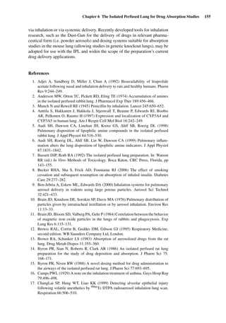
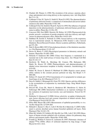

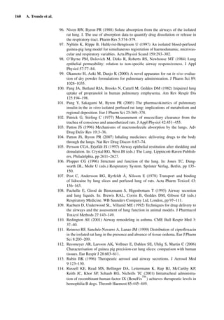
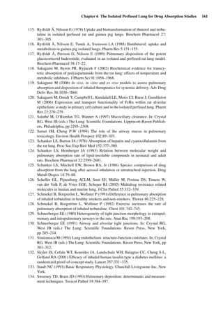
![7
In Vitro Models for Investigations of
Buccal Drug Permeation and
Metabolism
Tanja Obradovic and Ismael J. Hidalgo
Abstract Buccal drug delivery across mucosal lining of the cheek offers
a clear advantage over the peroral dosing route by avoidance of intestinal
and hepatic first-pass metabolism. However, despite offering the possibility
of improved systemic drug delivery, buccal administration has been utilized
for relatively few pharmaceutical products so far. One of the reasons is costly
preclinical development, which includes laborious pharmacokinetic evaluation
in animal models. Utilization of in vitro methodology can greatly aid in early
development by offering a significant increase in the number of compounds
and/or formulations that can be evaluated for their potential for buccal absorp-
tion. Currently, in vitro models used for the assessment of buccal drug delivery
utilize freshly isolated animal buccal mucosa as well as human buccal tissue
cultures. This chapter describes the different in vitro techniques used for the
evaluation of buccal drug absorption and indicates their advantages and disad-
vantages.
Keywords: Buccal; Mucosa; In vitro; Absorption; Permeability; Culture
7.1. Introduction
Buccal drug delivery offers several advantages over the peroral route. These
advantages include avoidance of presystemic drug elimination within the gas-
trointestinal tract and/or during the hepatic first-pass metabolism, and inde-
pendence from the potential variability of absorption caused by the gastric
emptying rate, or the presence of food in the upper region of the gastrointestinal
tract [1]. In addition, the buccal mucosa is relatively permeable with a rich
blood supply and has a substantial resistance to irritation or damage [1–3].
Structurally, the human buccal mucosa is a multistratified, nonkeratinized,
squamous epithelium lining the cheek area of the oral cavity [4]. Drug per-
meation across buccal epithelium has been suggested, like for other mucosal
barriers, to involve paracellular (between the cells) and transcellular (through
the cells) routes [5]. Hydrophilic compounds predominantly permeate via the
paracellular route, whereas hydrophobic compounds do sovia the transcellular
route [6]. Numerous compounds with diverse physicochemical properties have
167](https://image.slidesharecdn.com/drugabsorptionstudies-2008-150610022529-lva1-app6892/85/Drug-Absorption-Studies-2008-185-320.jpg)
![168 T. Obradovic and I. J. Hidalgo
been developed for buccal application targeting local or systemic delivery
including proteins such as insulin [7–13]. Since buccal administration is an
attractive way to deliver drugs, the feasibility of this route for a candidate
compound is investigated by in vivo or in vitro methodologies of buccal drug
permeation properties. This chapter will provide an insight into often employed
in vitro methodologies for the assessment of drug absorption potential across
buccal mucosa.
7.2. In Vitro Studies
7.2.1. Isolated Buccal Tissue Mounted in Diffusion Cell Apparatus
Currently, the most widely used in vitro methodology for assessing drug per-
meation across buccal mucosa is by testing drug permeation across isolated
buccal tissue mounted in diffusion chambers. Chapter 4 of this book focuses
exclusively on the available in vivo and ex vivo methods; thus, these will be
discussed only in brief in the context of this chapter. Human buccal mucosa
is scarcely available and if obtained, the size of the tissue available from one
donor is not sufficient for conducting a large number of in vitro experiments.
Therefore, most research efforts relied on the use of isolated animal buccal
tissue. Because of species differences in physiology and anatomy of buccal
mucosa, caution must be exercised during the process of selection of animal
species to be used in this type of studies. The only laboratory animal with a
nonkeratinized surface layer of the buccal mucosa similar to human tissue that
has been used in buccal absorption studies is the rabbit [14, 15]. However, in
the rabbit, the nonkeratinized area of the buccal tissue has a sudden transition
to keratinized regions, which limits the amount of suitable tissue available and
makes isolation of the desired region unreliable [16]. Therefore, buccal tissue
from other animal species tends to be used more often than that from rabbits. In
particular, the buccal mucosa of monkeys [17], dogs [17–21], and pigs [22–25]
has been extensively used for in vitro drug permeability studies. The porcine
buccal tissue is the most frequently utilized model due to relative similarity to
human tissue [3], and a low cost associated with tissue acquisition.
For in vitro experiments using isolated animal buccal mucosa, different types
of diffusion cells have been used for mounting tissue. The use of diffusion cells
makes it possible to determine the actual amount of drug that diffuses across the
mucosal barrier as well as the rate of drug diffusion. Various types of diffusion
cells are available. Among them is the Franz-type diffusion chamber that was
first utilized for the determination of buccal permeation by Kurosaki et al. [26].
The Franz-type diffusion chamber is a static, one-chamber diffusion cell where
compound in neat solution or in formulated form is applied to the mucosal
apical surface, which faces the air through the open cell-cap that constitutes
the donor compartment (Figure 7.1). The receiver chamber volume is typically
6–8 ml and a stirring bar maintains homogeneous temperature and mixes the
solution in the receiver compartment. The temperature of the apparatus is
maintained at 37◦C by placing the diffusion cell into a heated water bath.
Samples from the receiver solution are taken via the side port on the diffusion
cell typically for up to 180 min, a time interval much shorter (few hours) than](https://image.slidesharecdn.com/drugabsorptionstudies-2008-150610022529-lva1-app6892/85/Drug-Absorption-Studies-2008-186-320.jpg)
![Chapter 7 In Vitro Models for Investigations of Buccal Drug Permeation and Metabolism 169
Figure 7.1 Schematic of a Franz-type diffusion cell.
that at which isolated buccal mucosa preparation is viable in this system [27]. In
addition, the buccal tissue used in permeation studies should be fresh because
freezing and thawing has been shown to alter significantly the permeability
properties of porcine oral mucosa [28].
Figure 7.2 presents permeation across freshly isolated porcine buccal
mucosa of caffeine and atenolol. Caffeine and atenolol are compounds with
complete and poor human buccal absorption in vivo, respectively [10, 29],
Atenolol Caffeine
0.00
0.25
0.50
0.75
1.00
1.25
1.50
1.75
2.00
2.25
2.50
2.75
3.00
3.25
*
Compound
m-sPapp(x10
-6
cm
2
/s)acrossfreshlyisolatedporcinemucosa
Figure 7.2 Mucosal-to-submucosal (m-s) Papp values of
atenolol and caffeine obtained in freshly isolated porcine
buccal mucosa from ten animals (average ± SEM).
∗ (Asterisk) significantly higher than atenolol value (t-test,
p < 0.005). Results are from Ref. [42].](https://image.slidesharecdn.com/drugabsorptionstudies-2008-150610022529-lva1-app6892/85/Drug-Absorption-Studies-2008-187-320.jpg)
![170 T. Obradovic and I. J. Hidalgo
Table 7.1 Examples of atenolol and caffeine
mucosal-to-submucosal (m-s) permeation across
freshly isolated porcine buccal mucosa.
Porcine
Atenolol m-s Papp
(×× 10−6 cm/s)
Caffeine m-s Papp
(×× 10−6 cm/s)
1 0.01 1.89
2 0.02 2.46
3 0.04 1.56
4 0.04 1.61
5 0.07 4.31
6 0.22 5.90
7 0.05 1.97
8 0.10 1.14
9 0.12 1.53
10 0.23 3.32
Average (SD) 0.09 (0.08) 2.57 (1.51)
which correlates well with significantly higher caffeine than atenolol perme-
ation across porcine buccal epithelium in vitro (Figure 7.2). The transport of
caffeine and atenolol showed a significant difference in apparent permeability
coefficient (Papp) values of at least an order of magnitude in each of ten
animal tissue donors (Table 7.1). A clear difference existed between atenolol
and caffeine Papp in each donor, despite a large variability between different
animals (Table 7.1). The interindividual and intertissue variability in the buc-
cal epithelium thickness observed histologically may be a significant factor
contributing to the Papp variability shown in Table 7.1.
Squier and Hall [30, 31] have found that the thickness of the upper third
of the buccal epithelium, which contains membrane-coating granules, plays
a major role in limiting the drug permeation of hydrophilic compounds. In
addition, a recent study by Giannola et al. [32] demonstrated an extensive
effect of epithelial thickness on buccal in vitro permeation of carbamazepine,
a relatively lipophilic compound (c logP 2.7). Therefore, the determination
of statistically significant differences in permeation for compounds that have
relatively close rates of buccal permeation may be difficult in diffusion cells
due to large interanimal variability and it may require a large number of animal
tissue samples [33].
In general, drug permeability across buccal tissue, or any other mucosal bar-
rier, is dependent on physicochemical properties of the drug, such as lipophilic-
ity, molecular weight, and degree of ionization at physiological pH (pH 6.8–
7.4 for buccal tissue). Regardless of the transport route, small molecules tend
to be absorbed faster. In addition, a positive relationship between lipophilicity
and buccal in vitro permeability has been found on a set of acetanilides [34].
McElnay et al. [35] reported pH dependence of buccal captopril absorption
with good correlation between in vitro results and in vivo human absorption.
Current efforts to develop suitable drug delivery systems include permeation
enhancers and controlled release systems for local or systemic therapy that
rely substantially on the utilization of in vitro buccal tissue assays [36]. Only](https://image.slidesharecdn.com/drugabsorptionstudies-2008-150610022529-lva1-app6892/85/Drug-Absorption-Studies-2008-188-320.jpg)
![Chapter 7 In Vitro Models for Investigations of Buccal Drug Permeation and Metabolism 171
a few studies have shown a correlation between in vitro and in vivo buccal
drug absorption and most of them examined the effect of permeation enhancers
or release rate-modifying excipients [37]. For example, a good correlation
between in vitro permeation across porcine buccal mucosa and bioavailability
enhancement in rabbits has been observed with a triamcinolone acetonide
gel formulated with the permeability enhancer sodium deoxycholate [38, 39].
In addition, Degim et al. [40] recently observed good correlation between
effective delivery of acyclovir following buccal tablet dosing in dogs and the
acyclovir pH-dependant permeation profile across buccal tissue in vitro. In con-
trast, a poor correlation was found between in vitro permeation across porcine
buccal epithelium and bioavailability in pigs of fluorescein isothiocyanate
(FITC)-labeled dextrans, commonly used model compounds for peptides [41].
Therefore, caution needs to be exercised when extrapolating in vitro results to
in vivo absorption, including delivery in humans.
As noted earlier, the choice of species for experimentation is critical due to
anatomical differences and it may also reflect species, as well as individual,
differences in the expression/activity of transporter and metabolic proteins
[43, 44]. Since the fraction absorbed across buccal mucosa in vivo is not
established for many compounds in different species including humans, the
potential existence of a correlation between in vitro permeability coefficients in
freshly isolated pig, dog, monkey, and human buccal mucosa was investigated
(Figure 7.3). The correlation coefficient obtained for porcine and canine tissue
was poor (0.65 and 0.67, respectively, at the 95% confidence level). Results for
relatively high permeability compounds in porcine tissue resemble those previ-
ously reported where permeability coefficients were by an order of magnitude
0 1 2 3 4 5 6 7 8 9 10 11 12 13 14
0.5
1.5
2.5
3.5
4.5
5.5
6.5
Porcine R
2
= 0.65
Dog R2
= 0.68
Monkey R
2
= 0.87
Papp Across Fresh Human Buccal Mucosa
PappAcrossFreshAnimalBuccalMucosa
Figure 7.3 Correlation analysis for Papp values across freshly isolated animal buccal
mucosa and freshly isolated human buccal mucosa of Lucifer yellow, enalaprilat,
atenolol, caffeine, sumatriptan, and fentanyl. Results from internal study by Absorption
Systems Company.](https://image.slidesharecdn.com/drugabsorptionstudies-2008-150610022529-lva1-app6892/85/Drug-Absorption-Studies-2008-189-320.jpg)
![172 T. Obradovic and I. J. Hidalgo
lower across porcine buccal mucosa mounted in the Franz-type chamber than
across reconstituted human oral epithelium [32]. In addition, porcine buccal
mucosa showed consistently lower permeability than human tissue toward
compounds of hydrophilic and hydrophobic nature in vitro [45], which is in line
with observations for atenolol and caffeine (Tables 7.1 and 7.2). These findings
indicate that pig buccal tissue has a higher barrier property to drug permeation
than do human buccal mucosa. The strongest correlation observed was that
between permeation across monkey and human buccal mucosa (correlation
coefficient 0.87 at the 95% confidence level). These observations, obtained on
a small set of passively transported compounds with different physicochemical
properties, indicate that in vitro permeability determined using buccal tissue
mounted in the Franz-type cell varies across species. Observed correlation
results likely reflect physiological and anatomical differences between the
species [16], which also may contribute to the contradictory results between
in vitro experiments with animal buccal tissue and in vivo human studies [46].
Other factors that can cause an underestimation of the drug buccal penetration
potency in vitro include the lack of blood flow [47] and the existence of
an unstirred water layer on the mucosal surface that may pose a substantial
diffusion barrier especially for lipophilic compounds [26, 43].
In spite of the limitations associated with using the in vitro buccal tissue
system, this model appears to be a useful tool for evaluating drug permeation
across buccal mucosa. The main advantages include (1) assessment of buccal
delivery at the intended application site where, unlike the in vivo situation,
there is no compound loss due to swallowing; (2) tissue resistance to irritation
and loss of integrity, which allows for a wide-range of excipient testing; and (3)
the possibility to obtain in vitro animal buccal tissue data that can be correlated
to in vivo animal results prior to human testing.
7.2.2. Buccal Epithelial Cell Cultures
Although isolated animal buccal mucosa mounted in diffusion chambers is the
most used technique to estimate buccal absorption, this tissue model has several
practical disadvantages. These include a limited surface area, presence of tissue
damage from mastication, laborious and time-consuming excision procedure,
minimal potential for assay automatization, and large variability in permeation
results among replicates. These disadvantages make it impractical to use the
tissue model for testing large numbers of compounds and/or compound for-
mulations. For these reasons, attempts have been made to establish suitable
in vitro buccal epithelial cell culture models for testing drug permeation and
metabolism. Tavkoli-Saberi et al. [48] developed the hamster pouch epithelium
culture. In addition, a TR146 cell culture derived from human buccal metastasis
has been used to assess drug transport and metabolism [33, 49]. However,
the effect of parameters that can affect drug transport, such as the control of
culture growth rate, number of differentiated cell layers, lipid composition,
and optimum day(s) in culture, is still not well characterized in these models.
Therefore, these cultures have not been extensively used in the development
of drug candidates for buccal delivery. Recently, a new cultured human buc-
cal epithelium model EpiOralR
has become commercially available (MatTek,
Ashland, MA). This tissue model consists of normal human keratinocytes
cultured to form a three-dimensional differentiated tissue that histologically](https://image.slidesharecdn.com/drugabsorptionstudies-2008-150610022529-lva1-app6892/85/Drug-Absorption-Studies-2008-190-320.jpg)
![Chapter 7 In Vitro Models for Investigations of Buccal Drug Permeation and Metabolism 173
Table 7.2 Examples of atenolol and caffeine mucosal-to-submucosal (m-s)
permeation across cultured buccal epithelium.
Atenolol m-s Papp (× 10−6 cm/s) Caffeine m-s Papp (× 10−6 cm/s)
Batch Average ± SD, N = 3 Average ± SD, N = 3
1 1.89 ± 0.23 17.7 ± 1.1
2 2.02 ± 0.31 17.2 ± 0.5
3 1.67 ± 0.19 15.6 ± 0.9
Average (SD) 1.86 ± 0.18∗ 16.8 ± 1.1∗_
* Corresponding Papp value for atenolol and caffeine across freshly isolated human buccal
mucosa mounted in Franz-type cell is 1.16 (0.25) and 14.5 (1.6), respectively.
and biochemically resembles human buccal tissue [50]. So far, this model has
shown good correspondence to human buccal mucosa in terms of structure,
protein expression, and lipid content with good reproducibility of the tissue
cultures [51]. Also, a good correlation between the permeation of fentanyl
across cultured buccal epithelium, applied in different tablet formulations,
and bioavailability in humans has been found [52–54]. The Papp of atenolol
(poorly absorbed reference) and caffeine (highly absorbed reference), across
different cultured batches, are presented in Table 7.2. Results indicate that the
barrier function of this in vitro model appears reproducible across batches.
In addition, a relatively good correlation between drug permeabilities across
freshly isolated human buccal mucosa and human buccal culture is observed
(Figure 7.4, correlation coefficient 0.96 at the 95% confidence level). These
results indicate that human buccal epithelial culture may be a better predictor
of permeation across human buccal mucosa than animal buccal tissue models
(Figure 7.3).
However, the number of compounds tested in vitro on human EpiOralR
buccal culture is currently very limited [50]. Therefore, further studies are
needed to better evaluate usefulness of this novel model in testing drug buccal
delivery potential.
Of particular interest is a possible use of EpiOral R
human buccal cul-
ture for evaluation of drug metabolism and its effect on buccal drug perme-
ation. The use of previously frozen human mucosal specimens is an often-
employed method in the investigation of drug metabolism [55, 56]. However,
the loss of enzymatic activity in this tissue due to storage can be a limit-
ing factor in accurately determining drug metabolism. On the other hand,
metabolism in fresh human buccal tissue cannot be easily addressed due to
lack of readily available fresh mucosa in sufficient quantities for in vitro
testing. So far, the metabolic fate of drugs in the human buccal mucosa
is predominantly investigated in tissue homogenates obtained from the oral
cavity of different animal species [57–59]. However, due to well- known
interspecies differences in enzyme expression [44, 60], results in animals can
differ from human metabolism. The knowledge of drug enzymatic degra-
dation in the human buccal mucosa is very limited including the roles of
Cytochrome P450 enzymes (CYPs). The CYP enzymes are responsible for
oxidative metabolism in the liver and extrahepatic tissues including buccal
tissue [61, 62]. CYP3A4, CYP2C9, and CYP2D6 are the major human liver
CYPs that are known to limit bioavailability of numerous drugs [63]. The
functionality of these CYP enzymes in the EpiOralR
human buccal culture was](https://image.slidesharecdn.com/drugabsorptionstudies-2008-150610022529-lva1-app6892/85/Drug-Absorption-Studies-2008-191-320.jpg)
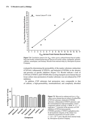
![Chapter 7 In Vitro Models for Investigations of Buccal Drug Permeation and Metabolism 175
0 10 20 30 40 50 60 70 80 90 100
0
5
10
15
20
25
30
35
40
45
50
55
60
65 Atenolol
Caffeine
Fentanyl
time (min)
Cummulativereceiverconcentration(µM)
Figure 7.6 Cumulative receiver
concentrations of fentanyl,
caffeine, and atenol vs time
across cultured human buccal
tissue (average ± SD, N = 3).
Results from internal study by
Absorption Systems Company.
compound in vivo. These results corroborate the findings of Vondracek et al.
[64] where, on a large set of human donors, buccal epithelium expression
and catalytic activity of CYP3A4, CYP2C9, and CYP2D6 was minimal or
nonexistent despite mRNA expression. Therefore, for compounds that may
be substantially degraded by CYP3A4, CYP2C9, and CYP2D6 following
peroral administration, a buccal route of delivery may be advantageous. For
example, fentanyl is a compound with very low oral bioavailability due to
a high first-pass metabolism that is mediated by CYP3A4 in the liver and
intestine [65]. Fentanyl permeation across cultured human buccal epithelium
is high (Figure 7.6), which is in accordance with similar fentanyl and caffeine
permeation in this model when fentanyl flux from the tablet form was tested
[52]. Furthermore, these results are in agreement with reported rapid and com-
plete fentanyl absorption when applied buccally in humans [53, 54]. Although
buccal mucosa appears to have low basal CYP activity [50, 64], other non-
CYP metabolic processes in this tissue have also been reported (Table 7.3).
Among them, different types of peptidases and esterases have been identified
in either human or animal buccal specimens [34, 49, 57, 66, 67]. In addition,
the buccal metabolism of drugs that are subjected to peptidase and/or esterase-
mediated degradation has been found to have substantial effect on their delivery
through the buccal route [49, 68, 69]. Therefore, buccal administration of
compounds that may be liable to these enzymatic processes may present a
challenge.
Currently, most strategies for buccal delivery of peptide drugs have focused
on the application of excipients that would shorten the time of absorption
and adhere drugs to a local site on the mucosa, thus decreasing exposure to
proteolytic degradation and possible release of drug back into the mouth cavity.
This strategy has been utilized in the buccal delivery of insulin, enkephalin, and
testosterone [37, 70].](https://image.slidesharecdn.com/drugabsorptionstudies-2008-150610022529-lva1-app6892/85/Drug-Absorption-Studies-2008-193-320.jpg)
![176 T. Obradovic and I. J. Hidalgo
Table 7.3 Enzymes present in the buccal mucosa.
Species Enzyme
Human Aminopeptidasea, b
Carboxypeptidaseb
Esteraseb
Dehydrogenasec
CYP1A1, 1A2, 2C, 2D6, 2E1, 3A4/7, 3A5d
Aminopeptidaseb
Pig Carboxypeptidaseb
Esterasesb
Aminopeptidasee
Rabbit Carboxypeptidasef
Proteaseg
Aminopeptidaseh
Rat Carboxypeptidasef
Esterasei
a Ref. [71]; b Ref. [49]; c Ref. [72]; d Ref. [64]; e Ref. [73];
f Ref. [74]; g Ref. [75]; h Ref. [66]; i Ref. [58].
7.3. Concluding Remarks
Considering the substantial use of excipients in buccal delivery systems, it
is important to use a suitable in vitro model that correlates well with human
results in vivo. Experiments on isolated animal buccal tissue provided suffi-
ciently predictive results of excipient effect on in vivo buccal absorption for
many drugs including many small molecules as well as peptides [37; 41].
Currently, buccal cell cultures have been utilized very scarcely to evaluate
the effect of formulation excipients on drug transport intended for transbuc-
cal delivery. So far, enhancing effects of chitosan [76], sodium glycocholate
[77], and bile salts [78] on drug permeation across TR146 buccal cell culture
have been reported while excipient effects in the EpiOralR
model have not
been investigated. In general, cell culture models tend to be less suitable than
isolated mucosal tissue for excipient testing because cultured cells are more
susceptible to formulation excipient effects on cell viability and functional
integrity [79–81]. Therefore, studies on the effects of permeation enhancers and
other excipients on the physical and biochemical integrity of cultured buccal
epithelium are needed in order to evaluate the potential utility of this model to
estimate buccal drug absorption under relevant conditions. Furthermore, such
studies may address the potential of excipients/enhancers to cause irritation in
humans.
At present, despite the advantages offered by the buccal delivery route,
such as the bypass of intestinal and hepatic first-pass metabolism for systemic
delivery, very few pharmaceutical products employ this route of administration.
The reasons that contribute to this situation include (1) high costs associ-
ated with development, (2) lack of standardized tests to identify drug candi-
dates suitability for this route, (3) the limited understanding of the impact of
metabolism and/or transporters found in the oral cavity mucosa on buccal deliv-
ery, and (4) the relatively small number of reports describing the usefulness and
safety of excipients/permeation enhancers in humans [82, 83]. Therefore, the](https://image.slidesharecdn.com/drugabsorptionstudies-2008-150610022529-lva1-app6892/85/Drug-Absorption-Studies-2008-194-320.jpg)
![Chapter 7 In Vitro Models for Investigations of Buccal Drug Permeation and Metabolism 177
development of an in vitro screening model derived from human tissue that can
address these issues may provide a useful tool in overcoming challenges during
preclinical development of candidates intended for buccal delivery.
References
1. Rathbone M, Drummond B, and Tucker I (1994) The oral cavity as a site for
systemic drug delivery. Adv. Drug Deliv. Rev. 13: 1–22.
2. Goodman CH, and Squier CA (1988) Blood flow in the oral mucosa of normal and
artherosclerotic rhesus monkeys. J. Oral Path. 17: 34–38.
3. Squier CA (1991) The permeability of oral mucosa. Crit. Rev. Oral. Biol. Med. 2:
13–32.
4. Squier CA and Finkelstein MW (1989) In: Oral Histology, Development, Structure
and Function. A. R. Ten Cate (Ed.), C. V. Mosby, St. Louis, pp 345–385.
5. Zhang H, and Robinson JR (1996) In vitro methods for measuring permeability
of the oral mucosa, In: Oral Mucosa Drug Delivery, Marcel Dekker, Inc., NY, pp
85–100.
6. Wertz PW, and Squier CA (1991) Cellular and molecular basis of barrier function
in oral epithelium. Crit. Rev. Ther. Drug Carrier Syst. 8: 237–269.
7. Oh CK, and Ritschel WA (1990) Biopharmaceutical aspects of buccal absorption
of insulin. Methods Find. Exp. Clin. Pharmacol. 12: 205–209.
8. Bray CL, Jain S, Faragher EB, Myers A, Myers P, MacIntyre P, Rae A, Goldman
M, and Alcorn M (1991) A comparison of buccal nitroglycerin and sublingual
nitroglycerin in the prophylaxis and treatment of exertional (situation-evoked)
angina pectoris. Eur. Heart J. 12 Suppl A: 16–20.
9. Venugopalan P, Sapre A, Venkatesan N, and Vyas SP (2001) Pelleted bioadhesive
polymeric nanoparticles for buccal delivery of insulin: Preparation and characteri-
zation. Pharmazie 56: 217–219.
10. Kamimori GH, Karyekar CS, Otterstetter R, Cox DS, Balkin TJ, Belenky GL, and
Eddington ND (2002) The rate of absorption and relative bioavailability of caffeine
administered in chewing gum versus capsules to normal healthy volunteers. Int.
J. Pharm. 234: 159–167.
11. Attia MA, El-Gibaly I, Shaltout SE, and Fetih GN (2004) Transbuccal permeation,
anti-inflammatory activity and clinical efficacy of piroxicam formulated in different
gels. Int. J. Pharm. 276: 11–28.
12. Cernea S, Kidron M, Wohlgelernter J, and Raz I (2005) Dose-response relationship
of an oral insulin spray in six patients with type 1 diabetes: A single-center,
randomized, single-blind, 5-way crossover study. Clin. Ther. 27: 1562–1570.
13. Mystakidou K, Katsouda E, Parpa E, Vlahos L, and Tsitas ML (2006) Oral trans-
mucosal fentanyl citrate: Overview of pharmacological and clinical characteristics.
Drug Deliv. 13: 269–276.
14. Siegel IA, and Gordon HP (1986) Surfactant-induced alterations of permeability of
rabbit oral mucosa in vitro. Exp. Mol. Path. 44: 132–137.
15. Dowty ME, Knuth KE, Irons BK, and Robinson JR (1992) Transport of tyrotropin
releasing hormone in rabbit buccal mucosa in vitro. Pharm. Res. 9: 1113–1122.
16. Squier CA, and Wertz PW (1996) Structure and function of the oral mucosa and
implications for drug delivery. In: Oral Mucosal Drug Delivery, Marcel Dekker,
Inc., NY, pp. 1–26.
17. Mehta M, Kemppaininen BW, and Stafford RG (1991) In vitro penetration of
tritium-labeled water (THO) ans [3H]PbTx-3 (a red tide toxin) through monkey
buccal mucosa and skin. Tox. Lett. 55: 185–194.
18. Ishida M, Machida Y, Nambu N, and Nagai T (1981) New mucosal dosage form of
insulin. Chem. Pharm. Bull. 29: 810–816.
19. Nagai T (1985) Adhesive topical drug delivery system. J. Control. Rel. 2: 121–134.](https://image.slidesharecdn.com/drugabsorptionstudies-2008-150610022529-lva1-app6892/85/Drug-Absorption-Studies-2008-195-320.jpg)
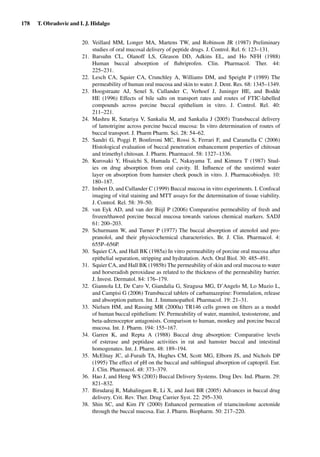
![Chapter 7 In Vitro Models for Investigations of Buccal Drug Permeation and Metabolism 179
39. Shin SC, Bum JP, and Choi JS (2000) Enhanced bioavailability by buccal admin-
istration of triamcinolone acetonide from the bioadhesive gels in rabbits. Int. J.
Pharm. 209: 37–43.
40. Degim T, Eglen B, and Ocak O (2006) A sustained release dosage form of acyclovir
for buccal application: An experimental study in dogs. J. Drug Target 14: 35–44.
41. Junginger HE, Hoogstraate JA, and Verhoef JC (1999) Recent advances in buccal
delivery and absorption: In vitro and in vivo studies. J. Control. Rel. 62: 149–159.
42. Obradovic T, Huynh T, Shingaki T, Strab B, Peck B, and Hidalgo JI (2005) Per-
meation across porcine buccal mucosa is correlated with absorption across human
buccal mucosa. American Association Pharm. Sci. Abs. W4101.
43. Lennernas H, Nylander S, and Ungell AL (1997) Jejunal permeability: A com-
parison between the Ussing chamber technique and the single-pass perfusion in
humans. Pharm. Res. 14: 667–671.
44. Ioannides C (2006) Cytochrome P450 expression in the liver of food-producing
animals. Curr. Drug Metab. 7: 335–348.
45. Van Eyk AD, and Van der Biijl P (2004) Comparative permeability of various
chemical markers through human vaginal and buccal mucosa as well as porcine
buccal and mouth floor mucosa. Arch. Oral Biol. 49: 387–392.
46. Wadell C, Bjork E, and Camber, O (2003) Permeability of porcine nasal mucosa
correlated with human nasal absorption. Eur. J. Pharm. Sci. 18: 47–53.
47. Ungell A L (1997) In vitro absorption studies and their relevance to absorption
from the GI tract. Drug Dev. Ind. Pharm. 23: 879–892.
48. Tavakoli-Saberi MR, and Audus KL (1989) Cultured buccal epithelium: An in
vitro model derived from the hamster pouch for studying transport and metabolism.
Pharm. Res. 6: 160–166.
49. Nielsen HM, and Rassing MR (2000b) TR146 cells grown on filters as a model
of human buccal epithelium: V. Enzyme activity of the TR146 cell culture model,
human buccal epithelium and porcine buccal epithelium, and permeability of leu-
enkephalin. Int. J. Pharm. 200: 261–270.
50. Walle T, Walle UK, Sedmera D, and Klausner, M (2006) Benzo[A]pyrene-induced
oral carcinogenesis and chemoprevention: Studies in bioengineered human tissue.
Drug Metab. Dispos. 34: 346–350.
51. Kubilus J, Ayehunie S, Breyfogle B, Dale DA, Kimball J, Wertz P, Klausner M
(2006) Characterization and testing of new buccal and gingival tissue models.
American Association of Dental Res. Meeting 399.
52. Agarwal V, Habib W, and Moe D (2005) Effervescent mediated transmucosal
enhancement of fentanyl permeability: Comparison across ex vivo porcine buccal
mucosa and in vitro cultured human buccal epithelium test. American Association
Pharm. Sci. Abs. T2126.
53. Darwish M, Kirby M, Robertson P, Tracewell W, and Jiang JG (2006) Pharma-
cokinetic properties of fentanyl effervescent buccal tablets: A phase I, open-label,
crossover study of single dose 100, 200, 400, and 800 micrograms in healthy adult
volunteers. Clin. Ther. 28: 707–714.
54. Portenoy RK, Taylor D, Messins J, and Tremmel L (2006) A randomized, placebo-
controlled study of fentanyl buccal tablet for breakthrough pain in opioid-treated
patients with cancer. Clin. J. Pain 22: 805–811.
55. Wynalda MA, Hutzler JM, Koets MD, Podoll T, and Wienkers LC (2003) In vitro
metabolism of clindamycin in human liver and intestinal microsomes. Drug Metab.
Disp. 31: 878–887.
56. Godoy W, Albano RM, Moraes EG, Pinho PRA, Nunes RA, Saito EH, Higa C,
Filho IM, Pinto Kruel CD, Schirmer CC, Gurski R, Lang MA, and Ribeiro Pinto
LF (2002) CYP2A6/2A7 and CYP2E1 expression in human oesophageal mucosa:
Regional and inter-individual variation in expression and relevance to nitrosamine
metabolism. Carcinogenesis 23: 611–616.](https://image.slidesharecdn.com/drugabsorptionstudies-2008-150610022529-lva1-app6892/85/Drug-Absorption-Studies-2008-197-320.jpg)
![180 T. Obradovic and I. J. Hidalgo
57. Kashi DS, and Lee VHL (1986) Enkephalin hydrolysis in homogenates of various
absorptive mucosal of the albino rabbit: Similarities in rates and involvement of
aminopeptidases. Life Sci. 38: 2019–2028.
58. Garren KW, and Repta AJ (1989) Buccal drug absorption. II: In vitro diffusion
across the hamster cheek pouch. J. Pharm. Sci. 78: 160–164.
59. Takagi Y, Kriehubner E, Imokawa G, Elias G, and Holleran WM (1999) ß-
Glucocerebrosidase activity in mammalian stratum corneum. J. Lipid Res. 40: 861–
869.
60. Nelson DR, Koymans L, Kamataki T, Stegman JJ, Feyerisen R, Waxman DJ, Water-
man MR, Gotoh O, Estabrook RW, Gunsalus IC, et al., (1996) P450 supefamily:
Update on new sequences, gene mapping, accession numbers and nomenclature.
Pharmacogenetics 6: 1–42.
61. Port JL, Yamaguchi K, Du BK, DeLorenzo M, Chang M, Heerdt OM, Kopelovich
L, Marcus CB, Altoki NK, Subbaramaiah K, et al., (2004) Tobacco smoke induces
CYP1B1 in the aerodigestive tract. Carcinogenesis 25: 2275–2281.
62. Wen X, and Walle T (2005) Preferential induction of CYP1B1 by benzo[a]pyrene
in human oral epithelial cells: Impact on DNA adduct formation and prevention by
polyphenols. Carcingenesis 26: 1774–1781.
63. Rendic S (2002) Summary of information on human CYP enzymes: Human P450
metabolism data. Drug Metab. Rev. 34: 83–448.
64. Vondracek M, Xi Z, Larson P, Baker V, Mace K, Pfeifer A, Tjalve H, Donato
MT, Gomez-Lechon MJ, and Grafstrom C (2001) Cytochrome P450 expres-
sion and related metabolism in human buccal mucosa. Carcinogenesis 22: 481–
488.
65. Kharasch ED, Whittington D, and Hoffer C (2004) Influence of hepatic and
intestinal cytochrome P4503A activity on the acute disposition and effects of oral
transmucosal fentanyl. Anesthesiology 101: 729–737.
66. Harris D, and Robinson JR (1992) Drug delivery via mucous membranes of the
oral cavity. J. Pharm. Sci. 81: 1–10.
67. Robinson DA, Bogdanoffy MS, and Reed CJ (2002) Histochemical localization
of carboxylesterase activity in rat and mouse oral cavity mucosa. Toxicology 180:
209–220.
68. Ho NF, Barshun CL, Burton SP, and Merkle HP (1992) Mechanistic insights
to buccal delivery of proteinaceous substances. Adv. Drug Deliv. Rev. 8:
197–235.
69. Veuillez F, Kalia YN, Jacques Y, Deshusses J, and Buri PA (2001) Factors and
strategies for improving buccal absorption of peptides. Eur. J. Pharm. Biopharm.
51: 93–109.
70. Wang C, Swerdloff R, Kipnes M, Matsumoto AM, Dobs AS, Cunningham G,
Katznelson L, Weber TJ, Friedman TC, Snyder P, and Levine HL (2004) New
testosterone buccal system (Striant) delivers physiological testosterone levels:
Pharmacokinetics study in hypogonadal men. J. Clin. Endocrinol. Metab. 89: 3821–
3829.
71. Audus KL, Wlliams A, and Tavakoli-Saberi MR (1991) A comparison of
aminopeptidases from excised human buccal epithelium and primary cultures of
hamster pouch buccal epithelium. J. Pharm Pharmacol. 43: 363–365
72. Hedberg JJ, Hoog J-O, Nilsson JA, Xi Z, Elfwing A, and Graftstrom RC (2000)
Expression of alcohol dehyrogenase 3 in tissue and cultured cells from human oral
mucosa. Am. J. Pathol. 157: 1745–1754.
73. Stratford RE, and Lee VHL (1986) Aminopeptidase activity in homogenates of
various absorptive mucosae in the albino rabbit: Implications in peptide delivery.
Int. J. Pharm. 30: 73–82.
74. Wearly LL (1991) Recent progress in protein and peptide delivery by non-invasive
routes. Crit. Rev. Ther. Drug Carrier 8: 331–394.](https://image.slidesharecdn.com/drugabsorptionstudies-2008-150610022529-lva1-app6892/85/Drug-Absorption-Studies-2008-198-320.jpg)
![Chapter 8 In Vitro Screening Models to Assess Intestinal Drug Absorption and Metabolism 183
MDCK Madin-Darby canine kidney cells
MDR Multidrug resistance
MRP Multidrug resistance-associated protein
MTT 3-(4,5-dimethylthiazol-2-yl)-2,5-diphenyltetrazolium bromide
NMP N-methyl-2-pyrrolidone
NRU Neutral red uptake
PAMPA Parallel artificial membrane permeation assay
PC Phosphatidylcholine
PD Potential difference
PE Phosphatidylethanolamine
PEG Polyethyleneglycol
P-gp P-glycoprotein
PI Phosphatidylinositol
SCC Short circuit current
TEER Transepithelial electrical resistance
UWL Unstirred water layer
8.1. Introduction
8.1.1. General Factors Influencing Intestinal Drug Absorption
Despite tremendous innovations in the field of drug delivery technology, oral
intake remains the preferred route of drug administration, for reasons of patient
convenience and therapy compliance. Compounds intended for oral adminis-
tration must have adequate biopharmaceutical properties in order to achieve
therapeutic concentrations at the targeted site of action.
The in vivo absorption process is the outcome of a complex series of events
and depends on a multitude of parameters related to drug characteristics as well
as to physiological aspects of the gastrointestinal (GI) tract [93]. A summary
of the most important factors influencing the rate and extent of intestinal
absorption is given in Table 8.1.
The parallel screening of drug candidates for their pharmacological activity
and for their physicochemical and biopharmaceutical properties has become
a major challenge in drug development and is performed at very early stages
Table 8.1 Overview of the main factors influencing the extent and rate of
intestinal drug absorption.
Physicochemical
factors
Physiological
factors
Formulation
factors
Biochemical
factors
Aqueous solubility
Molecular size and
weight
Aggregation/
Complexation
Charge (pKa)
H-bonding potential
Molecular surface area
Drug hydrophobicity
Crystal lattice energy
Stomach emptying
time
Intestinal motility
Membrane
permeability
Intestinal pH
Disease state
Blood flow
Luminal content and
composition
(including bile salts)
Dosage form
Drug release
Absorption
enhancers
Metabolism
Efflux
transporters
Active uptake
transporters](https://image.slidesharecdn.com/drugabsorptionstudies-2008-150610022529-lva1-app6892/85/Drug-Absorption-Studies-2008-201-320.jpg)
![184 S. Deferme et al.
Figure 8.1 (A) Cross-sectional view of the organization of the small intestine, illustrat-
ing the serosa, the longitudinal and circular muscle layers (=muscularis externa), the
submucosa, and the intestinal mucosa. The intestinal mucosa consists of four layers, the
inner surface cell monolayer of enterocytes, the basal membrane, the lamina propria
(connective tissue, blood capillaries), and the muscularis mucosae; (B) Schematic
representation of an enterocyte (small intestinal epithelial cell); (according to Tso and
Crissinger [151], with permission).
in drug development [50]. Numerous efforts have been made by the pharma-
ceutical industry and academia to identify the various molecular features that
are important for drug absorption and to implement appropriate, high-capacity,
and cost-effective screening models that are predictive for in vivo intestinal
absorption. It should be noted that the intestinal absorption process is the
result of interplay of various physicochemical, physiological, and biochemical
properties. Therefore, no single in vitro model will mimic the complexity of
the in vivo absorption process [22].
8.1.2. The Intestinal Mucosa as a Physical and Biochemical Barrier to
Drug Absorption
8.1.2.1. Physical Barrier Function
The small intestinal wall (Figure 8.1) consists of the serosa, the muscularis
externa (longitudinal and circular muscle layers), the submucosa, and the
intestinal mucosa. The latter forms the main barrier that controls drug and
nutrient uptake after dissolution in the luminal contents. The mucosa consists
of four layers: a surface monolayer, the basal membrane, the lamina propria
(containing connective tissue, nerves, lymphocytes, blood vessels, lymphatic
vessels, macrophages, and mast cells), and the lamina muscularis mucosa
(smooth muscle cells). The surface layer is a monolayer of columnar epithelial
cells (enterocytes; see Figure 8.1) and also contains mucus-producing cells,
lymphocytes, and microfold cells (M-cells) [134] Because of the presence of
tight junctions, the intercellular space is reduced to 0.8 nm (human jejunum)
and 0.3 nm (human colon) [8]. Only small, hydrophilic, and polar molecules
are able to cross the epithelial monolayer through the paracellular space
[8, 104]. The paracellular route (see Figure 8.2) generally results in low
oral absorption, due to the limited surface area. More lipophilic drugs will
distribute into the cell membrane and cross the epithelial monolayer by the](https://image.slidesharecdn.com/drugabsorptionstudies-2008-150610022529-lva1-app6892/85/Drug-Absorption-Studies-2008-202-320.jpg)
![Chapter 8 In Vitro Screening Models to Assess Intestinal Drug Absorption and Metabolism 185
Figure 8.2 Possible drug transport pathways across the intestinal mucosa, illustrating
transcellular (1) and paracellular (2) modes of passive transport, transcytosis (3), carrier-
mediated transport (4), and efflux transport (5). A combination of these routes often
defines the overall transepithelial transport rate of nutrients and drugs.
passive transcellular route [10]. The rate of transport through the epithelium
depends on the concentration gradient and the permeation characteristics of
the compound through the unstirred water layer at the luminal surface bound-
ary of the epithelium and through the apical and basolateral membranes.
Transcytosis, a form of transport in which the nutrient/drug is transported
through the enterocyte by small vesicles, is a third transport pathway across the
intestinal epithelium. Especially proteins and small nucleotides are believed
to cross the intestine in this way [155]. Some compounds are essential for
the functionality of the human body (e.g., glucose, amino acids) and can be
actively taken up from the intestinal lumen by specific carriers present in the
apical membrane of enterocytes. This carrier-mediated transport also follows
an electrochemical concentration gradient. Several drugs, such as dopamine,
cephalosporins, and angiotensin-converting enzymes (ACE)-inhibitors, are
actively absorbed by the enterocytes. Finally, efflux-mediated transport rep-
resents a fifth form of transport. In this case, drugs that entered the enterocytes
by passive diffusion are being actively pumped out of the enterocytes, back
into the intestinal lumen. During this transport process, an energy source such
as ATP, electron transport, or an electrochemical gradient of another ion is
required to move ions or molecules against their electrochemical potential
gradients [155].
8.1.2.2. Biochemical Barrier Function
8.1.2.2.1. Intestinal Metabolism: Intestinal drug metabolism can occur by
microflora present in the gut lumen, as well as by enzymes present in luminal
fluids and in the intestinal mucosa [166]. Metabolism of xenobiotics by gut
microflora is low in comparison to metabolism by the gut mucosa and liver
[62]. However, the intestinal microflora (e.g., Bacteroides and Bifidobacteria)
may play an important role in the first-pass metabolism of compounds that
are poorly or incompletely absorbed by the gut mucosa, especially in the
lower parts of the intestine. This bacterial metabolism is largely degradative,](https://image.slidesharecdn.com/drugabsorptionstudies-2008-150610022529-lva1-app6892/85/Drug-Absorption-Studies-2008-203-320.jpg)
![186 S. Deferme et al.
hydrolytic, and reductive, in contrast to the predominantly oxidative and con-
jugative metabolism that occurs in the intestinal mucosa.
Phase I metabolism by intestinal cytochrome P450 enzymes (CYP) has
long been considered a relatively minor determinant of oral drug absorption,
because concentrations normalized for the entire intestine are estimated to be
∼20- to 200-fold lower than those found in the liver [20]. However, enzymes
of the CYP3A subfamily, considered the major phase I drug metabolizing
enzymes in humans, have been described to be expressed at high levels in
the mature villus tip enterocytes of the small intestine [94, 73]. CYP3A4,
accounting for ∼70% of the CYP content in human enterocytes, and CYP3A5
are the major CYPs expressed in human intestine, whereas much lower levels
of CYP1A1, CYP2C8, CYP2C9, CYP2C10, CYP2D6, and CYP2E1 could be
detected [166]. Expression levels of intestinal CYP3A4 can be induced by
drugs and food components [170, 76]. CYP3A5 does not seem to be inducible
and, although it has similar substrate specificity to CYP3A4, demonstrates
significantly different catalytic activities [127].
Esterases form another important group of metabolizing enzymes. These
enzymes are characterized by a broad tissue distribution and play a role in the
biotransformation of a vast number of structurally diverse drugs (e.g., ester- and
amide-containing drugs). Besides arylesterases, lipases, acetylesterases, and
pancreatic hydrolases, the class of the carboxylesterases forms an important
group of esterases present in the gut. Carboxylesterases are often considered
to be the main esterases responsible for the activation of ester prodrugs,
which are mainly used to overcome the poor membrane permeability of a
pharmacologically active compound. After absorption by the intestine, the
prodrug can be hydrolyzed by esterases to restore the parent drug in the
bloodstream [163]. However, the luminal and mucosal esterase activity may
cause a presystemic activation and thus a decreased absorption of the lipophilic
prodrug.
The example of amprenavir, an HIV-1 protease inhibitor, shows that intesti-
nal metabolism can also be used as a strategy to enhance the bioavail-
ability of compounds. In the biopharmaceutics classification system (BCS),
amprenavir can be categorized as a class II compound: it is poorly solu-
ble but highly permeable [51]. Fosamprenavir, the water-soluble phosphate
salt of amprenavir, on the other hand, shows poor transepithelial trans-
port. However, after oral administration of fosamprenavir, this compound
is metabolized into amprenavir in the intestinal lumen and in the entero-
cytes mainly by alkaline phosphatases, resulting in an increased intestinal
absorption [51, 174].
Other drug-metabolizing enzymes of interest include monoamine oxidase
and a wide range of hydrolytic and phase II enzymes (e.g., acetyltrans-
ferases, glutathione-S-transferases, methyltransferases, sulfotransferases, and
UDP-glucuronosyltransferases) [62, 79]. An interesting example of intestinal
metabolism can be found in a study on dietary flavonoids [91]. The results of
this study indicate considerable differences among the various types of dietary
flavonoids. Small intestinal absorption ranged from 0% to 60% of the dose, and
elimination half-lives ranged from 2 to 28 h. Absorbed flavonoids underwent
extensive first-pass phase II metabolism in the small intestinal epithelial cells
and in the liver. It was concluded that the most abundant flavonoids in the diet](https://image.slidesharecdn.com/drugabsorptionstudies-2008-150610022529-lva1-app6892/85/Drug-Absorption-Studies-2008-204-320.jpg)
![Chapter 8 In Vitro Screening Models to Assess Intestinal Drug Absorption and Metabolism 187
did not necessarily produce the highest concentration of flavonoids (or their
metabolites) in vivo [91].
8.1.2.2.2. Intestinal Efflux: In addition to metabolism, intestinal efflux is a
limiting factor to the absorption of a variety of structurally unrelated com-
pounds. The intestinal efflux component is responsible for an active secretion
from the epithelial cells to the luminal side, or, in some cases, to the serosal
side. Different efflux transporters have been identified and have been classified
as ATP-binding cassette (ABC) proteins [74]. While P-glycoprotein (P-gp,
MDR1, ABCB1) is the most extensively studied ABC efflux transporter that
acts as a biological barrier by extruding toxic substances and xenobiotics out
of the cell, it was demonstrated that BCRP (breast cancer resistance protein;
ABCG2) and MRP2 (ABCC2) are more extensively expressed in human
jejunum than P-gp [141]. The in vivo effect of efflux proteins in modulat-
ing the oral absorption of drugs is not fully understood. A study in man
with 13 drugs that were all substrates for intestinal efflux transporters, such
as P-gp, showed no significant influence of efflux transport on the in vivo
absorption of the drugs [37]. However, other reports suggest that the role of
intestinal efflux transporters in modulating oral drug absorption can be impor-
tant and especially relevant for dual CYP3A/efflux transporter substrates. In
such cases, the metabolism and efflux-affinity can severely influence intestinal
absorption [28, 29, 42].
Different food components (e.g., fruit extracts) and commonly used phar-
maceutical excipients (e.g., Tween 80) can interfere with secretory transport
systems, leading to drug–food interactions [31, 43, 46, 167]. As for CYPs,
inhibition and induction of efflux transporters have been reported as possible
causes for drug–drug interactions [88].
8.2. In Vitro Models to Study Intestinal Absorption
As oral intake remains the preferred route of drug administration, it is important
to have a good understanding of the intestinal absorption characteristics of
drug candidates in an early stage of their development. Clearly, whole animal
studies cannot be used as a screening tool in an early development stage;
therefore, in vitro models of intestinal absorption have been developed [33].
Each of these models has its advantages and disadvantages, which are summa-
rized in Table 8.2. This overview confirms that there is no perfect absorption
model and stresses the importance of using and combining different absorption
models.
In this chapter, the focus will be on membrane-based, cell culture-based,
and ex vivo models that are currently used in the industry to predict intestinal
absorption of new compounds.
8.2.1. Membrane-Based Models (PAMPA)
8.2.1.1. Background
The use of artificial membranes to investigate passive permeation processes
has a long history, going back more than 40 years [68]. The parallel arti-
ficial membrane permeation assay (PAMPA) is an application of the filter-
supported lipid membrane system [149] and was first introduced by Kansy and](https://image.slidesharecdn.com/drugabsorptionstudies-2008-150610022529-lva1-app6892/85/Drug-Absorption-Studies-2008-205-320.jpg)
![188 S. Deferme et al.
Table 8.2 Strengths and weaknesses of models to study intestinal drug
absorption [157, 25, 154, 33, 159, 69, 70, 171].
Absorption
model Strengths Limitations
In silico No animals required
Prediction of intestinal
absorption before synthesis
of compound
Difficult to model active
processes
Difficult to model complex
interplay of a multitude of
factors
Physicochemical
methods (e.g.,
log P, log D)
No animals required
Easy, fast, and inexpensive
Only predictive for
transcellular pathway
Low predictive value
Brush border
membrane
vesicles
IAM
Both animal and human tissue
can be used
Useful for mechanistic studies
Nonspecific binding
Only predictive for a part of
absorption process
Articifical
membranes
(e.g., PAMPA
technique)
High throughput
Relatively inexpensive
Different lipid compositions
available
Good predictability
Only predictive for a part of
absorption process
Membrane retention of
lipophilic compounds
Dependent on lipid
composition and pH
Isolated intestinal
cells
Animal or human origin
Relatively easy to perform
Important intervariability in
preparation of cells
Need for radiolabeled or
fluorescent compound
Limited viability of primary
cells
Everted intestinal
rings/sacs
Easy and inexpensive to
perform
Both animal and human tissue
can be used
Any segment of intestine
Useful for mechanistic studies
Nonspecific binding
Viability of tissue (<30 min)
Suboptimal stirring
conditions
Caco-2 cell
system
Good screening model
No bioanalysis (simple salt
buffer solutions)
Evaluation of transport
mechanisms (e.g. polarity in
transport)
Evaluation of absorption
enhancing strategies on a
mechanistic basis
Evaluation of toxicity of
compounds
Reduction in the number of
laboratory animals
Techniques available to
increase biorelevance of
model
Human origin
Lack of mucus secreting cells
resulting in absence of a
mucus layer
Presence of a thicker
unstirred water layer
compared to human small
intestine
Cancer cells, with different or
no expression of metabolic
enzymes (e.g., absence of
CYPs)
“Tighter” monolayer
compared to human small
intestine
Inter- and intralaboratory
variability of permeability
data
Long differentiation period
Low expression of uptake
transporters
Static model
(cont.)](https://image.slidesharecdn.com/drugabsorptionstudies-2008-150610022529-lva1-app6892/85/Drug-Absorption-Studies-2008-206-320.jpg)
![Chapter 8 In Vitro Screening Models to Assess Intestinal Drug Absorption and Metabolism 189
Table 8.2 (Continued)
Absorption
model Strengths Limitations
Ussing chambers
(Diffusion
chambers)
Good screening model
Good correlation with in vivo
permeability data
No bioanalysis (simple salt
buffer solutions)
Possibility to evaluate different
regions of the gastrointestinal
tract
Evaluation of transport
mechanisms (e.g., polarity in
transport)
Evaluation of absorption
enhancing strategies on a
mechanistic basis
Well-defined absorptive area
Good oxygenation
Tissue viability
Presence of circular muscle
layers during transport studies,
resulting in possible
under-estimation of the
permeability
Difficulties with unstirred water
layer
Tissue availability (human)
In situ intestinal
perfusion
Closest model to in vivo
situation
Evaluation of intestinal
absorption without influence
of hepatic first-pass
metabolism
Intact blood flow
Implies anesthesia and surgery
No screening tool
More difficult analysis due to
biological media (e.g., blood)
Laborious and time-consuming
Relevance for human situation
not always clear
coworkers as a rapid in vitro assay of transcellular permeation [67]. To make
lipid membranes, phospholipids and other membrane constituents are added
to the filter support as a solution in an organic solvent. In this first setup, a
96-well microtiter plate was completely filled with aqueous buffer solutions
(pH 7.4/6.5) and was covered with a microtiter filterplate in a sandwich con-
struction [67]. In the initial PAMPA experiments, the phospholipid composition
of mammalian membranes was mimicked by using a 10% dodecane solution
of egg lecithin [a mixed lipid containing phosphatidylcholine (PC), phos-
phatidylethanolamine (PE), phosphatidylinositol (PI), and cholesterol] [16].
Kansy and coworkers were able to relate their measured fluxes to human
absorption values with a hyperbolic curve, resembling very much the relation-
ship obtained in Caco-2 screening [67]. However, they experienced difficulties
with polar compounds (Mw < 250) that are transported via the paracellular
pathway and with actively transported compounds. Another disadvantage was
the strong dependency of the membrane permeability on pH, especially with
compounds having pKa values near the pH of the incubation buffer used
[67]. Additionally, the permeability of some basic compounds at pH 6.5 was
underestimated.
8.2.1.2. Lipid Composition
As different tissues have different lipid compositions [112], several groups
have worked on the composition of the lipid solutions to overcome these initial
PAMPA issues. Sugano and coworkers investigated the influence of the chain
length of the organic solvent on the permeability of a small set of compounds
over a PC membrane [136]. They reported an enhanced permeability with](https://image.slidesharecdn.com/drugabsorptionstudies-2008-150610022529-lva1-app6892/85/Drug-Absorption-Studies-2008-207-320.jpg)
![190 S. Deferme et al.
shorter chain alkyldienes, probably due to their lower viscosity. They also
added an anionic compound to the lipid composition in order to mimic the
intestinal lipid membrane, thus increasing the affinity of basic compounds
to the membrane [136]. The best-performing lipid composition consisted of
PC (0.8%), PE (0.8%), phosphatidylserine (0.2%), PI (0.2%), and cholesterol
(1.0%). Sphingomyelin, one of the main constituents of the intestinal mem-
brane, could not be added, due to its low solubility in 1,7-octadiene. This
setup allowed performing the permeability measurements at pH 6.5 only, while
obtaining a better prediction of oral drug absorption [136]. In a further study,
the group of Sugano investigated the influence of pH conditions and cosolvents
in their PAMPA setup [137].
In a second approach, Sugano et al. [138] tried to consider paracellular
transport in addition to transcellular permeation. The prediction of the para-
cellular transport potential was based on size and charge parameters together
with artificial membrane permeability in relation to known human absorption
values. Other groups have focused on the paracellular route by modification of
the assay [26].
Wohnsland and Faller ([175] performed measurements using a thin (9–
10 µm) supported, phospholipid-free hexadecane layer. To validate their
model, they used 32 well-characterized chemically diverse compounds. The
permeability values obtained with their model could be correlated with known
human absorption values if the maximum permeability obtained at different
pH was taken into account. However, several disadvantages are related to this
method. For hydrophilic drugs, hexadecane by itself has an increased barrier
function in comparison with membranes. In addition, the hexadecane layers
are not very stable, which makes this assay difficult to apply as a routine
screening method. The advantage of this PAMPA setup is that it appears to
be a satisfactory substitute for obtaining alkane–water partition coefficients,
which are usually very difficult to measure directly, due to the poor solubility
of drug molecules in alkanes.
Zhu and coworkers used hydrophilic filters (low protein binding PVDF) in
their model [179]. This change in the setup resulted in a significant transport
time reduction to 2 h (compared with more than 10 h for a hydrophobic filter)
without loss of information for low permeability compounds.
Recently, the effect of the lipid composition on the permeability in PAMPA
was investigated [129]. By varying the phosphate head group and the saturation
degree of the acyl chain, differences in transport were observed for a set of five
model compounds, which was due to changes in membrane fluidity and ion
pairing [129].
Avdeef and coworkers continuously introduce new ideas to optimize
and extend the original PAMPA method [15, 119, 103, 17, 18, 19]. The
commercially available system reported by Avdeef and coworkers [13, 15, 16]
is an extension of the original approach by Kansy [68]. In this PAMPA assay,
a sandwich is formed from a 96-well microtiter plate and a 96-well microfilter
plate, such that each composite well is divided into two chambers: donor at the
bottom and acceptor at the top, separated by a 125-µm thick microfilter disc
(with 0.45 µm pores), coated with a 2% dodecane solution of dioleoylphos-
phatidylcholine (DOPC), under conditions that multilamellar bilayers form
inside the filter channels when the system contacts an aqueous buffer
solution.](https://image.slidesharecdn.com/drugabsorptionstudies-2008-150610022529-lva1-app6892/85/Drug-Absorption-Studies-2008-208-320.jpg)
![Chapter 8 In Vitro Screening Models to Assess Intestinal Drug Absorption and Metabolism 191
8.2.1.3. Applications of PAMPA
PAMPA can quickly provide information about passive-transport permeability
that is not complicated by other mechanisms (e.g., active transport, paracel-
lular transport, and metabolism) [103]. The procedure is used increasingly
in pharmaceutical research in major pharmaceutical companies as well as in
CROs [13, 137, 175, 179, 165, 119, 70]. However, it should be stated that
PAMPA to date comprises numerous methods applied in various laboratories
using different membrane constituents, sink conditions, permeation times, etc.,
which makes interlaboratory comparison extremely difficult. Therefore, stan-
dardization and validation methods of this technique should be introduced.
The assay is generally carried out in the 96-well format using the sandwich
construction. As PAMPA focuses on high throughput, the concentration of
the analytes in the acceptor wells after incubation is determined by rapid
UV measurements at various wavelengths using a UV plate reader. However,
for compounds lacking a UV chromophore or with a low UV extinction
coefficient, LC/MS has been introduced as a detection method for determi-
nation of PAMPA permeability values [89, 95]. LC/MS detection offers greater
sensitivity and selectivity as compared to UV-VIS detection for the PAMPA
assay. This application is very useful when determining permeability of poorly
soluble compounds in combination with solubilizer interfering with UV-VIS
detection [89].
PAMPA is often used at various pH values in order to measure the per-
meability pH profiles, as the permeability of ionizable compounds depends
heavily on the pH of the buffer. Since the pH range of the intestinal tract varies
between pH 6 and 8, this is the range of pH values most often used. Kerns
recommended to measure in a range from pH 4 to pH 7.4, in order to predict
both bases and acids correctly [72]. Ruell and colleagues used permeation pH
profiles from pH 4 to pH 9, together with the pKa values of the compounds
under investigation, to establish the optimum pH value for a single pH PAMPA
measurement [119]. The Double-sinkTM conditions are offered commercially
by pION. This method combines pH gradients between donor and acceptor
compartments, depending on the acid/base properties of the analytes, with the
use of a surfactant in the acceptor compartment, mimicking serum albumin and
trapping mainly neutral compounds [18].
8.2.1.4. Modifications to Further Optimize PAMPA
A major problem that is encountered with different absorption models is
membrane retention [173, 124, 13]. This phenomenon occurs especially with
lipophilic compounds, which represent an increasing collection of compounds
in current drug discovery programs. Unfortunately, this problem is often not
taken into account [177]. A simple method to correct for membrane retention
was introduced by Youdim and colleagues [177]. They suggested calculating
the mass of the compound lost in cells or membranes from the difference
between the total starting amount and the amounts in donor and receiver
compartments at the end of the experiment. However, this methodology can
be applied only for stable compounds. Compound degradation and compound
retention to filters and plastic would then affect the accuracy of the permeability
calculation. Additional experiments and analysis would be required to reduce
these errors and the use of plastics and filters should be consistent and well-
characterized.](https://image.slidesharecdn.com/drugabsorptionstudies-2008-150610022529-lva1-app6892/85/Drug-Absorption-Studies-2008-209-320.jpg)
![192 S. Deferme et al.
Problems with highly lipophilic compounds (log DpH7.4 > 2.5) might
also be related to reduced solubility and variable sink conditions [124]. Sink
conditions can hardly be achieved for lipophilic compounds because of the
difficulty to abolish the back-flux from the acceptor to the donor compartment.
Different strategies have been explored to overcome these problems. Addition
of glycocholic acid to the donor solution in concentrations below the
critical micelle concentration has been evaluated [68]. However, the observed
permeabilities were only slightly altered by the inclusion of glycocholic acid in
the test medium. This was probably due to solubilization effects as well as the
formation of poorly absorbable drug-bile complexes [68]. Similar results were
obtained for very lipophilic compounds in the 2% DOPC—dodecane lipid
set-up [15]. Addition of taurocholic acid to the donor solution was observed
to significantly increase permeabilities, but addition of sodium lauryl sulfate
arrested membrane transport for cationic surface active drugs [15]. Sugano
and coworkers studied the effect of PEG-400, dimethyl sulfoxide (DMSO),
and ethanol in PAMPA [137]. DMSO and PEG-400 dramatically reduced
the permeability of several of the molecules studied. The use of ethanol as
a cosolvent resulted in increased permeabilities for some compounds and
reduced permeabilities for other compounds. This effect of ethanol is probably
due to a combination of two events: (1) the lower membrane-medium partition
coefficient of water–cosolvent mixtures and (2) the decreased dielectric
constants of the cosolvent–water mixture resulting in a higher proportion of
the ionizable molecule in the uncharged state [137]. Another strategy is to use
micelles to trap compounds in the acceptor compartment [16]. The application
of sink conditions can also reduce membrane trapping by more than 50%
for highly lipophilic compounds [16]. Sink conditions can also be created by
applying serum proteins, usually bovine serum albumin (BSA), in the acceptor
compartment in a concentration range of 0.1–3% [69].
Another important aspect is the presence of the unstirred water layer (UWL).
Especially for lipophilic compounds, this UWL can function as the rate-
limiting barrier when assessing their permeability. In human intestinal tis-
sue, the thickness of the UWL has been estimated to be 30–100 µm [84].
However, in unstirred PAMPA assays, a value of 1900–3800 µm has been
estimated, depending on the type of experimental conditions used [103]. The
first attempts to reduce the thickness of the UWL in PAMPA did not work out
[14]. When the PAMPA sandwich was shaken at a rate of 50–100 rpm, the
thickness was reported to be 300 µm [175]. However, using another measuring
technique, the estimated UWL thickness was 500–1200 µm at 75 rpm [103].
By introducing individual-well magnetic stirring, Avdeef and coworkers could
reduce the size of the UWL in PAMPA to the in vivo range [17]. At a stirring
speed of 622 rpm, the thickness of the UWL was reduced to only 13 µm.
By applying this technique, the effective permeability of lipophilic compounds
was spectacularly increased and the assay time could be reduced from 15 h to
15 min [17].
8.2.2. Cell Culture-Based Models (Caco-2)
As primary cultures of enterocytes fail to form a polarized epithelial monolayer
and therefore, do not display an apical and basolateral surface, continuously
growing (tumor) cell cultures can be used to investigate the permeation and](https://image.slidesharecdn.com/drugabsorptionstudies-2008-150610022529-lva1-app6892/85/Drug-Absorption-Studies-2008-210-320.jpg)
![Chapter 8 In Vitro Screening Models to Assess Intestinal Drug Absorption and Metabolism 193
Table 8.3 Cell culture models commonly used for permeability assessments
[81, 23, 122].
Cell line Species or origin Specific characteristics
Caco-2 Human colon
adenocarcinoma
Well-established cell model
Differentiates spontaneously and expresses
some relevant efflux transporters (e.g., P-gp,
MRP1–2, BCRP)
Interlaboratory differences
MDCK Dog kidney epithelial
cells
Polarized cells with low intrinsic expression
of transporters
Suitable cell line for transfections
LLC-PK1 Pig kidney epithelial
cells
Polarized cells with low intrinsic expression
of transporters
Suitable cell line for transfections
2/4/A1 Rat fetal intestinal
epithelial cells
Temperature-sensitive differentiation
More suited to study paracellular transport
(leakier pores) than Caco-2
TC7 Caco-2 subclone High taurocholic acid transport
Stable expression of CYP3A4
Expression of CYP3A5
Low expression of P-gp
HT29 Human colon Co-culture containing mucus-producing
goblet cells
transport of drugs across the intestinal barrier. An overview of cell culture-
based models commonly used for permeability assessment can be found in
Table 8.3.
The Caco-2 cell model (derived from a human colon adenocarcinoma) has
been the most popular and the most extensively characterized cell-based model
in examining the permeability of drugs in the pharmaceutical industry and in
academia [24]. Caco-2 cells undergo spontaneous enterocytic differentiation in
culture to become polarized cells (i.e., having an apical and basolateral surface)
with well-established tight junctions, and exhibit many functions of normal
enterocytes. They express many brush border enzymes, some CYP isoenzymes
(but not CYP3A), and phase II enzymes, including glutathione S-transferase
and sulfotransferase [33]. Many active transport systems found in small intesti-
nal enterocytes have been characterized in Caco-2 cells, including transport
systems for glucose, peptides, amino acids, bile acids, and monocarboxylic
acids, as well as transport proteins for organic anions and cations, P-gp, and
MRPs [48, 91, 30].
Evaluation of the epithelial integrity can be performed by measuring the
transepithelial electrical resistance (TEER). TEER values ranging from 150
ohms.cm2 up to 600 ohms.cm2 have been reported. An alternative method for
assessing the monolayer integrity is to monitor the flux of hydrophilic marker
molecules that pass the monolayers by the paracellular route (e.g., mannitol,
Na-fluorescein, or atenolol).
8.2.2.1. Applications of Caco-2 Cells in Drug Discovery and Development
8.2.2.1.1. (High-Throughput) Screening of Drugs: Cell culture models have
been employed in the screening of the intestinal permeability of libraries of
new drug entities that have been generated through combinatorial chemistry](https://image.slidesharecdn.com/drugabsorptionstudies-2008-150610022529-lva1-app6892/85/Drug-Absorption-Studies-2008-211-320.jpg)
![194 S. Deferme et al.
and high-throughput pharmacological screening [9]. The biggest challenge lies
in accurately screening a large number of compounds in a short period of time.
This can be achieved by cassette dosing (i.e., simultaneous testing of different
compounds in 1 mixture, N > 1). The cassette dosing method can save valuable
time, money, and resources, and can be used to achieve structure–permeability
relationships, which can be useful in the design of bioavailable drugs.
However, care should be taken to avoid drug–drug interactions during the
coincubation and analysis. Another often applied technique is that of cassette
analysis: the permeability screening of the compounds is performed separately,
but after the screening experiment samples are pooled and the compounds are
analyzed together. Automation, miniaturization, shorter cell culturing time,
and analytical advances have also enabled the exploration of cassette dosing
in cell culture models to drive the efficiencies even higher (see further).
8.2.2.1.2. Evaluation of Prodrugs for Enhanced Oral Absorption: Prodrug
synthesis is one of the methods to enhance drug absorption of low permeability
compounds. Evaluation of the use of prodrugs in an in vivo model is difficult
and resource-intensive. Therefore, Caco-2 cells have been extensively used to
assess the relative ability of various prodrugs to enhance the transport of drugs
across the intestinal epithelium. In our laboratory, Caco-2 cells have been used
to investigate the absorption of antiviral prodrugs and to evaluate strategies to
enhance the absorption of esterase-sensitive prodrugs [11, 162, 6]. The addition
of fruit extracts containing various esters resulted in a threefold increase in the
transport of tenofovir disoproxil across Caco-2 monolayers [162]. Compared
with the transport of the active parent compound adefovir, the use of an ester
prodrug, adefovir dipivoxil, resulted in a 100-fold increase in transport of
adefovir in the Caco-2 model [6].
8.2.2.1.3. Evaluation and Selection of Absorption Enhancers: The Caco-2
model is routinely used for assessing the impact of excipients used in drug
delivery systems to enhance bioavailability and for mechanistic absorption-
enhancer studies. The effects of several excipients, including Cremophor
EL, Tween 80, and PEG-300, on the bidirectional transport of taxol were
evaluated in the Caco-2 system [59]. After inclusion of the excipients, an
increase in absorptive transport and a decrease in secretory transport of taxol
could be observed, which was due to the (partial) inhibition of P-gp [59]. In
another study, the effect of four different permeation enhancers (palmitoyl-
DL-carnitine chloride, N-trimethyl chitosan chloride, sodium caprate, and
EGTA) on the transport of clondronate was evaluated [114]. The addition
of several of these permeation enhancers to the transport medium led to an
increased transport of clondronate without reducing the viability of the cells.
Other examples of compounds that have been evaluated for their absorption-
enhancing properties in the Caco-2 system can be found in literature [115, 143,
110, 109].
8.2.2.1.4. Assessment of Drug–Drug, Drug–Food, and Drug–Excipient Inter-
action at the Level of Transporters: Carrier-mediated transport (influx and
efflux), which influences the absorption of drug molecules, is generally a
saturable process. This raises the possibility of competition for an enzyme or
transporter during coadministration of a drug and an excipient. Drug interac-
tions result in higher or lower exposure to drugs than would be predicted on
the basis of the absorption kinetics of the single agents. Since Caco-2 cells](https://image.slidesharecdn.com/drugabsorptionstudies-2008-150610022529-lva1-app6892/85/Drug-Absorption-Studies-2008-212-320.jpg)
![Chapter 8 In Vitro Screening Models to Assess Intestinal Drug Absorption and Metabolism 195
express different transporters, they can be used as an experimental tool for
drug–drug, drug–food, and drug–excipient interaction studies at the level of
transporters. Several examples of the interaction between P-gp substrates and
P-gp inhibitors are known in the literature [111, 166, 167, 106], as well as
between the substrates and inhibitors for other efflux mechanisms including
MRP1 (ABCC1), MRP2 (ABCC2), and BCRP (ABCG2) [1, 60, 96, 126,
148]. In our laboratory, we have used the Caco-2 cell model to screen for the
possible inhibitory effect of fruit extracts on P-gp-related efflux carriers [43].
In the first phase, the Caco-2 model was used to assess the effect of 68 fruit
extracts on the secretory transport of cyclosporin A (CyA), a well-known P-gp
substrate. In the second phase, nine extracts were retained to investigate their
effect on the bidirectional transport of CyA, showing that strawberry, orange,
apricot, and mint extract exert an inhibitory effect on intestinal P-gp-related
functionality without compromising the viability of the Caco-2 cells [43, 44].
The results of this study suggested that coadministration of fruit extracts might
be a conceptually safe and useful strategy to enhance the intestinal absorption
of P-gp substrates.
Rege and colleagues have studied the effect of nonionic surfactants (e.g.,
Cremophor EL, Tween 80, and TPGS) on membrane transporters [116]. Simi-
lar results for vitamin E TPGS using talinolol and rhodamine 123 as P-gp sub-
strates have been reported by Bogman et al. ([32] and Collnot and coworkers
[40], respectively.
8.2.2.1.5. Prediction of a Dissolution–Absorption Relationship: Instrumented
approaches for the assessment of drug transport across epithelial barriers are
discussed in detail in Chapter 19. Therefore, only a brief outlook on the subject
is given here. To predict dissolution–absorption relationships and the contri-
butions of dissolution and intestinal permeation to overall drug absorption,
Ginski and coworkers developed an integrated dissolution/Caco-2 system [52].
This integrated system consists of two steps. In the first step, a dosage form is
dissolved in a dissolution vessel and the resulting solution is transferred to the
Caco-2 system by a peristaltic pump. In the second step, the drug is transported
from the apical to the basolateral (A-to-B) chamber of the Caco-2 system. This
system showed a good correlation with data obtained from clinical studies.
However, the system does not take into account the pH change along the GI
tract. The system developed by Kobayashi and colleagues takes into account
the solid drug dissolution and pH change in the GI tract [75]. In this new sys-
tem, a dosage form is added into a drug-dissolving vessel filled with a solution
at pH 1.0. In the second step, the dissolved drug is transferred to a pH adjust-
ment vessel (pH 6.0) after which the drug solution is transferred to the apical
surface of a Caco-2 monolayer to determine the permeation rate of the drug. A
similar model was used to evaluate the pH-related changes in drug absorption
[54] and to evaluate the oral absorption of pH-independent controlled-release
preparations [139]. The integrated dissolution/Caco-2 system is useful for the
determination of the influence of dissolution and permeability on overall drug
absorption in humans. The system may prove to be a valuable tool in the
biopharmaceutical characterization of oral solid dosage forms.
8.2.2.1.6. Cytotoxicity Studies: Cell culture models have been employed to
determine the cytotoxicity of new chemical entities, drug delivery systems,
and excipients. Various techniques are available to assess cytotoxicity and cell](https://image.slidesharecdn.com/drugabsorptionstudies-2008-150610022529-lva1-app6892/85/Drug-Absorption-Studies-2008-213-320.jpg)
![196 S. Deferme et al.
viability, including TEER measurements, transport of trans- and paracellular
markers, confocal laser scanning microscopy, release of lactate dehydrogenase
(LDH), trypan blue exclusion assay, NRU (neutral red uptake), and MTT
((3-(4,5-Dimethylthiazol-2-yl)-2,5-diphenyltetrazolium bromide) assay. Very
often researchers use only the TEER values to estimate the possible cytotoxic
effects of the compound/excipient under investigation. However, the relation-
ship between TEER and cytotoxicity is not very clear and nonlinear: above
a certain level of TEER, very small increases in junctional permeability may
produce large decreases in resistance, while at low-resistance values relatively
large changes in transjunctional flux of tracer will often be associated with very
modest changes in TEER [121]. Additionally, a decrease in TEER is not neces-
sarily predictive for a cytotoxic effect as this decrease can be reversible. Kotze
and colleagues reported that chitosan hydrochloride, chitosan glutamate, and
N-trimethyl chitosan chloride are able to decrease TEER in a reversible man-
ner [78]. Sandri and coworkers demonstrated that the absorption-enhancing
properties, resulting in TEER decrease, of three grades of hyaluronic acid were
not accompanied by cellular toxicity [123]. Therefore, the best way to assess
cytotoxicity is to combine different techniques. For example, to evaluate the
compatibility of FaSSIF (Fasted state simulated intestinal fluid) and FeSSIF
(Fed state simulated intestinal fluid) with the Caco-2 model, a combination of
TEER measurement, MTT assay, and histological examination was used [63].
8.2.2.2. Limitations and Drawbacks
8.2.2.2.1. Analytical Limitations: A major challenge of cell-based permeabil-
ity models is the analysis. To meet the requirement of the analysis of a sig-
nificant number of samples generated in discovery, high-throughput, sensitive,
and accurate methods with a short analysis time are necessary. The high salt
content of phosphate-buffered transport media interferes with ionization, thus
complicating the development of LC/MS methods [23]. Traditional methods
might not be optimal when special solubilizers and additives are used in the
transport medium. Analytical methods with the speed of a UV-plate reader and
the sensitivity of LC/MS would be ideal for preventing the bottleneck at the
analytical end of permeability studies. Modifications to an analytical method
that simultaneously quantifies marker compounds (e.g., atenolol, propranolol,
and talinolol as marker compounds for paracellular, transcellular, and P-gp-
mediated efflux, respectively) and discovery compounds can improve the effi-
ciency of the analysis, as well as the quality of cell-based data [12].
8.2.2.2.2. Cacophilicity and Nonspecific Binding: Similar to the PAMPA
model, the unstirred water layer can become rate-limiting for the transport
of rapidly absorbed compounds, especially lipophilic compounds. Therefore,
stirring the plates during transport experiments is recommended to reduce the
effect of the UWL [55]. Besides the UWL, membrane retention, and perhaps
more important, nonspecific binding to the plastic and to the filter support are
significant factors to be considered when performing Caco-2 transport exper-
iments. These effects can lead to a physical loss of the compound, resulting
in a reduced concentration in the donor compartment (driving force) and a
reduction of the concentration in the receiver compartment. This will result in
an underestimation of transport across the monolayers and to false negatives.
Therefore, a mass balance is required to correctly interpret the outcome of the
experiments. A few approaches may be able to minimize nonspecific binding.](https://image.slidesharecdn.com/drugabsorptionstudies-2008-150610022529-lva1-app6892/85/Drug-Absorption-Studies-2008-214-320.jpg)
![Chapter 8 In Vitro Screening Models to Assess Intestinal Drug Absorption and Metabolism 197
Rather than using the initial donor concentration, the final donor concentration
at the termination of incubation can be used. Another approach to reduce non-
specific binding involves the addition of serum proteins [80, 120] or micelle-
forming excipients, such as Gelucire 44/14, Cremophor EL, or TPGS, [45] to
the receiver compartment, leading to an improved assay recovery and better
predictability of the model [64].
8.2.2.2.3. Low-Solubility Compounds: A significant percentage of new drug
candidates cannot be evaluated in permeability models due to their poor
aqueous solubility. Addition of organic cosolvents (e.g., DMSO, propylene
glycol, N-methyl-2-pyrrolidone, NMP) to the donor solution is a possible
way to overcome this problem. However, as the tight junctions are easily
compromised, even at low concentrations of cosolvents (<1%), this creates
only a limited solution. Inclusion of other solubility improving agents, such
as cyclodextrins or bile acids, can be another strategy to work around this
issue [64].
8.2.2.2.4. Expression of Transporters: Although pharmaceutically important
(uptake) transporters, including peptide transporters, organic cation transporter,
and organic anion transporter, are expressed in Caco-2 cells, they are quanti-
tatively underexpressed compared to the in vivo situation [140]. Several β-
lactam inhibitors and ACE inhibitors, which are known to be substrates of
dipeptide transporters, show a poor permeability across Caco-2 monolayers
despite being completely absorbed in vivo [38]). In addition to the lack of
expression of various uptake transporter proteins, another issue is the dif-
ferent expression levels of efflux transporters in Caco-2 cells compared to
human small and large intestine [141, 4, 128]. For instance, BCRP (ABCG2)
exhibited a 100-fold lower transcript level in Caco-2 cells compared with
jejunum [141].
8.2.2.2.5. Other Limitations: The small intestine is a very dynamic environ-
ment: the pH of the medium changes, the intestinal content is propelled by
muscular contractions, food and xenobiotics are being digested by different
enzymes and after absorption of compounds by the enterocytes, these com-
pounds reach the blood vessels to be transported throughout the body. In
contrast to the in vivo situation, the Caco-2 model is a static model consisting
of one single cell type which is unable to secrete mucus. The unstirred water
layer is thicker than the one in vivo and for some compounds it is difficult to
work under sink conditions.
Another drawback is the lack of correlation for paracellularly transported
compounds. For example, some low molecular weight hydrophilic compounds
(e.g., ranitidine, atenolol, furosemide, and metformin) showed poor permeabil-
ity in the Caco-2 model despite an absorption larger than 50% in humans [168].
This is due to the smaller size of the paracellular channels (controlled by tight
junctions) in the Caco-2 model compared to human small intestine [146].
8.2.2.3. Improvements
8.2.2.3.1. Reduced Culture Time: Caco-2 cells are usually grown for at least
20 days to form differentiated monolayers on a porous filter support, form-
ing an apical and a basolateral compartment. The 20-day culture time is
required to obtain tight junctions, cell polarity, and an adequate expression
of drug efflux mechanisms. For economical reasons, various attempts have](https://image.slidesharecdn.com/drugabsorptionstudies-2008-150610022529-lva1-app6892/85/Drug-Absorption-Studies-2008-215-320.jpg)
![198 S. Deferme et al.
been made to reduce culture time [39, 86]. Fast growth of the Caco-2 cells
can be obtained by altering the filter support, the filter coating, the seed-
ing density, and the cell culture medium [23, 39]. After growing the cells
for 3 days using the Biocoat R
technique, the permeability of 18 passively
absorbed compounds was found to correlate well with the extent of absorption
in man [39]. However, due to the short culturing time, the expression of
efflux transporters, such as P-gp, was significantly lower than in the origi-
nal model, making the model not suitable for mechanistic and bidirectional
studies.
8.2.2.3.2. Physiological Buffer Systems: Recently, a lot of efforts have been
made on how to increase the biorelevance of the Caco-2 model [63, 47, 64,
65, 105]. Historically, the media used for Caco-2 experiments were buffered at
pH 7.4 on both sides of the monolayer. The pH in the cellular interstice and
blood compartment is known to be 7.4. However, the pH in the upper GI tract
under fasted conditions ranges from 5.0 to 6.5, with an acidic microclimate
existing just above the epithelial cell layer estimated to be between 5.8 and 6.3
[90]. The pH of the apical medium can have a critical effect on the transport
of drugs, especially for drugs with a pKa close to 7, or when pH-dependent
transporters are involved.
Although useful for standard screening tests, the utility of a pH-gradient
setup can be questioned when performing mechanistic studies in the Caco-
2 system. Donor and acceptor solvents should indeed be similar or identical
when determining bidirectionality of transport in order to avoid bias from
external parameters [64]. Neuhoff and coworkers demonstrated the impact of
a pH gradient when performing bidirectional transport experiments of weak
bases and acids [101, 102]. The change of the unionized/ionized fraction of
the drugs in the presence of a pH-gradient may alter the observed transport
of the compounds by a “false” efflux component [101, 102]. Therefore, it
has been suggested to use the pH-gradient setup when performing standard
screening experiments for the absorptive ranking of compounds and to use an
iso-pH setup when performing mechanistic efflux studies or when compounds
are moving forward through the later development stages [64].
The inclusion of bile salts into the apical compartment has been proposed
to enhance the solubility of drug compounds, as well as to increase the phys-
iological relevance of the model. More complex media containing bile salts,
such as FaSSIF, have also been evaluated in the Caco-2 model [63, 65]. In a
study with 19 model compounds, no effect of FaSSIF was observed on the
total predictability of the model [65]. However, an impact was demonstrated
on the bidirectional transport of actively transported drugs and on the recovery,
permeability, and solubility of poorly water-soluble drugs [65]. The observed
differences were attributed to a P-gp inhibitory effect of sodium taurocholate,
micellar encapsulation, and/or increase of the solubility of lipophilic drugs.
When evaluating the possibility to use human intestinal fluid as a biorelevant
medium in the Caco-2 model, an important P-gp attenuating effect of human
intestinal fluid was observed [47].
8.2.2.3.3. Miniaturization: Currently, permeability studies using cell mono-
layers are conducted in automation-friendly 12- or 24-well multiwell plates.
Unfortunately, further miniaturization (96- or 384-well cell models) has been
technically difficult to achieve because of the small surface area of the](https://image.slidesharecdn.com/drugabsorptionstudies-2008-150610022529-lva1-app6892/85/Drug-Absorption-Studies-2008-216-320.jpg)
![Chapter 8 In Vitro Screening Models to Assess Intestinal Drug Absorption and Metabolism 199
monolayer. Recently, there have been reports of the successful implementa-
tion of Caco-2 cells in 96-well plate format for both permeability and P-gp
interaction studies [2, 92]. This will result in an increased throughput of the
assay, reduced cost, and a decrease in the amount of compound required for
permeability assessments.
8.2.2.3.4. Standardization: Despite the use of similar transport study proto-
cols, permeability values obtained with the Caco-2 model in different laborato-
ries differ [168, 35]. Small differences in culturing conditions (seeding density,
composition of culturing media), experimental conditions (composition and
pH of transport media, washing steps), and age of the cells (passage num-
ber and culture duration) can generate significant interlaboratory differences
[122]. The expression level of P-gp and other transport proteins is known to
vary significantly with the age of cell cultures [35, 3, 27, 128]. An improved
scenario would be if each laboratory incorporated a set of standard internal
reference compounds as controls and provided acceptance criteria for their cell
culture models [64]. Therefore, standardization of cell culture procedures and
protocols across all laboratories is required. A recent study has addressed these
problems and has produced standard operating procedures for passive perme-
ability testing (European Commission-Joint Research Centre, IHCP, ECVAM,
study contract “Development and refinement of a Caco-2 cell in vitro model
of intestinal barrier function,” contract ISS/ECVAM 17299–2000–12 FIED
ISP IT). This report is available upon request at http://ecvam.jrc.it/index.htm.
Parts of this report can also be found in a publication by Le Ferrec and
colleagues [81]. FDA recommends the use of compounds with high, low,
and zero permeability, passive and active transport, and use of efflux mark-
ers for this purpose. A list of suggested internal standards can be found on
http://www.fda.gov/cder/guidance/3618fnl.pdf (Guidance for Industry Waiver
of In Vivo Bioavailability and Bioequivalence Studies for Immediate-Release
Solid Oral Dosage Forms Based on a Biopharmaceutics Classification System).
The simultaneous use of model compounds requires that they do not express
cytotoxicity, do not interact with each other during permeation, and that they
are easily detected. Therefore, the use of different sets of model compounds
has to be validated before the actual experiments with drug candidates can be
performed.
8.2.2.4. Alternative Cell Lines to Caco-2
8.2.2.4.1. MDCK: Madin-Darby canine kidney (MDCK) cells have received
attention as an alternative to Caco-2 cells for permeability measurements.
When grown under standard culture conditions, MDCK cells develop tight
junctions and form monolayers of polarized cells. The main advantage over
Caco-2 cells is the shorter culture time to confluence (3–5 days). The transep-
ithelial electrical resistance of MDCK cells is lower than that of Caco-2 cells
and thus, closer to the TEER of the small intestine in vivo. The permeability
coefficients of hydrophilic compounds are usually lower in Caco-2 cells than
in MDCK cells, which is consistent with the lower TEER values for MDCK
cell monolayers. The nonhuman (canine) and nonintestinal (renal) origin of
MDCK cells is considered as a disadvantage. They have low expression lev-
els of transporter proteins and low metabolic activity [34]. MDCK cells that
are stably transfected with P-gp/MDR1 are often proposed as an alternative
for Caco-2 cells to study bidirectional transport of compounds and, more](https://image.slidesharecdn.com/drugabsorptionstudies-2008-150610022529-lva1-app6892/85/Drug-Absorption-Studies-2008-217-320.jpg)
![200 S. Deferme et al.
particularly, to evaluate the influence of P-gp on drug transport. However, it has
been suggested that the apparent kinetics, constants, and affinities of substrates
determined in the MDCK-MDR1 cell model may be different than the values
obtained in Caco-2 cells. These differences in substrate activity could result
from differences in the relative expression levels of total P-gp in Caco-2 and
MDCK-MDR1 cells and/or differences in the partitioning of substrates into
these two cell membrane bilayers [144].
8.2.2.4.2. LLC-PK1: Cells derived from pig kidney epithelial cells (LLC-
PK1) have been used as an alternative to Caco-2 cells for assessing the
permeability for test compounds [153, 85]. LLC-PK1 cells are more easily
transfected than Caco-2 cells. Especially the LLC-PK1 line stably transfected
with P-gp/MDR1 has been reported as a tool to study bidirectional transport of
compounds [142, 58, 180].
8.2.2.4.3. TC7: Clonal selection has been used to obtain Caco-2 cells with
stronger metabolic competence [36]. The TC7 subclone has been character-
ized for its ability to transport and metabolize endogenous compounds and
xenobiotics. After exposure to methotrexate, the TC7 subclone was shown to
acquire a stable expression of CYP3A4, the major small intestinal CYP3A
isoform [36]. A direct comparison of CYP3A isoforms mRNA expression
in parental Caco-2 cells and in TC7 cells, in the absence of specific induc-
ers, showed that TC7 cells expressed both CYP3A4 and CYP3A5 while the
parental Caco-2 cells expressed only CYP3A5 [113, 49]. It was therefore
suggested that the metabolic competence of TC7 cells makes this clone a valid
alternative for the parental cell line, particularly suitable for drug transport
and biotransformation studies [81]. However, the degree of expression of
CYP3A in TC7 cells still remains lower than the expression level in human
enterocytes.
8.2.2.4.4. 2/4/A1: An interesting cell model is 2/4/A1 which originates from
fetal rat intestine. This model might better mimic the permeability of the human
small intestine, particularly with regard to passive paracellular transport [145–
147]. The paracellular pore radius of 2/4/A1 was estimated to be 9.0 ± 2.0
Å, which is similar to the pores in the human small intestine (pore size in
Caco-2 is estimated to be 3.7 ± 0.1 Å) [146]. The transport rate of poorly
permeable compounds in 2/4/A1 monolayers was comparable with that in the
human jejunum and was up to 300-fold higher than that in the Caco-2 cell
monolayers. The transport of drugs with high permeability was comparable in
both models [146].
8.2.2.4.5. HT29: Wild-type HT29 cells form multilayers of undifferentiated
cells, which are not useful for intestinal permeability studies. Culturing wild-
type HT29 cells in media containing galactose instead of glucose, however,
leads to the selection of a subclone of HT29 that forms monolayers of polar-
ized cells [56]. Several HT29 clones differentiate into enterocytic cells or
mucus-secreting goblet cells. Two HT29 clones that secrete mucus are HT29-
H and HT29-MTX [71, 36]. To increase the biorelevance of the Caco-2 model,
cocultures with HT29-MTX and HT29-H cells have been developed [172, 57].
However, the expression of carriers involved in intestinal uptake of nutrients
and drugs is significantly lower in these cocultures compared to the Caco-2
culture [57].](https://image.slidesharecdn.com/drugabsorptionstudies-2008-150610022529-lva1-app6892/85/Drug-Absorption-Studies-2008-218-320.jpg)
![Chapter 8 In Vitro Screening Models to Assess Intestinal Drug Absorption and Metabolism 201
8.2.3. Ex Vivo Models (Ussing Chambers Technique)
8.2.3.1. Background
The Ussing chambers technique was originally developed by Ussing and Zer-
ahn [160] and has since been used frequently in physiological studies con-
cerning pharmacology and physiology of ion and water transport. Chapter 3
discusses available in situ and ex vivo models for the assessment of intestinal
drug absorption in more detail. The original setup was further refined for GI
permeability determinations by Grass and Sweetana [53]. The method utilizes
excised intestinal segments (in some cases stripped of the serosa and the
muscle layers), which are mounted between two diffusion cell compartments
[157]. The serosal and the mucosal compartments are usually supplied with
Krebs-Ringer bicarbonate buffer, which can be supplemented with additional
elements including glucose, glutamate, fumarate, and pyruvate [44]. The buffer
solution is continuously gassed with an O2:CO2 (95:5, carbogen) mixture to
provide sufficient oxygen tension for maintaining tissue viability. Additionally,
this gas lift [53] creates a fluid circulation in both compartments, thereby reduc-
ing the effect of the unstirred water layer on transport of lipophilic compounds.
In order to avoid sometimes annoying foaming, especially when high amounts
of proteins or surfactants are used in the buffer solutions, a stirring rotor system
can be employed [156]. The test compound is added either to the mucosal
or serosal side of the tissue to study transport in the absorptive or secretory
direction, respectively.
8.2.3.2. Viability
The viability and integrity of the intestinal tissue mounted in the Ussing cham-
bers remain controversial. Although drug transport in the Ussing chambers is
usually followed for up to 150 or 180 min, intestinal edema and disruption
of the villi have been reported to occur after only 20 min of incubation
[107]. The phenomenon of edge damage might be an important factor in
the loss of tissue viability [21], especially in chambers with a small exposed
tissue area [135]. Polentarutti and coworkers monitored different sections
of rat small intestine (duodenum, jejunum, ileum, and colon) over a time
period of 180 min looking at the permeability of propranolol and mannitol
in combination with the evolution of the electrical parameters and some mor-
phological parameters [108]. They showed that permeability coefficients for
propranolol and mannitol were significantly influenced by the duration of the
experiment and that these time-dependent changes were related to changes in
bioelectrical parameters and morphological alterations. However, other stud-
ies have demonstrated active transport of glucose for at least 120 min com-
bined with limited transport of paracellular leakage markers, and a preserved
ultrastructure [53, 132].
Electrophysiological parameters are widely accepted for monitoring the via-
bility and integrity of intestinal segments in the Ussing chambers. In general,
the potential difference (PD) is said to reflect the voltage gradient generated
by the tissue, the transepithelial resistance (TEER or RT) reflects the tissue
integrity, and the short circuit current (SCC or ISC) reflects the ionic fluxes
across the epithelium [108]. Excluding tissue for experimental use by mea-
suring the bioelectrical parameters of the intestinal segment before initiating
transport studies is a common strategy to ensure sufficient quality of the tissue
and to reduce variability [83, 158, 44, 152]. The bioelectrical parameters,](https://image.slidesharecdn.com/drugabsorptionstudies-2008-150610022529-lva1-app6892/85/Drug-Absorption-Studies-2008-219-320.jpg)
![202 S. Deferme et al.
and therefore the tissue viability and integrity, are dependent on the overall
prechamber treatment from the anesthesia of the rats to the mounting of the
intestinal segments in the Ussing chambers.
Extracellular marker molecules, such as mannitol, inulin, Na-fluorescein,
and PEG-400, have been used to verify a tight epithelium; they have also been
used for testing the effects of enhancers and increased fluid absorption [159].
The more viable the segment is the better is the quality of the results. Based
on the tissue viability data, extremes in permeability values can be discarded
from the data set, resulting in better and more reliable results and a better
overall understanding of drug transport [108].
In an attempt to increase the biorelevance of the Ussing chambers technique
even further, the use of simulated intestinal fluids (FaSSIF and FeSSIF) as
transport media was recently evaluated [105]. However, the potential difference
collapsed to zero after 120 min when exposed to FaSSIF solution and perme-
ability for mannitol was increased twofold. Electron micrographs revealed ero-
sion of the villi tips and substantial denudation of the microvilli after exposure
of the ileal tissue to FaSSIF and FeSSIF [105].
8.2.3.3. Applications of Ussing Chambers Technique
The major strength of the Ussing chambers technique is that it can be used
for multiple purposes, as shown by several examples in the literature. The
technique allows to determine transepithelial drug transport in combination
with intestinal metabolism [117, 131, 6], to study regional differences in
intestinal absorption, [91, 158, 100, 61, 21] or to study interspecies differences
in intestinal absorption [118, 100]. Ussing chambers have also been used to
assess the role of drug transporters in intestinal absorption. The functionality
of several efflux transporters including P-gp, organic cation transporters, and
MRPs in intestinal segments has been explored using the Ussing chambers
technique [99, 44, 97, 130, 178]. Investigation of the influence of surfac-
tants and excipients on the integrity of the tissue and on the permeability
for compounds is another application of the Ussing chambers technique [77,
7, 130]. However, it must be stated that surfactants can be used only at low
concentrations in systems equipped with the gas lift, due to foaming in the
chambers.
The major drawback of the system is the possible underestimation of drug
transport across the intestinal tissue due to membrane retention, which occurs
especially for lipophilic compounds [176]. On the other hand, Ussing chambers
are available which are manufactured from glass, which at least decreases drug
adsorption on the chambers’ walls. In the Ussing chambers, the transport of the
compound across the whole intestinal tissue is assessed, including the muscle
layers. This can be partly resolved by stripping the serosal muscle layer of the
intestinal segment before mounting.
Similar to the PAMPA and the Caco-2 models, the experimental pH of the
buffer solution can be changed in the Ussing chambers model. However, it
seems that the impact of changing the pH of the mucosal (= apical) buffer
solution is lower than for the other two systems [82]. This is probably due to
the presence of the mucus layer retaining the microclimate pH regardless of the
luminal pH using the Ussing chambers technique [82].
Recently, more importance is given to the link between the integrity
of the mucosal epithelium on one side and the immune system on the](https://image.slidesharecdn.com/drugabsorptionstudies-2008-150610022529-lva1-app6892/85/Drug-Absorption-Studies-2008-220-320.jpg)
![Chapter 8 In Vitro Screening Models to Assess Intestinal Drug Absorption and Metabolism 203
other. Intestinal models used to study the local changes in permeability
and the barrier function related to different diseases, such as Crohn’s dis-
ease and inflammatory bowel disease (IBD), are mainly in vivo models pre-
treated with an immunological challenge. However, ex vivo animal mod-
els such as the Ussing chambers technique appear particularly useful to
obtain insights in altered intestinal permeability to macromolecules including
proteins [133].
8.2.3.4. In Vitro-In Vivo Correlations
Only a few studies on the correlation between data obtained with rat intestinal
tissue in the Ussing chambers system and human absorption data have been
performed [83, 158, 100, 169]. In a first report, the permeability of 12 passively
transported compounds was compared using the Ussing chambers technique
with rat intestinal tissue and single-pass perfusion in human jejunum [83].
Although very similar rank orders were obtained with both techniques, per-
meability values were fivefold higher in humans compared to rats. However, in
a direct comparison of porcine, rat, and human intestinal tissue with the Ussing
chambers technique, Nejdfors and coworkers found a higher permeability in
rat compared to human intestinal tissue [100]. Watanabe and colleagues used
the Ussing chambers method with rat intestinal tissue to predict the absorption
of poorly water-soluble drugs in man [169]. Another study with 19 selected
drug compounds with a wide range of physicochemical properties showed that
the Ussing chambers model using rat intestinal segments was a very useful
method to classify compounds with high and low permeability according to the
BCS [158].
8.2.3.5. Concluding Remarks
The Ussing chambers technique is not designed as a high-throughput screening
tool. However, when correctly used as a mechanistic secondary screening tool,
well-suited for evaluation of new drug candidates, data from the Ussing cham-
bers technique are more closely related to the in vivo situation than many of the
other biological models available [159]. Clearly, none of the models discussed
above will qualify individually as a universally applicable drug absorption
screening tool.
8.2.4. Combination of Models
After discussing different models to predict the intestinal permeability, it is
clear that a universal model which includes all parameters does not exist.
Therefore, a combination of different in vitro models is generally required
to predict intestinal drug absorption. Often a tiered approach is applied,
which involves high-throughput (but less predictive) models for primary
screening followed by low-throughput (but more predictive) models for sec-
ondary screening and mechanistic studies. To obtain the maximum perme-
ability/absorption information in the least amount of time with minimal use
of resources, a combination of PAMPA along with unidirectional Caco-2
experiments (A-to-B permeability) is becoming increasingly popular [72, 24].
Kerns and colleagues made suggestions for a tiered approach: during early
drug discovery all compounds can be rapidly screened using PAMPA at a low
and neutral pH in order to assess passive diffusion permeability as a potential
for GI and cell assay permeation. During lead identification and optimization](https://image.slidesharecdn.com/drugabsorptionstudies-2008-150610022529-lva1-app6892/85/Drug-Absorption-Studies-2008-221-320.jpg)
![204 S. Deferme et al.
phase, selected compounds can be additionally assayed by A-to-B Caco-2
experiments, which, in combination with PAMPA data, indicate susceptibility
to additional permeation mechanisms (secretory and/or absorptive). Multiple
directional Caco-2 experiments including the use of transporter inhibitors for
complete characterization of permeation mechanisms can be conducted during
late phase discovery to examine the selected candidates [72]. Balimane and
colleagues recently recommended to simultaneously performing PAMPA and
A-to-B Caco-2 experiments [24]. Based on the outcome of this set of exper-
iments, a prioritization can be made for the compounds: if both PAMPA and
A-to-B Caco-2 cell permeability values are low, then the compound will have
a low intrinsic permeability and it is likely to show poor intestinal absorption
in man. However, if the A-to-B Caco-2 cell permeability is low, but PAMPA
values are high, then the compound is likely to be a substrate of an efflux
transporter and not necessarily a poorly permeable compound in vivo [24].
Therefore, the parallel combination assessment of PAMPA and A-to-B Caco-2
cell permeability followed by more detailed bidirectional Caco-2 cell assays
might be a path for research programs.
8.3. In Vitro Methods to Assess Intestinal Metabolism
Drugs can be metabolized either in the intestinal lumen or in the intestinal wall.
The most common approach to determine drug metabolism in the intestinal
lumen is by direct incubation with bacterial preparations, which can be gut
content or pure culture of organisms of the GI tract of various mammalian
species [62].
To determine the drug metabolism in the intestinal wall, a multitude of
methods is available. The simplest and probably most widely used prepara-
tions for studying metabolic reactions are tissue homogenates and subcellular
fractions. They provide data on pathways of intestinal xenobiotic metabolism
and are eminently suited to screening new drug compounds for pre-systemic
metabolism. This technique can also be used to gain information on the
subcellular localization of enzymes. In a study in our laboratory, intestinal
homogenates of different species were used in order to select a species for
drug absorption studies of ester prodrugs and to identify a possible absorption
window with low esterase activity [164]. A site-specific and species-dependent
degradation was observed. It was shown that rat ileum is a suitable model to
evaluate the influence of esterase activity on the oral absorption of the ester pro-
drug. Alternative, albeit less preferred models to study intestinal metabolism
include tissue slices, isolated intestinal mucosal cells, and everted intestinal
sacs.
Despite the lack of CYP enzymes in the parent, untreated Caco-2 cell
line, activity of other drug metabolizing enzymes including peptidases and
esterases has been demonstrated [5, 125, 162, 164]. Different attempts have
been made to up-regulate the expression of CYP3A4 enzymes in the Caco-2
cells. Addition of 1α-25-dihydroxy-vitamin D3 to the culture medium for 2
weeks post-confluence led to an increased expression of CYP3A4 [125, 150].
Another strategy to up-regulate CYP3A4 expression consisted in transfecting
the CYP gene and subsequent stimulation with butyrate to provide the added
CYP3A4 activity [41].](https://image.slidesharecdn.com/drugabsorptionstudies-2008-150610022529-lva1-app6892/85/Drug-Absorption-Studies-2008-222-320.jpg)
![Chapter 8 In Vitro Screening Models to Assess Intestinal Drug Absorption and Metabolism 205
The Ussing chambers technique provides an excellent ex vivo model to
study intestinal drug absorption in combination with intestinal metabolism.
In this model, all the different enzymes are present at their normal expression
levels. It has been used to assess segmental differences in intestinal metabolic
activity [98], to explore the dynamic relationship between CYP3A and P-gp
[66], to investigate human intestinal metabolism [132], and to investigate the
effect of fruit extracts on the absorption/metabolism of sulfasalazine [97].
Recently, the metabolic activity of human small intestinal slices was compared
with the metabolic activity of human intestinal segments using the Ussing
chambers technique [161]. Similar metabolic rates, which were more than
2-fold higher than published microsomal data, were observed for phase I and
phase II metabolism. The tissue remained viable and metabolically active for
up to 4 h in both set-ups.
8.4. Concluding Remarks
Although a lot of effort is currently being put in optimization and improvement
of different models to assess intestinal drug absorption, such as increasing the
biorelevance of the models, it is clear that the ideal model does not exist.
Based upon the goal of the study (e.g., screening, mechanistic study), the
development stage of the compound under investigation, and the available
resources, a choice has to be made between different models, taking into
account the strengths and limitations of every model. In order to be able to
draw the correct conclusions, a combination of different models should be
considered. To increase the quality of the data obtained and to improve the
intra- and interlaboratory comparison of these data, additional efforts are still
required on the standardization and validation of the different models.
References
1. Allen JD, Schinkel AH (2002) Multidrug resistance and pharmacological pro-
tection mediated by the breast cancer resistance protein (BCRP/ABCG2). Mol
Cancer Ther 1:427–434.
2. Alsenz J, Haenel E (2003) Development of a 7-day, 96-well Caco-2 permeability
assay with high-throughput of P-gp screening in drug discovery. Eur J Pharm
Biopharm 58:99–105.
3. Anderle P, Rakhmanova V, Woodford K, Zerangue N, Sadee W (2003) Messenger
RNA expression of transporter and ion channel genes in undifferentiated and
differentiated Caco-2 cells compared to human intestines. Pharm Res 20:3–15.
4. Anderle P, Huang Y, Sadee W (2004) Intestinal membrane transport of drugs and
nutrients: Genomics of membrane transporters using expression microarrays. Eur
J Pharm Sci 21:17–24.
5. Annaert P, Kinget R, Naesens L, de Clercq E, Augustijns P (1997) Transport,
uptake, and metabolism of the bis(pivaloyloxymethyl)-ester prodrug of 9-(2-
phosphonylmethoxyethyl)adenine in an in vitro cell culture system of the intesti-
nal mucosa (Caco-2). Pharm Res 14:492–496.
6. Annaert P, Tukker JJ, Van Gelder J, Naesens L, de Clercq E, Van den Mooter
G, Kinget R, Augustijns P (2000) In vitro, ex vivo, and in situ intestinal absorp-
tion characteristics of the antiviral ester prodrug adefovir dipivoxil. J Pharm Sci
89:1054–1062.](https://image.slidesharecdn.com/drugabsorptionstudies-2008-150610022529-lva1-app6892/85/Drug-Absorption-Studies-2008-223-320.jpg)
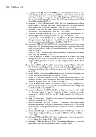
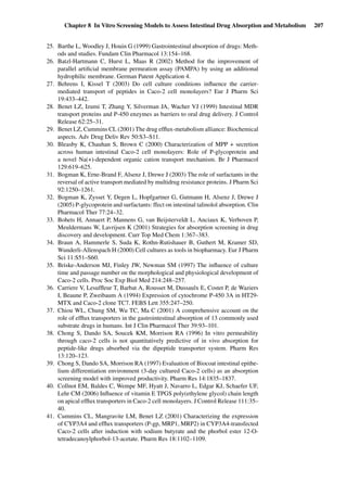
![Chapter 9 In Vitro Cellular Models for Nasal Drug Absorption Studies 217
log P Partition coefficient (octanol–water)
Papp Apparent permeability coefficient
RPMI Roswell Park Memorial Institute
SEM Scanning electron microscopy
TEER Transepithelial electrical resistance
TJM Tight junction modulating
9.1. Introduction
Nasal drug delivery has emerged as a noninvasive alternative route for drugs
that are susceptible to enzymatic or acidic degradation [1]. The sizable surface
area of the nasal cavity, avoidance of the hepatic first-pass effect, and relatively
high blood flow promoting rapid absorption are some of attractive features of
nasal drug delivery [2–4]. Conventionally, the nasal route has been used for
local delivery of drugs for treating nasal allergy, nasal congestion, or nasal
infections. However, systemic delivery through the nasal route has recently
begun to explore possibilities for those requiring a rapid onset of action or
necessitating avoidance of severe proteolysis involved in oral administration
(e.g., most peptide and protein drugs). Successful attempts to deliver corticos-
teroid hormones through the nasal route for systemic absorption [2, 5] have
triggered further studies in this area.
Today, drugs ranging from small chemicals to large macromolecules,
including peptide/protein therapeutics, hormones, and vaccines, are being
delivered through the nasal cavity [6]. Marketed products include a range of
antimigraine drugs (e.g., sumatriptan from GlaxoSmithKline, zolmitriptan
from AstraZeneca, ergotamine from Novartis, and butorphanol from
BristolMyersSquibb) as well as some peptides (e.g., calcitonin marketed by
Novartis, desmopressin from Ferring, and buserelin from Aventis) [7]. Lately,
the use of the nasal route for delivery of vaccines, especially against respiratory
infections such as influenza, is attracting interest from vaccine delivery
scientists [8].
For successful formulation of a nasal delivery system, testing with reliably
established in vivo and in vitro models are crucial. Because of methodological
and ethical limitations inherent in the use of human nasal specimens, in vivo
animal models or in vitro excised animal tissue models are frequently utilized
for nasal drug absorption studies. Several in vivo models of rat, rabbit, dog,
sheep, and monkey were reported for delivering hormones (e.g., testosterone)
and peptide/proteins (e.g., growth hormones and insulin) via nasal routes
[9–11]. Using rat and rabbit ex vivo (in situ) nasal perfusion models, phar-
macokinetics of drugs were studied following nasal instillation [9]. In vitro
models of the excised nasal mucosae of rabbit, sheep, and dog mounted on
diffusion chambers are frequently used for nasal transport and metabolism
studies [12]. However, there are several disadvantages in in vivo animal and
in vitro tissue/cell models, including species differences and peculiarities in
many anatomical features in various animal nasal cavities, compared with those
of the human.
In vitro cell culture models of human nasal epithelium based on primary
culture technologies have proven to be extremely useful for mechanistic studies
of nasal epithelial permeability and drug absorption [13]. However, efforts](https://image.slidesharecdn.com/drugabsorptionstudies-2008-150610022529-lva1-app6892/85/Drug-Absorption-Studies-2008-235-320.jpg)
![218 D.-D Kim
to develop and characterize various nasal culture systems are still in their
infancy. Moreover, difficulty in obtaining reliable tissue sources, especially if
human tissue is preferred, is a limiting factor that hinders the widespread usage
and usefulness of an in vitro primary nasal cell culture model. The apparent
shortage of human nasal tissue has prompted researchers to seek an alternative
to primary cultures of nasal epithelial cells, that is, the passage culture of nasal
epithelial cells. The importance of these types of in vitro cell culture model
is introduced in this chapter by briefly reviewing the biological barrier of the
nasal mucosa, followed by a summary of the currently available in vitro cell
culture models for nasal drug transport studies.
9.2. Structure and Physiology of the Nasal Cavity
In both humans and animals, primary functions of the nasal cavity are breathing
and olfaction. In addition, resonance of produced sounds, cleaning the air as it
is inhaled, mucociliary clearance, immunological activities, and heating and
humidification of the inspired air before it reaches the lungs are also important
functions of the nasal cavity [14]. Hairs in the nasal vestibule or mucus layer
covering the surface of nasal cavities trap inhaled particles or microorganisms.
Moreover, the nasal mucosa has the capacity to aid in the metabolism of
endogenous materials into compounds that can easily be eliminated from the
body.
9.2.1. Anatomy and Function
The human nose is divided by the median septum and each symmetrical half
opens to the face through nostrils, while extending posterior to the nasopharynx
[15]. The most anterior part of the nasal cavity is the nasal vestibule, adjacent
to the intermediate region called the atrium. Turbinates or conchae of the nasal
cavity considerably increase the surface area. The nasal cavity consists of three
regions, which are the vestibule, olfactory region, and respiratory region. The
human vestibule is the region just inside nostrils with an area of about 0.6
cm2. The olfactory region in humans is situated on the roof of nasal cavities
and covers only about 10% of the total nasal area of 150 cm2, of which the
respiratory region covers about 85%. The respiratory region contains three
nasal turbinates: the superior, middle, and inferior turbinates, which project
from the lateral wall of each half of the nasal cavity. The human nasal cavity
has a total volume of 15–20 ml. The presence of turbinates in the respiratory
region creates a turbulent airflow through nasal passages ensuring better con-
tact between the inhaled air and mucosal surface [16]. The anterior nasal cavity
is lined with a stratified, squamous, and transitional epithelium.
The highly vascularized respiratory epithelium is composed of five major
cell types: ciliated cells, nonciliated cells, columnar cells, goblet cells, and
basal cells. Low numbers of neurosecretory cells are present in the basement
membrane [17]. Approximately 20% of the total number of cells in the lower
turbinate area is ciliated with fine projections (∼100 per cell) on the apical cell
surface. Cilia are used to transport the mucus toward the nasopharynx. These
long (4–6 µm) and thin projections are mobile and beat with a frequency of
1,000 strokes per min. Ciliated and nonciliated columnar cells are populated
with about 300 microvilli per cell, which help in enlarging the surface area.](https://image.slidesharecdn.com/drugabsorptionstudies-2008-150610022529-lva1-app6892/85/Drug-Absorption-Studies-2008-236-320.jpg)
![Chapter 9 In Vitro Cellular Models for Nasal Drug Absorption Studies 219
Around 60–70% of the respiratory mucosa is lined with nonciliated cells that
are thought to be responsible for high metabolic activity and fluid transport into
and out of mucosal cells [17].
9.2.2. Physiology of Nasal Mucosa
Mucus gel covers the nasal epithelium, as in many other epithelia in contact
with the external environment. The mucus plays a number of important physi-
ological roles, including (a) entrapment of substances entering the nasal cavity
and participation in the removal of particulates via mucociliary clearance and
(b) humidification of inspired air to aid in heat transfer by holding water. Goblet
cells, which cover ∼10% of the mucosa in the turbinate area, contain numerous
secretory granules. The flow rate of human nasal mucus is in the order of 5
mm/min, renewing the mucus layer every 15–20 min. However, basal cells
(thought to be precursors of columnar and goblet cells) are poorly differentiated
and reside at the serosal aspect of the mucosa. It is believed that basal cells have
the ability to replace other cell types after differentiation [17]. The olfactory
mucosa is a pseudostratified, columnar epithelium which covers the superior
region of the human nasal cavity and composed of supporting cells, basal cells,
microvillar cells, and the typical receptor cells or olfactory cells [16, 18]. The
basal lamina or basement membrane is situated between the epithelium and
lamina propria. It is situated adjacent to underlying skeletal structures and is
composed of a loose type of connective tissue containing glands, subepithelial
cells, and vascular and nervous tissue [19].
9.3. Factors Affecting the Nasal Absorption
Drug transport across the nasal epithelium is assumed to occur by one or more
of the following mechanisms: transcellular passive diffusion, paracellular pas-
sive diffusion, carrier-mediated transport, and transcytosis. The factors influ-
encing nasal drug absorption are related to nasal physiology, physicochemical
characteristics of the drug, and formulation approaches used for specific drugs.
As with most sites of drug absorption, the bioavailability of nasally delivered
drug is affected by several factors, including the surface area available for
absorption, contact time between the drug and absorption site(s), metabolism
of the drug, and pathology of absorbing tissue(s) [20].
9.3.1. Physiological Factors
9.3.1.1. Mucociliary Clearance
Combined actions of the mucus layer and cilia dictate the mucociliary clear-
ance, which is an important factor in the physiological defense of the res-
piratory tract against inhaled hazardous substances including pathogens and
particles [21]. Although the speed of mucociliary clearance in healthy humans
is assumed to be about 5 mm/min [22], this is easily altered by pharmaceutical
excipients, airborne irritants, or nasal diseases. Generally, the length of time
that the drug is in contact with absorbing tissues influences the amount of drug
that crosses the mucosa. In the nasal cavity, this is influenced by the rate at
which the drug is cleared from the absorption site(s) by mucociliary clearance
as well as metabolism.](https://image.slidesharecdn.com/drugabsorptionstudies-2008-150610022529-lva1-app6892/85/Drug-Absorption-Studies-2008-237-320.jpg)
![220 D.-D Kim
9.3.1.2. Enzymes
Even though nasally administered drugs are thought to avoid first-pass hepatic
metabolism, bioavailability of nasally administered peptide or protein drugs
can be limited because of the broad range of metabolic enzymes localized in
the nasal mucosal cavity and epithelial cells lining the cavity [23, 24]. However,
the enzymatic degradation of peptide/protein drugs is generally smaller in
magnitude than that found in the gastrointestinal tract, although it has been
reported that nasally administered peptides resulted in low bioavailability due
to degradation by peptidases in the nasal cavity/epithelial cells. Nevertheless,
it is of note that up to 100% of relative bioavailability for nasal administration
of insulin was reported in a rat study [9]. Therefore, the nasal route is a
better alternative to oral delivery for some enzymatically labile drugs such
as insulin whose bioavailability via oral routes may be close to nil. It is of
importance to note that various pathophysiological conditions, including the
common cold, seasonal rhinitis, nasal polyps, and cancer, may alter absorption
from the nasal cavity by affecting the rate of mucociliary clearance, thereby
altering the rate of clearance of nasally administered drugs. However, a more
thorough investigation on this topic is still necessary.
9.3.2. Physicochemical Characteristics of the Drugs
Physicochemical characteristics of nasally administered drugs can influence
nasal absorption. These include molecular weight, solubility, dissolution rate,
charge, partition coefficient, pKa, particle size, and the presence of polymor-
phism [25]. Nasal absorption is known to be sharply decreased for a compound
with a molecular weight of greater than 1,000 Da [26]. Nasal absorption of
water-soluble, ionizable compounds (e.g., benzoic acid) is dependent on the
pH of the solution where the drug is dissolved. Nasal absorption of drugs
decreases as their ionized fractions increase. However, the role of lipophilicity
of compounds in their nasal absorption rates is controversial. Although the
lipophilicity measured as a simple octanol–water partition coefficient (log P)
may not be the best way to predict the nasal absorption, it is generally accepted
that permeability of lipophilic solutes increases as the log P value of the solute
increases. However, some studies report that the correlation between solute
lipophilicity and nasal absorption rate of the solute is not very significant. As
reported for the case of progesterone and its derivatives, systemic bioavailabil-
ity seems to correlate well with the partition coefficient determined using the
rabbit nasal mucosa-buffer system, where isotonic phosphate buffer (pH 8.0)
was used to mimic mucosal liquid [9].
9.3.3. Effect of Formulation
Formulation-related factors that should be taken into consideration to obtain
successful nasal absorption of drugs include the concentration of drugs, dose
and volume of administration, and pH, viscosity, and osmolarity of the solu-
tion in which the drug is dissolved. Moreover, excipients (e.g., preservatives
and absorption enhancers) in general affect the bioavailability. In addition,
dosage forms (e.g., drops, spray, and powder), administration techniques (e.g.,
inhalation, mechanically assisted and others), and devices used to administer
the drug will also affect the level of absorption. An important factor for
low drug absorption via the nasal cavity is the general rapid clearance of](https://image.slidesharecdn.com/drugabsorptionstudies-2008-150610022529-lva1-app6892/85/Drug-Absorption-Studies-2008-238-320.jpg)
![Chapter 9 In Vitro Cellular Models for Nasal Drug Absorption Studies 221
administered formulations due to the mucociliary clearance mechanism. This
has led to the development of a bioadhesive powder dosage form and mucoad-
hesive powder spray formulation for the nasal delivery [27].
For the nasal delivery of polar drugs, addition of absorption-promoting
agents called enhancers can be very helpful. Enhancers described in the litera-
ture include surfactants (e.g., laureth), bile salts and bile salt derivatives (e.g.,
sodium taurodihydrofusidate), fatty acids or fatty acid derivatives, phospho-
lipids, and various cyclodextrins [28]. These enhancer systems help increase
absorption via a variety of mechanisms but generally lead to changes in the per-
meability of epithelial cell layers by modifying phospholipid bilayers, leaching
out proteins through the cell membrane, or even stripping off the outer layer of
the mucosa. Some of these enhancers also have an effect on tight junctions
and/or act as enzymatic inhibitors. In animal studies with various enhancing
agents, a direct correlation between absorption-enhancing effect and damage
caused to the nasal epithelial barrier have been observed [29]. This is particu-
larly true for surfactants and bile salts. However, for other enhancers (e.g., some
of the cyclodextrins, chitosan, and selected phospholipids) the absorption-
enhancing effect outweighed any damage caused to the mucosa. Moreover, in a
Caco-2 cell culture study, chitosan has been shown to increase the paracellular
transport of polar drugs by transiently opening the tight junctions between the
adjacent epithelial cells [30]. As described above for surfactant-type enhancers,
a major limiting factor associated with the addition of enhancers to a nasal
formulation is the potential toxicity to the nasal mucosa. Thus, nasal absorption
enhancers should be chosen carefully, making sure that they are nonirritating,
nontoxic, and nonallergenic or at least able to demonstrate reversible effects in
a short time period. Needless to say, they should be potent and compatible with
the drug and other excipients in the formulation.
9.4. Transport Routes of Nasal Epithelial Barrier
There are two routes potentially involved in drug absorption across the nasal
epithelial barrier: the transcellular and paracellular routes [20]. Several experi-
mental evidences dealing with the mechanism of transnasal permeation support
the existence of both lipoidal pathyway (i.e., transcellular route) and an aque-
ous pore pathway (i.e., paracellular route).
9.4.1. Transcellular Route
This refers to the transport across the epithelial cells, which can occur by
passive diffusion, carrier-mediated transport, and/or endocytic processes (e.g.,
transcytosis). Traditionally, the transcellular route of nasal mucosa has been
simply viewed as primarily crossing the “lipoidal barrier,” in which the absorp-
tion of a drug is determined by the magnitude of its partition coefficient and
molecular size. However, several investigators have reported the lack of linear
correlation between penetrant lipophilicity and permeability [9], which implies
that cell membranes of nasal epithelium cannot be regarded as a simple lipoidal
barrier. Recently, compounds whose transport could not be fully explained by
passive simple diffusion have been investigated to test if they could be utilized
as specific substrates for various transporters which have been identified in the](https://image.slidesharecdn.com/drugabsorptionstudies-2008-150610022529-lva1-app6892/85/Drug-Absorption-Studies-2008-239-320.jpg)
![222 D.-D Kim
nasal mucosa [31–33]. The transcellular route can be categorized into three
different processes as described below.
9.4.1.1. Transcellular Passive Diffusion
For most conventional, small, and lipophilic drug molecules, absorption occurs
by transcellular passive diffusion across the epithelial cell. Molecular size of
drugs is known to be one of the important factors in determining the passive
diffusion. It was reported that nasal absorption sharply decreased for a drug
with molecular weight higher than 1,000 Da [9]. Because the degree of ioniza-
tion of a given drug is also an important property for absorption via this route,
transport is dependent on the pKa of the drug and pH of the environment (e.g.,
the pH of nasal secretions is normally in the region 5.5–6.5).
9.4.1.2. Carrier-Mediated Processes
The existence of a carrier-mediated transport in nasal mucosa was first sug-
gested by Kimura et al. [34]. P-glycoprotein, organic cation transporter,
dopamine transporter, and amino acid transporters have all been identified
in the nasal mucosa, especially in the olfactory mucosa [31, 32, 35, 36].
These transporters determine the polarized absorption and excretion of their
substrates including amino acids, amines, and cations.
9.4.1.3. Endocytic Processes
Since the uptake of particles in nasal epithelial tissue is known to be mostly
mediated by M cells, nasal administration has been investigated as a noninva-
sive delivery of vaccines [37]. However, since the uptake of naked DNA by
endocytocis is limited, use of either nanoparticles as mucosal delivery systems
[37] or hypotonic shock [38] is reported for the efficient transfection of gene
and vaccine into the nasal epithelium. It was also reported that polypeptides
and polypeptide-coated nanospheres (diameter: about 500 nm) are transported
through endocytic process in rat M cells [39].
9.4.2. Paracellular Route
This refers to the transport taking place between adjacent epithelial cells by
passive diffusion or solvent drag mechanisms. Paracellular permeability profile
of the nasal epithelium is similar to that of the intestinal epithelial barriers.
Thus, small hydrophilic molecules can passively diffuse through paracellular
routes between adjacent nasal epithelial cells. Passive diffusion of hydrophilic
solutes via paracellular routes is driven by a concentration gradient across
the epithelium with the rate of absorption governed by the Fick’s first law
of diffusion. Polar and charged drugs with molecular weights below 1,000
Da are thought to permeate the nasal epithelium via this paracellular route.
A major limiting site of paracellular pathways is known to be the tight
junctional region, which is characterized as the joining of contiguous cells
via various tight junctional proteins. There are many molecular components
of the tight junction such as occludin, various claudin isoforms, junctional
adhesion molecule (JAM), zonula occludens, and actin. Among these com-
ponents, the perijunctional actin is known to play a major role in controlling
the paracellular permeability [40]. Although tight junctions are dynamic struc-
tures and can open and close to a certain degree when appropriate signaling
mechanisms are activated [41], the average diameter of the equivalent pores
becomes less than 1 nm, considerably limiting the transport of larger molecules](https://image.slidesharecdn.com/drugabsorptionstudies-2008-150610022529-lva1-app6892/85/Drug-Absorption-Studies-2008-240-320.jpg)
![Chapter 9 In Vitro Cellular Models for Nasal Drug Absorption Studies 223
including peptide and proteins [26]. However, recent discoveries on the mole-
cular identity of various tight junctional proteins initiated new approaches for
enhancing nasal drug delivery, thereby improving the bioavailability of small,
polar molecules as well as biopharmaceutical products (e.g., peptide/protein
therapeutics, vaccines, and genes) [41]. It is reported that the modulation of
tight junctional routes is possible by using a toxin (e.g., cholera toxin) as an
adjuvant in mucosal vaccines [42], or using a tight junction modulating (TJM)
peptide (e.g., PN159) [43], or encapsulating drugs of interest in polymer-based
microspheres [44]. For example, when insulin was encapsulated in aminated
gelatin microspheres as a nasal delivery system, the hydrogel nature of the
microsphere resulted in temporary dehydration of the epithelial cells and as
a result loosening up the tight junctions, leading to an increase in insulin
absorption [45].
9.5. Models for Nasal Drug Transport
and Absorption Studies
Ethical issues as well as difficulty in obtaining enough human nasal tissue
specimens have called for the need to use alternative in vitro and in vivo
methods. Various in vivo animal models and in vitro excised tissue models
have been described in the literature for nasal drug transport studies. However,
due to the difficulty in both controlling the experimental conditions in in vivo
animal models and obtaining intact excised tissue samples, in vitro cell culture
models are also being actively developed.
Cultured nasal cells are reliable models for drug transport and metabolism
studies, since they are known to express important biological features (e.g.
tight junctions, mucin secretion, cilia, and various transporters), resembling
those found in vivo systems. Moreover, easy control of experimental conditions
as well as separation of the permeation step from the subsequent absorp-
tion cascade is also possible. A relatively simple primary culture condition
using human nasal epithelial cells for in vitro drug transport studies has been
established and applied in transport and metabolism studies of drugs. It is
known that the culture condition and/or selection of culture media are critical
in the recapitulation of well-differentiation features of in vivo nasal mucosal
epithelium [46].
9.5.1. In Vivo Animal Models
There are two types of animal models which can be used for nasal absorption
studies: the whole animal model and isolated organ perfusion model. The rat
model used for studying the nasal delivery of drugs was first presented in the
late 1970s [47]. The animal models using rat [2, 45], rabbit [43, 48], dog
[49], sheep [50], and monkey [51] have been reported for evaluation of nasal
absorption studies. Ex vivo nasal perfusion models using sheep [52] and rat
[9] are also reported. Although the rat model is easy to perform because of
the small animal size and low in overall cost (e.g., maintenance of the animal
and setup of the preparation), the small animal size is also a disadvantage
(e.g., limiting the number of blood samplings) and hence it is useful only
for preliminary studies of nasal drug absorption. The primate model would
be very useful, even though it is very expensive. Scientists are, however,](https://image.slidesharecdn.com/drugabsorptionstudies-2008-150610022529-lva1-app6892/85/Drug-Absorption-Studies-2008-241-320.jpg)
![224 D.-D Kim
hesitant to use this particular model due to the increasing pressure from animal
rights groups. Alternatively, rabbit, dog, and sheep models are excellent for
evaluating formulation and pharmacokinetic studies, although anatomical and
physiological differences in nasal mucosae of the human versus these animals
are very important issues that should be taken into account.
9.5.2. In Vitro Excised Models
Excised nasal mucosae obtained from various animal species are tools fre-
quently used to study nasal transport and metabolism ([53], Chap. 4). Main-
taining the viability of the excised nasal tissues during the experimental period
is crucial. Most studies were performed with epithelia excised from rabbits,
bovine, sheep, and dogs tissues [54–57]. This excised nasal tissue model has
been shown to be well suited for studies on nasal permeation and metabolism
of drugs. However, species differences in the activity of various enzymes found
in human versus these animal nasal mucosae have become an important issue.
9.5.3. Cell Line Models
RPMI 2650 is the only human nasal epithelial cell line derived from a spon-
taneously formed tumor. This particular cell line has been mostly used for
nasal metabolism studies, since it grows into a multilayer and does not form
confluent monolayers (although perijunctional actin rings are present). Thus,
RPMI 2650 cell monolayer is rarely used for nasal transport studies [44, 58].
9.5.4. In Vitro Primary and Passaged Cell Culture Models
In vitro cell culture models offer many advantages, including (a) a controlled
environment for the study of epithelial cell growth and differentiation, (b) the
elucidation of drug transport pathways and mechanisms, (c) rapid and conve-
nient means of evaluating drug permeability, and (d) opportunity to minimize
the expensive and limited controversial use of animals [12]. Although recent
advances in human nasal primary cell culture techniques appear to be very
promising, some hurdles remain to be resolved, including the relatively small
amount of obtainable cells and epithelial cell differentiation issues. Primary
culture of human nasal epithelial cells using tissue culture plates shows some
natural cell differentiation. The presence of tight junctions is confirmed by actin
staining. Using such primary culture systems, basic functional and biochemical
characteristics of the tissue have been demonstrated [59]. Primary culture
of cells and tissues obtained from differentiated human nasal epithelium is
an established tool for many studies, including fibrosis, electrolyte transport,
ciliogenesis, and ciliary movement [60, 61] as well as peptide transport and
metabolism [62, 63]. Primary cultures of human nasal epithelial cells on
TranswellR
insert as in vitro models are very promising for the study of nasal
drug absorption, especially for peptide transport studies [58, 64, 65]. Recently,
primary culture of nasal epithelial from a variety of animal species, including
human [62], bovine [59], rat, and rabbit [66], has provided valuable information
on nasal physiology in health and disease. Several research groups are actively
developing and validating human nasal cell culture systems as an alternative to
animal models.](https://image.slidesharecdn.com/drugabsorptionstudies-2008-150610022529-lva1-app6892/85/Drug-Absorption-Studies-2008-242-320.jpg)
![Chapter 9 In Vitro Cellular Models for Nasal Drug Absorption Studies 225
9.5.4.1. Liquid-Covered Culture Method
Human primary nasal epithelial cell culture on TranswellR
insert for in vitro
drug transport studies was first reported by Werner et al. [58, 64]. This culture
method has been applied in transport and metabolism studies of insulin and
other peptides [62, 63, 67]. Despite its advantages, it is difficult to obtain
adequate amounts of human nasal cells since most of the biopsies are used
for diagnostic examinations and yield only very limited amount of usable
nasal epithelial cells. The limited resources for in vitro primary cell culture
have been a serious obstacle for conducting diverse experiments, and thus,
high-throughput screening studies have not been possible. Other restrictions
of primary cell culture include contamination with pathogens and large donor-
to-donor variability [68]. To overcome these limitations of primary cell culture
of human nasal epithelial cells, a serially passaged culture system was con-
sidered as an alternative. Although studies on the development of a passaged
culture of primary human respiratory epithelial cells yielded information on
some physiological characteristics (e.g., mucin secretion) [68–72], there are
not many reports on serially passaged cells being cultured on a permeable
support for in vitro drug transport studies. The formation of functional tight
junctions would be the most essential factor to be considered for studies of drug
transport across the nasal epithelial cell layer derived from serially passaged
cells. Yoo et al. [40] reported that passage-cultured (e.g., up to passage number
4) human nasal epithelial cells can be grown as a confluent cell monolayer on
the TranswellR
insert utilizing the liquid-covered culture (LCC) method. Each
passaged culture formed a tight epithelial cell monolayer with high transep-
ithelial electrical resistance (TEER) value (up to 3,000 ohm cm2), enabling
drug transport studies. However, differentiation of cilia and mucin-secreting
cells was not deemed to be complete in these cultures. Of important note is
that the feasibility of the passage-cultured human nasal epithelial cell layer
for in vitro drug transport studies was confirmed by transport experiments
with 14C-mannitol and budesonide. Apparent permeability coefficient (Papp)
values of 14C-mannitol and budesonide at each passage (Figure 9.1) suggest
that the culture model would be suitable for reliable drug transport studies
up to passage-4 culture. Contrary to mannitol transport, budesonide showed
rather constant Papp values regardless of TEER values, suggesting that rather
high lipophilicity of budesonide may be the predominant factor governing
the permeation. In other words, the contribution of paracellular permeation
via tight junctions of the cultured cell layer does not seem to significantly
influence Papp of lipophilic solute(s), since the transcellular route is the major
transport route of such lipophilic drugs. When the TEER value was higher than
500 ohm cm2, constant Papp values were obtained regardless of the budesonide
concentration (10 and 20 µg/ml) and passage number. These data suggest that
the passage-cultured human nasal epithelial cell monolayer could be utilized
reliably for in vitro drug transport studies.
In order to verify the utility of this passaged culture model for actual
nasal transepithelial drug delivery, a series of antiallergic drugs were selected
for transport studies and evaluated for the influence of lipophilicity of these
drugs on their permeabilities observed across the cultured cell layer [73].
Permeability characteristics of model drugs across the cell layer were then
compared with those reported for excised porcine nasal mucosa to demonstrate
the feasibility of the passage-cultured nasal epithelial culture model for drug](https://image.slidesharecdn.com/drugabsorptionstudies-2008-150610022529-lva1-app6892/85/Drug-Absorption-Studies-2008-243-320.jpg)
![226 D.-D Kim
TEER (ohm . cm2
)
Papp(cm/sx105
)
0.01
0.1
1
10
500 1000 1500 2000 2500 30000
Figure 9.1 Relationship between the
transepithelial electrical resistance (TEER)
value of the passage-cultured human nasal
epithelial cell layer and permeability of
14C-mannitol (◦, passage-2; , passage-3;
, passage-4) and budesonide (•, passage-2;
, passage-3; , passage-4). (Data from
Ref. [40]).
transport studies [73]. As shown in Figure 9.2, a similar log–linear relationship
was reported for transport of various compounds across the excised porcine
nasal mucosa (r2 = 0.81, when lidocaine was ignored as an outlier) [55,
74]. Papp values of antiallergic drugs across the passage-cultured human nasal
epithelial cell layer were significantly lower than those estimated across the
excised porcine nasal mucosa. For example, mannitol Papp across the passage-
cultured human nasal epithelial cell layer was 0.90 ± 0.30 (×10−6 cm/s) [40],
while the excised porcine nasal mucosa showed mannitol Papp of 3.9 ± 2.2
(×10−6 cm/s) [55]. This discrepancy could be due to the species differences
between human versus porcine tissues/cells and/or to differences in tight junc-
tional properties of the two nasal models. TEER values of 40–120 ohm cm2
reported for the nasal mucosa excised from various animals (e.g., sheep, rabbit,
and cow/ox) and human [12] are considerably lower than that obtained in
passage-cultured human nasal epithelial cell layer studies (>1,000 ohm cm2).
However, a good log–linear relationship between lipophilicity index and Papp
found in both nasal models indicates that lipophilicity index (i.e., log P) of
compounds is the most important factor that determine the nasal permeability
and that the passage-cultured human nasal epithelial cell layer model is indeed
quite useful for predicting the nasal permeability of small molecular drugs.
logP(Oct/water)
Papp(cm/sX10−5
)
0.01
0.1
1
10
100
1
3
4
5
6
2
a
b c
d
e
f
g
−4 −2 0 2 4
Figure 9.2 Relationship between log P
values of various drugs and Papp across the
passage-cultured human nasal epithelial
cell monolayer using LCC method.
1 = albuterol hemisulfate, 2 = albuterol,
3 = fexofenadineHCl, 4 = dexamethasone,
5 = triamcinolone acetonide,
6 = budesonide, a = mannitol,
b = melagatran, c = sumatriptan,
d = propranolol, e = nicotine,
f = lidocaine, g = testosterone. ◦:
transport across the human nasal epithelial
cell monolayer passage-cultured on
Transwell, •: data observed using excised
porcine nasal mucosa mounted in Ussing
chambers (Data from Ref. [73]).](https://image.slidesharecdn.com/drugabsorptionstudies-2008-150610022529-lva1-app6892/85/Drug-Absorption-Studies-2008-244-320.jpg)
![Chapter 9 In Vitro Cellular Models for Nasal Drug Absorption Studies 227
9.5.4.2. Air-Interfaced Culture Method
In nasal epithelium, important properties of cultured epithelial cells include the
formation of confluent cell layers functionally interconnected by tight junc-
tional proteins, expression of apical cilia, and mucin production and secretion.
Among them, ciliated cells of nasal epithelium play an important role in phys-
iological function (e.g., mucocilliary clearance), which may also be relevant to
nasal drug delivery [21]. Additionally, the expression of various transporters
and presence of enzymatic systems in in vitro cell culture models will increase
the predictive potential of such models. However, compared to the in vivo
nasal epithelium, the passaged nasal culture model has been reported to exhibit
relatively undifferentiated epithelial cells with few ciliated cells when cultured
with LCC method [40]. The attainment of differentiated phenotype as well as
morphological and functional features of cells akin to those found in in vivo
setting may be greatly affected by specific culture conditions. These conditions
are the substratum on to which cells are cultivated, culture media including
appropriate additives for induction of cell differentiation (e.g., retinoic acid),
and liquid-covered versus air-interfaced (AIC) culture conditions. Moreover,
the development of serum-free (but hormone-supplemented) culture medium
has made it possible to induce the differentiation of airway epithelial cells
with morphologically resembling the epithelium in vivo [69, 75]. Thus, AIC
culture condition coupled with defined, serum-free medium is commonly used
for epithelial cell cultures [65, 76, 77], resulting in more ciliated cells and/or
enhanced mucin secretion.
For AIC conditions, the apical surface of the epithelial cell layer is exposed
to air after the nasal cells reached confluence on the TranswellR
insert, while
the basolateral side is fed with culture fluid. Figure 9.3 shows TEER changes
in epithelial cell layers cultured up to 20 days in LCC versus AIC methods
[46]. In AIC condition (initiated from day 3 after seeding), TEER peaked on
day 5 and maintained above the TEER values observed for LCC counterparts.
By contrast, TEER observed for LCC conditions peaked on day 2 and declined
toward zero by day 15. These data indicate that human nasal epithelial cells at
an air interface culture exhibit better electrophysiological characteristics than
those cultured by the conventional liquid-covered conditions.
Days after seeding
TEER(ohm.cm2)
0
1000
2000
3000
4000
0 5 10 15 20
Figure 9.3 Changes in transepithelial
electrical resistance (TEER) of human
nasal epithelial cell layers grown under
LCC (•) versus AIC ( ) conditions.
Each data point represents the
mean ± SD of three determinations.
(Data from Ref. [46]).](https://image.slidesharecdn.com/drugabsorptionstudies-2008-150610022529-lva1-app6892/85/Drug-Absorption-Studies-2008-245-320.jpg)
![228 D.-D Kim
Morphological characteristics of nasal epithelial cells, especially the devel-
opment of cilia, were observed by scanning electron microscopy (SEM) studies
[46]. The most prominent morphological difference between LCC versus AIC
cultures was the development of ciliated cells. The presence of cilia on the
apical surface of AIC culture was evident on day 7 in culture. The number
of ciliated cells increased on the 14th day. Markedly increased numbers of
ciliated cells matured with long and beating healthy cilia were observed after
3 weeks of AIC culture. In short, AIC conditions allow growth of epithelial
cells in a configuration resembling that in in vivo conditions. It has also shown
that cells grown in AIC conditions not only mimic the morphological features
of the epithelium but also exhibit many of the physiological and biochemical
activities observed in vivo [68]. In LCC conditions, by contrast, flattened
ciliated cells and numerous microvilli were observed after 3 weeks. It can be
stressed that the selection of culture medium used in AIC cultures may also
have played an important role. The bronchial epithelial cell growth medium
(BEGM), which is commonly used for AIC cultures, is reported to be suitable
for obtaining more homogeneous cell morphology than Dulbecco’s modified
Eagle’s medium (DMEM), which was used in the LCC method [77]. DMEM
with 10% FBS seems to be critical to stimulate cell proliferation in the early
stage of the culture [40], while retinoic acid in BEGM seems to be an impor-
tant factor for the growth and differentiation of airway epithelial cells [78,
79]. Therefore, the mixture of BEGM and DMEM/F12 (Dulbecco’s modified
Eagle’s medium/F12 Ham’s) (1:1) with all supplements may be suitable for
AIC cultures of human nasal epithelial cells, as greater and prolonged TEER
values of AIC cultures are found (Figure 9.3).
It can be noted that airway mucin gene expression was more significant
in AIC culture on 21 days than LCC on 7 days, perhaps confirming the
importance of retinoic acid in BEGM and AIC conditions on the growth
and differentiation of airway epithelial cells [78, 79]. We surmise that AIC
cultures probably can deliver oxygen much more efficiently across the thin
film of liquid to the cells, subsequently changing cellular respiration to a more
aerobic nature in AIC conditions [76] and improving the differentiation of
nasal epithelia cells than LCC conditions. These factors may have led to higher
TEER values for up to 21 days of human nasal epithelial cell culture in AIC
conditions.
Because mucin and/or cilia systems of AIC cultured epithelial cells may
work as a barrier for drug transport, lower Papp values are expected in cell
layers cultured in AIC than in LCC methods. However, it was interesting to
note that no significant differences in Papp values were observed between the
cell layers cultured with the two methods (Table 9.1). This is in contrast to
solute permeabilities reported previously for cell layers cultured with LCC
versus AIC methods [76, 80]. For example, Yang et al. reported that Papp
of lipophilic solutes (e.g., various β-blockers) across the primary cultured
conjunctival epithelial cell layer are about threefold lower when cultured under
AIC than LCC conditions, suggesting that the permeability of AIC cultured
cell layers generally better reflects that of the excised tissue than LCC coun-
terparts. Mathias et al. [76] also reported that the permeability of hydrophilic
solutes across the primary rabbit tracheal epithelial cell layer cultured under
AIC conditions was only half of that observed for cell layers cultured under](https://image.slidesharecdn.com/drugabsorptionstudies-2008-150610022529-lva1-app6892/85/Drug-Absorption-Studies-2008-246-320.jpg)
![Chapter 9 In Vitro Cellular Models for Nasal Drug Absorption Studies 229
Table 9.1 Apparent permeability coefficients (Papp)
of 14C-mannitol and budesonide across the passage-
cultured human nasal epithelial cell layer grown under
AIC or LCC conditions for 7 days.
Papp(×10−6cm/s)
Culture method 14C-Mannitol Budesonide
LCC 0.90 ± 0.30 15.34 ± 0.66
AIC 0.86 ± 0.04 16.53 ± 1.58
Source: Data from Ref. [46].
AIC, Air-interfaced culture.
LCC, Liquid-covered culture.
LCC conditions, whereas the permeability of lipophilic solutes was similar
to each other for cell layers grown in either AIC or LCC method. These
investigators suggested that the tight junctional arrangement (e.g. number of
strands, junctional length, presence of pores or channels within the junction, or
length of lateral intercellular space) might be widely different, depending on
specific culture conditions. The higher than expected Papp of mannitol found
in the human nasal epithelial cell layer cultured using AIC method may be due
to factors related to characteristics of the human nasal cells which differs from
the conjunctival cells used by Yang et al. [80] or tracheal cells used by Mathias
et al. [76]. However, the exact nature of factor(s) that would have caused
these disparate results would require further investigations. Nevertheless, these
data imply that the intact tight junction was well developed in both AIC and
LCC methods of culturing human nasal epithelial cell layers, enabling useful
mechanistic studies using both hydrophilic and lipophilic drugs. In a recent
study, the enhancing effect of various surfactants on the nasal permeability
of fexofenadine-HCl was evaluated using AIC-cultured human nasal epithelial
cell monolayer model [81]. It was demonstrated that this model is a useful tool
not only for evaluating the in vitro nasal drug transport but also for studying
the mechanism and toxicity of enhancers.
9.6. Conclusions
Recent development of an in vitro model of human nasal epithelial cells cul-
tured on permeable supports may be useful for various mechanistic studies of
nasal transport/physiology in health and disease. Although the model is promis-
ing and important, problems including the limited amount of available cells
and issues pertaining to epithelial cell differentiation in culture remain to be
resolved further. Cultures of human nasal epithelial cell layers on TranswellR
insert under AIC or LCC conditions may be particularly useful for in vitro drug
transport studies. Serially passaged culture under AIC conditions allows the
preparation of a comparably larger number of highly differentiated epithelial
cell layers with cilia on the apical surface and mucin gene expression, com-
pared to cells cultured in LCC conditions. Higher TEER value observed in
AIC conditions was maintained for a longer duration than that in LCC. Thus,
the AIC culture of passaged human nasal epithelial cell layers seems to be](https://image.slidesharecdn.com/drugabsorptionstudies-2008-150610022529-lva1-app6892/85/Drug-Absorption-Studies-2008-247-320.jpg)
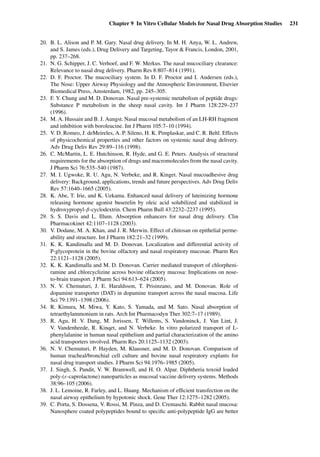
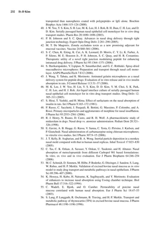
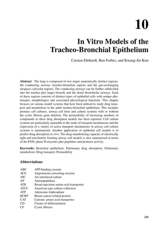
![236 C. Ehrhardt et al.
CFTR Cystic fibrosis transmembrane conductance regulator
COPD Chronic obstructive pulmonary disease
CP Carboxypeptidase
CYP Cytochrome P450
DPP Dipeptidyl peptidase
FMO Flavin monooxygenase
GSH Glutathione
GST Glutathione S-transferase
GTP Glutamyltranspeptidase
HBE Human bronchial epithelium
HIV Human immunodeficiency virus
Isc Short-circuit current
LAT L-amino acid transporter
LCC Liquid-covered culture
LRP Lung resistance protein
MRP Multidrug resistance-associated protein
MT Methyltransferase
NAT N-acetyl transferase
NEP Neutral endopeptidase
NHBE Normal human bronchial epithelial cells
OCT Organic cation transporter
OCTN Novel organic cation transporter
Papp Apparent permeability coefficient
PEPT Di/Tri-peptide transporter
P-gp P-glycoprotein
Rh123 Rhodamine 123
RT-PCR Reverse transcription-polymerase chain reaction
SLC Solute carrier protein
SULT Sulfotransferase
TEER Transepithelial electrical resistance
UGT UDP-glucuronosyl transferase
10.1. Introduction
10.1.1. Anatomy of the Lung Airways
The respiratory system can be separated anatomically into the conducting and
respiratory regions. The conducting airways consist of the air-transmitting
passages of the nose, nasopharynx, larynx, trachea, bronchi and bronchioles
[1, 2], where the latter three entities form the tracheo-bronchial region of the
lung (generations 1–16 in the bifurcating airway model; Figure 6.1, Chap.6).
The actual exchange of gases (i.e., fresh oxygen into and carbon dioxide out
of the body) occurs across the distal lung respiratory epithelium, comprising
the alveolar ducts and saccules (i.e., the alveoli, generations 17–23 in the
bifurcating airway model; Figure 6.1, Chap. 6). The main function of the
conducting airways is to act as conduit for air movement and in addition
to provide filtration, warming and humidification of inhaled air. Specialised
extra-pulmonary parts of the conducting system also serve other functions, for
example the nose with the sense of smell, the pharynx in alimentation and the
larynx in phonation [3].](https://image.slidesharecdn.com/drugabsorptionstudies-2008-150610022529-lva1-app6892/85/Drug-Absorption-Studies-2008-254-320.jpg)
![Chapter 10 In Vitro Models of the Tracheo-Bronchial Epithelium 237
Trachea/Bronchi Bronchioles
C
Br
S
N
G
C
Cl
B
Sec
Ci
K
B
Muc
Figure 10.1 Typical tracheo-bronchial and bronchiolar epithelia showing the major
cell types. The tracheo-bronchial epithelium showing the pseudostratified nature of
the columnar epithelium, principally composed of ciliated cells (C), interspersed with
goblet cells (G), brush cells (Br), serous cells (S), Kulchitsky’s cells (K) and basal
cells (B). The bronchiolar epithelium showing the cuboidal nature of the epithelium,
principally composed of ciliated cells (C), Clara cells (Cl) and infrequent basal cells.
Muc = mucus; Ci = cilia; N = nucleus; Sec = secretory granules.
Gradual changes in the structure of the conducting airways occur as the
diameter of the respiratory tubes becomes smaller [4]. The epithelium grad-
ually changes from being pseudostratified and ciliated with goblet cells and
submucosal glands interspersed to becoming a flattened squamous epithe-
lium devoid of goblet cells or glands (Figure 10.1). The lamina propria also
decreases in thickness and elastic fibres become more numerous, as the airways
bifurcate. A smooth muscle layer forms a conspicuous component of the subep-
ithelial wall of the higher generation conducting airways but becomes less
prominent in the alveolar ducts and is absent in alveolar walls. The cartilage
around the bronchi is also gradually reduced and finally disappears when the
airway diameter decreases to about 1 mm in size. At this point, the tube is
termed a bronchiole and the lack of cartilage enables bronchioles to constrict
to the point of closure in asthmatic patients [5].
The tracheo-bronchial epithelium forms the interface between the conduct-
ing airways and inspired air. As described above, the epithelium is layered
upon a connective tissue substratum consisting of a basement membrane,
lamina propria, and submucosa [6]. The submucosa contains elastic fibres, a
muscularis mucosa, connective tissues and seromucous glands. Lymphatics are
also located in the bronchial walls [2].
In the trachea and bronchi, the pseudostratified, ciliated, columnar epithe-
lium changes to a ciliated simple columnar one in smaller airway branches and
seromucous glands decrease in number. Goblet cells are present in the tracheo-
bronchial epithelium at all levels. The bronchioles (1 mm or less in diameter)
possess a ciliated epithelium of columnar to cuboidal shape (Figure 10.1).
The number of goblet cells in the bronchioles is reduced further compared
to the proximal bronchi, and Clara cells are present [2]. The sheathing spirals
of smooth muscles in this region are thicker than those found elsewhere in
the lung, and are thought to stabilise the tissue in absence of cartilage. In the](https://image.slidesharecdn.com/drugabsorptionstudies-2008-150610022529-lva1-app6892/85/Drug-Absorption-Studies-2008-255-320.jpg)
![238 C. Ehrhardt et al.
smaller bronchioles, which represent a transitional zone from the airway to the
respiratory region, the epithelium is cuboidal with less ciliated cells and more
Clara cells are present while goblet cells are absent. Thin connective tissues
and an incomplete layer of smooth muscles form the supporting wall of the
distal bronchioles.
10.1.1.1. Cell Types of Tracheo-Bronchial Epithelia
Several morphologically distinct cell types comprise the human tracheo-
bronchial epithelium. Ciliated cells are terminally differentiated columnar cells
that are thought to originate from basal or secretory cells. As a component of
the mucociliary escalator system, their main function is to remove inhaled par-
ticulate matters, including viruses and bacteria, which have been trapped in the
mucus [7]. There are also ciliated-secretory cells which bear fully developed
cilia and contain mucus granules, albeit these are seen only occasionally. In
general, secretory cells bear microvilli, but not cilia, on their apical surface.
Secretory cells comprise 15–25% of the bronchial epithelium and are present in
several forms. Mucous cells (also known as goblet cells) are the main producers
of airway mucus and the predominant secretory cell type in the larger airways;
neuroendocrine cells (Kulchitsky’s cells, K-cells or small granule cells) contain
amines and peptide hormones and rarely reach the lumen. Clara cells are the
predominant cell type in the bronchioles and produce the surfactant apoproteins
A and B and secretory leukoprotease inhibitors [3, 8–13]. The Clara cells
may also participate in the clearance of noxious agents via the metabolic
detoxification of inhaled agents. The contribution of Clara cells to the gen-
eration of normal human bronchiolar epithelium is substantial, demonstrating
a role of the Clara cell in maintaining the normal epithelium of the distal
airway [14].
While all the aforementioned cell types form the epithelial surface of the
airways, basal cells reside deep in the tracheo-bronchial epithelium and are not
directly in contact with the airway lumen. Basal cells are considered as the
stem cell or progenitor cell of the bronchial epithelium and are pyramidal in
shape with a low cytoplasmic/nuclear volume ratio [8, 15–17].
10.1.1.2. Barrier Function of the Airways
Bronchial epithelial cells are part of the non-specific immune system and
they not only defend against the noxious substances gaining access into the
body across the airway epithelial barriers but also impede the absorption of
therapeutically useful agents [18]. This defence is mediated via the integrity
of the airway epithelium that incorporates the physical barrier and secretory as
well as ciliary functions. This provides the effective mucociliary clearance and
secretion of mediators, protecting against a wide range of potentially injurious
agents. Thus, the integrity of airway epithelial function is important for host
defence.
10.1.1.2.1. Integrity of tracheo-bronchial epithelia: The bronchial epithelium
forms a continuous layer, its integrity maintained by several cell–cell adhe-
sion mechanisms [19]. The desmosomes (macula adherens) and the inter-
mediate junctions (adherence junctions or zonula adherens) are involved in
cell–cell adhesions. The tight junctions (zonula occludens) are narrow, belt-
like structures surrounding each cell at the apical pole. These tight junctions
provide a physical barrier, thereby preventing ‘leakage’ of water, ions and other](https://image.slidesharecdn.com/drugabsorptionstudies-2008-150610022529-lva1-app6892/85/Drug-Absorption-Studies-2008-256-320.jpg)
![Chapter 10 In Vitro Models of the Tracheo-Bronchial Epithelium 239
molecules (e.g., serum proteins). The epithelial cells are all anchored to the
basement membrane by hemidesmosomes and other focal adhesion molecules
(e.g., integrins, vinculin and radixin).
10.1.1.2.2. Mucociliary clearance: Inhaled particles are cleared from the air-
ways through trapping of the particles in mucus upon deposition and subse-
quent clearance of the mucus (with trapped particles) which is propelled by the
coordinated beating of cilia towards the throat. This is termed the mucociliary
escalator system. The mucociliary function is regulated by a variety of factors,
such as bradykinin, histamine and cytokines [20–24].
10.1.1.2.3. Secretion of protective mediators: To provide protection against
potentially injurious agents which enter the respiratory tract from the
environment during inspiration, the bronchial epithelium secretes a num-
ber of mediators, including antibacterial substances (e.g., lactoferrin and
lysozyme), anti-proteases (e.g., alpha1-protease inhibitor, secretory leuko-
protease inhibitor, alpha1-anti-chymotrypsin, alpha2-macroglobulin and tissue
inhibitors of metalloproteinases) and anti-oxidant molecules plus other mole-
cules involved in redox regulation (e.g., glutathione, GSH, enzymes involved in
GSH redox cycle, superoxide dismutase, ceruloplasmin, trasferrin and catalase)
[25–28]. The bronchial epithelium also produces components of the com-
plement system, which act as opsonins, facilitating efficient phagocytosis by
macrophages and eventual clearance [29]. In addition, bronchial epithelial cells
transport secretory immunoglobulin A into the bronchial lumen [30].
10.1.2. Drug Delivery to the Tracheo-Bronchial Region
Aerosolised medicines have been used for centuries to treat respiratory dis-
eases, with inhalation therapy for the airways focused primarily on the treat-
ment of asthma and chronic obstructive pulmonary disease (COPD). The devel-
opment of new products for delivery to the lungs for these respiratory diseases
includes new steroids and beta agonists plus combination products featuring
both agents. New classes of anti-asthma medication are also being developed
for inhalation with the aim of delivering them directly to the inflamed airways.
With the recent advances in biotechnology, many companies have been
formed with the goal of finding ways to deliver new drugs to the lung, with
the target being the systemic circulation and not the lung itself [31, 32]. In
particular, biopharmaceutical therapeutics based on proteins and peptides are
expensive to produce and often have narrow therapeutic windows, requiring
them to be delivered efficiently and reproducibly. The alveolar epithelium is
obviously a more attractive target as a portal for drug entry into the systemic
circulation from the standpoints of vast surface area coupled with an extremely
thin barrier thickness, However, tracheo-bronchial targeting of biopharma-
ceuticals might benefit from delivery to the airways where the epithelium is
leakier compared to the tighter alveolar epithelium and relatively free from
macrophage surveillance.
Two specialised delivery strategies which target inhaled therapies to the lung
are gene delivery and vaccination. These have been investigated widely with
some degree of success [33]. The goal of aerosolised gene therapy in treating
cystic fibrosis (CF) patients is to reconstitute the cystic fibrosis transmembrane
conductance regulator (CFTR) function and normal chloride channel function](https://image.slidesharecdn.com/drugabsorptionstudies-2008-150610022529-lva1-app6892/85/Drug-Absorption-Studies-2008-257-320.jpg)
![240 C. Ehrhardt et al.
in the lung and in the airways in particular. Over 20 clinical trials have been car-
ried out since the cloning of the CFTR gene in 1989 using three gene-transfer
platforms: adenovirus, adeno-associated virus 2 and cationic liposomes. All
three vectors have demonstrated proof-of-principle for gene transfer to airway
epithelia and efforts to develop the first gene delivery product for inhalation are
progressing. [33].
Another disease that has been targeted for treatment via aerosolised gene
therapy is lung cancer [34]. Studies have demonstrated that inhaled delivery
of polyethyleneimine DNA complexes results in a substantial gene expres-
sion level in mice lungs, with polyethylenimine-p53 and polyethyleneimine-
interleukin 12 therapy both significantly reducing the number and size of
osteosarcoma lung metastases in mice. Studies are underway to clarify the
response to long-term repeated exposures of these aerosol gene therapies in
lung cancer models [35].
The rationale for the inhaled delivery of vaccines is based on a number of
advantages over injection therapy [36]. First, vaccination via inhalation avoids
the need for disposal strategies for the large number of needles that would
be used in mass vaccination campaigns in developing countries. Second, it
prevents the spread of blood-borne diseases such as human immunodeficiency
virus (HIV), which can be transmitted by improper use and handling of used
sharps that may have been contaminated with the virus. Third, protection can be
induced by exposing the airway mucosa to a variety of viruses, as the airways
are the natural route of infection for many diseases. Finally, inhaled vaccination
may work better than subcutaneous vaccination with young children, in whom
the persistence of maternal antibodies does not appear to interfere with inhaled
mucosal immunisation but does interfere with subcutaneous immunisation.
Vaccination by inhalation was pioneered in the 1980s by Albert Sabin and his
colleagues who were vaccinating children in Mexico against measles using an
aerosolised vaccine [37]. By the mid-1990s, it was clear that this is an effective
means of measles vaccination and accordingly several investigators began to
consider the possibility of mass vaccination campaigns with an aerosolised
measles vaccine in other developing countries [38, 39].
10.2. In Vitro Models of the Tracheo-Bronchial Epithelium
10.2.1. Primary Cell Cultures
Protocols for the isolation and culture of primary tracheo-bronchial epithelial
cells obtained from the lungs of many species have been developed over the last
30 years. These include primary cultures of airway epithelial cells of the mouse
[40], hamster [41], guinea pig [42], rat [43], ferret [44], rabbit [45, 46], dog
[47], pig [48], cow [49], horse [50] and human [51–53]. Most of the protocols
result in well-differentiated epithelial cells with mixed phenotypes and these
primary cell cultures are generally good for drug absorption studies for the first
couple of subcultures after which they lose their ability to form tight junctions
and to generate high transepithelial electrical resistance (TEER). A linear drop
in short-circuit current (Isc) was observed over successive passages in human
tracheal epithelial cells in primary culture [54]. These subcultures exhibited
minimal rates of active Na + and Cl− transport by passage 3 and beyond
passage 4, the cells failed to generate any active ion transport when studied](https://image.slidesharecdn.com/drugabsorptionstudies-2008-150610022529-lva1-app6892/85/Drug-Absorption-Studies-2008-258-320.jpg)
![Chapter 10 In Vitro Models of the Tracheo-Bronchial Epithelium 241
using Ussing chamber techniques. These data suggest that it is important
to develop airway epithelial cell lines that retain the ability to differentiate,
form tight junctions and maintain ion channel/pump physiology when grown
in vitro [54].
Ready-to-use culture systems of human tracheo-bronchial cell layers
exhibiting well-differentiated ciliated and goblet cell phenotypes are com-
mercially available (EpiairwayTM system, Mattek Corporation, Ashland, MA,
USA) [55]. Although the EpiairwayTM is marketed for use in drug delivery
studies, little data has been reported to date, perhaps reflecting low uptake or
due to cost or usage in industry that is subject to confidentiality. In general,
primary culture is less convenient and economical than the use of cell lines.
Hence, primary cultured tracheo-bronchial cell layers have not been widely
utilised for biopharmaceutical purposes [56].
10.2.2. Tracheo-Bronchial Epithelial Cell Lines
In addition to the significantly reduced costs, a major advantage of using
immortalised airway epithelial cell models over primary lung airway epithelial
cells is the ability to reduce the variability in cultures that arises from donor to
donor differences. In contrast to gastrointestinal in vitro testing where Caco-2
cells have emerged as the gold standard, there is no such consensus to date
on the preferred cell line(s) for modelling the bronchial epithelium in vitro.
Several detailed protocols for culture, maintenance, growth and permeability
assessment of tracheo-bronchial epithelial cell lines have been published in
recent years [56], with the most commonly used systems being the Calu-3 and
16HBE14o- cell lines. Additionally, BEAS-2B are frequently used for studies
of metabolism and the interaction of cells with xenobiotics, and the use of the
NuLi-1 cell line has recently been explored [57, 58].
Calu-3 (American type culture collection; ATCC HTB-55) is a human
bronchial epithelial cell line derived from an adenocarcinoma of the lung
[59]. This cell line has been shown to exhibit serous cell properties and
form confluent monolayers of mixed cell phenotypes, including ciliated and
secretory cell types [60], but the cilia are formed very irregularly and seem
to disappear with increasing passage number (unpublished observations, C.E.
and B.F.). Calu-3 cells have shown utility as a model to examine transport [61–
63] and metabolism in human bronchial epithelial cells for many therapeutic
compounds [64]. Furthermore, they have been used in a number of particle–
cell interaction studies [65–67]. The interactions between respiratory epithelial
cells and particulates are discussed more in detail in Chap. 19.
Another continuous bronchial epithelial cell line, 16HBE14o-, was gen-
erated by transformation of normal bronchial epithelial cells obtained from
a 1-year-old male heart-lung transplant patient. Transformation was accom-
plished with SV40 large T antigen using the replication defective pSVori-
plasmid [68]. 16HBE14o- cells can be obtained from Dieter C. Gruenert, Ph.D.,
at the California Pacific Medical Center (see also Chap. 26 for more details of
the immortalisation procedures). The non-commercial availability of the cell
line might be one of the reasons they have been less widely used than Calu-3.
16HBE14o- cells have a non-serous, non-ciliated phenotype and are generally
rounder in shape and smaller in size than Calu-3. When grown under liquid-
covered culture (LCC) conditions, 16HBE14o- form confluent, polarised cell](https://image.slidesharecdn.com/drugabsorptionstudies-2008-150610022529-lva1-app6892/85/Drug-Absorption-Studies-2008-259-320.jpg)
![242 C. Ehrhardt et al.
layers with functional tight junctions and the expression of several drug trans-
port proteins [69, 70]. In contrast, air-interfaced culture (AIC) conditions lead
to cell layers of less desirable phenotypic and morphological traits. Since most
epithelial cells, which are normally located at an air interface, in vivo, function
optimally under AIC conditions for cultivation, the superior performance of the
16HBE14o- cell line under LCC conditions is an exception for which the exact
reasons or mechanism are currently unknown. Like Calu-3, the 16HBE14o-
cell line has been utilised for drug absorption and particle–cell interaction
studies [71, 72].
The BEAS-2B cell line was derived from normal human epithelial cells that
were immortalised using the hybrid virus of adenovirus 12 and simian virus 40
[73]. BEAS-2B is available from the ATCC (CRL-9609) and has been popular
in studies of airway epithelial cell structure and function, including phenotyp-
ing and mechanistic investigation of cytokine regulation [74]. BEAS-2B cells
have also been used to evaluate responses to challenges such as tobacco smoke
[75], environmental particles [76, 77] and hyperoxia [78]. However, as it is
challenging to generate TEER values higher than 100 ohm cm2 using BEAS-
2B cells [79], they have not been used much in drug permeability studies, but
have frequently been utilised to investigate the expression and activity of drug
metabolising enzymes [80, 81].
A relatively new cell line that has not to date been characterised for its use
in biopharmaceutics is based on primary airway epithelial cells infected with
retroviruses expressing hTERT and HPV-16 E6/E7 (NuLi-1) [54]. NuLi-1 cells
were cultured on plastic up to passage 30. When grown on collagen-coated,
semi-permeable membranes (Millicell-PCF), NuLi-1 TEER decreased only
slightly over the 30 passages from 685 ± 31 to 389 ± 21 ohm.cm2. The TEER
of NuLi-1 is similar to that observed with the primary bronchial cultures of
532 ± 147 ohm.cm2. Thus, NuLi-1 cells can form an electrically tight airway
epithelial barrier that mimics active and passive ion transport properties of
primary human bronchial epithelial cells [54].
10.2.3. In Vitro Models of CF Airway Epithelium
Cystic fibrosis (CF) is an autosomal recessive disease and is the most common
lethal genetic disease. There are ∼30,000 patients in the USA and Europe [33,
82]. CF is caused by mutation in the CFTR gene located at chromosome 7
and is associated mostly with defective chloride transport in airway epithelial
cells. Lung pathology in CF includes abnormally low to nil chloride trans-
port, increased mucus viscosity, reduced mucociliary clearance rates, recurrent
infections, chronic inflammations and airway damage. Because of the signifi-
cance of the disease, the most visible gene therapy schemes in development are
inhalable regimens (containing complementary DNA of CFTR) to treat CF by
restoration of CFTR function in the airways of the lung.
In vitro models based on CF airway epithelium have been used widely in
order to better understand the CF pathophysiology, to have a tool to study
alterations in airway permeability and to assess the efficiency of gene vectors.
Utilising approaches similar to those for cultivation of cells from healthy
tissues, several protocols have been developed to culture airway epithelial cells
harvested from CF patients [83, 84]. The development of immortalised cell
lines with a CF phenotype has been a significant benefit for investigators in](https://image.slidesharecdn.com/drugabsorptionstudies-2008-150610022529-lva1-app6892/85/Drug-Absorption-Studies-2008-260-320.jpg)
![Chapter 10 In Vitro Models of the Tracheo-Bronchial Epithelium 243
the CF field, alleviating the major limitation inherent in primary cell culture
models, that is the very limited availability of suitable CF tissues [85, 86]. A
number of these CF cell lines have been reported to show the ability to form
polarised cell layers with the necessary bioelectrical tightness that makes them
potentially useful for drug permeability studies. Examples of human immor-
talised CF airway epithelial cell lines are NCF3 [87], CFT1 [88], CFBE41o-
[89] and CuFi-3 and -4 [54]. It should be noted that only the CFBE41o- and
CuFi cell lines have been characterised and validated regarding their long-term
stability as reliable in vitro models.
10.3. Use of In Vitro Models of Tracheo-Bronchial Epithelial
Barriers in Biopharmaceutical Research
10.3.1. Drug Absorption Studies
Despite the fact that the alveolar epithelium with its extremely thin barrier
(<0.5 µm) and vast surface area (>100 m2) is regarded as the most favourable
site for achieving delivery of therapeutic molecules into the systemic circu-
lation following inhalation, several cell lines of the tracheo-bronchial origin
have been employed to gain mechanistic information on drug transport and
metabolism in the lung. This can be explained, in part, by the relative ease
and low costs by which continuously growing tracheo-bronchial cell lines can
be maintained, compared to primary cultures of alveolar epithelial cells. The
airways also constitute the target for therapeutic compounds for asthma and
COPD and, depending upon the site of deposition and physicochemical prop-
erties of an inhaled compound, may contribute significantly to the pulmonary
absorption of inhaled therapeutics.
10.3.1.1. Drug Permeability
The permeability of a large number of compounds has been measured in
airway epithelial cell culture models and the results of these studies have been
reviewed [56], with dependence on molecular size and lipophilicity being seen
in the Calu-3 and 16HBE14o- cell lines. Observations in these cell lines were
similar to those made in primary rabbit tracheo-bronchial cell cultures. The use
of airway epithelial cell models in pulmonary biopharmaceutical research in
general appears to be well justified, since permeability (Papp) values observed
in such in vitro cell culture models appear to be predictive of absorption from
intact lungs [58]. Drug delivery applications such as absorption enhancement
and the effects of inhaled formulation excipients on drug permeability have
also been investigated using these two cell lines [56].
10.3.1.2. Active Transport Mechanisms in Tracheo-Bronchial
Epithelial Cells
10.3.1.2.1. ATP-binding cassette Transporters: P-glycoprotein (P-gp) expres-
sion and functionality have been determined in several lung cell culture models.
In freshly isolated primary human bronchial epithelial cells, P-gp was present
and efflux of the P-gp substrate, rhodamine 123 (Rh123), was measured, con-
firming functional activity of P-gp, albeit whether the transporter is localised
to the apical or basolateral membrane is not investigated [90]. In 16HBE14o-
cells, P-gp was expressed at the apical membrane. Functional activity was](https://image.slidesharecdn.com/drugabsorptionstudies-2008-150610022529-lva1-app6892/85/Drug-Absorption-Studies-2008-261-320.jpg)
![244 C. Ehrhardt et al.
measured with Rh123 and the transport was inhibited by the P-gp inhibitor,
verapamil [70]. For Calu-3 cells, published data are somewhat conflicting in
that some groups observed net secretion of the P-gp substrate [91, 92], others
observed P-gp-mediated absorption [93], while another could not detect P-
gp expression at all [94]. Net secretion of Rh123 across Calu-3 cells may be
explained further by multidrug resistance-associated protein 1 (MRP1) activity
in these cells (most likely with a basolateral orientation), because Rh123 is
also an MRP1 substrate [95]. However, it appears that different Calu-3 clones
may have been utilised in the various studies, whereby the cell line may yield
different phenotypes, dependent upon the culture conditions used by investi-
gators. In CFBE41o- cells, P-gp was expressed at both message and protein
levels, although it lacked functionality in the efflux or secretion of Rh123 [89].
Whether this is a result of the CF phenotype or a finding specific to the cell line
is unclear.
MRP1 protein has been shown to be expressed in freshly isolated human
bronchial epithelial cells, where the efflux of carboxy-dichlorofluorescein (an
MRP1 substrate) was inhibited by MK571, a specific MRP inhibitor [90]. Both
16HBE14o- and Calu-3 cell lines have also been reported to express MRP1
basolaterally, which is thought to contribute to the net absorption of certain
substrates, for example monochlorobimane GSH conjugate [95, 96]. Real-time
PCR analysis of Calu-3 cells for ATP-binding cassette (ABC) transporters
confirmed the presence of transcripts for MRP1 and MRP2, although the
expression of P-gp, MRP3 or breast cancer-related protein (BCRP) was not
found [94].
10.3.1.2.2. SLC-Transporters: OCT1 and OCT2 may mediate acetylcholine
release and re-absorption from epithelial cells [97] and involvement of OCTN1
and OCTN2-mediated drug absorption from the airways in a pH-dependent
manner has been demonstrated [98]. With respect to carrier-mediated cationic
drug absorption in functional (i.e. electrically tight) cell layer cultures, how-
ever, only limited information on transporter expression and drug absorption
profiles is available. Net absorption of salbutamol (albuterol) across both
Calu-3 and 16HBE14o- cell layers has recently been shown and at the same
time, expression of OCT1 and OCT2 as well as OCTN1 and OCTN2 mRNA
transcripts in both cell types were confirmed [99, 100].
The presence of a high-affinity peptide transporter PEPT2 in bronchial
epithelium was demonstrated by measuring the uptake of peptides into
bronchial cells following intra-tracheal instillation [101]. The functional
expression of PEPT2 in primary cultured human lung cells from multiple donor
subjects has also been demonstrated [102]. Reverse transcription-polymerase
chain reaction (RT-PCR) analysis of the gene for cationic amino acid trans-
porters (CATs) expressed in Calu-3 cells detected amplicons of the expected
size for SLC7A1 (coding for hCAT1 transporter), SLC7A2 (for transcripts
hCAT2B and hCAT2A), SLC7A4, SLC7A6 (for y+ LAT2), SLC7A7 (for
y+ LAT1), SLC3A2 (for 4F2hc/CD98) and SLC6A14 (for ATB0+) [103].
10.3.1.2.3. Lung resistance protein: Lung resistance protein (LRP) is
expressed in normal human bronchial epithelium (NHBE) as well as in other
tissues potentially exposed to toxins [104]. LRP was reported to be expressed
in both Calu-3 cells [105] and 16HBE14o- model [70]. However, the role of
LRP in different cellular locations is not clear to date.](https://image.slidesharecdn.com/drugabsorptionstudies-2008-150610022529-lva1-app6892/85/Drug-Absorption-Studies-2008-262-320.jpg)
![Chapter 10 In Vitro Models of the Tracheo-Bronchial Epithelium 245
In vivo log Ka (min−1
)
InvitroPapp
(x10−6
cms−1
)
0.0001
0.001
0.01
0.1
1
10
100
1000
Calu-3
16HBE14o-
r2
= 0.8868
−5 −4 −3 −2 −1 0 1
Figure 10.2 Apparent permeability
coefficients (Papp) observed in vitro
across Calu-3 (•) [62] and
16HBE14o- (◦) [72] cell layers vs.
in vivo rate constants (Ka)
determined for absorption from the
rat lung after intratracheal delivery
of various molecules.
10.3.1.3. In Vitro–In Vivo Correlation Using Tracheo-Bronchial
Epithelial Cells
Data are emerging slowly on the use of in vitro respiratory epithelial models
as predictive screens for drugs administered via pulmonary routes [62, 72].
Although excellent predictive power has been achieved (Figure 10.2), the
experimental conditions are far from the reality where a patient would be
administered with the drug as an aerosol cloud and a significant proportion
of absorption from the lung is likely to occur from the alveolar region.
10.3.2. Drug Metabolism Studies Using Tracheo-Bronchial
Epithelial Cells
The human lung and the lining fluid in its lumen are frequently exposed to
all sorts of aerosolised compounds (among them drug molecules). Therefore,
the lung has a significant capability to biotransform such compounds to reduce
their potential toxicity. Biotransformation of lipophilic xenobiotics enables
the elimination of substances that might accumulate otherwise in tissues and
cause (cyto)toxic effects. Chemical modification of the compounds results
in the appearance of more polar and reactive functional groups in the mole-
cule (phase I reactions). These are followed by conjugation with endogenous
molecules such as glucuronic acid, GSH, sulphate and amino acids (phase II
reactions). Most phase I reactions consist of oxidation reactions catalysed by
two groups of monooxygenases, that is cytochrome P450 (CYP)-depending
monooxygenases and flavin monooxygenases (FMO).
In general, biotransformation reactions are beneficial in that they facilitate
the elimination of xenobiotics from pulmonary tissues. Sometimes, however,
the enzymes convert a harmless substance into a reactive form. For example,
CYP-mediated oxidation often results in the generation of more reactive inter-
mediates. Thus, many compounds that elicit toxic injury to the lung are not
intrinsically pneumotoxic but cause damage to target cells following metabolic
activation. A classic example of this is the activation of benzo(a)pyrene,
which is a constituent of tobacco smoke and combustion products, and is](https://image.slidesharecdn.com/drugabsorptionstudies-2008-150610022529-lva1-app6892/85/Drug-Absorption-Studies-2008-263-320.jpg)
![246 C. Ehrhardt et al.
transformed into reactive forms capable of binding to DNA with neoplastic
consequences.
Lung metabolism studies would greatly benefit from the availability of reli-
able in vitro experimental models that are able to reproduce biochemical and
toxicological processes that occur in the human lung. Because of differences in
the expression profiles of biotransformation enzymes between animal species,
conclusions drawn from experimental animals may not be representative of
what occurs in the human lung.
The bioactivation and biological effects of a xenobiotic can be investigated in
metabolically competent cells (i.e., cells expressing the xenobiotic biotransfor-
mation enzymes), but not reliably using subcellular fractions (microsomes con-
taining the enzyme activities). The spectrum of effects elicited by xenobiotics
interacting with cells (bioactivation of toxins, effects on cellular metabolism,
regulation of CYP expression) can be properly observed only in a biological
system fully capable of transcribing and translating genes in response to the
action of toxins. Human cells isolated from pulmonary tissues meet such
requirements, although the feasibility of establishing a physiologically faithful
in vitro model is hampered by the difficulties in isolating and culturing func-
tional cells from human lungs [106]. Even when human bronchial epithelial
cells are obtained from biopsy specimens, a major drawback is their limited
lifespan [107]. Although attempts have been made to immortalise human
bronchial epithelial cells, the phenotype of transformed cells inevitably differs
from that of normal cells in a variety of ways [107–109].
10.3.2.1. Expression of P450 Enzymes in Tracheo-Bronchial Mucosa
and Cell Cultures
The proteins encoded by the CYP superfamily (P450 monooxygenases) are
responsible for the oxidative metabolism of the vast majority of xenobiotics
(including drugs, environmental pollutants, carcinogens) as well as endogenous
compounds. The human P450 system consists of ∼50 isoforms that exhibit
major differences with respect to their catalytic specificities and tissue patterns
of expression. P450 enzymes act as monooxygenases, that is, they use one atom
of molecular oxygen to oxidise xenobiotics and require the aid of NADPH-
CYP reductase to provide the electrons required for the reduction of the second
oxygen atom to H2O. CYP enzymes are grouped in families and subfamilies
according to their amino acid sequence homology. The families CYP1, CYP2
and CYP3 are the main players in the oxidative metabolism of xenobiotics
[110]. Although the liver is the organ with the highest expression of most
CYP isoforms, other tissues have their own particular profiles of CYP enzyme
expression, which ultimately determines their capability for biotransformation
(and bioactivation).
The limited availability of suitable human tissue for experimental studies
and the rather low CYP expression level in the human lung, compared to
that found in rodents [111], have notably limited identification of the lung
CYP isoforms. By means of RT-PCR, it has been possible to qualitatively
demonstrate the presence of several CYP mRNAs in the human bronchial
mucosa, namely CYP1A1, 2A6, 2B6, 2C, 2E1 and 3A5, whereas the expres-
sion of CYP1A2, 2D6, 3A4 and 3A7 could be detected only in some samples
(Table 10.1) [109, 111, 112]. Immunohistochemical analysis has shown the
significant expression of CYP enzymes in different pulmonary cells including](https://image.slidesharecdn.com/drugabsorptionstudies-2008-150610022529-lva1-app6892/85/Drug-Absorption-Studies-2008-264-320.jpg)
![Chapter 10 In Vitro Models of the Tracheo-Bronchial Epithelium 247
Table 10.1 Expression profiles of CYP P450 mRNA and protein
in human lung tissue and cell culture models.
Enzyme Bronchial mucosa NHBE Calu-3 BEAS-2B
CYP1A1 +a,b +b/#d #d,d #d
CYP1A2 +b,c +b
CYP1B1 +a
CYP2A6 +b +b,e
CYP2B6/7 +a,b +b #f
CYP2C +b +b/##d ##d #d
CYP2D6 +b,c −b
CYP2E1 +a,b +b,e #f
CYP3A4 +b,c +b/###d #f,d/−g #d
CYP3A5 +b +b #g
CYP3A7 +b,c −g
CYP4B1 +a
− not detectable; + detectable (not quantified); # low intensity; ## medium
intensity; ### high intensity.
aRef. [112] (RT-PCR); bRef. [109] (RT-PCR); cNot in all samples;
dRef. [116] (RT-PCR); eRef. [115] (RT-PCR);
fRef. [61] (Western blot and EROD/PROD assays); gRef. [117] (RT-PCR).
alveolar type I and type II cells, Clara cells, ciliated columnar epithelial cells
and macrophages [113]. Nevertheless, the vast majority of data published
on CYP expression in human lung is of a qualitative nature. Real-time PCR
methods can now be used for a precise quantitative assessment of CYP mRNA
levels. The analysis of the various CYP mRNA in pooled human lung tissues
revealed that the most abundant transcripts were CYP1A1, 1B1, 2A6, 2B6, 2E1
and 3A5. Other minor, but measurable, CYP mRNA were CYP2C, 2D6 and
3A4 [114].
When the CYP expression in normal human bronchial epithelial cells
(NHBE cells) at passages 1 and 2 was compared with the expression previously
observed in the bronchial mucosa, a general reduction in CYP levels was
observed in the cultured cells [109, 115, 116]. Moreover, the BEAS-2B cell line
displayed limited CYP expression, since only low levels of CYP1A1, 2C and
4A4 mRNAs were detected [116]. In the case of Calu-3 cells, the presence of
CYP1A1, CYP2B6, CYP2E1 and CYP3A5 was confirmed, while conflicting
data were reported for CYP3A4 (Table 10.1) [61, 117]. However, all enzymes
were detected with low to moderate activities [61].
10.3.2.2. Expression of Phase II Enzymes in Tracheo-Bronchial Mucosa
and Cell Cultures
In phase II metabolism, conjugation reactions of phase I metabolites with
endogenous substance occur, increasing the hydrophilicity of the compound
and/or eliminating its biological activity. Phase II metabolism is energy-
and/or energy equivalent-requiring and can be attributed to the families
of UDP-glucuronosyl transferases (UGT, conjugation with glucuronic acid),
glutathione S-transferases (GST, conjugation with GSH), N-acetyl transferases](https://image.slidesharecdn.com/drugabsorptionstudies-2008-150610022529-lva1-app6892/85/Drug-Absorption-Studies-2008-265-320.jpg)
![248 C. Ehrhardt et al.
Table 10.2 Expression profiles of phase II metabolic enzymes in
human bronchial mucosa and in vitro cell culture models.
Enzyme Bronchial mucosaa,b NHBE Calu-3 BEAS-2B
GSTM1 ##a ##c #c
GSTM3 ###a
NAT1 ###a/#b
NAT2 −a/#b
UGT1A ####d ####d #d
UGT2B4 #d #d ###d
UGT2B10 −d −d −d
UGT2B11 #d ##d ###d
UGT2B17 ###d ###d ####d
− not detectable; # low intensity; ## medium intensity; ### high intensity; ####
very high intensity.
aRef. [109]; bRef. [120]; cRef. [122]; dRef. [116].
(NAT, acetylation of nitrogen), sulfotransferases (SULT, formation of sulfate
esters) and methyltransferases (MT, addition of methyl group).
In the human lung cytosol, SULT1A1 and SULT1A3 proteins are detectable
and histological studies have shown that these proteins are present in the epithe-
lial cells of respiratory bronchioles [118]. Immunohistochemical localisation of
SULT2B1b in human tissues indicated expression in the cytoplasm of ciliated
columnar or cuboidal epithelial cells in terminal bronchioles [119]. NAT1
and NAT2 have also been found in the bronchial mucosa; however, there is
some inconsistency in the data reported. One report describes abundant NAT1
expression and no trace of NAT2 [109], while another paper reports the absence
of NAT1 and a low activity of NAT2 (Table 10.2) [120].
The BEAS-2B cell line has been described previously as retaining the
expression of most of the phase II enzymes (i.e., epoxide hydrolase, GST pi,
GSH peroxidase and catalase) [121]. However, when Kuzuya and co-workers
quantitatively compared UGT levels amongst NHBE, Calu-3 and BEAS-2B
cells, the Calu-3 cell line showed expression levels most comparable to NHBE,
while results from BEAS-2B cells were significantly different from others
(Table 10.2) [116].
10.3.2.3. Expression of Peptidases and Proteinases in Tracheo-Bronchial
Mucosa and Cell Cultures
Proteolytic enzymes in the respiratory mucosa play important role(s) in the
regulation of lung inflammation and remodelling [123, 124]. Pulmonary prote-
olytic enzymes, however, also comprise one of the barriers which pulmonary-
administered protein/peptide drugs have to overcome in order to achieve
adequate bioavailability [125]. Intriguingly, the pulmonary enzymatic barrier
is an aspect that has been little investigated and is poorly understood. Incon-
sistencies in the data available to date are most likely a result of the use
of different techniques (e.g., PCR, immunotechniques and enzyme activity
assays), different species and different cell (pheno)types, for example primary
cells vs. cell lines.](https://image.slidesharecdn.com/drugabsorptionstudies-2008-150610022529-lva1-app6892/85/Drug-Absorption-Studies-2008-266-320.jpg)
![Chapter 10 In Vitro Models of the Tracheo-Bronchial Epithelium 249
By PCR and immunohistochemistry, neutral endopeptidase 24.11 (NEP
24.11) expression was detectable in both alveolar and airway epithelial tis-
sues [126, 127]. These findings were replicated using BEAS-2B cells [81,
128]. Aminopeptidases M (APM) and N (APN) are functionally expressed in
BEAS-2B cells, while no signal was obtained for aminopeptidase P (APP),
caboxypeptidase M (CPM), angiotensin-converting enzyme (ACE) or dipep-
tidyl peptidase IV (DPP IV) [81, 128]. The presence of APN and the absence
of ACE in BEAS-2B were confirmed by Juillerat-Jeanneret and co-workers
[129], who additionally reported DPP IV and γ -glutamyltranspeptidase (γ -
GTP) activities. DPP IV was also positively identified in NHBE cells at both
mRNA and protein levels [130]. Proteolytic activity of Calu-3 cells towards
cell-penetrating peptides has also been reported recently [131].
10.3.2.4. Other Metabolic Activities in Tracheo-Bronchial Mucosa
and Cell Cultures
Calu-3 cells have shown the ability to perform fatty acid esterification of budes-
onide [132]. In pre-clinical studies, this esterification results in a prolonged
local tissue binding and efficacy, which is not found when the esterification is
inhibited [133]. The precise mechanism remains undefined in that the identity
of specific enzyme(s) responsible for this metabolic reaction is unclear [134].
Assessment of the potential toxicity and metabolism of pharmaceuticals and
other xenobiotics using in vitro respiratory models is still at its infancy. The
development of robust in vitro human models (i.e., cell lines from human pul-
monary origin) has the potential to contribute significantly to better understand-
ing the role of biotransformation enzymes in the bioactivation/detoxication
processes in the lung.
10.4. Concluding Remarks
A variety of cell culture systems for the modelling of the tracheo-bronchial
epithelium are available. These include primary cultures and cell lines of
human and animal origins, plus airway cells with characteristics of lung disease
such as CF. The advantages and limitations of using a simple culture system
compared to one that recreates to a greater extent the epithelial structure and
function in vitro should be considered according to the pre-clinical application
required. However, this choice is complicated by the lack of comparative data,
both between the different cell systems and for in vitro–in vivo correlation,
upon which to base such decisions.
For permeability screening, the barrier function of the cells in vitro is
essential and only cell lines and primary culture systems that form tight junc-
tions in culture are suitable. For passively transported compounds, it appears
that in vitro–in vivo correlation with absorption from the rat lung can be
obtained with cell lines (16HBE14o- and Calu-3 cells) as well as primary
cultures. However, predictive relationships are not exclusively obtained by
airway cell cultures and may be obtained using non-respiratory epithelial
cells (e.g., Caco-2). The extent to which cell lines or indeed primary cultures
can model the absorption of actively transported compounds remains to be
shown. For metabolic or toxicological studies, the requirement for an organ-
specific cell system that is of human origin is paramount. Existing data in](https://image.slidesharecdn.com/drugabsorptionstudies-2008-150610022529-lva1-app6892/85/Drug-Absorption-Studies-2008-267-320.jpg)
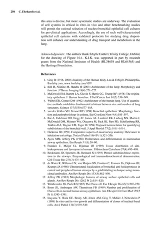
![Chapter 10 In Vitro Models of the Tracheo-Bronchial Epithelium 257
125. Yang X, Rojanasakul Y, Wang L, Ma JY, Ma JK (1998) Enzymatic degradation of
luteinizing hormone releasing hormone (LHRH)/[D-Ala6]-LHRH in lung pneu-
mocytes. Pharm Res 15(9):1480–1484.
126. Cohen AJ, Bunn PA, Franklin W, Magill-Solc C, Hartmann C, Helfrich B,
Gilman L, Folkvord J, Helm K, Miller YE (1996) Neutral endopeptidase: Variable
expression in human lung, inactivation in lung cancer, and modulation of peptide-
induced calcium flux. Cancer Res 56(4):831–839.
127. Baraniuk JN, Ohkubo K, Kwon OJ, Mak J, Ali M, Davies R, Twort C, Kaliner M,
Letarte M, Barnes PJ (1995) Localization of neutral endopeptidase (NEP) mRNA
in human bronchi. Eur Respir J 8(9):1458–1464.
128. van der Velden VH, Naber BA, van der Spoel P, Hoogsteden HC, Versnel MA
(1998) Cytokines and glucocorticoids modulate human bronchial epithelial cell
peptidases. Cytokine 10(1):55–65.
129. Juillerat-Jeanneret L, Aubert JD, Leuenberger P (1997) Peptidases in human
bronchoalveolar lining fluid, macrophages, and epithelial cells: Dipeptidyl
(amino)peptidase IV, aminopeptidase N, and dipeptidyl (carboxy)peptidase
(angiotensin-converting enzyme). J Lab Clin Med 130(6):603–614.
130. Wesley UV, Tiwari S, Houghton AN (2004) Role for dipeptidyl peptidase IV in
tumor suppression of human non small cell lung carcinoma cells. Int J Cancer
109(6):855–866.
131. Trehin R, Nielsen HM, Jahnke HG, Krauss U, Beck-Sickinger AG, Merkle HP
(2004) Metabolic cleavage of cell-penetrating peptides in contact with epithelial
models: human calcitonin (hCT)-derived peptides, Tat(47–57) and penetratin(43–
58). Biochem J 382(Pt 3):945–956.
132. Borchard G, Cassara ML, Roemele PE, Florea BI, Junginger HE (2002) Trans-
port and local metabolism of budesonide and fluticasone propionate in a human
bronchial epithelial cell line (Calu-3). J Pharm Sci 91(6):1561–1567.
133. Miller-Larsson A, Mattsson H, Hjertberg E, Dahlback M, Tunek A, Brattsand
R (1998) Reversible fatty acid conjugation of budesonide. Novel mechanism for
prolonged retention of topically applied steroid in airway tissue. Drug Metab
Dispos 26(7):623–630.
134. Sudsakorn S, Liu H, LeDuc B, Williams D (2005) In vitro oleate esterification of
budesonide. AAPS J 7(S2):R6244.](https://image.slidesharecdn.com/drugabsorptionstudies-2008-150610022529-lva1-app6892/85/Drug-Absorption-Studies-2008-275-320.jpg)
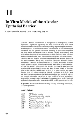
![Chapter 11 In Vitro Models of the Alveolar Epithelial Barrier 259
CAT Cationic amino acid transporter
CPM Carboxypeptidase M
CYP Cytochrome P450
DC Dendritic cells
ENaC Epithelial sodium channel
FITC Fluorescein isothiocyanate
GABRP Gamma-aminobutyric acid receptor pi subunit
GLUT Glucose transporter
GST Glutathione S-transferases
hAEpC Human alveolar epithelial cell
ICAM-1 Intercellular adhesion molecule-1
IVIVC In vitro/in vivo correlation
LRP Lung resistance protein
MLE Mouse lung epithelium
MRP Multidrug resistance-associated protein
NAT N-acetyl transferases
OCT Organic cation transporter
Papp Apparent permeability coefficient
PEPT2 peptide transporter
P-gp P-glycoprotein
RAGE Receptor for advanced glycation end products
RT-PCR Reverse transcription-polymerase chain reaction
SLC Solute carrier protein
SP Surfactant protein
SVCT Sodium-dependent vitamin C transporter
TEER Transepithelial electrical resistance
UGT UDP-glucuronosyl transferases
VC Vitamin C
ZO Zonula occludens
11.1. Introduction
11.1.1. The Alveolar Epithelium
The respiratory system consists of the nasopharynx, trachea, bronchial tree
and terminal air saccules. The nasopharynx, trachea and bronchi conduct air
into respiratory parts of the lung, that is the alveoli [1]. The human alveoli
are small, round or polyhedric chambers with an average diameter of about
250 µm, which form connected groups at the end of bronchoalveolar ductules.
The wall of an alveolus is lined by the alveolar epithelium with underlying
connective tissues and capillary networks. Two neighbouring alveoli form a
common septum with the alveolar epithelium on each side, with connective
tissues and capillaries in between. The average surface area covered by the
alveolar epithelium is reported to be about 102 m2 in humans [2]. Because of
their smaller body sizes, rodents have much smaller alveolar epithelial surface
areas than larger mammals (e.g., mice: 0.05 m2; rats: 0.4 m2; and dogs: 52
m2). However, there is a very good correlation between the lung size and cell
numbers when analysed using data from various mammalian species [2].
The human alveolar epithelium consists of two cell types: type I (alveolar
epithelial type I [ATI], pneumocyte I) and type II (alveolar epithelial type II](https://image.slidesharecdn.com/drugabsorptionstudies-2008-150610022529-lva1-app6892/85/Drug-Absorption-Studies-2008-277-320.jpg)
![260 C. Ehrhardt et al.
A B
C D
2 µm
5 µm 1 µm
0,5 µm
er
en
ai
ct
ATI
n
ct
ATI
en
er
n
ai
ai
ctATI
ATII
n
mlb
ai
ai
mi
gc
Figure 11.1 Ultrastructure of the human lung alveolar barrier. The tissue specimen is
obtained via lung resection surgery. (A) Section through a septal wall of an alveolus.
The wall is lined by a thin cellular layer formed by alveolar epithelial type I cells (ATI).
Connective tissues (ct) separate ATI cells from the capillary endothelium (en) within
which an erythrocyte (er) and granulocyte (gc) can be seen. The minimal distance
between the alveolar airspace (ai) and erythrocyte is about 800–900 nm. The endothelial
nucleus is denoted as n. (B) Details of the lung alveolar epithelial and endothelial barri-
ers. Numerous caveolae (arrows) are seen in the apical and basal plasma membranes of
an ATI cell as well as endothelial cell (en) membranes. Caveolae may partake transport
of some solutes (e.g., albumin). (C) ATII cells (ATII) are often localised in the corners
of alveoli where septal walls branch off. (D) ATII cells are characterised by numerous
multilamellar bodies (mlb) which contain components of surfactant. A mitochondrion
is denoted as mi.
[ATII], pneumocyte II) (Figure 11.1). ATI cells are very flat, spread out and
cover >93% of the alveolar surface [3], while ATII cells are cuboidal and
cover the remainder of the alveolar airspace. Although ATII cells are much
more numerous than ATI cells (67% vs. 33%; [4]), ATI cells provide most of
the surface area in alveoli, due to their larger cell surface of ∼7,000 µm2 versus
∼250 µm2 for ATII cells [2]. The height of ATI cells varies from about 2 µm to
3 µm in the nuclear region to about 0.2 µm in the cytoplasmic extensions. In
many regions of the ATI cell, the apical and basal plasma membranes form
caveolae, omega-shaped invaginations, which further decrease the distance
between the apical and basal plasma membranes to a few tens of nanometres
[5]. Caveolae are also a characteristic feature of the microcapillary endothelial
cells which, in many parts of the alveolar wall, are localised close to the
alveolar epithelium, separated from each other only by a thin layer of basement
membrane. Thus, alveoli form an extremely thin barrier, allowing efficient gas
exchange between the air and blood compartments (Figure 11.1). This thin
barrier may also be utilised for fast absorption of therapeutics.](https://image.slidesharecdn.com/drugabsorptionstudies-2008-150610022529-lva1-app6892/85/Drug-Absorption-Studies-2008-278-320.jpg)
![Chapter 11 In Vitro Models of the Alveolar Epithelial Barrier 261
ATII cells are taller and cuboidal, with a height of ∼10 µm [4, 6]. The cells
often reside at positions in the alveolar wall where three alveoli are connecting
each other. Occasionally, ATII cells can be found in groups, which may repre-
sent daughter cells from a preceding cell division of an ATII mother cell. Char-
acteristic structural features of ATII cells are the (multi)lamellar bodies. These
structures are membrane-bound, intracellular compartments (up to 1 µm in
size [7]) which contain densely packed membrane materials appearing as
lamellae in cross sections. The membrane content of lamellar bodies is secreted
onto the surface of the alveolar wall where it forms a mostly single layer
of surfactant. The surfactant consists of a mixture of phospholipids (∼90%)
and surfactant-associated proteins (SP, ∼10%): SP-A, SP-B, SP-C and SP-
D. The main function of the surfactant layer is to reduce the surface ten-
sion and thereby preventing the alveoli from collapsing and help expand
them with inspiration of air. Moreover, the surfactant contains molecules
(e.g., immunoglobulins, proteases and ceruloplasmin) for the defence against
microorganisms and antioxidants (e.g., catalase) which prevent the oxidation
(= inactivation) of proteins [8]. The surfactant proteins, SP-A and SP-D, belong
to the family of collectins which bind with broad specificities to a variety
of microorganisms and possess antimicrobial properties [9]. These proteins
also exhibit both inflammatory and anti-inflammatory functions, which occur
through interactions with pattern recognition receptors, including toll-like
receptors and CD14, signal inhibitory regulatory protein alpha and a receptor
complex of calreticulin and CD91 [10]. While the composition of the surfactant
is well known, the structural organisation of the surfactant and the aqueous
hypophase is still under debate. Major points of the discussion are whether or
not the alveolar epithelium is lined by a continuous fluid layer and to what
extent the surfactant covers the alveolar epithelium [11, 12].
Alveolar epithelial cells (AECs) are connected with each other by various
epithelial cell–cell contacts (i.e., tight junctions, adherens junctions and oth-
ers). These contacts comprise several groups of proteins, such as occludin,
zonula occludens (ZO-1, -2, -3), claudins, tricellulin, E-cadherin and inter-
cellular adhesion molecule-1 (ICAM-1) [13–15]. Their regulation and inter-
play, which has influence on epithelial barrier properties, however, is largely
unknown to date.
11.1.1.1. Molecular Markers of Alveolar Epithelial Cells
While morphological features of AECs are well characterised, only few mole-
cules have been identified which can be used as clear signatures for the respec-
tive cell type. These markers are summarised in the following paragraphs. The
best marker for AT II cells is SP-C, which is exclusively expressed in this cell
type. All other SP, such as SP-D, are also expressed in cells of the bronchial
epithelium or even other epithelial cells of the body [16].
There is still some discordance about markers for ATI cells [17, 18]. Vari-
eties of glycocalyx appear to be expressed in ATI versus ATII cells, which
can be distinguished by the binding of specific lectins. Lycopersicon esculen-
tum lectin, for example, appears to adhere specifically to the apical plasma
membrane of ATI cells [19, 20]. Other lectin markers that have been reported
in AEC typing are Ricinus communis agglutinin, Erythrina cristagalli lectin,
soybean lectin and Bauhinia purpurea agglutinin (all for the ATI phenotype),](https://image.slidesharecdn.com/drugabsorptionstudies-2008-150610022529-lva1-app6892/85/Drug-Absorption-Studies-2008-279-320.jpg)
![262 C. Ehrhardt et al.
and Maclura pomifera agglutinin, Helix pomatia lectin and Sambucus nigra
agglutinin (for the ATII phenotype) [21].
T1α was the first protein marker observed to have a high specificity for
mammalian ATI cells [22]. Based on its DNA sequence, T1α is projected to
be an integral membrane protein of ∼18 kDa which is also expressed in cells
of the choroid plexus of the central nervous system and the ciliary epithelium
of the eye in both foetuses and adults. However, physiological function of T1α
remains largely unknown to date [23]. Very recently, mice lacking the T1α
gene [T1α (–/–)] were reported to fail formation of expanded alveolar sacs,
resulting in respiratory failure at birth [24]. It might be of interest to note that
an attempt to generate antibodies recognising homologous structures in human
tissue against T1α has not been successful (personal communication with
Dr. Michael Kasper). The expression of caveolin-1, the main structural protein
of caveolae, has also been used to distinguish ATI from ATII cells, since ATI
(but not ATII) cells contain numerous caveolae [25]. Caveolin-1, however, has
only a limited potential as a differentiation marker in the alveolus, as it is highly
expressed in lung microcapillary endothelial cells as well [26].
Furthermore, aquaporin-5 (AQP-5) has received some attention as an ATI
cell marker. Of all mammalian cells studied to date, ATI cells reveal the highest
water permeability in in vitro isolated cell studies [27], perhaps reflecting the
expression of various water channels in alveolar epithelium, such as AQP-5,
in addition to the active ion transport proteins, including epithelial sodium
channels (e.g., ENaC) and Na +/K +-ATPase [28]. AQP-5 belongs to the
aquaporin family of transmembrane proteins, which form water permeable
channels and is also expressed in several other tissues (e.g., salivary and
lacrimal glands, cornea and parts of the upper airway). In the human and
rat alveolar epithelium, AQP-5 is localised specifically in the apical plasma
membrane of ATI cells [29, 30]. Recent studies also indicate the expression
of other members of the aquaporin family, such as AQP-3 and AQP-4, in the
alveolar epithelium of the human lung. Although initially attributed to upper
airway epithelia, immunolabelling of aquaporins in human lung tissue sections
suggest that AQP-3 is expressed in ATII cells, whereas AQP-4, like AQP-5, is
localised in ATI cells [31].
The transmembrane receptor for advanced glycation end products (RAGE)
is a member of the immunoglobulin superfamily and highly expressed not only
in the lung but also in other organs, including blood vessels. RAGE interacts
with a broad spectrum of molecules, such as advanced glycation end products,
amyloid beta fibrils, amphoterin and members of S100/calgranulin family,
by recognising pattern of the tertiary protein structure. RAGE is involved
in inflammatory processes, diabetic vascular disease and Alzheimer’s disease
[32]. Different reports from in situ studies as well as in vitro data have shown
that RAGE is expressed in the alveolar epithelium. Although initial reports
were controversial regarding the exact cellular localisation, a growing amount
of experimental data supports the localisation of RAGE to the basolateral
plasma membrane of ATI cells [33, 34]. Very recent studies suggest that RAGE
expressed in the alveolar epithelium might play a role in inflammation and cell–
matrix interactions [30, 35].
In the healthy lung, ICAM-1, an adhesion molecule that is involved in migra-
tion and activation of T-cells and macrophages, is highly expressed in the ATI
cell [36]. In comparison, the expression of ICAM-1 in ATII and other cells in](https://image.slidesharecdn.com/drugabsorptionstudies-2008-150610022529-lva1-app6892/85/Drug-Absorption-Studies-2008-280-320.jpg)
![Chapter 11 In Vitro Models of the Alveolar Epithelial Barrier 263
Table 11.1 Constituents/Compartments of the Distal Blood–Air Barrier
of the Lung.
Air
Surfactant
Hypophase
Epithelium (cells and basement membrane)
Connective tissue (collagen and elastic fibres)
Free (unattached to basement membranes) cells: fibroblasts, macrophages,
granulocytes and lymphocytes
Endothelium (cells and basement membrane)
Blood plasma
Blood cells
the lung, such as endothelial cells, usually is low. In response to inflammatory
stimulation and hyperoxia, expression levels of ICAM-1 increase in these other
cell types, rendering distinction between cell types based on ICAM-1 levels
rather difficult [37]. Expression of this immunologically important adhesion
molecule also involves complex spatial interactions of the cells with the basal
lamina and other epithelial cells [38].
Another protein which has been suggested as a differentiation marker
between the ATI and ATII phenotypes is HTI56, a membrane protein which
seems to be expressed exclusively in ATI cells [39]. In addition, Chen and
co-workers confirmed four new ATI genes and one ATII gene by quantitative
real-time PCR and immunostaining of both cultured alveolar cells and rat
lung tissue: fibroblast growth factor receptor-activating protein 1, P2 purinergic
receptor 7 (P2X7), interferon-induced protein and Bcl2-associated protein were
found in ATI cells, while the gamma-aminobutyric acid receptor pi subunit
(GABRP) was associated with ATII cells [40].
11.1.1.2. Functions of the Blood–Air Barrier of the Alveolus
In the alveolus, gases, liquids and solutes (e.g., metabolites) are exchanged or
transported between the blood and air compartments. Moreover, the surfactant
and exogenously presented material is taken up for degradation or inactivation
into various cells. The trafficking molecules have to travel through different
extracellular and cellular compartments by passive and sometimes also active
processes. Table 11.1 gives a list of the components which molecules translo-
cating from the blood to air and vice versa may encounter.
While gases and other small molecules dissolved in the extracellular fluid
in the airspace of the alveolus or the blood are predominantly transported by
passive diffusion, larger molecules in some cases traverse the distal blood–
air barrier of the lung using active processes, for example cell-energy requiring
transcytosis [41, 42]. Translocation of molecules can involve passive paracellu-
lar and/or transcellular pathways. Receptor-mediated endocytosis (via clathrin-
coated pits or caveolae), adsorptive endocytosis and pinocytosis (non-specific
fluid-phase endocytosis) of various molecules across the alveolar epithelium
are expected. For larger particles/molecules, phagocytosis may also occur. Bar-
rier properties of the alveolar epithelium are mainly determined by the tightness
of alveolar epithelium and endothelium for passively transported molecules.](https://image.slidesharecdn.com/drugabsorptionstudies-2008-150610022529-lva1-app6892/85/Drug-Absorption-Studies-2008-281-320.jpg)
![264 C. Ehrhardt et al.
This tightness depends on the quality of cell–cell contacts (and apical and
basolateral plasma membranes, in part). In particular, cell–cell contacts can
be regulated, allowing certain molecules or even cells (e.g., macrophages,
lymphocytes and/or granulocytes) to cross the blood–air barrier in the
lung [43, 44].
Almost all transport studies for the alveolar epithelial barrier have been per-
formed in animal models or cell lines, making the relevance of observed results
in relation with the human situation somewhat questionable [45]. Observations
on the clearance of lung oedema, which involves the removal of liquid and
macromolecules from alveoli, revealed significant species differences in the
regulation of the process, indicating that transport pathways might be different
among the mammalian lungs and/or that the experimental parameters (e.g.,
concentrations and administration modality for molecules) studied may need
further refinement for each species studied [46]. Our current understanding is
that many molecules are transported across the alveolar epithelial barrier into
the interstitial space or blood [41, 42]. Paracellular transport seems to be pre-
dominant for most small hydrophilic molecules (≤6 nm Stokes-Einstein radius
for hydrophilic molecules ≤40 kDa) under normal physiological conditions.
Larger molecules are usually transported by transcytosis, albeit their trans-
port rates may vary significantly. Proteolytic degradation in alveolar airspaces
and/or epithelial/endothelial cells seems to contribute to the observed variable
fractions of intact peptides/proteins appearing in the systemic circulation fol-
lowing pulmonary delivery.
Caveolae might serve as a main transport conduit across alveolar epithelial
and endothelial barriers, since they are abundant in both ATI and microvascular
endothelial cells in the lung. Although the function and modus operandi of
caveolae are still under debate, they seem to be involved in various transport
processes (e.g., endocytosis and transcytosis of various molecules [e.g., folates
and albumin]). A growing body of evidence suggests that caveolae are more
stable structures than coated pits, but also pinch off from the plasma mem-
brane and form small vesicles in the cytoplasm. Furthermore, caveolae are
the site where several receptors are expressed/congregated, which might act
as the centre controlling the activity of receptors and molecules involved in
cell signalling (see Chap. 22 for a detailed description of caveolae function).
Insights into the importance of caveolin-1 for the function of the distal lung
blood–air barrier have mainly emerged from knockout mice studies, where the
loss of caveolin-1 resulted in severe lung abnormalities and thickened alveolar
septa [47]. Under pathophysiological conditions, AEC damage is associated
with lower levels of caveolin-1, which is probably mainly due to the high
vulnerability of ATI cells and consequently the significant loss of ATI cells
in response to several sources of injury [26].
Regulation of the intra- and extracellular fluid homoeostasis is an important
challenge. Several ion and water channels are involved in this regulation.
However, the significance of a few individual molecules playing the field has
been reported to date. In ATII and ATI cells, Na+ channels are localised at
apical cell membranes, while Na+/K+-ATPases are found at the basolateral cell
membranes [28, 48]. Na+/K+-ATPases extrude sodium ions out of the cell into
the interstitium, thereby building up a sodium ion gradient across the apical cell
membranes, setting up an electrical gradient across the epithelium as a result.
This electrochemical gradient drives transport of the counter ion, Cl−, from](https://image.slidesharecdn.com/drugabsorptionstudies-2008-150610022529-lva1-app6892/85/Drug-Absorption-Studies-2008-282-320.jpg)
![Chapter 11 In Vitro Models of the Alveolar Epithelial Barrier 265
the alveolar airspace across the alveolar epithelium into interstitium, creating
an osmotic gradient which in turn drags water into the interstitium as well. The
importance of aquaporins for this water transport has been elucidated in AQP-5
knockout mice, where a markedly reduced water flow was observed in response
to an artificially imposed osmotic pressure in the lung. Interestingly, however,
the near iso-osmolar water flux (secondary to active Na+ absorption in AQP-5
knockout mice) was not affected at all [49]. Elucidation of transport mech-
anisms in the human alveolus is crucial, since pathophysiological conditions
(e.g., lung oedema and acute respiratory distress syndrome) are demonstrated
in part to be associated with abnormal and severely impaired transport across
the alveolar epithelial barrier.
In addition to movement of water, solutes and proteins across alveolar
epithelium, intact cells are known to cross the blood–air barrier of the lung.
Interstitial and/or vascular macrophages are able to travel to the alveolar lumen
where they may take up exogenously instilled molecules, for example microor-
ganisms. After an initial contact with a potential pathogen, macrophages then
migrate back into the connective tissue (or stay in the airspace) and recruit
lymphocytes via cytokine release [50]. The alveolar epithelium might also be
an important source of chemokines, such as CCL20. ATII cells may play a
critical role in controlling the movement of dendritic cells (DC) through the
distal lung blood–air barrier under both normal and inflammatory conditions.
In vitro studies showed that cultured DC start migrating in response to con-
ditioned media from human ATII cell cultures (but not those from alveolar
macrophages) [51].
11.1.1.3. Developmental Biology and Wound Repair of the Alveolus
During human embryogenesis, the lung bud is formed by a diverticulum of
the foregut. In the initial phase, development of the lung bud resembles that of
sub-epithelial tubulo-acinar gland. Epithelial stalks grow and branch into the
embryonic mesenchyme and finally widen to form tubes. The period between
weeks 17 and 26 of gestation is characterised by a further growth of the
system, the formation of terminal saccules and thinning of the mesenchymal
structure. In addition, epithelial cells differentiate into type I and type II cells
and surfactant production (by ATII cells) starts at the end of this period. Groups
of alveoli first appear from about week 29 of gestation as indentations of the
saccules reaching full functional development by 34 weeks of gestation. At
birth, about one third to one half of the alveoli are present, steadily increasing
in number during the first 2–3 years with their size and surface area continuing
to increase until after adolescence. [52, 53]
In the mature, that is, adult alveolar epithelium, ATII cells serve as a progen-
itor with the ability to restore AEC populations by proliferation, especially
when injury occurs. Daughter cells may proliferate and/or some may differ-
entiate into ATI cells [54]. This process, under the control of still largely
unknown regulatory mechanisms, is the basis for the normal cell turnover in
the alveolar epithelium under physiological conditions. In case of an injured
alveolar epithelium, the ability of ATII cells to proliferate and differentiate into
ATI cells is thought to be a key factor in alveolar epithelial repair following
injury. Nevertheless, recent findings also imply a possible role of bone marrow
derived stem cells in the repair process or cell turnover in alveolar epithelium,
although the relative contribution of bone marrow derived stem cells to these](https://image.slidesharecdn.com/drugabsorptionstudies-2008-150610022529-lva1-app6892/85/Drug-Absorption-Studies-2008-283-320.jpg)
![266 C. Ehrhardt et al.
processes, especially under normal physiological conditions, is unknown at
best [55, 56].
The differentiation process of ATII daughter cells or progenitor cells towards
the ATI cell phenotype has only been characterised largely in cell culture set-
ting. In a human cell culture model, the morphology of isolated ATII cells shifts
towards ATI cell morphology within 5–10 days [30, 57, 58]. The molecular
signatures also change concomitantly, for example SP-C expression declines
while levels for caveolin-1, RAGE and AQP-5 expression increase with time
in culture. In parallel, typical epithelial cell–cell contacts are formed, accom-
panying the expression of typical cell–cell adhesion molecules (e.g., occludin,
ZO-1 and ICAM-1). These molecules may be used as indicators/markers for
the epithelial development/injury/repair. For a general overview on alveolar
cell biology, various articles by Fehrenbach [59], Williams [17] and Mason
[60] may be consulted.
11.2. In Vitro Models of the Alveolar Epithelial Barrier
for Drug Absorption Studies
Although there is consensus about the crucial role of the distal lung epithelial
tract in systemic drug absorption, not many useful in vitro model systems exist
to date [61]. Because of the intricate and delicate network of the alveoli, an ex
vivo approach (e.g., mounting excised mammalian lung alveolar epithelium
in a diffusion chamber set-up) is impossible, although amphibian alveolar
epithelial tissues have been studied as such to provide some useful information
on transport properties in the past [62]. Furthermore, neither in vivo nor in
situ studies (as discussed in Chap. 6) allow for precise assessment/prediction
of the actual absorption sites involved. Therefore, cellular models based on
primary or cell line cultures or co-culture of epithelial cells with either alveolar
macrophages, capillary endothelial cells or DC are the only tools available to
investigate drug absorption mechanisms in the alveolar epithelial barrier [63].
11.2.1. In Vitro Models of the Alveolar Epithelial Barrier
11.2.1.1. Alveolar Epithelial Cell Lines
While a number of immortalised cell lines emanating from different cell types
of the airway (i.e., tracheo-bronchial) epithelium of lungs from various mam-
malian species are available (see Chap. 10), reliable and continuously growing
cell lines that possess AEC morphology and phenotype are not reported to date.
Most studies use cell lines of alveolar epithelial origin for drug absorption
studies, while the observations are hard or meaningless to extend to in vivo
human situation.
Probably the most frequently used alveolar epithelial model is the A549 cell
line (American Type Culture Collection, ATCC CL-185), continuously grow-
ing cells derived from a human pulmonary adenocarcinoma that have some
morphologic and biochemical features of the human pulmonary alveolar type II
cell in situ [64]. A549 cells contain multilamellar cytoplasmic inclusion bodies,
like those typically found in human lung ATII cells, although these hallmarks
disappear as culture time increases. At early and late passage levels, the cells
synthesise lecithin with a high percentage of disaturated fatty acids utilising](https://image.slidesharecdn.com/drugabsorptionstudies-2008-150610022529-lva1-app6892/85/Drug-Absorption-Studies-2008-284-320.jpg)
![Chapter 11 In Vitro Models of the Alveolar Epithelial Barrier 267
A
C
B
D
Figure 11.2 Morphological differences between human alveolar epithelial cells in
primary culture (A and C) and the A549 cell line (B and D). Cells are visualised by
light microscopy (A and B) and immunofluorescence microscopy (C and D) using an
antibody against a tight junctional protein, occludin.
the cytidine diphosphocholine pathway [64]. The cell line has been utilised
for many biopharmaceutical studies, albeit A549 cells lack the ability to form
tight monolayers of polarised cells, due to the inability to form functional tight
junctions [61, 63, 65, 66].
Figure 11.2 shows marked differences in morphology between primary
human alveolar epithelial cells (hAEpCs) that started as ATII cells and dif-
ferentiated into ATI-like morphology on day 8 in primary culture (Panel A)
and A549 cells on day 5 in culture (Panel B) by light microscopy. In addition,
Figures 11.1C and 11.1D show results of immunofluorescence microscopy
using an antibody against occludin (a tight junctional protein) for the hAEpC
monolayer and A549 cells, respectively. Despite their obvious unsuitability,
due to their lack of good transepithelial electrical resistance (TEER), some
investigators still employed A549 cell layers in drug absorption studies [67,
68]. The absorption data reported by these groups were not able to clearly
show differences in transport rates between proteins and peptides of various
sizes where the published permeability values were 2–4 orders of magnitude
higher than those obtained in tight monolayer systems (e.g., hAEpC monolay-
ers) using identical compounds [63]. Moreover, the reported A549 TEER of
∼600 ohm·cm2 by one group [67] may be in gross error. Their data indicate
that large hydrophilic molecules are translocated across A549 cell layers at
rates approaching near-free diffusion limits. In other words, such large fluxes
probably took place via large gaps between A549 cells, contradicting to the
very large TEER they reported. It should be noted that TEER of A549 cells
is usually in the range of 20–60 ohm·cm2, which makes much more sense,
as it is known that no functional tight junctions are present in A549 cell
layers. Notwithstanding, A549 cells might be a useful model in other areas](https://image.slidesharecdn.com/drugabsorptionstudies-2008-150610022529-lva1-app6892/85/Drug-Absorption-Studies-2008-285-320.jpg)
![268 C. Ehrhardt et al.
of biopharmaceutical research, including metabolism studies or cytotoxicity
studies for various substances [65, 69, 70].
Other cell lines of an alveolar epithelial origin that can be found in the
literature include R3/1 and L-2 (rat), mouse lung epithelium (MLE-12 and 15)
(mouse) and H441 (human). Of these, NCI-H441 cell line (ATCC HTB-174),
which emanated from a human lung adenocarcinoma, has been described to
have characteristics of both ATII [71, 72] and bronchiolar (i.e., Clara) epithelial
cells [73, 74]. Studies can thus be designed to characterise metabolism and
transport properties of these two particular cell types. There is an emerging
evidence that H441 cells are capable to form monolayers of polarised cells,
exhibiting a significant TEER [75, 76], although the cell line has not been used
for drug absorption experiments so far.
Wikenheiser and co-workers generated a series of continuous AEC lines
[MLE-7, -12 and -15] from transgenic mice harbouring the SV40 large T
antigen under the control of the human SP-C promoter region. These MLE cell
lines show morphological and functional characteristics of distal respiratory
epithelial cells normally lost after isolation and primary culture [77], which are
consistent with those seen in non-ciliated bronchiolar and ATII cells. However,
morphological and functional characteristics associated with an individual cell
type do not appear to always coexist in a clonal cell line. For example, MLE-
12 cells express SP-C mRNA (i.e., indicative of ATII cells in the adult mouse),
but other characteristics (e.g., SP-A mRNA expression or presence of lamellar
bodies) are missing in MLE-12 cells. The heterogeneity of cellular markers
thought to be characteristic of ATII cells may be related to the immortalisation
of cells at various stages of lung development. Alternatively, MLE cell lines
may represent distinct subtypes of distal respiratory epithelial cells. MLE-12
can be obtained from the American Type Culture Collection (ATCC CRL-
2110).
L-2 cells (ATCC HTB-149) have been isolated by clonal culture techniques
from the adult rat lung. These cells appear to retain differentiated functions that
are present in ATII cells of intact rat lungs. L-2 cells are diploid, epithelial cells.
They contain osmiophilic lamellar bodies in their cytoplasm and synthesise
lecithin by the same de novo pathways as in a whole lung [78]. It is not known
if L-2 cells are capable to form confluent and electrically tight monolayers. L-2
cells have not been systematically investigated regarding their suitability as a
model for absorption studies.
The rat cell line R3/1 was established from cells obtained from broncho-
alveolar tissues of foetal Wistar rats at 20 days of gestation. This cell line
displays a phenotype with several characteristic features of ATI cells. R3/1 cells
were analysed to show a positive expression for mRNA and protein content
of markers related to the ATI cell type (e.g., T1α, ICAM-1, connexin-43 and
caveolin-1 and -2) [79]. Whether or not this cell line can form functional tight
junctions is currently under investigation in our laboratories.
11.2.1.2. Primary Alveolar Epithelial Cell Culture Models
Because of the paucity of appropriate AEC lines that form functional tight
junctions, primary cultures of AEC are used for most in vitro studies of alveolar
epithelial function (e.g., solute transport and metabolism). Primary mammalian
AEC techniques involve isolation, purification and culture of ATII cells from
tissues obtained either after lung resections or isolated perfused lungs. These](https://image.slidesharecdn.com/drugabsorptionstudies-2008-150610022529-lva1-app6892/85/Drug-Absorption-Studies-2008-286-320.jpg)
![Chapter 11 In Vitro Models of the Alveolar Epithelial Barrier 269
ATII cells, when plated on permeable supports or plastics under appropriate
culture conditions, acquire features of type I cell-like phenotype and mor-
phology [30, 57, 80]. Although isolation of ATI pneumocytes from rat lungs
has recently been reported with some success [28, 48, 81], development of
confluent ATI cell monolayer with electrically tight characteristics has not been
reported yet. It should be noted that unlike many other cells in primary culture,
AEC exhibits generally a very limited proliferation profile and is therefore not
suitable for passaging. Thus, a new preparation of cells has to be used for each
data set, which drives the costs up tremendously, and a reliable normalisation
scheme of data observed from each set of cell preparations is needed.
Because of the limited availability of human lung tissues and some ethical
issues pertaining to use of human tissues, most studies were based on isolation
and culture of cells from the lungs of animals, including mouse [82], rat [83],
rabbit [84] and pig [85]. In the Appendix, a universal protocol has been devel-
oped by us for the isolation and purification of mammalian ATII cells, followed
by monolayer cultures of ATII or ATI cell-like phenotype and morphology.
However, since evidence for species differences between human and rodents
might be more significant than once assumed [45], confirmation using human
pneumocyte cultures has been performed for various aspects [58, 86, 87].
11.2.2. The Use of Alveolar Epithelial Cells in
Biopharmaceutical Research
With the lung emerging as an attractive non-invasive delivery routes for sys-
temic absorption of biopharmaceuticals and fast-acting conventional drugs, it
appears pivotal to gather knowledge about modes of transport and the fate of
these compounds at the alveolar epithelial barrier. Although an in vitro/in vivo
correlation (IVIVC) using AEC monolayers could not be established defini-
tively currently, various in vitro models can help elucidate underlying transport
mechanisms of drug molecules, as well as their metabolic stability in the lung.
Therefore, the available in vitro systems need to be carefully characterised and
validated prior to using them in the context of biopharmaceutical research.
11.2.2.1. Drug Absorption Studies
Absorptive processes have first been studied in the context of ion and fluid
clearance from the lung in health and disease (e.g., oedema), which were
conducted largely using monolayers of rat pneumocytes [e.g., 88]. Following
studies of active and passive ion movement across alveolar epithelium using the
in vitro model, translocation of plasma proteins (which are commonly found
in the alveolar lumen, e.g., albumin, immunoglobulin G and transferrin) was
investigated [41]. Since rat-based monolayers have been considered the gold
standard in elucidation of AEC physiology/biology for several decades, it is not
surprising that the same well-established primary rat pneumocyte monolayer
model was also employed for drug delivery studies [89–91]. However, as stated
above, newer models based on different animal species (which might more
closely resemble human, e.g., derived from porcine lung [85]) were developed
and a number of laboratories also started to use hAEpC monolayers for drug
absorption studies [66, 92].
Effros and Mason showed an apparent inverse relationship between mole-
cular weights and rates of in vivo lung clearance of molecules of different
sizes, both in log scales [93]. A similar relationship has also been found in](https://image.slidesharecdn.com/drugabsorptionstudies-2008-150610022529-lva1-app6892/85/Drug-Absorption-Studies-2008-287-320.jpg)
![270 C. Ehrhardt et al.
−9.5
−9.0
−8.5
−8.0
−7.5
−7.0
−6.5
−6.0
−5.5
−5.0
Log molecular weight (Da)
LogPapp(cms−1)
3.5 4.0 4.5 5.0
Figure 11.3 Permeability to FITC-labelled
dextrans across rat (♦, [94]) and human ( ,
[66]) alveolar type I (ATI) cell-like
monolayers compared with the permeability
across A549 cell layers (•, [67]).
two in vitro alveolar epithelial models of primary rat [94] and human [66]
monolayers (Figure 11.3). By comparison, Fluorescein isothiocyanate (FITC)-
dextran permeabilities observed with A549 cells [67] are over two orders of
magnitude greater [63] (Figure 11.3).
Active transport mechanisms by which some compounds traverse the distal
lung blood–air barrier are more likely to provide new insights and improved
strategies for pulmonary delivery of drugs into the systemic circulation and/or
targeting drugs to lung parenchymal cells. The expression pattern of ATP-
binding cassette (ABC)-transporters in both the healthy and pathological
lung has recently been reviewed by van der Deen et al. [95]. Several trans-
porters have been identified in AEC, which might have an impact on pul-
monary bioavailability of drugs, especially lipophilic drugs (e.g., cytostatics).
P-glycoprotein (P-gp, MDR1) is probably the best characterised efflux pump in
the body. In 2001, Lehmann and co-workers reported the presence of P-gp and
multidrug resistance-associated protein 1 (MRP1) in rat ATII cells in primary
culture [96]. These findings were later confirmed using both human and rat lung
tissue sections as well as ATI cell-like monolayers by immunohistochemistry
and efflux studies using vinblastine and rhodamine 123 as respective substrates
for P-gp and MRP1 [97, 98]. MRP1 and lung resistance protein (LRP) were
also identified in cultures of human peripheral lung epithelial cells [99].
ABCA1 and ABCA3 gene transcripts were found in cell lines (e.g., A549) of
human lung origin, in ATII cells of human, rat, and mouse, as well as different
tissues of rat, but the highest expression of ABCA1 and ABCA3 was observed
in murine ATII cells [100, 101]. Molecular and biochemical studies show
that ABCA1 mediates basolateral surfactant efflux, while ABCA3 is found
in the limiting membrane of lamellar bodies. Because both transporters are
members of a subfamily of ABC-transporters that are predominantly known
to be involved in the regulation of lipid transport and membrane trafficking, it
can be speculated that these transporter proteins may play a key role in lipid
organisation during the formation of lamellar bodies. It should also be noted
that there are some conflicting reports regarding the activity and localisation of
ABC-transporters in mammalian AEC [95].
Other solute transporters (solute carrier protein [SLC]) are also expressed in
the alveolus. The mRNA transcripts of glucose transporters, GLUT1, GLUT4,
GLUT5 and SGLT1, have been detected in freshly isolated rat ATII cells by](https://image.slidesharecdn.com/drugabsorptionstudies-2008-150610022529-lva1-app6892/85/Drug-Absorption-Studies-2008-288-320.jpg)
![Chapter 11 In Vitro Models of the Alveolar Epithelial Barrier 271
reverse transcription-polymerase chain reaction (RT-PCR) and Western blot
analyses. However, a gradual loss of sodium-glucose co-transport activity and
transcription during transdifferentiation of ATII cells towards the type I-like
cell phenotype in culture has been reported [102].
Efficient gas exchange and other functions (e.g., active Na+ absorption)
in alveoli are thought to be tied to transepithelial clearance of luminal pro-
teins, albeit little is known about alveolar epithelial mechanisms of protein
transport. Protein degradation followed by transport of resultant peptides and
amino acids may play an important role in this process. Groneberg reported
the expression and activity of the high-affinity proton-coupled peptide trans-
porter, PEPT2, in rat and murine ATII cells [103]. The neutral and cationic
amino acid transporter (CAT), ATB0+, is expressed in mouse ATI cells in
situ [104]. The important role of peptide and amino acid removal was further
supported by the recent finding of Rothenberg et al. that the arginine transport
protein, CAT2, has a critical role in regulating lung inflammatory responses,
in that CAT2-deficient mice revealed spontaneous inflammation in the
lung [105].
Vitamin C (VC, L-ascorbic acid) is known to be essential for many enzy-
matic reactions. Sodium-dependent VC transporters (SVCT), SVCT1 and
SVCT2, were recently identified and reported to be localised in the apical cell
membrane of AECs in the lung of adult rats. These results suggest that SVCT
proteins could transport the reduced form of VC from the airway/alveolar
surface liquid into respiratory epithelial cells [106].
Primary rat and mouse ATII cells exhibit pH-sensitive, reversible transport
of choline, a process blocked pharmacologically with phenoxybenzamine, an
inhibitor of organic cation transporters (OCT). RT-PCR products for choline
transporters, OCT1 and OCT2, were detected in AEC, with only OCT2 pro-
tein being robustly expressed [107]. In 1999, Shen and co-workers published
evidence that rabbit AEC in primary culture shows a concentration-dependent
net absorption of organic cations [84], although the molecular identity of
the OCT involved was lacking. Our laboratories recently published data on
net absorption of salbutamol across hAEpC monolayers [108]. Since salbu-
tamol carries a positive charge at physiological pH, involvement of OCT
family member(s) in the alveolar epithelial absorption of salbutamol can be
surmised.
Investigations utilising tight monolayers of either rat or human AEC showed
that clearance of a number of endogenous proteins (e.g., immunoglobulin G,
albumin and transferrin) occurs in a cell energy-dependent manner [92, 109–
111]. These observations strongly suggest that transcytotic processes (e.g.,
caveolae-mediated or via clathrin-coated pits) may be responsible for removal
of these endogenous proteins from alveolar lining fluid. Intriguingly, these
proteins were net absorbed as intact molecules with variable degrees of degra-
dation [41]. Table 11.2 summarises the current literature data on absorption of
various peptides and proteins across tight monolayers of primary human and
rat AEC.
Although there appears to be no significant species differences in absorption
rates for small lipophilic drugs, some interspecies differences are noted with
water-soluble drugs absorbed from distal airspaces of in vivo mammalian lungs
[112]. These species differences have not been systematically studied yet.
However, some marked differences were reported for protein absorption rates](https://image.slidesharecdn.com/drugabsorptionstudies-2008-150610022529-lva1-app6892/85/Drug-Absorption-Studies-2008-289-320.jpg)
![272 C. Ehrhardt et al.
Table 11.2 Molecular Weights and Apparent Permeability Coefficients (Papp) of Various Proteins and
Peptides Across Primary Human Versus Rat AEC Monolayers.
Molecular
weight (Da)
Papp a-to-b
(human)
Papp b-to-a
(human)
Papp a-to-b
(rat)
Papp b-to-a
(rat) References
Gly-L-Phe 222 na na 3.29/1.38a 1.58/1.29a [89]
Gly-D-Phe 222 na na 1.55 1.60 [89]
TRHb 362 na na 3.27 3.80 [113]
acXASNH2 491 na na 1.19 Na [114]
Vasopressin 1,056 na na 0.45 0.45 [115]
acXAS(GAS)7NH2 1,998 na na 0.25 Na [114]
GLP-1(7-37) 3,355 6.13 1.91 na Na [92]
PTH(1-38) 4,458 2.20 1.80 na Na [92]
Insulin 5,800 0.77 0.72 0.21 0.12 [92, 116]
Cytochrome c 12,300 na na 0.48 0.44 [109]
G-CSF 20,000 na na 0.029 0.034 [109]
Growth hormone 22,125 8.33 9.02 0.041 0.074 [92, 117]
HRP 40,000 na na 0.069 0.073 [90]
Ovalbumin 43,000 na na 1.09 0.58 [109]
Albumin 65,000 2.45 0.21 0.77 0.39 [92, 109]
Transferrin 76,500 0.88 0.30 1.10 0.47 [91, 92, 109]
IgG 150,000 0.36 0.15 0.91 0.17 [92, 109, 111]
Notes: All Papp values are expressed as mean (×10−7 cm/s). The abbreviations of a-to-b and b-to-a denote the apical-to-
basolateral and basolateral-to-apical directions, respectively.
a Concentrations were 1 mM/10 mM. b Based on 3H-labelled TRH. The entry ‘na’ denotes ‘not available’.
found in primary human versus rat AEC monolayers [92]. Absorption rates
for most peptides and proteins in primary rat versus human AEC monolayers,
however, remain largely unknown.
11.2.2.2. Drug Metabolism Studies
While most textbooks and review articles on pulmonary drug delivery appear
to assume that lungs have a low metabolic activity and consider this as one
of the major advantages for pulmonary drug delivery (of peptide and protein
therapeutics), little is known about the pulmonary metabolism of endogenous
compounds and exogenously administered xenobiotics. As Chap. 10 contains a
general introduction to pulmonary metabolism, only a concise overview of the
metabolic activity in the alveolus is given here.
Some features, such as morphology and functionality of A549 cells, are
consistent with those of ATII cells in vivo, for example surfactant synthesis
and oxidative metabolism [65, 118]. Qualitative RT-PCR analysis revealed the
presence of several transcripts of cytochrome P450 (CYP) 1A1, CYP1B1,
CYP2B6, CYP2C, CYP2E1, CYP3A5 and CYP3A7 mRNA in A549 cells
[119]. More recently, quantitative RT-PCR studies revealed the presence of
CYP1A1, CYP1B1, CYP2B6, CYP2C, CYP3A4 and CYP3A5 messages in
A549 cells, whereas no detectable CYP1A2, CYP2A6, CYP2A13 and CYP2F1
transcripts were found [120]. Compared to the profile found in human distal
lung tissues, CYP1B1 mRNA was the most abundantly expressed transcript in](https://image.slidesharecdn.com/drugabsorptionstudies-2008-150610022529-lva1-app6892/85/Drug-Absorption-Studies-2008-290-320.jpg)
![Chapter 11 In Vitro Models of the Alveolar Epithelial Barrier 273
Table 11.3 Summary of the Expression Pattern and Activities of Phase I
and Phase II Metabolic Enzymes and Various Peptidases/Proteases in Human
Peripheral Lung Tissues, Primary Rat AEC Culture and Human Alveolar
Epithelial Cell Line, A549.
Enzyme
Lung
mRNAa
Lung
proteina,e
Peripheral
tissueg,h
Rat AECi,e
ATII/ATI A549j,k,l,h,e
CYP1A1 ? (∗∗∗)b − (∗∗∗)b + + +
CYP1A2 ? ? + −
CYP1B1 ∗∗c ?c + + +
CYP2A6 ∗∗ ? − +/−
CYP2A7 −
CYP2A13 + −
CYP2B1/2 +
CYP2B6/7 ∗∗∗ ∗∗∗ + +m/−
CYP2C ?d ?d −/+ +
CYP2D6 ? ? − +
CYP2E1 ∗∗∗ ∗∗∗ + + +/−
CYP2F1 ∗∗∗ −
CYP2J3 +
CYP3A2 −
CYP3A4 ? ∗f + −
CYP3A5 ∗∗∗ ∗∗ + +
CYP3A7 ? +
CYP4B1 ∗∗∗ −
GST + +
UGT + +
NAT1 +
NAT2 +
APN 24.77e 16.16/23.99e 47.74e/8.8k
DPP IV 69.26e 36.65/16.29e 24.24e/4.6k
NEP 24.11 0.99e 1.81/0.96e 2.41e
ACE 10.80e 4.29/nde −
CPM 4.63e nd/5.20e 4.28e
∗: weak evidence; ∗∗: moderate evidence; ∗∗∗: strong evidence; ?: conflicting evidence; −: no
evidence/undetectable; +: detectable (but not quantified); nd: not detected.
a [123]; b Strong evidence in smokers; c In alveolar macrophages; d In bronchial cells; e [69]; f In
∼20% of samples; g [122]; h [120]; i [125]; j [65]; k [124]; l [119]; m not inducible.
A549 cell line, followed by CYP3A5. Although the amount of specific CYP
transcripts in A549 cells is markedly lower than that found using lung lysates,
the profile of CYP expression shows a similar trend in overall profiles in
both systems. These results show that the A549 lung adenocarcinoma cell line
expresses limited numbers of phase I enzymes and responds to P450 inducers,
but at much lower levels than those observed in lung lysates.](https://image.slidesharecdn.com/drugabsorptionstudies-2008-150610022529-lva1-app6892/85/Drug-Absorption-Studies-2008-291-320.jpg)
![274 C. Ehrhardt et al.
The phase II metabolising step of conjugation reactions can be attributed
to the families of UDP-glucuronosyl transferases (UGT), glutathione S-
transferases (GST) and N-acetyl transferases (NAT) [121]. Using specific
primers for the detection of GST and NAT isoenzymes, it was reported that
GSTM1 is expressed in 25% of the peripheral lung tissues, whereas GSTM3
and NAT1 and NAT2 mRNAs are found in all samples [122]. Castell et al.
found very similar results in samples of human lung tissues versus A549 cells
for UGT and GST activities [120].
It has been suggested that the lung may possess relatively low prote-
olytic activities, but the inaccessibility of the peripheral lung for experimental
manoeuvres and/or manipulations may have confounded the correct assessment
of such data regarding the extent of metabolism in the deep lung for exoge-
nously inhaled peptides/protein therapeutics. Nevertheless, only 60% of insulin
was still intact after transport across rat AEC monolayers in vitro [116]. This
might be an indication of an underestimated protease activity in the alveolar
epithelium and lining fluid/surfactant, which could account for low systemic
bioavailabilities of some protein drugs after inhalation. The activity of individ-
ual peptidases in A549 cells and primary rat ATII or ATI-like pneumocytes has
been studied by Forbes and co-workers [69]. The pattern of peptidase expres-
sion appears to change over time in primary rat AEC culture. Interestingly,
A549 cells revealed levels of activity closest to those of primary ATI-like cells,
further complicating the pulmonary metabolic research (Table 11.3).
11.3. Concluding Remarks
As seen above, the lung alveolar epithelium serves an essential and efficient
function of providing fresh oxygen in exchange for the waste product of
carbon dioxide across its very thin and wide expanse of epithelial barrier.
This latter arrangement of thinness and enormous surface area of the alve-
olar epithelium has been considered by many as the advantage for systemic
delivery via pulmonary routes of therapeutic drugs, especially peptides and
protein molecules that show very poor bioavailability via oral routes because
of extensive degradation at stomach and intestines. However, currently we
have to rely on primary cell culture models, as many cell lines developed to
date for alveolar epithelium failed to recapitulate what has been shown for in
vivo properties of AECs. Most formidable may be the lack of genuine type II
cell models, although some success using keratinocyte growth factor to keep
the ATII cell morphology and phenotype has been documented for generating
monolayer cultures with functional tight junctions. For non-transport studies
(e.g., metabolism of certain classes of xenobiotics) may be performed using
cell line culture models (e.g., A549) for alveolar epithelium. Major hurdles,
however, still remain for successful development of passageable alveolar type
I and type II cell (lines), either as separate or mixed cell types of ATI and
ATII coexisting in one monolayer setting, thus mimicking the in vivo set-
ting. Moreover, the co-culture models of alveolar epithelium and capillary
endothelium of the mammalian lung are in their infancy at best. With further
refinement of such co-culture systems, additional cell types (e.g., macrophages,
lymphocytes, DC and/or polymorphonuclear cells) can be added to the culture
to study various cell biology-related questions in the lung field (including drug](https://image.slidesharecdn.com/drugabsorptionstudies-2008-150610022529-lva1-app6892/85/Drug-Absorption-Studies-2008-292-320.jpg)
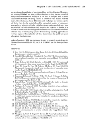
![278 C. Ehrhardt et al.
50. Center DM (2000) A role for the alveolar epithelium in recruitment of mononu-
clear cells into the lung. J Clin Invest 106(6):741–743
51. Thorley AJ, Goldstraw P, Young A, Tetley TD (2005) Primary human
alveolar type II epithelial cell CCL20 (macrophage inflammatory protein-
3alpha)-induced dendritic cell migration. Am J Respir Cell Mol Biol 32(4):
262–267
52. Burri PH (1984) Fetal and postnatal development of the lung. Annu Rev Physiol
46:617–628
53. Burri PH (2006) Structural aspects of postnatal lung development—alveolar for-
mation and growth. Biol Neonate 89(4):313–322
54. Adamson IY, Bowden DH (1974) The type 2 cell as progenitor of alveolar
epithelial regeneration. A cytodynamic study in mice after exposure to oxygen.
Lab Invest 30(1):35–42
55. Kotton DN, Summer R, Fine A (2004) Lung stem cells: new paradigms. Exp
Hematol 32(4):340–343
56. Neuringer IP, Randell SH (2006) Lung stem cell update: promise and controversy.
Monaldi Arch Chest Dis 65(1):47–51
57. Fuchs S, Hollins AJ, Laue M, Schaefer UF, Roemer K, Gumbleton M, Lehr CM
(2003) Differentiation of human alveolar epithelial cells in primary culture—
Morphological characterisation and expression of caveolin-1 and surfactant
protein-C. Cell Tissue Res 311:31–45
58. Wang J, Edeen K, Manzer R, Chang Y, Wang S, Chen X, Funk CJ, Cosgrove
GP, Fang X, Mason RJ (2007) Differentiated human alveolar epithelial cells and
reversibility of their phenotype in vitro. Am J Respir Cell Mol Biol [Epub ahead
of print] doi:10.1165/rcmb.2006-0410OC
59. Fehrenbach H (2001) Alveolar epithelial type II cell: defender of the alveolus
revisited. Respir Res 2(1):33–46
60. Mason RJ (2006) Biology of alveolar type II cells. Respirology 11 Suppl:S12-S15
61. Kim KJ, Borok Z, Crandall ED (2001) A useful in vitro model for transport studies
of alveolar epithelial barrier. Pharm Res 18:253–255
62. Crandall ED, Kim KJ (1982) Transport of water and solutes across bullfrog
alveolar epithelium. J Appl Physiol 52(4):902–909
63. Forbes B, Ehrhardt C (2005) Human respiratory epithelial cell culture for drug
delivery applications. Eur J Pharm Biopharm 60(2):193–205
64. Lieber M, Smith B, Szakal A, Nelson-Rees W, Todaro G (1976) A continuous
tumor-cell line from a human lung carcinoma with properties of type II alveolar
epithelial cells. Int J Cancer 17(1):62–70
65. Foster KA, Oster CG, Mayer MM, Avery ML, Audus KL (1998) Characterization
of the A549 cell line as a type II pulmonary epithelial cell model for drug
metabolism. Exp Cell Res 243(2):359–366
66. Elbert KJ, Schäfer UF, Schäfers HJ, Kim KJ, Lee VHL, Lehr CM (1999) Mono-
layers of human alveolar epithelial cells in primary culture for pulmonary absorp-
tion and transport studies. Pharm Res 16(5):601–608
67. Kobayashi S, Kondo S, Juni K (1995) Permeability of peptides and proteins
in human cultured alveolar A549 cell monolayer. Pharm Res 12(8):1115–
1119
68. Wang Z, Zhang Q (2004) Transport of proteins and peptides across human cul-
tured alveolar A549 cell monolayer. Int J Pharm 269(2):451–456
69. Forbes B, Wilson CG, Gumbleton M (1999) Temporal dependence of ectopepti-
dase expression in alveolar epithelial cell culture: implications for study of peptide
absorption. Int J Pharm 180(2):225–234
70. Anabousi S, Bakowsky U, Schneider M, Huwer H, Lehr CM, Ehrhardt C (2006)
In vitro assessment of transferrin-conjugated liposomes as drug delivery systems
for inhalation therapy of lung cancer. Eur J Pharm Sci 29(5):367–374](https://image.slidesharecdn.com/drugabsorptionstudies-2008-150610022529-lva1-app6892/85/Drug-Absorption-Studies-2008-296-320.jpg)
![Chapter 11 In Vitro Models of the Alveolar Epithelial Barrier 279
71. Duncan JE, Whitsett JA, Horowitz AD (1997) Pulmonary surfactant inhibits
cationic liposome-mediated gene delivery to respiratory epithelial cells in vitro.
Hum Gene Ther 8(4):431–438
72. Rehan VK, Torday JS, Peleg S, Gennaro L, Vouros P, Padbury J, Rao DS,
Reddy GS (2002) 1Alpha,25-dihydroxy-3-epi-vitamin D3, a natural metabolite
of 1alpha,25-dihydroxy vitamin D3: production and biological activity studies in
pulmonary alveolar type II cells. Mol Genet Metab 76(1):46–56
73. Newton DA, Rao KM, Dluhy RA, Baatz JE (2006) Hemoglobin is expressed by
alveolar epithelial cells. J Biol Chem 281(9):5668–5676
74. Zhang L, Whitsett JA, Stripp BR (1997) Regulation of Clara cell secretory pro-
tein gene transcription by thyroid transcription factor-1. Biochim Biophys Acta
1350(3):359–367
75. Shlyonsky V, Goolaerts A, van Beneden R, Sariban-Sohraby S (2005) Differ-
entiation of epithelial Na + channel function. An in vitro model. J Biol Chem
280(25):24181–24187
76. Woollhead AM, Baines DL (2006) Forskolin-induced cell shrinkage and apical
translocation of functional enhanced green fluorescent protein-human alphaENaC
in H441 lung epithelial cell monolayers. J Biol Chem 281(8):5158–5168
77. Wikenheiser KA, Vorbroker DK, Rice WR, Clark JC, Bachurski CJ, Oie HK,
Whitsett JA (1993) Production of immortalized distal respiratory epithelial cell
lines from surfactant protein C/simian virus 40 large tumor antigen transgenic
mice. Proc Natl Acad Sci USA 90(23):11029–11033
78. Douglas WH, Kaighn ME (1974) Clonal isolation of differentiated rat lung cells.
In Vitro 10(3–4):230–237
79. Koslowski R, Barth K, Augstein A, Tschernig T, Bargsten G, Aufderheide M,
Kasper M (2004) A new rat type I-like alveolar epithelial cell line R3/1: bleomycin
effects on caveolin expression. Histochem Cell Biol 121(6):509–519
80. Danto SI, Shannon JM, Borok Z, Zabski SM, Crandall ED (1995) Reversible
transdifferentiation of alveolar epithelial cells. Am J Respir Cell Mol Biol
12(5):497–502
81. Chen J, Chen Z, Narasaraju T, Jin N, Liu L (2004) Isolation of highly pure
alveolar epithelial type I and type II cells from rat lungs. Lab Invest 84(6):727–735
Erratum in: Lab Invest 85(9):1181 (2005)
82. Corti M, Brody AR, Harrison JH (1996) Isolation and primary culture of murine
alveolar type II cells. Am J Respir Cell Mol Biol 14:309–315
83. Goodman BE, Crandall ED (1982) Dome formation in primary cultured mono-
layers of alveolar epithelial cells. Am J Physiol 243:C96–C100
84. Shen J, Elbert KJ, Yamashita F, Lehr CM, Kim KJ, Lee VH (1999) Organic cation
transport in rabbit alveolar epithelial cell monolayers. Pharm Res 16(8):1280–
1287
85. Steimer A, Franke H, Haltner-Ukomado E, Laue M, Ehrhardt C, Lehr CM (2007)
Monolayers of porcine alveolar epithelial cells in primary culture as an in vitro
model for drug absorption studies. Eur J Pharm Biopharm [Epub ahead of print]
doi:10.1016/j.ejpb.2006.11.006
86. Bingle L, Bull TB, Fox B, Guz A, Richards RJ, Tetley TD (1990) Type II
pneumocytes in mixed cell culture of human lung: a light and electron microscopic
study. Environ Health Perspect 85:71–80
87. Ehrhardt C, Kim KJ, Lehr CM (2005) Isolation and culture of human alveolar
epithelial cells. Methods Mol Med 107:207–216
88. Cheek JM, Kim KJ, Crandall ED (1989) Tight monolayers of rat alveolar epithe-
lial cells: bioelectric properties and active sodium transport. Am J Physiol 256
(3 Pt 1):C688–C693
89. Morimoto K, Yamahara H, Lee VH, Kim KJ (1993) Dipeptide transport across rat
alveolar epithelial cell monolayers. Pharm Res 10(11):1668–1674](https://image.slidesharecdn.com/drugabsorptionstudies-2008-150610022529-lva1-app6892/85/Drug-Absorption-Studies-2008-297-320.jpg)
![280 C. Ehrhardt et al.
90. Matsukawa Y, Yamahara H, Lee VH, Crandall ED, Kim KJ (1996) Horseradish
peroxidase transport across rat alveolar epithelial cell monolayers. Pharm Res
13(9):1331–1335
91. Widera A, Kim KJ, Crandall ED, Shen WC (2003) Transcytosis of GCSF-
transferrin across rat alveolar epithelial cell monolayers. Pharm Res 20(8):1231–
1238
92. Bur M, Huwer H, Lehr CM, Hagen N, Guldbrandt M, Kim KJ, Ehrhardt C (2006)
Assessment of transport rates of proteins and peptides across primary human
alveolar epithelial cell monolayers. Eur J Pharm Sci 28(3):196–203
93. Effros RM, Mason GR (1983) Measurements of pulmonary epithelial permeabil-
ity in vivo. Am Rev Respir Dis 127(5 Pt 2):S59–S65
94. Matsukawa Y, Lee VH, Crandall ED, Kim KJ (1997) Size-dependent dextran
transport across rat alveolar epithelial cell monolayers. J Pharm Sci 86(3):305–
309
95. Van der Deen M, de Vries EG, Timens W, Scheper RJ, Timmer-Bosscha H,
Postma DS (2005) ATP-binding cassette (ABC) transporters in normal and patho-
logical lung. Respir Res 6:59
96. Lehmann T, Kohler C, Weidauer E, Taege C, Foth H (2001) Expression of MRP1
and related transporters in human lung cells in culture. Toxicology 167(1):59–72
97. Campbell L, Abulrob AN, Kandalaft LE, Plummer S, Hollins AJ, Gibbs A,
Gumbleton M (2003) Constitutive expression of p-glycoprotein in normal lung
alveolar epithelium and functionality in primary alveolar epithelial cultures.
J Pharmacol Exp Ther 304(1):441–452
98. Endter S, Becker U, Daum N, Huwer H, Lehr CM, Gumbleton M, Ehrhardt C
(2007) P-glycoprotein (MDR1) functional activity in human alveolar epithelial
cell monolayers. Cell Tissue Res [Epub ahead of print] doi:10.1007/s00441-006-
0346-6
99. Lehmann T, Torky AR, Stehfest E, Hofmann S, Foth H (2005) Expression of
lung resistance-related protein, LRP, and multidrug resistance-related protein,
MRP1, in normal human lung cells in long-term cultures. Arch Toxicol 79(10):
600–609
100. Mulugeta S, Gray JM, Notarfrancesco KL, Gonzales LW, Koval M, Feinstein SI,
Ballard PL, Fisher AB, Shuman H (2002) Identification of LBM180, a lamellar
body limiting membrane protein of alveolar type II cells, as the ABC transporter
protein ABCA3. J Biol Chem 277(25):22147–22155
101. Agassandian M, Mathur SN, Zhou J, Field FJ, Mallampalli RK (2004) Oxysterols
trigger ABCA1-mediated basolateral surfactant efflux. Am J Respir Cell Mol Biol
31(2):227–233
102. Mamchaoui K, Makhloufi Y, Saumon G (2002) Glucose transporter gene expres-
sion in freshly isolated and cultured rat pneumocytes. Acta Physiol Scand
175(1):19–24
103. Groneberg DA, Paul H, Welte T (2006) Novel strategies of aerosolic pharma-
cotherapy. Exp Toxicol Pathol 57 Suppl 2:49–53
104. Sloan JL, Grubb BR, Mager S (2003) Expression of the amino acid transporter
ATB 0 + in lung: possible role in luminal protein removal. Am J Physiol
284(1):L39–L49
105. Rothenberg ME, Doepker MP, Lewkowich IP, Chiaramonte MG, Stringer KF,
Finkelman FD, MacLeod CL, Ellies LG, Zimmermann N (2006) Cationic amino
acid transporter 2 regulates inflammatory homeostasis in the lung. Proc Natl Acad
Sci USA 103(40):14895–14900
106. Jin SN, Mun GH, Lee JH, Oh CS, Kim J, Chung YH, Kang JS, Kim JG, Hwang
DH, Hwang YI, Shin DH, Lee WJ (2005) Immunohistochemical study on the
distribution of sodium-dependent vitamin C transporters in the respiratory system
of adult rat. Microsc Res Tech 68(6):360–367](https://image.slidesharecdn.com/drugabsorptionstudies-2008-150610022529-lva1-app6892/85/Drug-Absorption-Studies-2008-298-320.jpg)
![284 S. Reichl and U. Becker
LCC Liquid-covered culture
MTT 3-(4,5-Dimethylthiazol-2-yl)-2,5-diphenyltetrazolium bromide
Papp Apparent permeability
P-gp P-glycoprotein
PHCl Pilocarpine hydrochloride
RCE Rabbit corneal epithelium
SIRC Statens Seruminstitut rabbit cornea
SV40 Simian virus 40
TEER Transepithelial electrical resistance
12.1. Introduction
Dosing substances to the eye is a technique performed since the start of civiliza-
tion. First eye cosmetics have been described around 1550 BC by the ancient
Egyptians. Later, eye preparations for women (e.g., containing belladonna
extracts) have been described during the Baroque period of Europe. Major
importance for eye medications was gained through the discovery of alkaloids
in the nineteenth century. Since then, eye medications always played a major
role in therapeutics.
Targeting drugs to the mammalian eye via topical application is a formidable
task and of major interest for ophthalmic research. A major advantage is the
easy application for the patient and considerably low systemic availability,
which might alleviate unwanted side effects. In addition, to opt for the ocular
route of drug delivery might avoid drug degradation caused by the first-pass
effect occurring in the liver. It is also possible to target drugs to several ocular
sites or tissues of interest [1], a technique that is generally applied in many
pharmaceutical applications.
Potential disadvantages of ocular drug delivery may include the rather low
applicable volume and rapid drainage of the drug solution through nasolacrimal
ducts, resulting in a low ocular bioavailability. Moreover, all pharmacopoeias
require a low irritant potential for eye medications. To assess the irritancy
of drugs and vehicles as well as acceptable transcorneal drug delivery, test
systems suitable for prescreening are needed.
For a long time, drugs have been tested using animals or animal tissues
as means to determine ocular delivery, toxicity, and eye irritation. But since
such animal-based testing is controversial and often met with resistance from
some sectors of the society, great efforts have been made into establishing
other more accepted and easier methods. One such effort resulted in the
introduction of cell culture techniques in the early part of the twentieth cen-
tury [2, 3]. Major drawbacks for animal-based testing are high maintenance
costs and efforts. Scientifically, complexity of the “test” system and high
variation of the outcome are major points for criticism. Last, but not least,
animal testing does have a bad reputation and low acceptance in the mod-
ern society. Therefore, lots of efforts have been put into the development of
cell culture systems that replace laboratory animals in scientific studies. This
chapter provides a thorough review about the efforts made on the rewarding
field of in vitro models suitable for studies of transcorneal drug delivery and
metabolism.](https://image.slidesharecdn.com/drugabsorptionstudies-2008-150610022529-lva1-app6892/85/Drug-Absorption-Studies-2008-302-320.jpg)
![286 S. Reichl and U. Becker
that is exposed to the external environment, joining the opaque sclera at the
corneoscleral limbus. The anterior corneal surface is covered by the tear film
while the posterior surface is directly bathed in the aqueous humor. Average
horizontal and vertical diameters of the human cornea are 12.6 and 11.7 mm,
respectively. The central cornea of the human eye is ∼520 µm thick, whereas
in the periphery a thickness of about 700 µm is found. The shape of the anterior
corneal surface is convex and nonspherical. Its average radius of curvature is
7.8 mm, providing ∼48 diopters for the human eye [4].
Microscopically, the cornea shows a rather simple and multilayered struc-
ture that can be divided into six layers: the epithelium, basement membrane,
Bowman’s layer, stroma, Descemet’s membrane, and endothelium. The corneal
tissue consists of three different cell types: epithelial cells, keratocytes (corneal
fibroblasts), and endothelial cells. The outermost corneal surface is covered
with the preocular tear film, which is functionally associated with the cornea.
The epithelial surface must be kept moist and smooth, a role played by the tear
film in conjunction with a spreading function of the eyelids during blinking
motions. Furthermore, the tear film provides a protection against infectious
agents that may gain access into the eye.
The human tear film is ∼7 µm thick and consists of three layers: the most
superficial lipid layer (∼0.1 µm), the underlying aqueous layer (∼7 µm),
which forms the bulk of the tear film, and the innermost mucous layer, which
is intimately attached to the corneal epithelium (0.02–0.05 µm). These three
different layers of the tear film are produced by various cells and glands in
the eye. The lipid layer is primarily secreted by the Meibomian glands located
in a row near the edge of upper and lower eyelids, while the aqueous layer
is produced by main and accessory lacrimal glands. The mucus is excreted by
conjunctival goblet cells. The normal tear volume is about 7 µl with an average
aqueous tear flow rate of about 1.2 µl/min (ranges from 0.5 to 2.2 µl/min) in
humans. The osmolarity of normal human tear film averages to be 302 mOsm/l.
The aqueous layer of the tear film contains many biologically important factors,
including electrolytes, glucose, immunoglobulins, lysozyme, albumin, and
oxygen [5].
The corneal epithelium (∼50 µm) represents about 10% of the thickness of
corneal tissue and is a nonkeratinized, noncornified, stratified, and squamous
epithelium. None of the other multilayered human epithelia show a more
regular structure than the corneal epithelium, which is composed of five to
six cell layers in total and three cell types: basal, wing, and superficial cells.
Only basal cells show a mitotic activity, in that during a differentiation process
(taking 7–14 days) the cells continue to move anteriorly and become superficial
cells before they disintegrate and desquamate into the tear film. Thus, corneal
epithelium continuously renews its surface. The presence of a centripetal move-
ment of corneal epithelial cells (the “XYZ” hypothesis) has been demonstrated
[6]. Furthermore, in this context, localization of epithelial stem cells at the
limbus (the region where the corneal epithelium graduates into transitional
epithelium and then into conjunctival epithelium) has been described [7–9].
At the corneal surface, two to four layers of polygonal, extremely flat, and
terminally differentiated superficial cells can be found. The cell diameters are
40–60 µm with about 2–6 µm thickness. The corneal surface is populated with
microvilli and microplicae, resulting in surface area enlargement. These struc-
tures are associated with the tear film. Superficial corneal epithelial cells are](https://image.slidesharecdn.com/drugabsorptionstudies-2008-150610022529-lva1-app6892/85/Drug-Absorption-Studies-2008-304-320.jpg)
![Chapter 12 Cell Culture Models of the Corneal Epithelium and Reconstructed Cornea Equivalents 287
joined by desmosomes and ribbon-like tight junctional complexes, surrounding
the entire cell at the most apical region [10]. Desmosomal attachments provide
lateral mechanical stability. However, presence of tight junctional complexes
(zonula occludens) prevents the movement of substances from the tear film
into intercellular spaces of the epithelium as well as the deeper layers of corneal
tissues. Tight junctions are designed to resist the flow of fluid (and other molec-
ular substances, e.g., pharmaceutical compounds) through the epithelial barrier.
Hence the epithelium is the main barrier for topically applied hydrophilic drugs
(see Sect. 12.3). It has been shown that the removal of corneal epithelium per
se results in a significant increase in transcorneal drug permeability [11].
The wing cells, possessing a characteristic wing-like shape, are arranged
in two to three layers. They are located between the basal and superficial
cells, exhibiting an intermediate state of differentiation. These cells have an
extensive cytoskeletal network consisting of intermediate filaments. The 64-
kDa keratin (K3) is specific to corneal epithelial cells [12]. Furthermore,
numerous desmosomes and gap junctions are present between wing cells, but
functional tight junctional proteins have not yet been established in this state of
differentiation.
The columnar, cuboidal basal cells are 18–20 µm in height and 8–10 µm
in diameter. They form a monolayer adhered to a basement membrane by
hemidesmosomes which are linked to anchoring fibrils of collagen type VII. At
the lateral borders, basal cells are extensively interdigitated and joined together
in places only by junctional complexes (zonula adherens), desmosomes, and
gap junctions.
The basement membrane, 40–60 nm in thickness, is produced by basal
epithelial cells and composed of the lamina lucida (adjacent to the cell mem-
brane of basal cells) and lamina densa where the main components include
collagen type IV, laminin, and fibronectin. As in other epithelial and endothe-
lial barriers, the basement membrane in the corneal epithelium seems to be
important for the maintenance of a multilayered, well structured epithelium
and its adhesion to the corneal stroma. The basement membrane may also play
a significant role in epithelial wound healing and re-epithelialization. Using the
rat cornea, Suzuki et al. [13] found that the presence of a basement membrane
may be required for the re-formation of intercellular junctions during wound
repair. This matches with findings made in the clinic during laser treatment
[14]. It was noticed that patients with basement membrane dystrophy show
a higher tendency for postsurgical sloughing of the corneal epithelium than
do patients with intact basement membrane. Furthermore, a careful literature
review done by Saika et al. [15] also supports the hypothesis of the importance
of the extracellular matrix and its metabolism in the repair process of the
corneal epithelium.
As another extracellular component in the cornea, the Bowman’s layer is
an acellular and amorphous band between the corneal epithelium and stroma.
The layer is about 8–12 µm thick and consists of randomly arranged colla-
gen fibers (types I and III) and proteoglycans. The physiological function of
Bowman’s layer is not yet completely understood, since not all animal species
exhibit this membrane in the corneal structures, but an important role in the
maintenance of the corneal epithelial structure is expected or probable, since
a damaged Bowman’s membrane usually results in scarring during wound
repair [16].](https://image.slidesharecdn.com/drugabsorptionstudies-2008-150610022529-lva1-app6892/85/Drug-Absorption-Studies-2008-305-320.jpg)
![288 S. Reichl and U. Becker
Stacked lamellae of collagen fibrils are the most obvious morphological
characteristics of the stroma, which comprises more than 90% of the whole
cornea. The stroma consists of about 78% water, an extracellular matrix, and
about 3 to 5% cellular components (keratocytes). Keratocytes, also known
as stromal or corneal fibroblasts, are spindle-shaped cells that are embedded
network-like in the stromal matrix. They produce the extracellular matrix that
mainly consists of collagen (predominantly type I and a minor content of
type V) and glycosaminoglycans (predominantly keratan sulfate, followed by
smaller fractions of dermatan sulfate and chondroitin sulfate proteoglycans).
Collagen constitutes about 71% of the corneal dry weight, contributing to
the corneal transparency and mechanical resistance against the intraocular
pressure. Stromal collagen fibers are remarkably uniform in their diameter. The
distance between collagen fibers is also remarkably constant. These regular
arrangements of collagen fibers as well as the “relatively” constant water
content in the stroma are essential requirements for corneal transparency. Any
disturbance in the uniformity of the fixed interfiber distance may cause a loss
of corneal transparency.
The Descemet’s membrane, which is also termed as “basement membrane
of corneal endothelium,” is a 7 µm thick assembly of collagen fibrils secreted
by endothelial cells. It is an acellular layer like the Bowman’s layer and mainly
consists of collagen type IV and laminin. Beneath the Descemet’s membrane,
corneal endothelial cells form a single layer of ∼400,000 cells with a total
thickness of about 5 µm. The endothelium represents the innermost surface
of cornea and acts as a barrier to the aqueous humor. Endothelial cells exhibit
predominantly a hexagonal shape and are ∼20 µm wide. The most impor-
tant physiological functions of the corneal endothelium are formation of an
effective barrier against the penetration of the aqueous humor into the cornea
and a pump function, therefore preserving the corneal transparency. Barrier
properties of the endothelial cell layer are mainly of the physiological aspect
and not considered critical for drug permeation. Because of the less pronounced
formation of tight cell–cell connections and also its low total thickness, the
corneal endothelium exhibits less resistance to substance diffusion than the
corneal epithelium. The basis for corneal clarity is an intact endothelial layer
with a cell density of 2,000–3,000 cells/mm2 in adults, ensuring the formation
of numerous cell–cell contacts (i.e., via tight junctions and gap junctions). The
presence of a pump mechanism afforded by Na+-K+-ATPase is also important
for the maintenance of the corneal clarity. Na+-pumps (about 3,000,000 per
cell [17]) are mainly localized at the lateral part of endothelial cell mem-
branes (i.e., facing intercellular spaces [18]). Damage or dysfunction of the
endothelium (e.g., by cell loss; so-called “endothelial decompensation” occurs
at a critical cell density below 500 cells/mm2), traumatic disease, reduction of
pump function or tight junctions, or both, will all lead to corneal edema (i.e.,
resulting in opacity of stroma). Thus, destruction of the corneal endothelium
is one of the major causes for corneal transplantation. Several studies focusing
on transplantation of cultured endothelial cells have been recently performed
[19, 20]. Current results in the field of corneal equivalents for transplantation
will be described in Sect. 12.4.3. More details about the anatomy and function
of the cornea are available in articles written by Nishida [21] and Klyce and
Beuerman [22]. A review on the ultrastructure of the human cornea written by
Beuerman and Pedroza [23] can also be consulted for details.](https://image.slidesharecdn.com/drugabsorptionstudies-2008-150610022529-lva1-app6892/85/Drug-Absorption-Studies-2008-306-320.jpg)
![Chapter 12 Cell Culture Models of the Corneal Epithelium and Reconstructed Cornea Equivalents 289
12.3. Transcorneal Drug Absorption into the Eye
Since most eye medications need to enter the eye for a pharmacological effect,
ocular drug delivery is a major issue in ocular research. Generally, three routes
into the eye have been described in the literature [24]:
1. Drug distribution in the eye through the blood vessels, either by systemic
dosing or by local treatment. Because the blood route always involves a
distribution of the drug in the whole human body, the ocular level of drugs
achieved by this way of application is much less than that achievable by
ocular topical treatment. Thus, this way of drug distribution is not preferred
for ocular dosing.
2. Drug entry into the eye region utilizing the conjunctival-scleral pathway,
with topical dosing. Even though this pathway can be of high efficiency for
intraocular drug delivery and does bypass cornea and local vasculature, only
a few (high molecular) substances were delivered using this route.
3. The transcorneal route. This is still the major pathway for ocular drug
delivery, representing the direct pathway into the eye and is applicable for
most of drug substances.
If the medical drug is applied topically to the eye, another important factor
for ocular drug delivery and penetration has to be taken in account: that is,
the precorneal clearance which rapidly reduces the drug amount available
for penetration. Thus, it is a major factor to take into consideration while
evaluating (transcorneal) absorption. So far, due to experimental limitations,
not much thought has been given to precorneal clearance in cell-based in vitro
models.
For absorption through the cornea, which is the major pathway for drug
absorption into the eye, certain physicochemical properties are required for
the substance to cross the corneal barrier. Since the cornea, as described
in Sect. 12.2, is divided into three parts with different characteristics, only
amphiphilic substances can easily penetrate all corneal layers. Purely lipophilic
substances, in contrast, will not face much hindrance in the epithelial and
endothelial cell layers but will be unable to cross the aqueous stroma. The same
considerations also apply for purely hydrophilic drugs that are not withheld
from penetration by the stroma but are unable to cross the lipid bilayers of
cells (i.e., cell plasma membranes) and tight junctions in the epithelium and
endothelium. Thus, the ability to switch between the lipophilic and hydrophilic
states is indispensable for ocular absorption. Other factors can also influence
ocular penetration, for example, charge and molecular weight, which have to
be taken into consideration when opting for the ocular route of drug delivery.
Generally, the main barrier in the cornea is represented by the corneal epithe-
lium. It is a multilayered epithelium with progressive states of differentiation
from basal to superficial cells. The superficial cells adhere to each other through
desmosomes and are surrounded by tight junctions [25], creating a tight cellular
barrier against xenobiotics and limiting permeation of hydrophilic substances.
Stroma and endothelium, on the other hand, do not represent a critical barrier
for transcorneal permeation, compared to the corneal epithelium. Only highly
lipophilic substances show limited diffusion due to the hydrophilic character-
istics of the stroma.](https://image.slidesharecdn.com/drugabsorptionstudies-2008-150610022529-lva1-app6892/85/Drug-Absorption-Studies-2008-307-320.jpg)
![290 S. Reichl and U. Becker
12.4. Corneal Cell Culture Models
For in vitro toxicity studies and assessment of the barrier function, drug trans-
port, cell physiology, and metabolism as well as the development of delivery
systems, cell culture models provide powerful systems for scientific research.
As the corneal epithelium is the main barrier for ocular penetration, various
corneal epithelial cell cultures were established besides the corneal constructs
that mimic the whole cornea and serve as reductionist models for the ocular
barrier. In general, two types of cell culture models are available: primary cell
cultures and immortalized, continuous cell lines.
12.4.1. Primary Corneal Cell Cultures
Since the cell culture technique was introduced in the early twentieth century,
many attempts have been undertaken to mimic the human organism in simpli-
fied in vitro cell systems. First approaches were performed with tissue explants,
which later led to more refined cell culture systems. Among the first tissues to
be examined and applied in cell culture was the cornea. With the development
of enzymatic isolation protocols in the 1960s [26, 27], cultivation of corneal
cells (covering epithelial, stroma, and endothelial cells) started in a greater
extent. Since the establishment of the 3R principle (Replace, Reduce, Refine)
in 1959 by Russell and Burch [28] and guidelines setup banning animal-based
drug tests by the cosmetics industry in the European Union in 2003, immense
efforts have been made to improve these cell cultures and substitute animal
experiments by suitable in vitro methods. A variety of corneal epithelial cell
cultures have therefore been established in recent years. These models were
developed in most part to serve as a tool for toxicity and eye irritation testing,
but in recent years some models for drug transport and permeation studies have
been made available as well. Besides the immortalized cell culture models that
have an extended life span and improved cell growth characteristics with easy
handling [29], various attempts have been made to establish primary corneal
epithelial cell cultures that better resemble the in vivo situation and have
features of original cell properties [30]. Different techniques have been applied
to generate primary cell culture models, including explantation techniques
[31, 32], enzymatic treatments [33, 34], and mechanical processing [35, 36].
Successful primary cultures of the corneal epithelium have been generated for
following species: rabbit [31, 33], cow [37], guinea pig [38], rat [39], mouse
[40], and pig [41]. Even though several attempts have been undertaken, a
successful setup of a human corneal epithelial cell model in a bigger scale
for permeation studies has not been achieved yet.
The main aspect of the primary cell cultures was to authentically mimic
the ocular surface in eye irritation and toxicity testing. In addition, issues of
fundamental research were addressed with these models as well, for example,
investigating cell proliferation in the cornea and location of the corneal stem
cells [42]. Two cell culture models of rabbit origin have mainly influenced
ocular research and cell culture efforts. For example, Kawazu et al. [43] were
among the first to introduce a primary corneal epithelial model that could
be grown in a larger scale. This cell culture system has been widely used
to examine permeation, drug absorption, and P-glycoprotein (P-gp) activity
[43–45]. The cultured cells morphologically resemble those found in intact](https://image.slidesharecdn.com/drugabsorptionstudies-2008-150610022529-lva1-app6892/85/Drug-Absorption-Studies-2008-308-320.jpg)
![Chapter 12 Cell Culture Models of the Corneal Epithelium and Reconstructed Cornea Equivalents 291
rabbit cornea, but a major drawback of the model is the lack of reasonable
electrical resistance of the cultured corneal barrier. In other words, a measure
for the tightness of the cell–cell connections or tight connections between cells
(thought to be established mainly by tight junctions) is very low. In an ideal
model, electrical resistance of cultured cell layers should be comparable to
excised corneal tissues. These primary cultured corneal cell layers do form
multiple cell layers, but only a maximum transepithelial electrical resistance
(TEER) of about 150 ohmcm2 has been reported. This value is generally con-
sidered insufficient for a tight cell layer, which usually exceeds 400 ohmcm2 in
tight cell culture models [46]. Another primary corneal epithelial cell culture
model is described by Chang and coworkers [33]. This latter corneal cell
culture model of rabbit origin was designed for in vitro drug permeation
studies and exhibits tight cell layers (TEER > 1,000 ohmcm2). The nature of
the cultured tight cell layer is also reflected from the apparent permeability
coefficients (Papp) reported by Chang et al. [33] and Scholz et al. [47], using
the same cell culture model. For hydrophilic mannitol and fluorescein, Papp
was 0.044 and 0.32 (×10−6 cm/s), respectively [33], while the more lipophilic
pilocarpine-HCl showed a higher Papp of 2.19 (×10−6 cm/s) [47], as expected.
The model was also used to study ion fluxes across corneal epithelium, where
Chang-Lin et al. [48] reported that the cultured rabbit corneal epithelial cell
layers exhibit active ion transport properties similar to those of excised rabbit
corneal tissues (focusing on Na+ absorption, Cl− secretion, and K+ exchange).
This model has thus been widely used and proved to be useful in scientific
research and pharmaceutical applications. An isolation and culture protocol for
rabbit primary corneal epithelial cells is described in the appendix (available
online) for this chapter. Isolation and culture of these cells are rather easy even
for novices in cell culture techniques.
12.4.2. Immortalized Continuous Cell Lines for Corneal Epithelial Cells
Since Gey et al. successfully established a human cell line derived from tumor
tissues (HeLa cells) in 1952 [49], the interest in human tissues as basis for
continuous cell lines was intense. Today, imitating the human organism as
closely as possible by means of human cell culture is still a major task. After
the hybridoma technique for fusing functional cells (e.g., B cells) with tumor
(e.g., myeloma) was discovered [50] and later refined by the development of
viral vectors for transfection and immortalization, the field of continuous cell
lines exploded in scope and usage. The extended life span and the improved
growth characteristics are considered major advantages of continuous immor-
talized cell lines. In the last decade, several continuous cell lines represent-
ing the corneal epithelium have been reported. Application topics of these
models include drug permeation, eye irritation testing, basic research, and
characterization.
12.4.2.1. Rabbit Corneal Epithelial Cell Line
The first immortalized cell line to gain major impact in the development of
corneal epithelial cell clones was the rabbit corneal epithelial cell line (RCE,
also known as K’RCE) introduced by Araki et al. in 1993 [51]. Corneal epithe-
lial cells from a New Zealand albino rabbit were transfected with an SV40-
vector. After transfection, the cells grew with cobblestone-like appearance
and showed stratification, desmosomes, microvilli, and some typical types of](https://image.slidesharecdn.com/drugabsorptionstudies-2008-150610022529-lva1-app6892/85/Drug-Absorption-Studies-2008-309-320.jpg)
![292 S. Reichl and U. Becker
keratin. The cell line has recently been used for drug transport studies [52].
These studies revealed that the cell line exhibits only low TEER values of about
150 ohmcm2, suggesting that RCE may not be suitable for drug permeation
studies due to lack of functionally tight barrier properties. This cell line also has
the disadvantage of being nonhuman and being derived from an albino rabbit.
The albino origin and thus the absence of pigments in the eye are sometimes
considered a blemish by investigators in the field. Still, this model paved the
way for the important human corneal epithelium (HCE-T) cell line. The RCE
cell line can be obtained from the European Cell Culture Collection (ECACC,
Cat. No. 95081046).
12.4.2.2. Statens Seruminstitut Rabbit Cornea
The Statens Seruminstitut rabbit cornea (SIRC) cell line was derived by M.
Volkert of the Statens Seruminstitut, Copenhagen, Denmark, from the cornea
of a normal rabbit in 1957. Little is recorded relating to the history of this
cell line for approximately the first 400 passages. It was originally used for
the cultivation of rubella viruses. In 1990, the cells were morphologically
characterized by Niederkorn and coworkers [53]. The SIRC cells do not exhibit
an epithelial, cobblestone-like morphology but do have a fibroblast phenotype,
which is rated as a major drawback of this particular culture system. In the
early 1990s, the cells were utilized for examination of their suitability for
drug transport studies by Hutak and coworkers [54]. Even though the reported
TEER values in this study were very promising (about 2,233 ohmcm2 on
day 8 in culture), recent attempts failed to reproduce these results [55; Becker
et al., in press]. Notwithstanding, the cell line has been used for uptake exper-
iments in some studies [55], in which no tight cell–cell connections may be
absolutely required. This cell line can be obtained from American Type Culture
Collection (ATCC, Cat. No. CCL-60).
There are many immortalized human corneal epithelial cell models that
gained high acceptance for various ocular studies (e.g., toxicity testing and
corneal epithelial transport; Table 12.1).
12.4.2.3. HCE-T Cells
The HCE-T model was established in 1995 by Araki-Sasaki and coworkers
[56]. It was formed by transfection of human corneal epithelial cells from a 47-
year-old female donor with a recombinant SV40-adenovirus, which lacked the
origin of viral replication (i.e., not able to produce viral particles). The cell line
has good growth characteristics and shows a cobblestone-like appearance. The
cell line is intensively used by Urtti and coworkers and represents a standard
Table 12.1 Immortalized human corneal epithelial cell culture models.
Cell model Supplier (or introduced by)
HCE-T (human corneal epithelium, SV 40) Araki-Sasaki [56]
Reconstituted human corneal epithelium Skinethic
EpiOcular-Model MatTek
Clonetics Human Corneal Epithelium Cambrex
HCE-T (10.014 pRSV-T) Gillette Medical Evaluation Laboratories
CEPI-17-CL 4 cell line Nestec
tet HPV16-E6/E7 transduced HCE Mohan [64]](https://image.slidesharecdn.com/drugabsorptionstudies-2008-150610022529-lva1-app6892/85/Drug-Absorption-Studies-2008-310-320.jpg)
![Chapter 12 Cell Culture Models of the Corneal Epithelium and Reconstructed Cornea Equivalents 293
tool for drug permeation, bioavailability prescreening, and toxicity assessment
[57–59]. The cell culture model exhibits tight cell layers under air-interface
conditions with TEER >400 ohmcm2. These findings were verified by drug
transport studies with mannitol (low-permeability marker) and rhodamine B
(high-permeability marker). The respective Papp values were 1.42 and 16.3
(×10−6 cm/s) [57]. This confirms that HCE-T cell layers discriminate well
between the hydrophilic and lipophilic substances (that are thought to cross the
epithelial barrier via passive diffusion) and do indeed form a tight barrier. It
should, however, be noted that some drawbacks of HCE-T system regarding
ABC-transporters have recently been reported [60].
12.4.2.4. Reconstituted HCE
Reconstituted HCE (order no. RHC/S/5) is available from Skinethic, Nice,
France. It consists of transformed human corneal epithelial cells immortalized
by Roger Beuerman from the Louisiana State University–Eye Center, New
Orleans, USA. This reconstituted human corneal epithelium forms a multi-
layered cell culture model that does not exhibit tight junctions and is therefore
unsuitable for in vitro drug transport studies. Main focus of the model is eye
irritation and toxicity testing and it is commonly used in this area with good
prediction qualities. Major advantage of this model is the high reproducibility
and uniform appearance. The commercial availability provides a ready-to-use
model that is easy to handle.
12.4.2.5. EpiOcular Cell Culture System
The corneal model provided by MatTek (order no. OCL-200) consists of
normal, human-derived epidermal keratinocytes which have been cultured to
form a stratified, squamous epithelium similar to that found in the cornea.
Thus, this cell model does not provide “real” corneal epithelial cells, but
cells derived from skin, which resemble corneal epithelial cell-like structures.
Certain cell culture conditions allow these cells to turn into a multilayered
structure which is corneal epithelium-like. In histological staining, clear
differences in cell structure and arrangement of the cell layers to corneal
epithelium can be seen. These differences can be construed as serious
drawbacks for the model. Similar to the Skinethic system discussed above, the
EpiOcular system provides a well-defined model whose main applications may
include toxicity testing and (partly) substitution of the Draize test. Even though
the model does not feature the signs of redness and tear flow, the number
of Draize test runs can be reduced. Using a corneal epithelial cell culture
as prescreening tool for drug substances, one can screen various eye-irritant
substances in an early development stage, thus reducing the number of animal
preparations (e.g., rabbit eye test).
12.4.2.6. Clonetics Corneal Epithelial Cell Line
The “Clonetics” cell line from Cambrex Bio Science represents a cultured
human corneal epithelial model (order no. CMS-2015; Cambrex Bio Science,
Walkersville, MD). The culture model was generated by isolation of cells
from normal human corneal tissues. The cells were then infected with an
amphotropic recombinant retrovirus containing HPV-16 E6/E7 genes to extend
the useful cell life span. The Clonetics cell model is a very recent entry into
the immortalized corneal cell line field, but it has been proved to be useful
for toxicity testing as well as in vitro drug permeation studies so far. Because
of its very recent introduction, further examinations have to be undertaken to](https://image.slidesharecdn.com/drugabsorptionstudies-2008-150610022529-lva1-app6892/85/Drug-Absorption-Studies-2008-311-320.jpg)
![294 S. Reichl and U. Becker
fully evaluate the benefits of this particular model for in vitro corneal drug
delivery research.
12.4.2.7. HCE-T (10.014 pRSV-T)
This human-derived cell line was established by Kahn and coworkers at the
Gillette Medical Evaluation Laboratories [61]. Further evaluation of the cell
line was performed by Kruszewski et al. [62]. This corneal cell culture was
generated through transfection with plasmid pRSV-T that contains the SV40
early region genes and the Rous sarcoma virus long terminal repeat. The cell
line has been found to exhibit a typical cobblestone morphology and optimal
growth characteristics. It was designated to serve as a model for toxicity testing
and tool to investigate corneal epithelial cell biology. The cell line is available
via ATCC (Cat. No. CRL-11515).
12.4.2.8. CEPI-17-CL 4
This cell line was developed by Nestec (Nestlé Research Center, Lausanne,
Switzerland) and described by Offord et al. [63]. Originally, this cell model was
used for testing toxicity and ocular irritation. The cell line also is intensively
used in organotypic cornea constructs (vide infra for details).
12.4.2.9. tet HPV16-E6/E7 Transduced HCE
This cell line was recently established by Mohan et al. [64]. Intensive testing
of this cell line regarding its suitability for pharmaceutical purposes has not
been performed yet. The cell line is highly interesting, however, because of
its controlled cell growth and differentiation capabilities. An integrated DNA
section allows cell growth to be “switched” on or off. Therefore, further exam-
ination of this cell line should be of major interest and importance for various
pharmaceutical researches.
Thus far, a wide array of useful cell culture models of the corneal epithelium
has been established. Many of these cell culture models focus on toxicity
testing and ocular irritation, but some cell layer models for drug permeation
studies are also available. Indispensable for successful drug penetration testing
is a cell layer that exhibits a tight epithelial barrier. This latter requirement of
tight barrier properties disqualifies some of the models that were established
as substitutes for the Draize test. At least two cell lines are available for
pharmaceutical studies and some newer models may qualify as a useful tool,
once they are characterized for their barrier properties.
12.5. Organotypic Equivalents
During the cultivation of differentiating cells, including corneal cells, an
approximation or recapitulation of the in vivo situation such as expression
of typical histological parameters was attempted by variation of culture con-
ditions at an early stage. With this approach, it has been shown that an
improvement of growth behavior and cell differentiation can be achieved by
using extracellular matrices that resemble the basement membrane, especially
type I collagen, or attachment factors, for example, laminin and fibronectin,
as growth substrates [33, 65]. A further adaptation or mimicking of in vivo
conditions was attempted by coculture of corneal epithelial cells with ker-
atocytes or endothelial cells [66]. Corneal epithelial cells were cocultivated](https://image.slidesharecdn.com/drugabsorptionstudies-2008-150610022529-lva1-app6892/85/Drug-Absorption-Studies-2008-312-320.jpg)
![Chapter 12 Cell Culture Models of the Corneal Epithelium and Reconstructed Cornea Equivalents 295
with primary cultures of keratocytes as well as cell growth on 3T3 feeder
layers [67, 68]. Typical characteristics of cells were found more frequently
by coculturing schemes. Furthermore, these systems as well as the use of
keratocyte-conditioned media allowed mechanistic investigations of cell–cell
interactions [69]. An improvement of barrier properties by coculturing with
keratocytes could be shown for permeation models of epithelial cells as well
[57]. Toropainen and coworkers showed that the TEER value was increased,
while the permeation rate of mannitol reduced in the keratocyte coculturing
schemes, suggesting an increased tightness of the cultured corneal epithelial
barrier. With air-interfaced culture (AIC) of several cells (e.g., dermal, nasal,
lung, and other epithelial cells) which are normally in contact with the air in
an in vivo setting, a stronger expression of in vivo phenotypic features was
also found in cultured cells. This has also been found to be applicable to the in
vitro cultivation of corneal epithelium, establishing AIC as a standard condition
for corneal epithelial cell cultures. Especially, certain histological markers for
the multilayered corneal epithelium, for example, K3 expression [70], and in
particular the formation of a tight barrier of corneal epithelial cells [33] could
be promoted. For example, Chang and coworkers [33] showed the necessity of
AIC conditions in their primary cultures of rabbit corneal epithelial cells for
developing tight epithelial cell layers. Toropainen et al. [57] also demonstrated
an increase in TEER values, when immortalized human corneal epithelial
cells were grown under AIC conditions in comparison to the liquid-covered
culture (LCC) method. It is now well accepted by the scientific community
that the formation of functional multilayered epithelial barriers for both corneal
epithelial cell models and organotypic constructs requires AIC conditions.
At the beginning of the 1990s, due to the more rapid expansion of tissue-
engineering methods, first important steps were laid down for development
of a three-dimensional (3-D) model of the cornea. Models composed of both
epithelial cells and stroma were established, which later led to more elaborate
constructs composed of all three cell types (i.e., epithelial cells, keratocytes,
and endothelial cells). The usage of such corneal equivalents is quite varied
in scope. The 3-D constructs can be applied in various ways: (a) investiga-
tions regarding cell–cell interactions, (b) a replacement for the Draize test
for toxicological studies, (c) in vitro permeation studies, and (d) transplan-
tation (profoundly challenging but envisioned to be a possibility in the near
future). The aims of studies were very different and the used cells (animal or
human, primary or immortalized cells) and methods are widely different. In
the following section, some of the models that gained major importance in the
development of organotypic constructs are described.
The first 3-D cornea equivalent was characterized in 1993 by Minami and
coworkers [35]. This in vitro corneal construct model, containing bovine pri-
mary cells, was developed by a stepwise reconstruction and final cultivation of
the whole construct under AIC conditions. This design was further modified
and used by many other groups in subsequent studies. First, pure primary
cultures of the different corneal cell types were established. The cultivation was
performed in permeable membrane inserts (e.g., made from nitrocellulose). A
stroma-biomatrix containing incorporated keratocytes was formed from gelled
type I collagen. Endothelial and epithelial cells were seeded and cultivated
on the top and bottom side, respectively, of the collagen gel matrix. After
reaching confluence, the levels of culture medium was adjusted in such a way](https://image.slidesharecdn.com/drugabsorptionstudies-2008-150610022529-lva1-app6892/85/Drug-Absorption-Studies-2008-313-320.jpg)
![296 S. Reichl and U. Becker
that the epithelial cell layers were further cultivated under AIC conditions.
The resulting construct possessed a three-layered structure with a multilayered
epithelium comparable to the in vivo situation. In addition, the 55/64-kDa
keratin pair was detected in the epithelium, indicating corneal epithelial cell
differentiation. However, no further pharmaceutical/bioelectrical investigations
were performed utilizing this bovine corneal equivalent. Thus, it remains
unclear to what extent the construct could be used (e.g., as permeation model).
Another 3-D cornea model, comprising rabbit primary cultures of epithe-
lial and stromal cells as well as mouse immortalized endothelial cells, was
described in 1994 by Zieske and coworkers [70]. They showed the influence of
endothelial cells on the formation of a tightly packed, multilayered epithelium
as well as the expression of laminin, type VII collagen, α6 integrin, keratin
K3, and α-enolase. Furthermore, their findings suggested that the formation of
an in vivo-like epithelium requires the cultivation of the 3-D corneal construct
under AIC conditions. By contrast, LCC methods of cultivating corneal equiv-
alents in the absence of endothelial cells failed to promote the expression of
differentiation markers and basement membrane components.
With the objective of creating an in vitro corneal model for an alternative
to the Draize test, Schneider and coworkers developed a 3-D cornea con-
struct, which also comprised all three cell types from fetal porcine primary
cultures [71]. In contrast to other studies, they cultivated this cell culture
model under LCC methods. They validated the cornea equivalent regarding
cell materials and culture conditions (especially the growth media). They
found an in vivo-like cell morphology as well as the expression of basement
membrane components like laminin and fibronectin. In addition, the formation
of glycosaminoglycans was detected in the artificial stroma. In another study,
they reported, using a modified MTT cytotoxicity test, that the data of five
chemical test compounds observed with this in vitro corneal model correlate
well with the Draize test classification [72]. This model was the first 3-D
organotypic cornea construct for in vitro toxicity studies and therefore seems
to be an interesting and useful approach for an alternative to the Draize test.
Unfortunately, no further investigations on this model are reported and thus its
usefulness as an in vitro cornel model for drug absorption studies remains to be
confirmed.
In another approach, Parnigotto and coworkers reconstructed corneal struc-
tures in vitro by using corneal stroma containing keratocytes to which corneal
epithelial cells from bovine primary cultures were overlaid [73]. However,
this particular corneal model did not contain an endothelial layer. This model
was histochemically characterized and the toxicity of different surfactants was
tested using MTT methods. This stroma-epithelium model has been reported
to show a cornea-like morphology, where a multilayered epithelial barrier
composed of basal cells (of a cuboidal shape) and superficial cells (of a flat-
tened shape) is noted. Furthermore, the formation of a basement membrane
equivalent and expression of the 64-kDa keratin were reported, indicating
the presence of differentiated epithelial cells. The toxicity data for various
surfactants obtained with this model correlate well with those seen by the
Draize test [73]. However, this corneal equivalent was not further validated
or used as a model for permeation studies.
In 1999, Germain and coworkers developed a corneal stroma-epithelium
equivalent without endothelial cell coculture, using human primary cultures](https://image.slidesharecdn.com/drugabsorptionstudies-2008-150610022529-lva1-app6892/85/Drug-Absorption-Studies-2008-314-320.jpg)
![Chapter 12 Cell Culture Models of the Corneal Epithelium and Reconstructed Cornea Equivalents 297
[34]. They isolated human epithelial cells from the limbus of human donor
corneas and cultivated them on a collagen gel containing keratocytes under
AIC conditions. This in vitro corneal model was investigated to exhibit an
in vivo-like morphology as well as expression of basement membrane com-
ponents when studied using histological and immunohistochemical methods.
Germain’s corneal equivalent possesses epithelial cell layers (four to five
layers), including cuboidal and basal cells. In addition, laminin, type VII
collagen, fibronectin, and β1α3,5,6 integrin subunits were detectable. Germain
and coworkers described their in vitro corneal model as a promising tool
for further physiological, toxicological, and pharmacological investigations
as well as a useful model for gene expression studies. Although Germain’s
group is known for its wide expertise in the field of tissue engineering
(in particular, corneal tissue engineering) [74, 75], toxicity or permeation
data using their model of stroma-corneal epithelium equivalent were lacking
to date.
The first 3-D corneal construct composed of all three cell types of human
cells has been described by Griffith and coworkers [76]. Each cellular layer
was fabricated from immortalized human cells that were screened for use on
the basis of morphological, biochemical, and electrophysiological similarity to
their in vivo counterparts. This in vitro human corneal model exhibits simi-
larities with respect to biophysical and physiological functions, morphology,
biochemical marker expression, transparency, ion and fluid transport as well
as gene expression profiles observed in vivo. In addition to the fact that a
whole human cornea was reconstructed in vitro for the first time, this human
corneal equivalent was entirely formed from several immortalized cell lines,
using HPV E6/E7. This maneuver allows the production of a great number of
cornea equivalents with much decreased variability due to the extended life
span of cells showing the same properties over many population doublings.
However, as is well known, immortalized cells may represent features of a
less differentiated state compared to primary cells in culture. Nonetheless, the
human corneal equivalent showed morphology similar to the in vivo features,
including well-defined epithelial, stromal, and endothelial cell layers. The
transparency of the artificial stroma should be pointed out as a big advantage,
as the transparency of corneal models is an absolute requirement for possible
transplantation. In contrast to previously developed corneal equivalents, the
stroma-biomatrix was formed from a collagen-chondroitin sulfate substrate
that was cross-linked by treatment with glutaraldehyde. The in vitro human
corneal model was cultivated under AIC conditions, similar to those corneal
equivalents mentioned above. Besides morphology, this particular construct
possesses a similar degree of stromal swelling as well as a physiologically
active endothelium, mimicking those observed in an in vivo setting. After
exposure to sodium dodecylsulfate, the construct showed a wound-healing
progress that was comparable to healing found in eye bank donor corneas in the
same experimental setup. Furthermore, this construct exhibits the same opaci-
fication after treatment with different surfactants, as found in rabbit and human
corneas. Thus, Griffith and coworkers described their model as a promising
tool for irritancy testing, wound healing, and other biomedical research (e.g.,
cell-matrix interactions), and as basis for the development of temporary or
permanent cornea replacements. Unfortunately, this human corneal construct
has not been tested further as an in vitro model for both toxicity and drug](https://image.slidesharecdn.com/drugabsorptionstudies-2008-150610022529-lva1-app6892/85/Drug-Absorption-Studies-2008-315-320.jpg)
![298 S. Reichl and U. Becker
absorption studies yet. However, Griffith’s group has acquired a strong exper-
tise in the field of corneal tissue engineering, particularly in the development
of stroma-biomatrices (e.g., collagen-based polymers) for both transplantation
and keratoprostheses [77–80]. Subsequent studies from Griffith and coworkers
have also been published, dealing with innervated human corneal equivalents
as in vitro models for studying interactions between nerve cells and their target,
that is, corneal epithelial cells [81, 82].
In the last decade, several investigations were performed with regard to
in vitro reconstruction of corneal epithelium for transplantation. Meanwhile,
many studies have also been reported, dealing with the cultivation and trans-
plantation of corneal epithelium grown on amniotic membranes (i.e., the
innermost membrane of the placenta), fibrin gels, or temperature-responsive
culture dishes [83–85]. However, none of these models has been examined for
suitability as a model for drug absorption studies.
Following the report by Griffith et al. [76–79] on the human 3-D cornea
model, two more human models were reported. One model was described as
an alternative to the Draize test [86, 87] while the other as an in vitro model for
permeation studies [88]. Both will be presented in more details in this section
due to their major impacts. Engelke and coworkers [86] developed an in vitro
corneal model that was also reconstructed using immortalized cells (in this
case, using SV-40). The cultivation was performed according to the methods
established by Minami et al. [35], using a type I collagen stroma-matrix
and AIC methods. This construct comprised the well-characterized epithelial
cell line HCE-T [56] (see Sect. 12.4.2), endothelial cell line HENC (estab-
lished by Bednarz and coworkers [89]), and keratocyte cell line HCK (gener-
ated in-house from primary cultures by pSV40-dN-Plasmid DNA-transfection
[86]). This cornea equivalent exhibited an in vivo-like morphology, includ-
ing multilayered epithelial cells. Engelke et al. [87] further investigated the
usefulness of their in vitro cornea model in toxicological studies. The quan-
tification of cell damage caused by different detergents was carried out via
confocal laser scanning microscopy using simultaneous vital dye staining
by calcein AM and ethidium homodimer-1. This human cornea equivalent
is still under investigation, regarding its suitability as an alternative to the
Draize test.
Three-dimensional cornea constructs, comprising all three cell types present
in corneal tissues in vivo and possessing an in vivo-like structure, were
described specifically as a model for in vitro permeation studies for the first
time by Müller-Goymann’s group [90]. First, a cornea construct from bovine
primary cultures in the style of that reported by Minami et al. [35] was estab-
lished and characterized. Similar techniques were then applied to develop a
porcine primary construct. The current construct is composed of a combination
of immortalized and native human cells. The bovine cornea construct exhibited
an in vivo-like morphology including a multilayered epithelium as well. In
addition to the typical corneal epithelial morphology, exhibiting basal cells of
cuboidal shapes and superficial cells of flattened architectures, the expression
of the 64-kDa keratin K3 was demonstrated [91]. The permeation through this
corneal construct of pilocarpine hydrochloride (PHCl) from either an array
of commercially available aqueous and oily eye drop solutions or an inverse
micellar solution (i.e., an oily solution containing an emulsifier that forms
inverted micelles with drugs trapped inside) was compared with permeation](https://image.slidesharecdn.com/drugabsorptionstudies-2008-150610022529-lva1-app6892/85/Drug-Absorption-Studies-2008-316-320.jpg)
![Chapter 12 Cell Culture Models of the Corneal Epithelium and Reconstructed Cornea Equivalents 299
coefficients from experiments using excised bovine corneas. Comparison of
permeation rates through the excised bovine cornea versus this in vitro cornea
construct model showed the same rank order for the different formulations.
Permeation coefficients obtained with this cornea construct were about twofold
to fourfold higher than those observed in the excised bovine cornea. Similar
results were also described for permeation studies including timolol maleate,
a frequently applied antiglaucoma drug [91]. The porcine cornea construct
appears to exhibit similar results on permeation of PHCl from different oph-
thalmic formulations in comparison to excised porcine cornea [92]. About
twofold to fourfold greater permeability indices for the porcine cornea con-
struct than those found in the excised porcine cornea were demonstrated
for different formulations of befunolol hydrochloride, another standard drug
in glaucoma therapy. Furthermore, a keratin pattern (mimicking the in vivo
cornea), basement membrane components (such as laminin and fibronectin) as
well as surface structures (microvilli and microplicae) like those found in the
in vivo setting were also observed in the porcine cornea construct [41, 93].
In order to avoid species-specific differences and get a more precise predic-
tion of transcorneal drug absorption into human eye, Müller-Goymann and
coworkers additionally developed a human cornea construct (HCC). The HCC
was reconstructed from native keratocytes and immortalized cells of corneal
epithelium and endothelium. The poor availability of human corneal materials
for isolating primary cells and a better standardization of the equivalent through
the use of immortalized cell lines both influenced the choice of the cellular
components. The corneal epithelial cell line, CEPI-17-CL 4, consists of SV-40
transfected human corneal epithelial cells and was established by Sharif and
coworkers [94]. This cell line is well characterized with respect to expression of
keratins, cytokines, growth factors, and cytochrome P450 enzymes and used for
ocular toxicity and inflammation studies [63]. The endothelial cell line HENC
established by Bednarz et al., already mentioned above, was used to form the
endothelial barrier in this HCC model. An in vivo-like morphology was found
in this HCC model. About 1.6-fold to 1.8-fold higher permeabilities were found
in this HCC model, compared to those observed in the excised porcine cornea,
when aqueous formulations of three model drugs with various physicochemical
properties (log Poctanol/water ranged between 1.6 and –1.3) were studied [88].
In another study, the permeation of six different ophthalmic drugs through the
HCC model and human donor corneas was evaluated [95]. The HCC model
showed a similar permeation behavior comparable to that found in the human
donor corneas for all substances studied and permeation coefficients varied by a
factor of 0.8 to 1.4, when compared to native human cornea. These data indicate
that Papp values correlate well with those of human corneas, even though some
substances did not match their Papp with that of the human cornea exactly. In
a follow-up permeation study using hydrocortisone acetate and p-nitrophenyl
acetate, the HCC was demonstrated to have esterase enzyme activities similar
to those observed in both the excised porcine cornea and human donor cornea.
Hence the HCC model could be used for permeation studies, including ester
prodrugs [96].
As can be seen above, it has been shown that cultivation of 3-D in vitro mod-
els of animal and human cornea is possible and the model resembles the in vivo
cornea of the animal and human. In order to use these cornea constructs as mod-
els for in vitro drug absorption studies and alternative to the Draize test, further](https://image.slidesharecdn.com/drugabsorptionstudies-2008-150610022529-lva1-app6892/85/Drug-Absorption-Studies-2008-317-320.jpg)
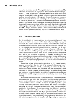
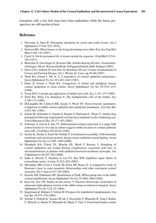
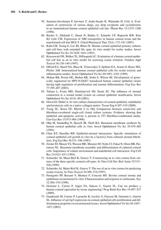
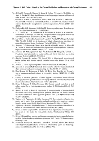
![308 H. J. Gukasyan et al.
L-NA NG-nitro-L-arginine
NO Nitric oxide
NOS Nitric oxide synthase
PD Potential difference
P-gp P-glycoprotein
RCEC Rabbit conjunctival epithelial cells
TEER Transepithelial electrical resistance
13.1. Introduction to the Ocular Surface and the Relative
Contribution of the Conjunctiva
The conjunctiva is a thin, mucus-secreting, relatively well vascularized (com-
pared to avascular cornea) tissue that covers ∼80% of the ocular surface. It
comprises the inner surface of the eyelids and is part of the anterior sclera
ending where the cornea begins. While the conjunctiva has been long thought
to play a simple protective role in the eye by functioning as a passive physical
barrier, it is an intricate and heterogeneous tissue which participates in the
maintenance of tear film stability due to the mucus secreted by its resident
goblet cells [1, 2]. Studies performed in recent years have characterized addi-
tional functional and practical features of the conjunctiva, that is, contributing
to the regulation of electrolyte and fluid balance in the microenvironment of
its mucosal surface as well as providing a unique conduit for drug delivery
to the anterior and posterior segments of the eye when drugs are applied
topically to ocular surfaces [3, 4]. The dynamic nature of conjunctiva was
demonstrated from the identification of several transport mechanisms for Na+
absorption from the conjunctival mucosa: Na+-glucose, Na+-amino acid, and
Na+-nucleoside cotransporters, in addition to the predominant and active Cl−
secretion. Permeability of the conjunctiva to a wide variety of hydrophilic
and lipophilic molecules and drugs was also characterized in the recent past,
which is detailed in a recent review [5]. Furthermore, multiple reports sub-
stantiate mechanisms of conjunctival ion transport, a key defining property of
this tissue. Pharmacological fingerprints of conjunctival ion transport include
significant reduction by the serosal application of bumetanide, an inhibitor
of Na+/K+/2Cl− cotransporter, the only known serosal entry pathway for
Cl− into conjunctival cells to date [6–8]. Net conjunctival Cl− secretion is
modulated by cyclic adenosine monophosphate (cAMP), protein kinase C, and
Ca2+, suggesting the presence of at least three mucosal Cl− exit pathways
[8]. Moreover, the existence and apical localization of CFTR (cystic fibrosis
transmembrane conductance regulator) was confirmed by electrophysiological,
molecular, biological, and immunocytochemical methods in the pigmented
rabbit conjunctival epithelium [9, 10]. By contrast, metabolic enzymes or bio-
chemical elimination mechanisms in the conjunctiva remain largely unknown.
In addition to acting as a highly specialized secretory epithelium within the
context of biochemical and metabolic capabilities, the conjunctiva also displays
esterase activity [11], and is likely to express isoforms of drug efflux pumps
(e.g., P-glycoprotein, P-gp) [12]. Lastly, the vectorial secretion of glutathione
(GSH, an abundant and ubiquitous antioxidant tripeptide) from conjuncti-
val epithelial cells has been functionally characterized in both the primary](https://image.slidesharecdn.com/drugabsorptionstudies-2008-150610022529-lva1-app6892/85/Drug-Absorption-Studies-2008-326-320.jpg)
![Chapter 13 The Conjunctival Barrier in Ocular Drug Delivery 309
Fornicial
conjunctiva
Bulbar
conjunctiva
Palpebral
conjunctiva
Sclera
Cornea
Lacrimal gland
Uppereyelid
Low
ereyelid
Anterior
segment
Posterior
segment
Figure 13.1 A cross-sectional
representation of the ocular surface,
highlighting the conjunctival tissue as a
thick black contour. Three distinct areas
of the conjunctiva are labeled along
with adjacent ocular tissues.
culture model and excised tissues of conjunctivas obtained from the pigmented
rabbit [13].
The ocular epithelium, obtained from other mammalian epithelial tissues
that serve as primary barriers from the outside environment, is atypical in
structure and function. Comprising relatively and proportionally a small area
of the total body surface, the ocular surface includes two continuous but very
different tissues of the conjunctiva and cornea. While the conjunctiva covers
most of the ocular surface area, only a small fraction (called the bulbar region)
covers the anterior sclera, which has an air interface exposed to the environment
during “open-eye” intervals (Figure 13.1). It continues through the fornicial
and palpebral regions as highly vascularized, thin/semitransparent, elastic,
and heterogeneous tissue (Figure 13.1). The cornea is a hydrophobic barrier
continuous with the bulbar conjunctiva (Figure 13.1), has a smaller relative
surface area, shows a clear transparency to light, exhibits a characteristic
high resistance to passive diffusion of ions and molecules, and withstands
against the intraocular pressure. Both the corneal and conjunctival epithe-
lial tissues are constantly subjected to light irradiation, atmospheric oxygen,
environmental chemicals, and physical abrasion. Each of these factors poses
stress of primarily oxidative nature that can ultimately contribute to ocu-
lar surface damage and disease, if left unchecked. Natural protective com-
ponents like water-soluble antioxidants (e.g., vitamin C, L-cyst(e)ine, GSH,
uric acid, pyruvate, and tyrosine), lipid-soluble antioxidants (e.g., tocopherols
and retinols), and highly specialized enzymes (e.g., superoxide dismutase,
catalase, and GSH peroxidase) have all been identified in human tear fluid
collected at normal and stimulated secretion rates and are thought to serve
as a frontline defense for the ocular surface tear film and underlying tis-
sues. While mechanisms or glandular sources of these antioxidants have not
been identified definitively, the conjunctiva may be a possible source due
to its dynamic properties of biochemical, electrophysiological, and secretory
functions.](https://image.slidesharecdn.com/drugabsorptionstudies-2008-150610022529-lva1-app6892/85/Drug-Absorption-Studies-2008-327-320.jpg)
![310 H. J. Gukasyan et al.
13.2. Trans- and Sub-Conjunctival Ocular Drug Delivery
Although the cornea is the primary route of drug entry into the eye follow-
ing topical administration, applied drugs also reach intraocular tissues by
conjunctiva–scleral pathway. About 1–5% of a topically applied drug dose
often reaches the anterior segment of the eye. Therefore, subsequent diffusion
of absorbed drugs to the posterior segment of the eye will often be relatively
insignificant. Eye drops are eliminated from the precorneal area within 90 s
and absorbed systemically by way of the highly vascular conjunctival stroma
and nasolachrymal ducts. Despite several anatomical hurdles for intraocular
drug delivery by the conjunctival pathway, a number of critical discoveries
have been reported which highlight the importance of this route. Nipradilol,
a β-blocker, and iganidipine, a Ca2+ antagonist, are good examples that have
been reported to reach the posterior segment of the eye in preclinical species,
respectively [5].
Improving the conjunctival drug permeability is one of the major challenges
in ocular drug delivery. Enhancing transcellular drug penetration by increasing
drug lipophilicity through the use of prodrugs or analogs or improving para-
cellular penetration by using enhancers to open tight junctions was reported
with some success. The transcellular route significantly contributes toward the
absorption of lipophilic drugs, but is less significant for hydrophilic species.
Propranolol, with a log partition coefficient between octanol and water (log PC)
of 3.21, is absorbed through cornea and conjunctiva up to tenfold greater than
a hydrophilic drug of similar size, for example, sotalol with a log PC of −0.62.
A sigmoidal relationship, reported in various other tissues and in vitro epithelial
cell culture systems, describes penetration of compounds through the conjunc-
tiva and cornea, in a manner that an increase in lipophilicity is seen with a
rise in permeation, followed by apparent saturation at high lipophilicity. Since
hydrophilic drugs penetrate primarily via the paracellular pathway between
epithelial cells through the tight junctions, their penetration area is extremely
small compared to the surface area offered by transcellular route for absorp-
tion of lipophilic species. However, most lipophilic drugs are prone to efflux
mechanisms afforded by pumps (e.g., P-gp) localized on the apical side of
conjunctiva [5].
Delivery of biologic pharmaceuticals via the conjunctival route has been
studied as an alternative to injections. Because of their hydrophilic nature,
peptide and protein drugs permeate across ocular epithelia predominantly
via the paracellular route. In conjunctival tissues, penetration through tight
junctions comprising the paracellular pathway is the rate-limiting step for
diffusion of macromolecule drugs. When this pathway is modeled to be pop-
ulated with equivalent pores having a cylindrical shape, analysis suggests
that conjunctiva may allow the passive restricted diffusion via such pores of
hydrophilic substances <20,000 Da. The theoretical radius of such equivalent
pores is predicted to be ∼5.5 nm [14]. In addition, an external enzymatic
barrier also restricts the penetration of peptide drugs across the conjunctiva,
where enkephalins, substance P, and insulin have been shown to significantly
degrade during penetration across the tissue when instilled to the mucosal
fluid of conjunctivas mounted in modified Ussing chambers [15–17]. Within
this context, the coapplication of protease inhibitors (e.g., camostat mesylate
and leupeptin) in the mucosal fluid afforded the absorption of an increased](https://image.slidesharecdn.com/drugabsorptionstudies-2008-150610022529-lva1-app6892/85/Drug-Absorption-Studies-2008-328-320.jpg)
![Chapter 13 The Conjunctival Barrier in Ocular Drug Delivery 311
fraction of intact vasopressin across the excised pigmented rabbit conjunctiva
[18]. Recent reports indicate that the conjunctival tissue may also be endowed
with endocytic vesicular routes for absorption of macromolecules [19, 20]. It
is important to note that the roles and their respective contributions of cave-
olae, clathrin-coated pits, and other subcellular vesicle-associated elements in
absorption/secretion of protein drugs remain ambiguous at best [5].
Most of the prominent carrier-mediated transport systems, which have been
extensively characterized in the gastrointestinal tract, have been reported for
ocular absorption of amino acids, small peptides, sugars, monocarboxylates,
organic cations, phosphates, bile acids, and several water-soluble vitamins
[5]. These carrier-mediated transport systems can contribute toward the ocular
absorption of mimetic drugs. Since paracellular transport of drugs in ocular
epithelia, especially the cornea and conjunctiva, is relatively limited in its
capacity, carrier-mediated drug transport systems endowed in the conjuncti-
val epithelium (which has a larger surface area than the cornea) offer great
advantages. Na+-coupled cotransport of D-glucose, L-arginine, nucleosides,
and monocarboxylates as well as proton-dependent dipeptide cotransport have
all been reported in the conjunctival mucosa [5]. Utilizing these transporters
for absorption of (pro)-drugs has been proven conceptually feasible in local,
ocular drug delivery. Nitric oxide synthase (NOS) inhibitors (e.g., NG-nitro-
L-arginine methyl ester, NG-monomethyl-L-arginine, and NG-nitro-L-arginine,
L-NA) are absorbed by the same active mechanism of the B0,+(ATB0,+) which
utilizes the essential amino acid L-arginine in the excised pigmented rabbit
conjunctiva [21, 22]. As nitric oxide (NO) plays a cytostatic or cytotoxic
role in the conjunctiva and anterior uveal tract, transconjunctival delivery of
NOS inhibitors is a practical strategy to alleviate symptoms associated with
NO overproduction. Since ATB0,+ is abundantly expressed in the mucosal
aspect of conjunctiva and recognizes almost all of naturally occurring neu-
tral and cationic amino acids as substrates, it may be a major drug delivery
facilitator to target by rational design approaches. Therefore, there are a wide
range of options for chemical modification of drugs through the application
of prodrug approaches. For example, herpes simplex virus inhibitor, valacy-
clovir, is transported as a substrate through mouse and human ATB0,+, in
various tissues including the eye. By virtue of analogous or similar amino
acid structures, ATB0,+ also affords the transport of acyclovir glutamic acid
γ -ester [23]. Dipeptide transporters known to play a key role in the oral
absorption of certain β-lactam antibiotics and angiotensin-converting enzyme
inhibitors are also present in the mucosal aspect of the conjunctiva (albeit
with relatively lower expression levels) [24–26]. Nucleoside transporters are
reported to be expressed in the conjunctival mucosa [27], offering the potential
for the targeting of nucleoside mimetic antiviral drugs. However, this class
of transporter proteins exhibit narrow specificity for substrates, which may
entail a comparatively narrow option for chemical design in targeted drug
delivery [5].
The use of polymeric nanoparticles in the eye has gained considerable inter-
ests in recent years. Ocular disposition, safety, efficacy, and pharmacokinetic
profiles of various nanoparticles offer a wide range of application for the
delivery of many drugs used to treat common ocular disorders. Polymeric
nanoparticles have been utilized to enhance the performance of ibuprofen and
cyclosporine, while reducing systemic side effect of carteolol compared with](https://image.slidesharecdn.com/drugabsorptionstudies-2008-150610022529-lva1-app6892/85/Drug-Absorption-Studies-2008-329-320.jpg)
![312 H. J. Gukasyan et al.
commercial aqueous eye drops [28]. Poly-D,L-lactide-co-glycolide nanoparti-
cles have been shown to be internalized (albeit direct proof of endocytosis is
lacking) in primary cultures of pigmented rabbit conjunctival epithelial cells
(RCEC) [19]. As alluded above, although endocytic mechanisms of nanopar-
ticles in the conjunctiva have not been fully characterized, this drug delivery
strategy may be a useful tool to apply in ocular delivery of novel therapeutics
through this tissue. The use of polymeric nano- and microparticles in subcon-
junctival route of ocular drug delivery offers a unique method for enhancing
local exposure level of drugs by maximizing and maintaining intraocular con-
centrations with minimal frequency of dosing. Compared to direct intravitreal
injection, this approach is less invasive, although it requires the rational formu-
lation design/small-scale engineering of both dimension and surface properties
of the drug. Subconjunctival injection of polylactide microparticles (3.6 µm)
led to a much higher budesonide concentration in retina and vitreous humor
over 14 days, compared to solutions of dosing nanoparticles of smaller (345
nm) size [29]. Collagen matrix and fibrin-based sealants provided a better
controlled release of cisplatin and carboplatin, compared to a simple drug solu-
tions, attaining higher drug concentrations after subconjunctival administration
of rabbits in several intraocular tissues [30]. Since the sclera, an immediate
underlying tissue, is significantly more permeable than the conjunctiva, physi-
cal and biological barriers of both cornea and conjunctiva are circumvented in
subconjunctival injections [5].
Iontophoresis is a relatively noninvasive technique that uses electric currents
to drive introduction of ionized drug substances into tissues or across the tissue
barriers. Transcorneal or transscleral/conjunctival iontophoresis has been uti-
lized in ocular drug delivery. Transcorneal iontophoresis effectively enhances
aqueous humor concentrations of drugs including gentamicin, tobramycin, and
vidarabine monophosphate, but falls short of delivering these therapeutics in
high enough concentrations to the posterior segment of the eye due to the
presence of the iris-lens barrier [31–35]. In contrast, transscleral/conjunctival
iontophoresis affords higher concentrations of ticarcillin, cefazolin, and gen-
tamicin in the vitreous humor as a function of applied current density and
duration [36]. Iontophoretic drug penetration heavily depends on the intensity
and duration of applied currents, perhaps two of the major limitations of
this technique from practical and commercial points of view. Recently, this
technique was successfully applied for intraocular gene delivery of fluorescein-
labeled anti-NOS oligonucleotides to the retina with an efficacy in the rat
model of endotoxin-induced uveitis, suggesting that transscleral/conjunctival
iontophoresis facilitates the topical penetration of intact oligonucleotides into
intraocular tissues [5].
13.3. An Overview of Conjunctival Disorders
Conjunctivitis, or “pink eye,” is an inflammation of the conjunctiva and prob-
ably the most widespread form of ailment in this tissue. If left untreated,
it can have serious consequences on vision and overall health. There are
many forms of conjunctivitis, and symptoms can range from itchy, burning,
or teary eyes (allergic conjunctivitis) to more severe cases of bacterial and
viral conjunctivitis (commonly known as “pink eye”). Each type requires](https://image.slidesharecdn.com/drugabsorptionstudies-2008-150610022529-lva1-app6892/85/Drug-Absorption-Studies-2008-330-320.jpg)
![Chapter 13 The Conjunctival Barrier in Ocular Drug Delivery 313
different treatment regimen and is highly contagious except for allergic con-
junctivitis. Because of the similarity of the symptoms, only medical doc-
tors trained well in ophthalmology can definitively distinguish between var-
ious types of conjunctivitis and could determine the appropriate course of
therapy [37].
A number of ocular surface disorders collectively termed as “Dry Eye
Syndromes” have also been associated with the conjunctiva. For example, a
deficiency and/or imbalance in compositions of the tear film is often found
on the ocular surface during keratoconjunctivitis sicca. Since the conjunctiva
plays a direct role in the maintenance of the tear fluid stability via secretion
of mucin [1] by its resident goblet cells [4] and basal fluid secretion driven
by electro-osmotic gradients across the tissue [3], the conjunctiva is a well
deserved, but not intensively studied, target of interest in research efforts aimed
against combating Dry Eye Syndromes.
Anatomical characteristics of disease in conjunctival inflammatory condi-
tions have been reported. In contrast to the bacterial form of conjunctivitis, viral
conjunctivitis shows a clear, watery discharge. The affected eye is irritated and
red with a swollen eyelid. This condition is also highly contagious and while
onset is in one eye it can quickly infect the other eye. There are a number of
viruses that can trigger this form of conjunctivitis. Some viral infection is a rel-
atively mild form that clears up on its own, while other viruses produce a more
severe form of the infection, such as herpes simplex conjunctivitis frequently
seen in children or herpes zoster conjunctivitis which can lead to serious eye
damage. Adenovirus (Ad) is the most frequent viral cause of epidemic ocular
infections. Ocular Ad infection may lead to disseminated disease, which can be
fatal in children or in individuals with acquired immunodeficiencies. A wealth
of knowledge about the relationship (that can be applied to improve treatment)
between Ad and human ocular tissues is made possible with the development
of cotton rat and rabbit models, Ad-induced pulmonary and ocular infections,
together with the availability of human Ad mutants [38–40]. The adenovirus
type 5 (Ad5) rabbit ocular infection model has been established for evalua-
tion of various antiviral and anti-inflammatory agents in Ad5-induced ocular
disease treatment [38].
13.4. Models for Studying Conjunctival
Transport Properties
13.4.1. Excised Conjunctival Tissues
The Ussing chamber technique, originally engineered to study transepithelial
ion transport, has been optimized to study drug transport and electrophysiology
of excised mammalian conjunctivas. This technique utilizes small tissue sheets
that are mounted between two compartments of mucosal and serosal aspects
of the isolated conjunctival tissue, which are bathed with a physiological
buffer that is continuously bubbled with a stream of gas (compressed air or air
balanced with 5% CO2). In addition to providing sufficient oxygen tension for
maintaining tissue viability, this configuration creates essential fluid circulation
in each reservoir. A schematic diagram of Ussing chamber approaches is shown
in Figure 13.2. Compounds of interest are added to either the mucosal or serosal](https://image.slidesharecdn.com/drugabsorptionstudies-2008-150610022529-lva1-app6892/85/Drug-Absorption-Studies-2008-331-320.jpg)
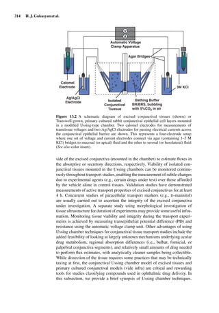
![Chapter 13 The Conjunctival Barrier in Ocular Drug Delivery 315
Conjunctival short-circuit current, termed Isc, describes the cumulative sum of
all active, energy-driven, and electrogenic transport processes occurring across
the conjunctiva. The Ussing chamber apparatus can impose an experimental
condition called “zero voltage clamp”, where the electrical PD developed
across the conjunctival tissue is forced to zero (i.e., offset to zero) by imposing
an electrical PD of the same magnitude but opposite sign. The resultant current
flowing across the tissue is termed “short-circuit current” (Isc), bringing down
the spontaneously developed PD to zero (thus the name short-circuiting is
used to describe the procedure). Under short-circuited conditions, there are no
electrical gradient across the tissue and by using the same Ringer’s solution
or culture medium on both sides of the barrier, the chemical gradients are
also zero. Thus, asymmetric fluxes of ions or solutes measured under such
zero gradient conditions across the barrier have to be energized by the barrier
itself (e.g., via utilization of cell energy in the form of ATP consumption; e.g.,
sodium pumps and/or vesicular transport dependent on cytoskeletal network-
based movements of cell organelles and so forth). Surgically excised pigmented
rabbit conjunctival tissues display a tear side negative PD of 15.5 ± 1.5
mV (mean ± S.E.M. of 70–80 determinations) when mounted in a modified
Ussing-type chamber [6–8, 41]. The voltage detection apparatus is composed
of two matched calomel electrodes positioned very closely to the tissue sur-
faces. A pair of Ag/AgCl electrodes is used to send and receive electrical cur-
rents (that are generated by imposing an electrical PD of the same magnitude
with opposite sign of the observed PD), which are positioned away from the
tissue surfaces in order to ensure uniform current density across the tissue
and short the bath-tissue-bath circuit (Figure 13.2). Overall tissue resistance
(transepithelial electrical resistance, TEER) can then be estimated by taking
the ratio between the observed PD and Isc, or TEER = PD/Isc (which describes
the Ohm’s law). Conjunctival TEER ranges from 0.75 to 1.5 k ·cm2 [6, 7].
A variety of automatic voltage clamp devices with special modifications
have been extensively utilized in electrophysiological studies of Isc in sev-
eral ocular tissues including the amphibian corneal epithelium [42] and
human fetal retinal pigment epithelium [43, 44], as well as non-ocular tissues
like the rat tracheal epithelium [45]. A strong temperature dependency and
inhibitory effect of serosally instilled ouabain on the rabbit conjunctival Isc
are characteristic of active ion transport driven by Na+/K+-ATPases in the
conjunctiva [6, 7].
Utilizing healthy conjunctival tissues under various ex vivo configurations
has allowed for mechanistic studies of potential drug transport routes. More-
over, it is just as feasible to surgically isolate conjunctivas from ocular disease
models for transport studies under pathological conditions with some limita-
tions. Ocular infection models have been established using competent human
Ad and thoroughly characterized in pigmented and albino rabbits [38, 39]. As
compared to excised conjunctival tissues in health or disease, in vitro cultiva-
tions of respective epithelial cells on permeable matrices under primary culture
conditions allow further investigative control on many additional dimensions.
Studies of the conjunctival epithelial barrier alone, i.e., excluding the partic-
ipation of systemic and lymphatic routes, provide a unique opportunity for
a greater throughput in simultaneous transport investigations that consume
minimal animal resources with lowest data variability [26].](https://image.slidesharecdn.com/drugabsorptionstudies-2008-150610022529-lva1-app6892/85/Drug-Absorption-Studies-2008-333-320.jpg)
![316 H. J. Gukasyan et al.
PIGMENTED
MALE
LIQUID COVERED
CONDITION
CULTURED CELLS ARE
PLACED UNDER
AIR-INTERFACE
CONDITION FROM DAY 4
AIR INTERFACE
CONDITION
COATED FILTERS
CULTURE MEDIUM
RESUSPEND CELLS IN
GROWTH MEDIUM
SEED ON TRANSWELLS®
40 µm CELL
STRAINER
FILTRATION
WASH
CELLS
EXCISE EYEBALL
FROM SOCKET &
SEPARATE THE
CONJUNCTIVA
TREATMENT WITH A
PROTEASE SOLUTION
HARVEST
CELLS
TR
EATM
EN
T
W
ITH
A
D
N
ase
SO
LU
TIO
N
SACRIFICE
Figure 13.3 A flowchart illustrating various steps for the preparation and maintenance
of primary rabbit conjunctival epithelial cell layers, cultured under liquid-covered and
air-interfaced conditions (See also color insert).
13.4.2. Primary Culture Models of Conjunctival Epithelial Cell Layers
Cell cultures are a useful, high throughput means for evaluating drug transport
mechanisms, impact of formulations, and physiological factors influencing
absorption across biological barriers (e.g., epithelia). Functional primary cul-
tures of RCEC under conventional liquid-covered or advanced air-interfaced
culture (AIC) conditions are available (Figure 13.3) [26, 46]. Conjunctival
epithelial cell cultures exhibit morphological as well as functional similarities
to the tissue in vitro or in vivo. Cultured cells form functional tight junc-
tions and express various enzymes (e.g., esterases, phosphodiesterases, and
GSH metabolizing enzymes) and transporters (for amino acids, dipeptides, and
nucleosides), similarly to those found in tissues in vitro or in vivo. Primary iso-
lates of conjunctival epithelial cells achieve differentiated phenotypes and form
multilayer of cells, displaying epithelial characteristics with isles of mucin-
secreting goblet cell populations. Culture time-dependent organization of tight
junctions, development of cell polarity, and expression of various transport
mechanisms have been reported [26, 46]. Test compounds are typically added
to the apical side of the multilayer of cultured cells, while the cumulative
appearance in the basolateral compartment is measured using appropriate mea-
surement schemes (e.g., radioactivity assays or fluorescence determinations
depending on the tags on the drugs of interest). Evaluation of in vitro sys-
tem integrity is performed through measuring electrical resistance across the
multilayered cells cultured on permeable supports (e.g., TranswellTM systems)
using specially engineered voltage-sensing and current-passing electrodes,
attached to a portable voltohmeter (e.g., MilliCell device, Millipore Corp.,
Billerica, MA, or EVOM device, World Precision Instruments, Sarasota, FL).](https://image.slidesharecdn.com/drugabsorptionstudies-2008-150610022529-lva1-app6892/85/Drug-Absorption-Studies-2008-334-320.jpg)
![Chapter 13 The Conjunctival Barrier in Ocular Drug Delivery 317
An alternative method for assessing cell layer integrity is through the use
of hydrophilic paracellular transport markers (e.g., radiolabeled D-mannitol
or fluorescein-Na+), which passively traverse cells by the paracellular route.
Small amounts of compound required for in vitro conjunctival cell culture
transport experiments make this approach well suited for screening purposes.
Relative absorption index of a series of pharmacologically active molecules
can be ranked against known markers for the identification of candidates with
potential absorption problems, which is a reliable tool to select drug candidates
with optimal characteristics.
13.4.3. Conjunctival Disease Models
Understanding differences in drug transport and metabolism in diseased con-
junctival tissue is also important. A number of critical and physiological char-
acteristics of conjunctival epithelial cells are altered in inflammatory states.
An Ad5 infection model of the conjunctiva has been developed for utility
in studying modulations of transport mechanisms in health and disease [38].
This virus-inoculated model has been adapted for tissue studies in the Ussing
chamber configuration, as well as for primary cultures of conjunctival epithelial
cells. The Ad5 infection model is a pertinent and critical tool for understanding
the ophthalmic drug delivery paradigm across the conjunctival tissue in (health
and) disease. Detailed protocols for this and other related subjects described
above are included as appendix to this chapter.
13.5. Summary and Conclusions
The conjunctival tissue covers ∼80% of the ocular surface in higher mam-
malian species and offers a unique route of delivering drugs locally to the
eye. Paracellular, transcellular, active, and endocytic routes of drug absorp-
tion play a key role in topical ocular delivery via the conjunctiva. Transscle-
ral/conjunctival iontophoresis offers an alternative noninvasive topical delivery
strategy for charged drugs including macromolecular biologics. Lastly, the
subconjunctival route of drug delivery provides critical space and capacity for
minimally invasive, safe, and sustained exposure of therapeutics to intraocular
compartments. The conjunctiva plays an important role in the etiology of a
number of ocular surface disorders as potential drug targets and an absorption
conduit. Multiple methods (see details in appendix) are available to study the
preclinical biopharmaceutics of the conjunctiva, facilitating physiological traits
of the conjunctiva, and improving the design of ocular drug delivery strategies
to the eye in health and disease.
Acknowledgments: Supported by grants HL38658 (KJK), HL64365 (KJK and
VHLL), EY12356 (VHLL), and the Hastings Foundation. HJG thanks the
AFPE, PhRMA, and USC Town and Gown. The authors appreciate contribu-
tions by scientists who have worked on conjunctival transport in collaboration
or directly in our laboratories (in the alphabetical order of last names): Drs P.
Ashton, S. K. Basu, E. Hayakawa, Y. Horibe, K. I. Hosoya, S. D. Kashi, U. B.
Kompella, A.A. Kulkarni, M. A. Narawane, M. G. Qaddoumi, J. R. Robinson,
P. Saha, M. H. Shiue, R. E. Stratford Jr., L. Sun, H. Ueda, W. Wang, and J. J.
Yang.](https://image.slidesharecdn.com/drugabsorptionstudies-2008-150610022529-lva1-app6892/85/Drug-Absorption-Studies-2008-335-320.jpg)
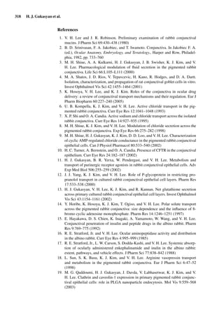

![322 K.-i. Hosoya and M. Tomi
GSH Gluthathione
HBSS Hank’s balanced salt solution
Kin,retina Apparent retinal influx clearance
LAT L-type amino acid transporter
MCT Monocarboxylate transporter
MRP Multidrug resistance-associated protein
OAT Organic anion transporter
Oatp Organic anion-transporting polypeptide
PAH p-aminohippuric acid
RPE Retinal pigment epithelial cells
RUI Retinal uptake index
RVEC Retinal vascular endothelial cells
TAUT Taurine transporter
tsA58 SV 40 Temperature-sensitive Simian virus 40
Vd Apparent retina-to-plasma concentration ratio
Vi Vascular volume of the retina
14.1. Introduction
This chapter will describe the inner blood–retinal barrier (BRB) transport
methodology used for in vitro, ex vivo, and in vivo studies. Transporters
expressed in the inner BRB and drug delivery to the retina will also be dis-
cussed. The retina is a thin tissue (∼0.5 mm in thickness) which lines the
interior of the posterior globe of the eye, is highly organized as about ten
layers, and plays an important role in vision. Retinal diseases (e.g., age-related
macular degeneration, diabetic retinopathy, glaucoma, and retinitis pigmen-
tosa) accompany severe vision loss and pose a serious socioeconomic burden.
The treatment of these retinal diseases is challenging, since the ocular barriers
that effectively protect the eye from foreign materials also hinders efficient
absorption of pharmaceuticals.
In addition, the BRB maintains a constant milieu and provides shields for
the neural retina from the circulating blood. The BRB forms complex tight
junctions of retinal capillary endothelial cells (inner BRB) and retinal pigment
epithelial cells (RPE; outer BRB) (Figure 14.1) [1, 2]. The inner two thirds
of the human retina is nourished by retinal capillaries and the remainder is
covered by choriocapillaris via the outer BRB [3]. The BRB is thought to play
an essential role in supplying nutrients to the neural retina to maintain visual
functions. It must be responsible for the efflux of neurotransmitter metabolites
from the retina; otherwise, metabolites are accumulated in the retina. Most
of current understanding of BRB functions is primarily based on in vivo data
obtained using the retinal uptake index (RUI) method, where a rapid intrac-
arotid injection of test substrates followed by sampling from the retina was
used extensively to assess the permeability properties of the rat BRB [4–7].
For example, Törnquist and Alm reported that transport of D-glucose, amino
acids, and L-lactate from the blood to the retina across the BRB takes place
via carrier-mediated processes [5–7]. In relation to this maneuver, we recently
developed an in vivo microdialysis method to evaluate the efflux transport of
test substrates across the rat BRB by monitoring the vitreous concentration of
a test substrate and bulk flow marker in rat vitreous humor [8].](https://image.slidesharecdn.com/drugabsorptionstudies-2008-150610022529-lva1-app6892/85/Drug-Absorption-Studies-2008-340-320.jpg)
![Chapter 14 Inner Blood–Retinal Barrier: Transport Biology and Methodology 323
Figure 14.1 Schematic diagram of the blood–retinal barrier (BRB). The retinal cell
layers seen histologically consist of retinal pigment epithelium (RPE); photoreceptor
outer segments (POS); outer limiting “membrane” (OLM); outer nuclear layer (ONL);
outer plexiform layer (OPL); inner nuclear layer (INL); inner plexiform layer (IPL);
ganglion cell layer (GCL); nerve fiber layer (NFL); inner limiting “membrane” (ILM).
Unfortunately, information specific to each barrier (i.e., the inner BRB and
outer BRB) and details of the transport mechanisms are difficult to obtain using
the in vivo approaches described above. Therefore, in vitro models of retinal
endothelial cells (i.e., the inner BRB) and RPE (i.e., the outer BRB) have
been developed by us and others. Although many RPE models for the primary
culture of different species [9–11] and the human RPE cell line (ARPE-19)
[12] have been used to investigate ion and solute transport properties, the
use of primary cultures of retinal capillaries is not so easy due to the very
small amount of cells/tissues that can be obtained from a single eye of the
experimental animal. For example, only 170–250 µg protein (capillary) can be
obtained from a single bovine eye [13]. In order to circumvent such paucity of
cells/tissues that can be obtainable from the eye of the experimental animal(s),
conditionally immortalized cell lines for rat retinal capillary endothelial cells,
pericytes, and Müller cells have been successfully developed and used for the
acquisition of information involving transport, metabolism, and other cellular
functions (transporter gene regulation and cell-to-cell interaction) [14–18]. In
addition, for the purpose of measuring reliably the expression levels of various
transporter genes in the inner BRB in vivo, we have used magnetic beads coated
with anti-rat CD31 antibodies to isolate rat retinal vascular endothelial cells and
quantify transporter gene levels in the inner BRB using real-time quantitative
polymerase chain reaction (PCR) analysis [19]. This latter ex vivo approach
provides specific information on expression levels for transporter genes in the
inner BRB.](https://image.slidesharecdn.com/drugabsorptionstudies-2008-150610022529-lva1-app6892/85/Drug-Absorption-Studies-2008-341-320.jpg)
![324 K.-i. Hosoya and M. Tomi
14.2. Conditionally Immortalized Cell Lines as a Novel in
Vitro Inner Blood–Retinal Barrier Model (Uptake Studies)
A transgenic rat harboring the temperature-sensitive Simian virus 40 (tsA58
SV 40) large T-antigen (tsA58 Tg rat) has been developed as a source of
conditionally immortalized cell lines [20]. Several conditionally immortalized
cell lines with differentiated functions, such as brain capillary endothelial cells
(TR-BBB), brain pericytes (TR-PCT), astrocytes (TR-AST), choroid plexus
epithelial cells (TR-CSFB), syncytiotrophoblasts (TR-TBT), small intestine
epithelial cells (TR-SIE), and lymphatic endothelial cells (TR-LE) have all
been derived from tsA58 Tg rats [21–23]. The tsA58 Tg rats have several
advantages as far as establishing immortalized cell lines are concerned (see
other reviews [1, 24]). The tsA58 SV 40 large T-antigen gene is stably
expressed in all tissues, and cultured cells can be easily immortalized by
activation of the SV 40 large T-antigen at 33◦C. The activated large T-antigen is
thought to induce cell proliferation by interacting with the retinoblastoma gene
products and p53, which normally regulate cell proliferation and inactivate the
growth-suppressive function of these two proteins [24].
The retinal capillary endothelium is surrounded by pericytes and Müller cell
foot processes as shown in Figure 14.1. The overall retinal microvascular biol-
ogy is thought to be governed, if not entirely, by paracrine interaction between
endothelial cells, pericytes, and Müller cells. In order to provide mechanistic
information on how the transporters function as well as paracrine cell-to-cell
interactions take place, we have established conditionally immortalized cell
lines for rat retinal capillary endothelial cells (TR-iBRB), retinal pericytes
(TR-rPCT), and Müller cells (TR-MUL) from tsA58 Tg rats (Figure 14.2A)
[14–16, 18]. TR-iBRB, TR-rPCT, and TR-MUL cells express typical mark-
ers for endothelial, perycyte, and Müller cells, respectively [14–16]. TR-
iBRB cells were shown to express facilitative glucose transporter 1 (GLUT1,
Slc2A1), P-glycoprotein (ABCB1), creatine transporter (CRT, Slc6A8), and
L-type amino acid transporter 1 (LAT1, Slc7A5), which are expressed in
the inner BRB in vivo. Accordingly, TR-iBRB cells maintain certain in vivo
functions and are considered to be a suitable and useful in vitro model for the
inner BRB [1, 14, 25, 26].
For uptake studies, TR-iBRB cells are seeded on rat tail collagen type I-
coated 24-well plates at a cell density of 1 × 10⁵cells/cm2. Typically, cells
are cultured at 33◦C for 48 h to reach confluence for uptake studies. Upon
confirming confluence using an inverted microscope under phase-contract
optics, cells are washed with 1 mL extracellular fluid (ECF) buffer consisting
of 122 mM NaCl, 25 mM NaHCO3, 3 mM KCl, 1.4 mM CaCl2, 1.2 mM
MgSO4, 0.4 mM K2HPO4, 10 mM D-glucose, and 10 mM HEPES (pH 7.4)
at 37 ◦C. After washing, uptake is initiated by incubating cells with a suitable
ECF buffer containing an appropriate concentration of the test substrate (e.g.,
[3H]- or [14C]-labeled compounds). In order to test if the test substrate is
taken up into the cells via transporter(s), uptake studies are carried out in
the presence of various concentrations of unlabeled substrates and/or poten-
tial competitors/inhibitors/stimulators. Uptake studies are also carried out at
4◦C, in the presence of metabolic inhibitors, or in the presence or absence
(or substitutions) of any particular ion(s) that are required for ion-coupled](https://image.slidesharecdn.com/drugabsorptionstudies-2008-150610022529-lva1-app6892/85/Drug-Absorption-Studies-2008-342-320.jpg)
![Chapter 14 Inner Blood–Retinal Barrier: Transport Biology and Methodology 325
Figure 14.2 Phase contrast microscopic images of conditionally immortalized cells
forming the inner blood–retinal barrier (A) and time-course of [3H]adenosine uptake
by TR-iBRB cells (B). A: Conditionally immortalized rat retinal capillary endothelial
cell line; TR-iBRB, retinal pericyte cell line; TR-rPCT and Müller cell line; TR-MUL.
B: The [3H]adenosine (14 nM) uptake was performed at 37◦C in the presence (closed
circle) or absence (open circle) of Na+.
transport process(es) in the ECF buffer. Uptake is usually measured for the
range of 10 s–10 min at 37◦C (Figure 14.2B), in order to ensure the kinet-
ics of uptake is within a linear range, which makes the determinations of
the kinetic parameters for uptake meaningful. After a predetermined period,
uptake is terminated by removing the solution, followed by immersing cells
in ice–cold ECF buffer to stop uptake process(es). The cells are then sol-
ubilized in 1 N NaOH and subsequently neutralized with 1 N HCl. Cell-
associated radioactivity and protein content are assayed by liquid scintillation
spectrometry and detergent compatible protein assay (a DC protein assay
kit, Bio-rad, Hercules, CA) with bovine serum albumin used as a standard,
respectively.
An example of this analysis method is shown in Figure 14.2B for
[3H]adenosine, which is taken up into TR-iBRB cells. [3H]Adenosine uptake
increases linearly for at least 5 min in the presence or absence of Na+.
Concentration-dependent adenosine uptake is known to take place with](https://image.slidesharecdn.com/drugabsorptionstudies-2008-150610022529-lva1-app6892/85/Drug-Absorption-Studies-2008-343-320.jpg)
![326 K.-i. Hosoya and M. Tomi
a Michaelis-Menten (Km) constant of 28.5 µM in a nitrobenzylmercap-
topurine riboside-insensitive manner, suggesting that ENT2 is involved in
[3H]adenosine uptake by TR-iBRB cells. Quantitative real-time PCR data
indicate that ENT2 mRNA is predominantly expressed in TR-iBRB cells [27].
14.3. In Vivo Blood-to-Retina Influx Transport
14.3.1. Integration Plot Analysis
The primary objective of integration plot analysis is to analyze the data on
influx of the test substrate from the circulating blood to the retina (i.e., blood-
to-retina direction) across the BRB after intravenous administration of the test
substrate. The advantage of this approach is that it allows reliable determination
of the retinal uptake (i.e., clearance) of the test substrate which has a slow
permeability across the BRB [28]. On the other hand, due to the intravenous
injection, interference by endogenous substrates and plasma–protein binding
of the test substrate can produce an unseemingly low estimate of the retinal
uptake.
The apparent retinal influx clearance, Kin,retina, expressed as mL/(min · g
retina), of the test substrate labeled with either [3H] or [14C] from the cir-
culating blood to the retina is determined by integration plot analysis. In
brief, rats are anesthetized, followed by injection of the test substrate (e.g.,
an [3H]-labeled compound, about 10 µCi/head) into the femoral vein. After
collection of plasma samples, rats are decapitated and the retinas removed.
The retinas are dissolved in 2 N NaOH and subsequently neutralized with 2 N
HCl. The radioactivity of retinal cell lysates is measured by liquid scintillation
spectrometry. As an index of the retinal distribution characteristics of the radi-
olabeled test substrate, the apparent retina-to-plasma concentration ratio (Vd)
as a function of time is used. This ratio [Vd(t)] (mL/g retina) is defined as the
amount of [3H] per gram retina divided by that per milliliter plasma, calculated
over the time-period of the experiment. The Kin,retina can be described by the
following relationship:
Vd(t) = Kin, retina ×
AUC(t)
Cp(t)
+ Vi (14.1)
where AUC(t) (dpm · min/mL), Cp(t) (dpm/mL), and Vi (mL/g retina) rep-
resent the area under curve showing time course of the plasma concentration
of the [3H] test substrate from time 0 to t, the concentration of plasma [3H]
test substrate at time t, and the rapidly equilibrated distribution volume of the
[3H] test substrate in the retina, respectively. Vi is usually comparable with the
vascular volume of the retina, while Kin,retina can be obtained from the initial
slope of a plot of Vd(t) versus AUC(t)/Cp(t), as an integration plot.
An example of this integration plot analysis using [3H]adenosine is
shown in Figure 14.3A. The estimated Kin,retina of [3H]adenosine is
2.58 × 10⁻² ± 0.07 × 10⁻² mL/(min · g retina) (mean ± S.D.) from the
slope of Figure 14.3A (27), which is far greater than that for nonpermeable
paracellular markers (e.g., [14C]sucrose [2.6 × 10⁻⁴ mL/(min · g retina)] and
[3H]D-mannitol [7.5 × 10⁻⁴ mL/(min · g retina)]) [28].](https://image.slidesharecdn.com/drugabsorptionstudies-2008-150610022529-lva1-app6892/85/Drug-Absorption-Studies-2008-344-320.jpg)
![Chapter 14 Inner Blood–Retinal Barrier: Transport Biology and Methodology 327
Figure 14.3 Integration plot of the initial uptake of [3H]adenosine by the retina
after intravenous administration (A) and retinal uptake index (RUI) of [3H]adenosine
and [3H]D-mannitol (B). A: [3H]Adenosine (10 µCi/head) was injected into the
femoral vein. B: A test compound, [3H]adenosine or [3H]D-mannitol (10 µCi/head),
and a reference compound, [14C]n-butanol (0.1 µCi/head), were injected into the
common carotid artery in the presence or absence of 2 mM inhibitors. *p < 0.05,
significantly different from the control. Data from Biochimica et Biophysica Acta,
1758, Nagase et al., Functional and molecular characterization of adenosine trans-
port at the rat inner blood–retinal barrier. 13–19, 2006, with permission from
Elsevier.
14.3.2. Retinal Uptake Index Method
The RUI method is a modification of the brain uptake index method [29] which
was first described by Alm and Törnquist [4]. The RUI approach consists
of a single-arterial (e.g., intracarotid) injection technique, where the primary
objective is to analyze the influx of the test substrate from the circulating
blood to the retina through the BRB (i.e., blood-to-retina direction). This
approach, for example, has been used to determine the retinal uptake of the
test substrate which has a relatively high permeability across the BRB. The
advantage of this approach is that it avoids the effect of plasma–protein bind-
ing of the test substrate and allows the retinal uptake of the test substrate](https://image.slidesharecdn.com/drugabsorptionstudies-2008-150610022529-lva1-app6892/85/Drug-Absorption-Studies-2008-345-320.jpg)
![328 K.-i. Hosoya and M. Tomi
to be investigated in the presence of unlabeled competitor, since there is
less than 5% mixing of the injected bolus (e.g., around 0.2 mL) with the
plasma [29].
In brief, the rats are anesthetized, followed by an injection of 0.2 mL of the
test solution into the common carotid artery. The injection solution consists
of a HEPES buffered Ringer’s solution (containing 141 mM NaCl, 4 mM
KCl, 2.8 mM CaCl2, and 10 mM HEPES, pH 7.4) which contains both the
test substrate (e.g., a [3H]-labeled compound, about 10 µCi) and a reference
compound, which is highly extracted by the tissue (e.g., 0.1 µCi [14C]n-
butanol) in the presence or absence of transport inhibitors. If a [14C]-labeled
compound is used as a test substrate, [3H]H2O can be selected as a reference
compound. Rats are decapitated at 15 s after injection and the retina is removed.
The retina is dissolved in 2 N NaOH and subsequently neutralized with 2 N
HCl. The radioactivity is measured by liquid scintillation spectrometry. The
RUI value, an index of the retinal distribution characteristics of the [3H] test
substrate, is estimated using the following relationship:
RUI =
([3H]/[14C] (dpm in the retina))
([3H]/[14C] (dpm in the injection solution))
× 100 (14.2)
An example of RUI analysis method using [3H]adenosine is shown in
Figure 14.3B. The RUI value of [3H]adenosine is greater than that of [3H]D-
mannitol (used as a paracellular transport marker) and significantly reduced by
∼30% in the presence of 2 mM unlabeled adenosine and thymidine, while 2
mM cytidine has no effect. Thus, the nature of the inhibition shown by this
approach confirms the carrier-mediated transport of adenosine from the blood
to the retina across the BRB in vivo [27].
14.4. In Vivo Vitreous/Retina-to-Blood Efflux Transport
(Microdialysis Study)
Vitreous fluorophotometry has been used to determine the transport of fluo-
rescein across the BRB in the blood-to-vitreous humor direction as well as
in the opposite direction [30, 31]. Although this method is very useful for
monitoring the integrity of the BRB, it is of limited value for studying the efflux
mechanisms for other test substrates due to the required use of a fluorescent
probe. In order to circumvent such limitations, microdialysis has been used
for sampling the vitreous fluid in the rabbit and monitoring drug concentra-
tions in the vitreous humor [32], as well as estimation of neurotransmitter
concentrations in the vitreous/retina tissue [33]. It is of importance to note that
experimental animal species should be chosen carefully for these experiments.
For example, although rabbits are generally used to evaluate the ocular kinetics
of drugs due in part to the fact that rabbits have eyes whose size is similar
to that of human eyes [32], the anatomy of the rabbit eye is unusual among
mammals in that the retina in rabbits is almost avascular [34]. In contrast,
the retinas in rats are well vascularized, similar to humans [34]. Therefore,
microdialysis applied to the rat eye is preferable to evaluate the vitreous/retina-
to-blood efflux mechanisms for the test substrate taking place via
the BRB.](https://image.slidesharecdn.com/drugabsorptionstudies-2008-150610022529-lva1-app6892/85/Drug-Absorption-Studies-2008-346-320.jpg)
![Chapter 14 Inner Blood–Retinal Barrier: Transport Biology and Methodology 329
We insert a vertical probe (O.D. 0.5 mm, the length of the tubing, which
consists of cellulose membrane, is 2.0 mm, and the molecular cutoff for the
dialysis tubing is 50 kDa., TEP-50, Eicom, Kyoto, Japan), which is designed
specifically to accommodate experiments intended for the rat eye, into the
vitreous humor [8] to assess the efflux rates of the test substrate (e.g., a [3H]-
labeled compound) from the vitreous humor/retina to the blood across the
BRB. We monitor the concentrations of both [3H] test substrate and [14C]-
labeled bulk flow marker (e.g., [14C]](https://image.slidesharecdn.com/drugabsorptionstudies-2008-150610022529-lva1-app6892/85/Drug-Absorption-Studies-2008-347-320.jpg)
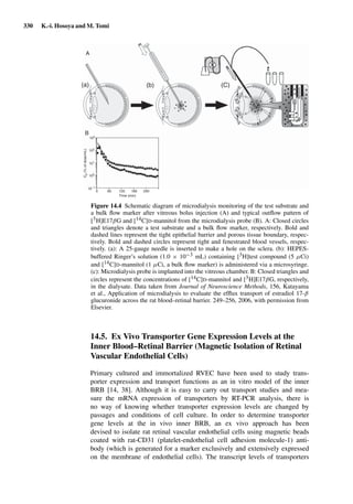
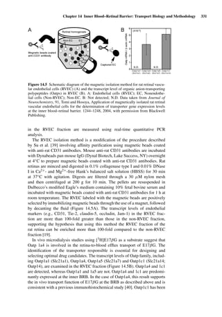
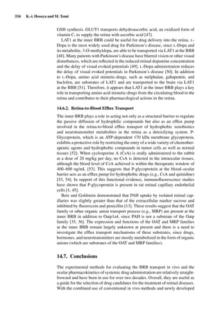
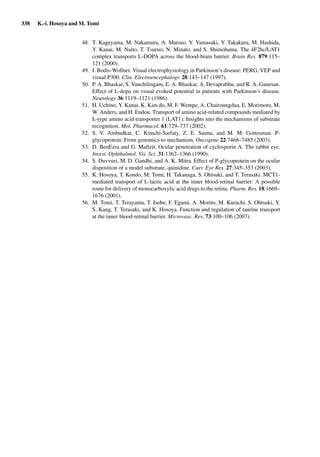
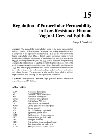
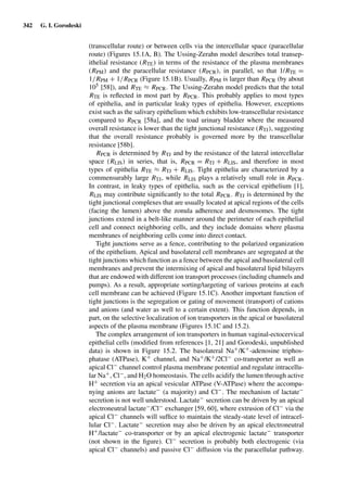
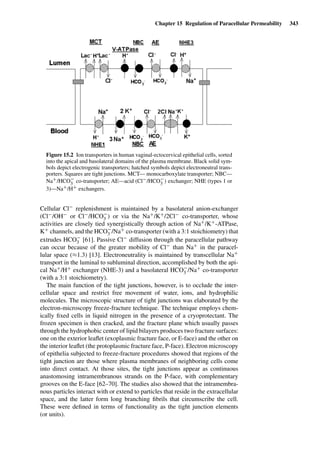
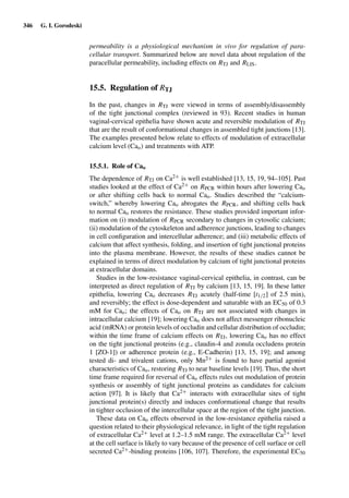
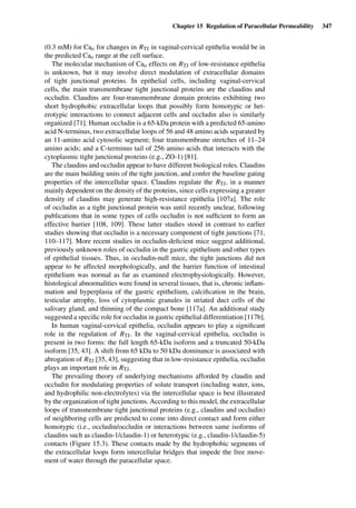
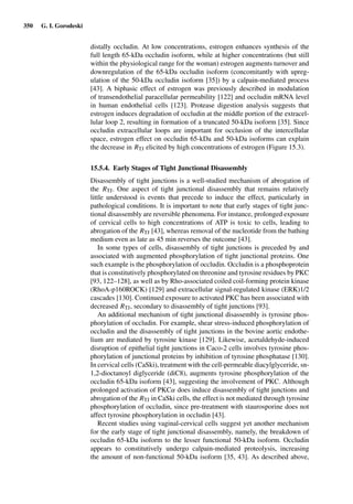
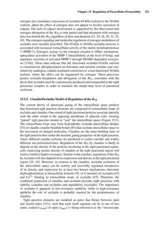
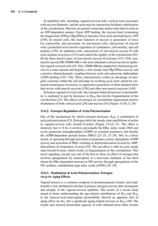
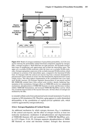
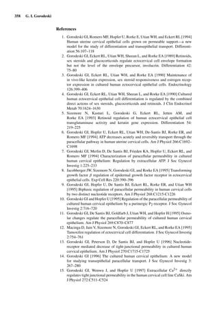
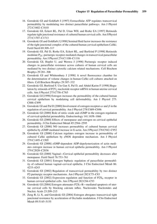
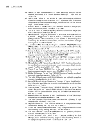
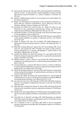

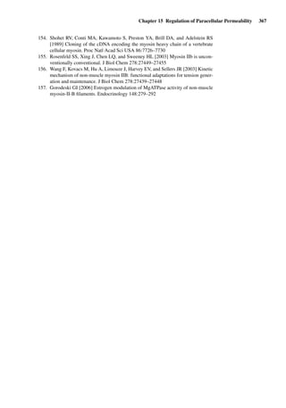
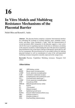
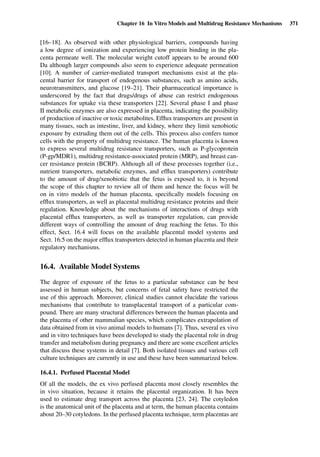
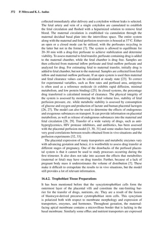
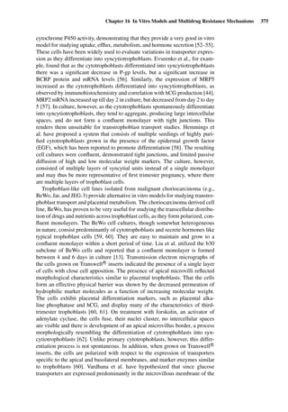
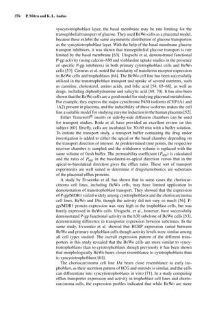
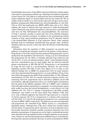
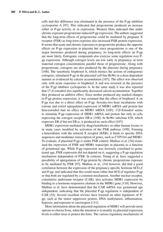
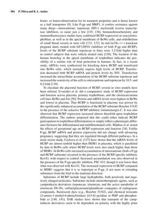
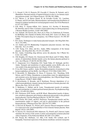
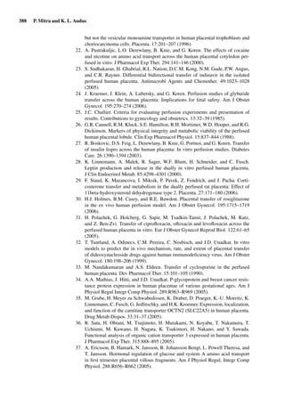
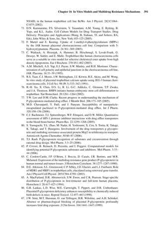
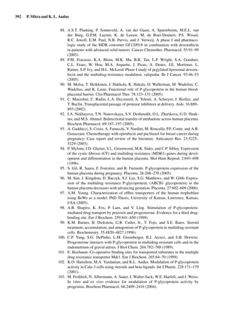
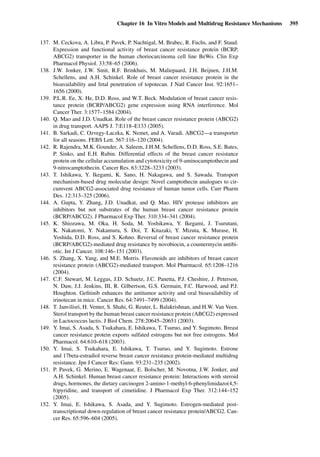

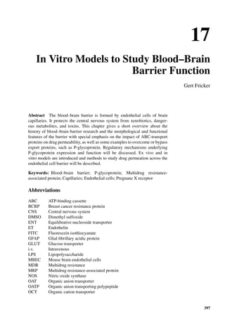
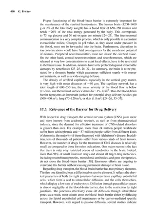
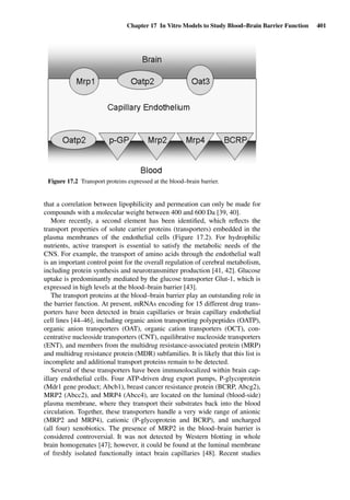
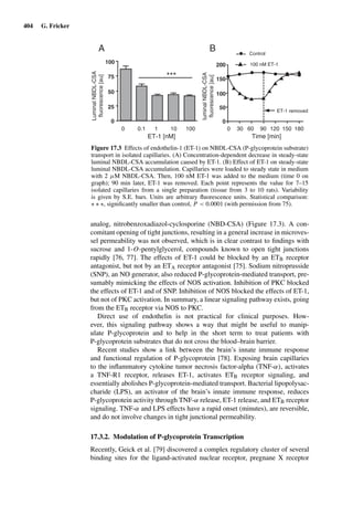
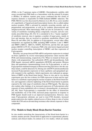

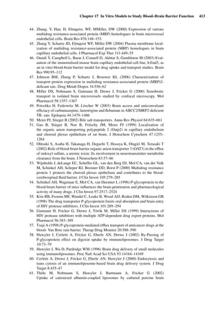
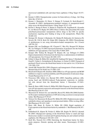

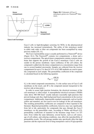
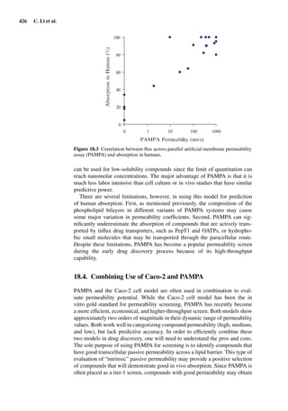
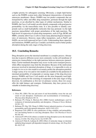
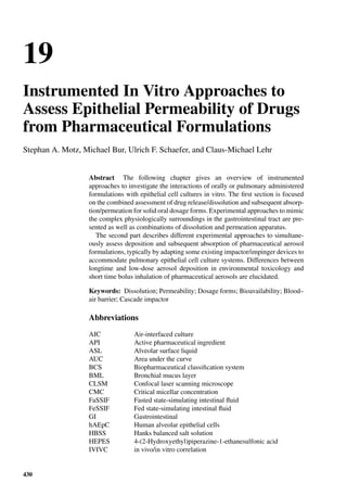
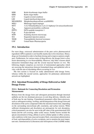
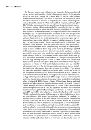
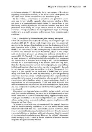
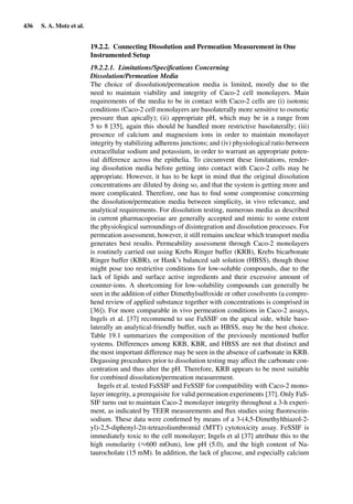
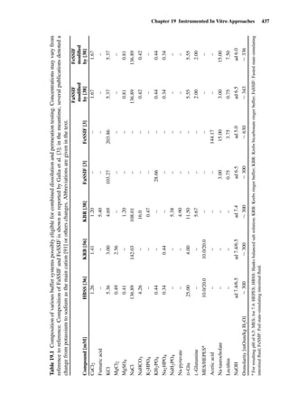
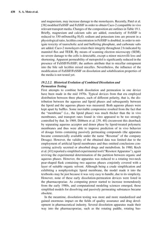
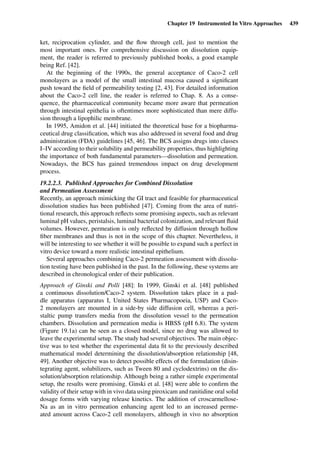
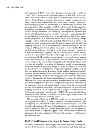
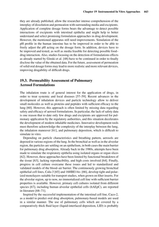
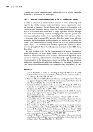
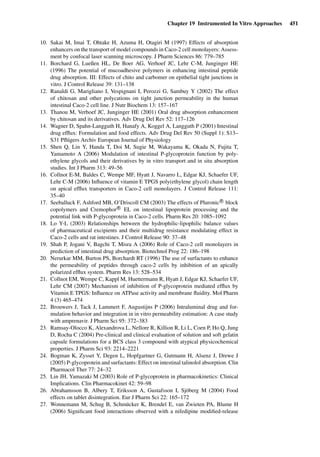
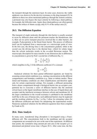
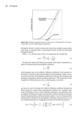
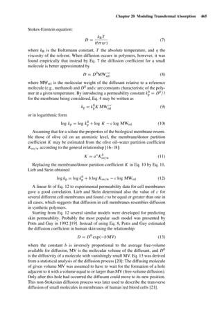
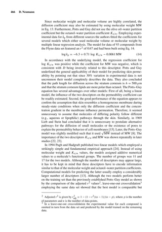
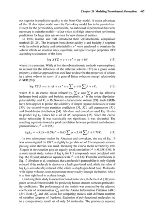
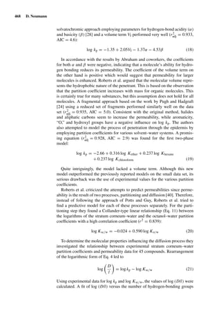
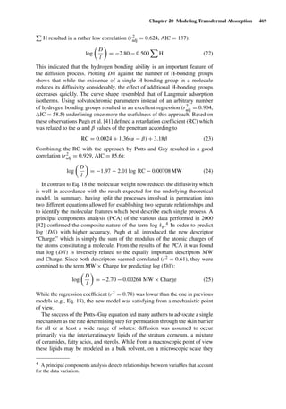
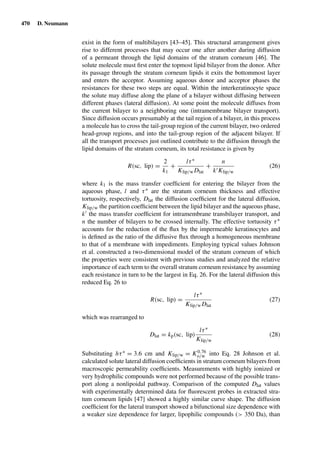


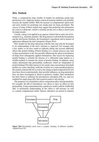
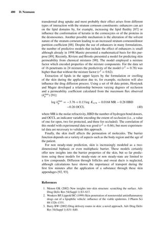
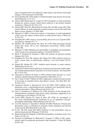
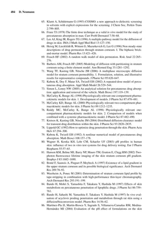
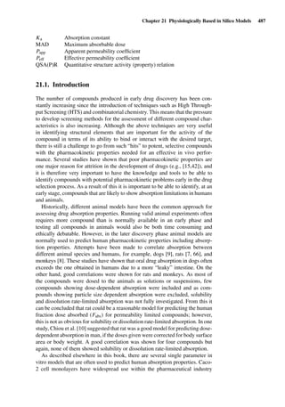
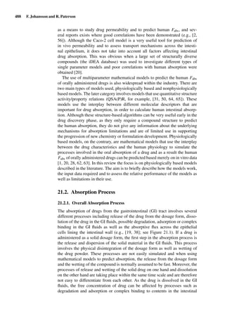
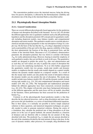
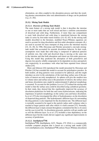
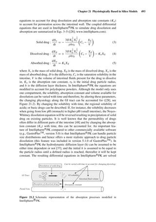
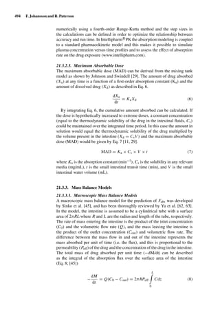
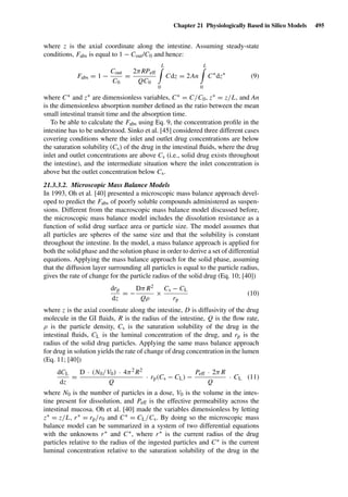
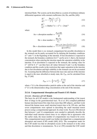
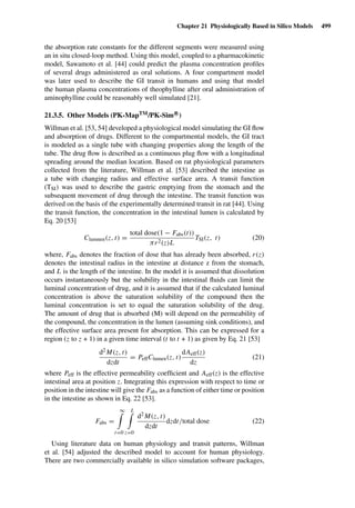
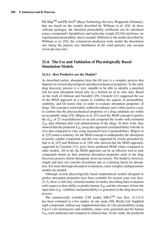
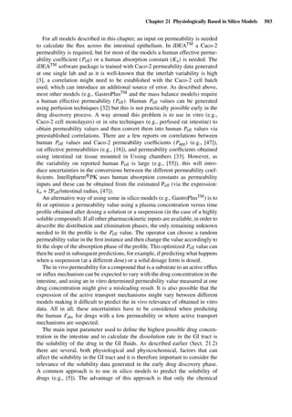
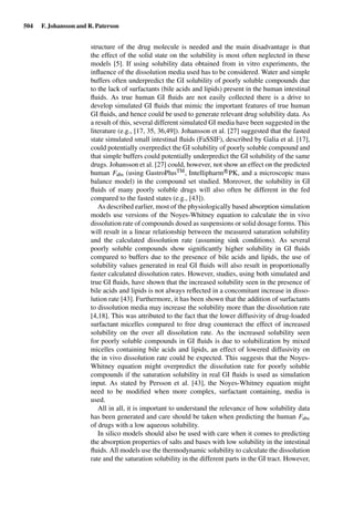
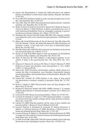
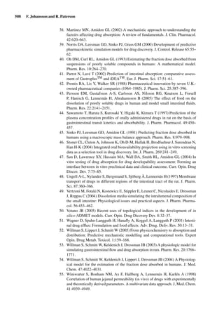
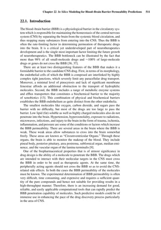
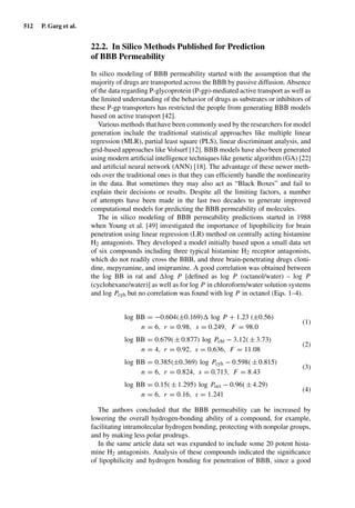
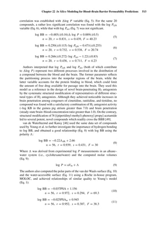
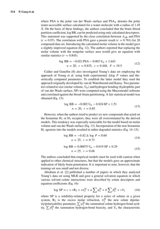
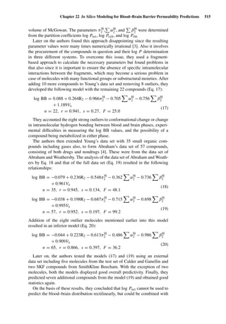
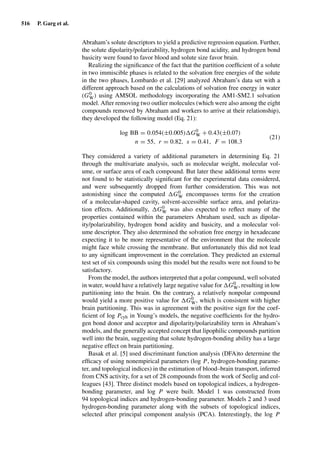
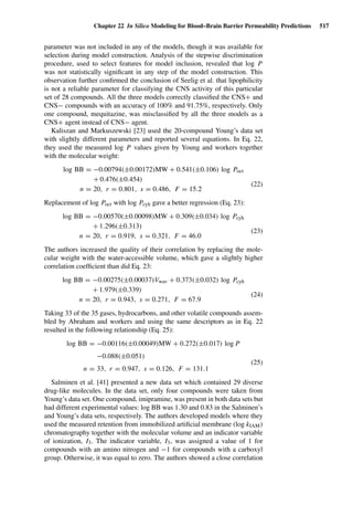
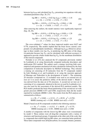
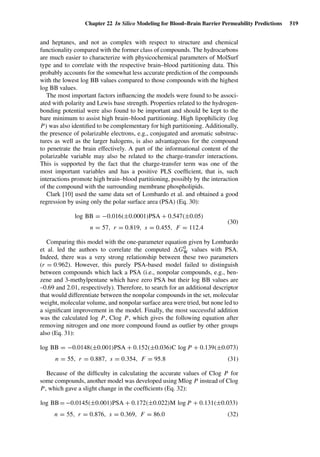
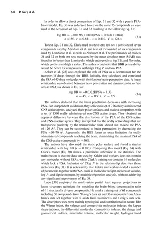
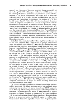
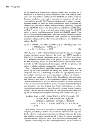
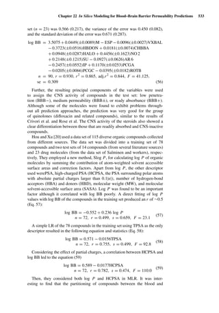
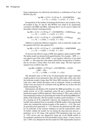
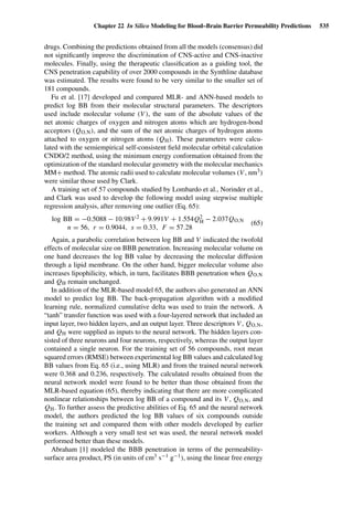
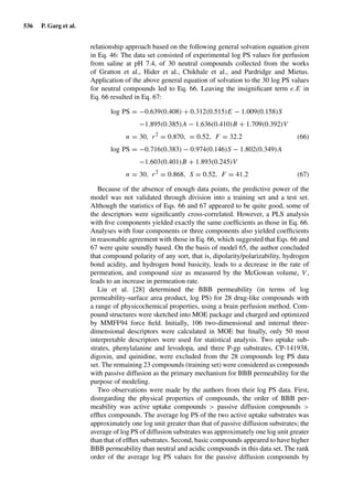
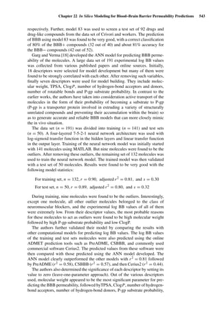
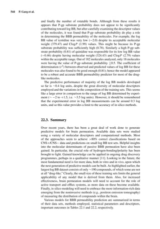
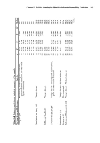
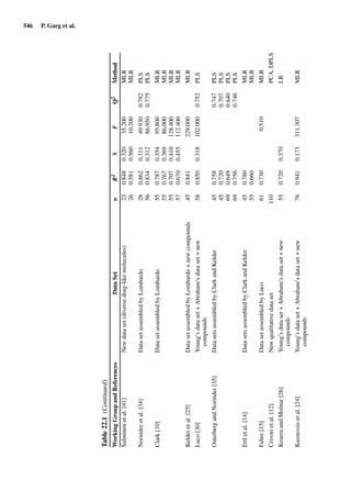
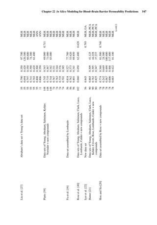
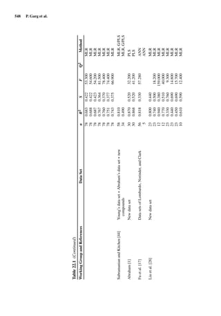
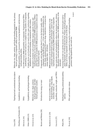
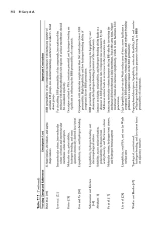
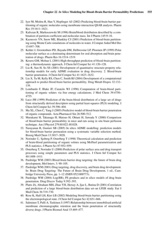
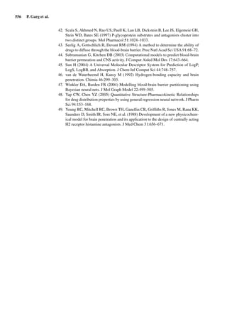
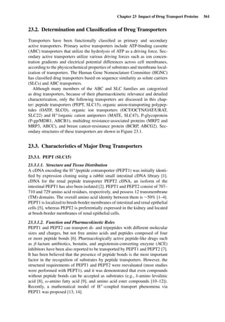
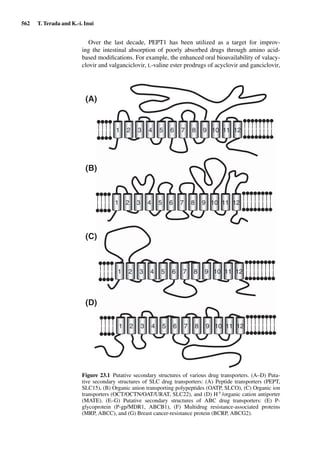
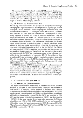
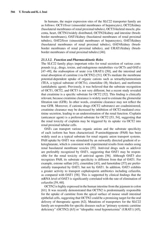
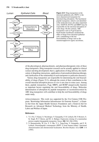
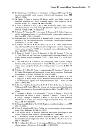
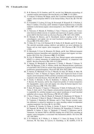
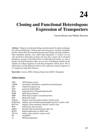
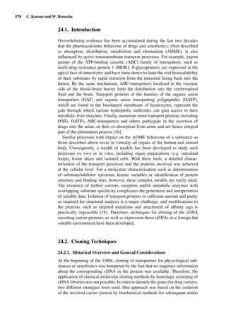
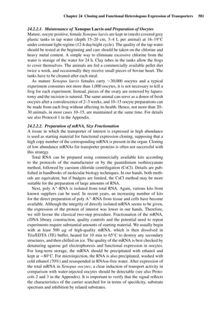
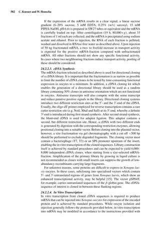
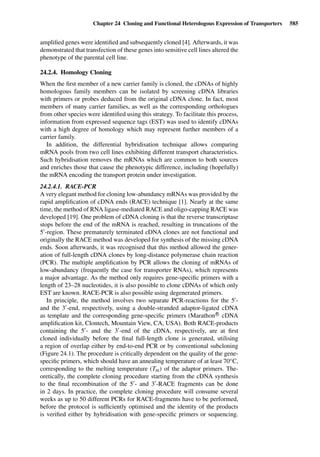
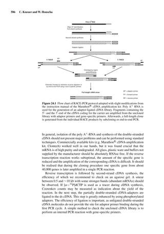
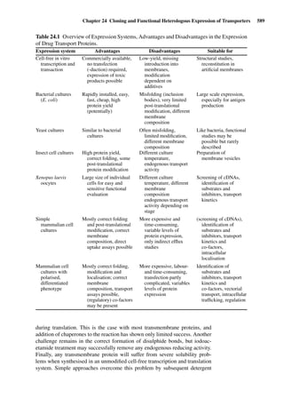
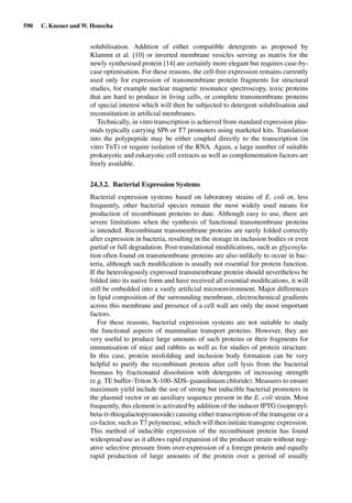
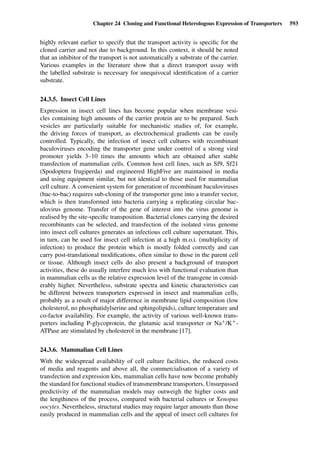
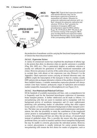
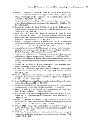
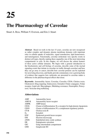
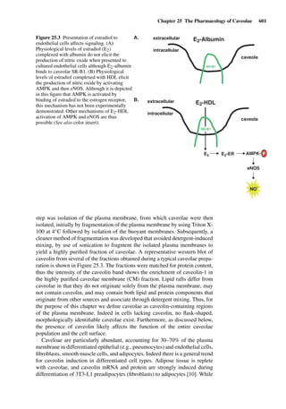
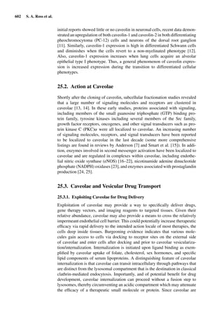
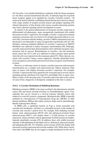
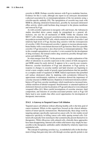
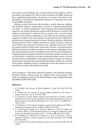
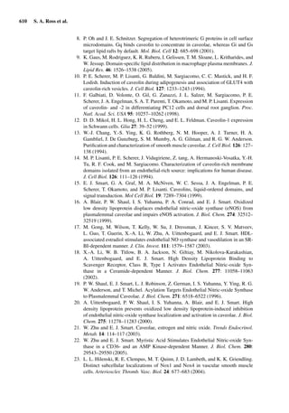
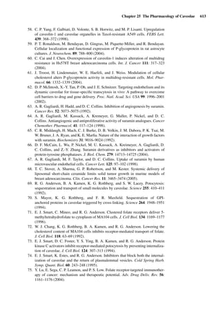
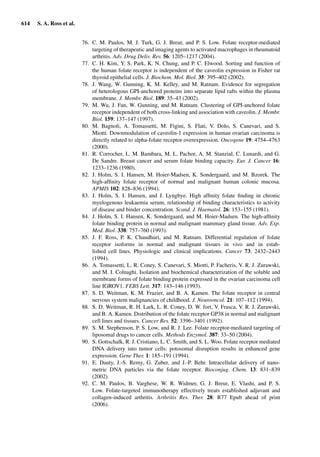
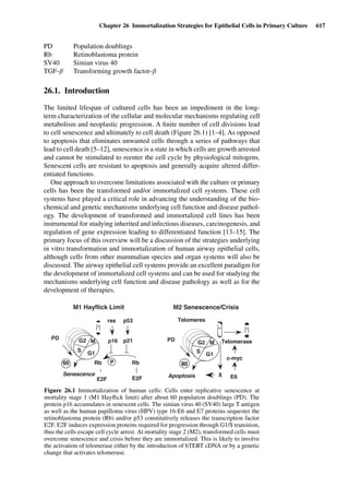
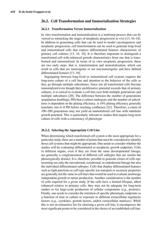
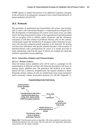
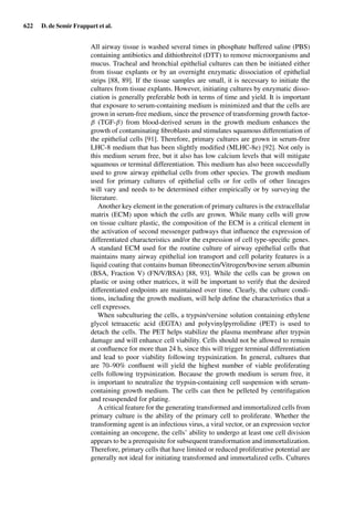
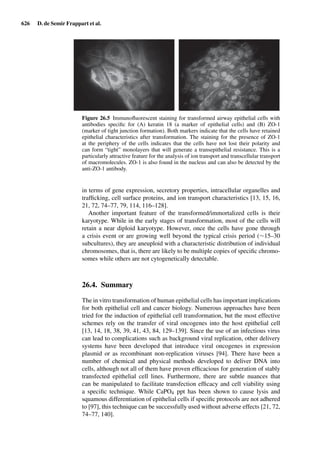
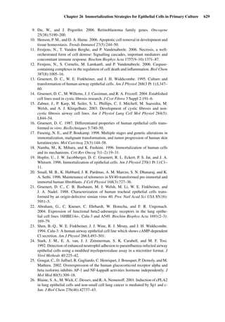
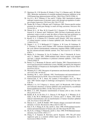
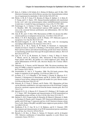
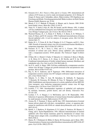
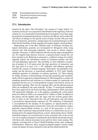
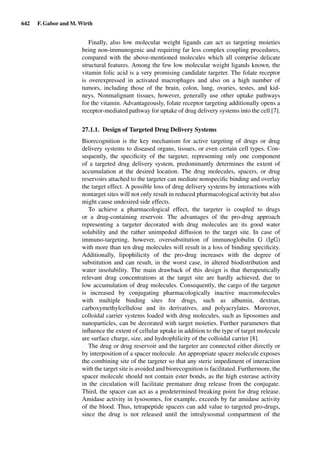
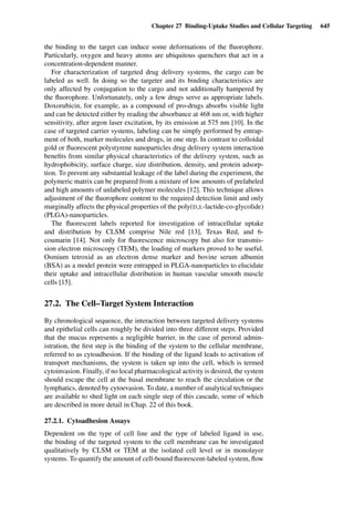
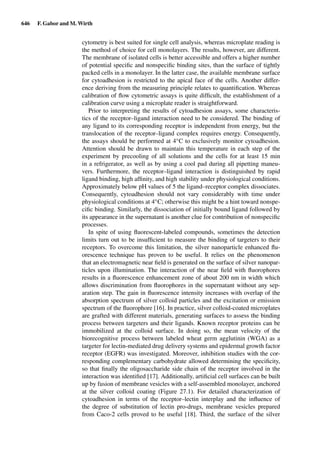
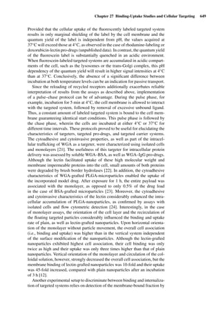
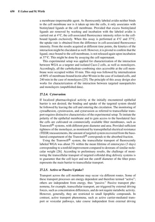
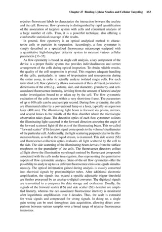
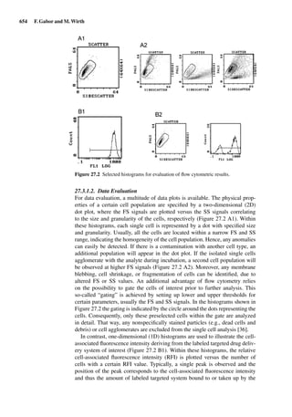

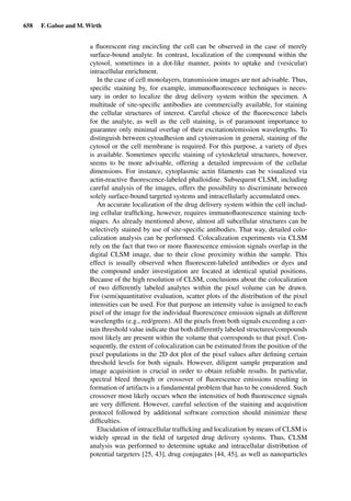
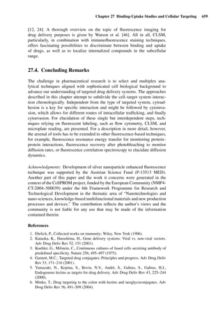
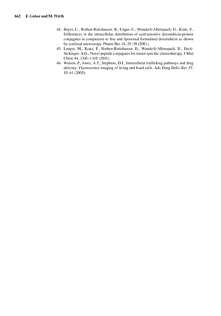
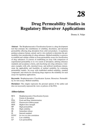
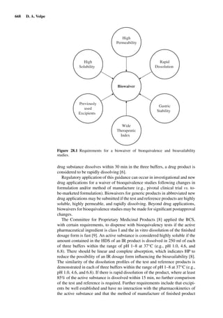
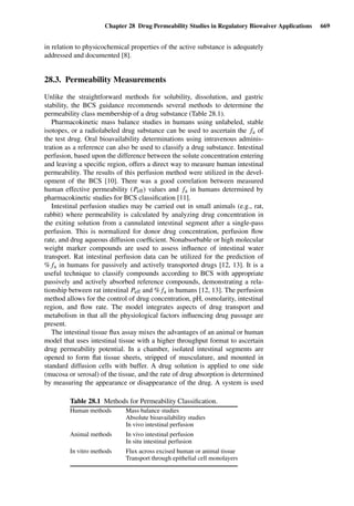
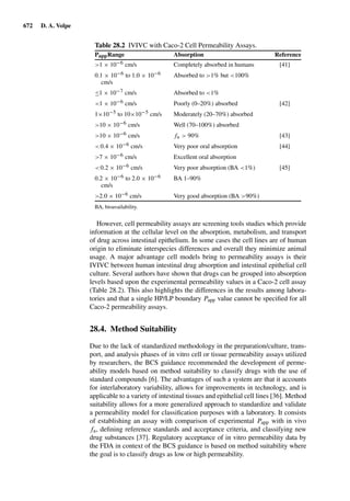
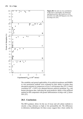
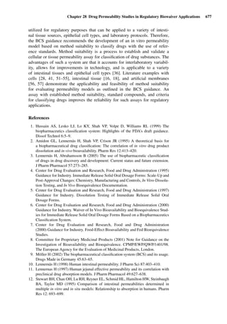
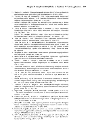
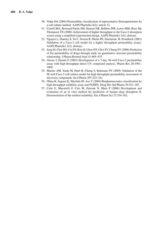

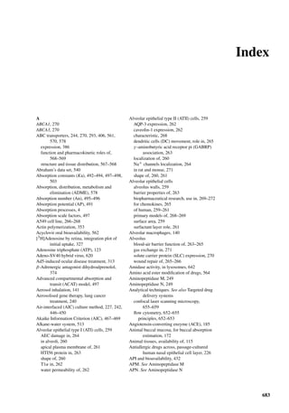
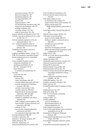
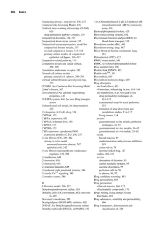
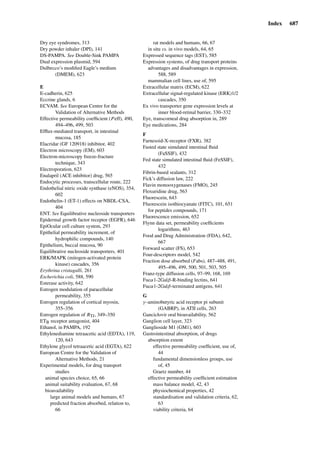
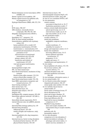
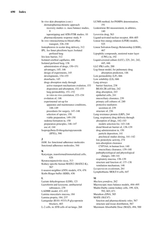
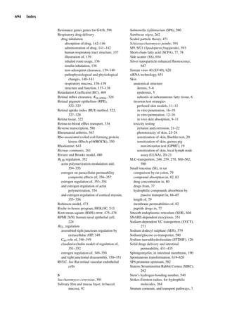
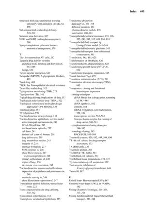


This document provides an overview of 7 chapters focused on in situ and in vitro models for assessing drug absorption. The chapters cover models of the skin, small intestine, colon, buccal mucosa, nasal cavity, lung, and various epithelial and endothelial barriers. A variety of in situ and in vitro techniques are discussed, including perfused organ models, cell culture systems, and excised tissue preparations. The models vary in their complexity and ability to predict in vivo absorption, with in situ techniques generally providing a more physiologically relevant system compared to simpler in vitro methods.


![Bio pharmaceutical classification System [BCS]](https://cdn.slidesharecdn.com/ss_thumbnails/biopharmaceuticalclassificationystembcs-160328061345-thumbnail.jpg?width=640&height=640&fit=bounds)


![physicochemical principles of drug action 01-02 2014-2015 [Autosaved].pptx](https://cdn.slidesharecdn.com/ss_thumbnails/physicochemicalprinciplesofdrugaction01-022014-2015autosaved-250702011653-1a031bc7-thumbnail.jpg?width=640&height=640&fit=bounds)


