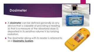
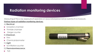
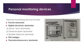
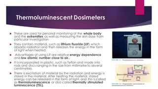
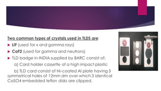
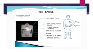
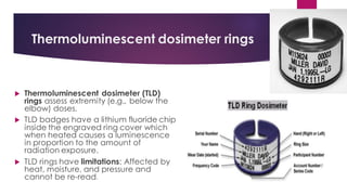
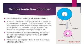
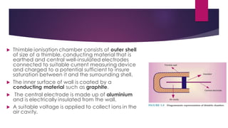
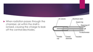
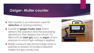
DOSIMETRY
Contents
 Definition
 Measures of dosimetry
 Dosimeter
 Properties of dosimeter
 Thermoluminescent dosimeter
 Ionisation chamber
 Rate meter
 Thimble ionisation chamber
 Condenser ionisation chamber
 Geiger- Muller counters
 Pocket dosimeter
 Radiographic film
 Radiochromic film
 Conclusion
Introduction
Definition: Dosimetry is the measurement of quantity of radiation
exposure or amount of energy absorbed per unit mass at an interest
site. Radiation cannot be perceived by our normal senses, such as
sight, touch or smell.
Effects on which detection of dose is possible are:
 Ionisation
 Luminescence
 Themoluminescence
 Chemical effect
 Biological effect
 Calorimetry
Measures of Dosimetry
The various measures of Dosimetry are as follows:
 Absorbed dose
 Erythema dose
 Exposure
 Equivalent dose (H)
 Radioactivity
 MPD
 Maximum accumulated dose
Measures of Dosimetry
 Absorbed dose: The amount of radiation at a given point to the
amount of energy absorbed per unit mass at the site of interest is
known as dose. Or Dose can be defined as the amount of energy
absorbed by a tissue. The radiation absorbed dose or rad is the
traditional unit of dose.
 Erythema dose :The dose which produces in one sitting a reversible
reddening of the skin (3-4 Gy) is known as erythema dose.
 Exposure It is a measure of radiation quantity or the capacityof the
radiation to ionize air. It is measured as the amount of charge per
mass of air namely Coulombs/kg.
 Equivalent dose(H): It is used to compare the biologic effects of different
types of radiation on a tissue or organ.. It is the sum of absorbed dose and
radiation weighing factor.
 Effective dose. The dose used to estimate the risk in humans is known as
effective dose. It is the sum of products of equivalent dose to each organ
or tissue and the tissue weighing factor. The unit of effective dose is Sievert
(Sv).
 Radioactivity: The measurement of radioactivity (A) describes the decay
rate of a sample of a radioactive material.
 MPD: It is the amount of radiation that an individual is allowed to receive
from artificial sources of radiation such as x-ray machine.
 As per the most recent report of NCRP
a) MPD for occupationally exposed: 5rem/year or 0.05Sv /year
b) MPD for non-occupationally exposed: 0.1rem/year or
0.001SvIyear
c) MPD for occupationally exposed pregnant woman is same as
that for non occupationally exposed.
Dosimeter
 A dosimeter can be defined generally as any
device that is capable of providing a reading
(r) that is a measure of the absorbed dose D,
deposited in its sensitive volume V by ionizing
radiation.
 The dosimeter along with its reader is referred to
as a Dosimetry System.
 Dosimeter is a device that measures directly or indirectly
a) Absorbed dose
b) Exposure
c) Kerma
d) Equivalent dose
e) Or other related quantities.
Properties of dosimeter
Properties of an useful dosimeter are as follows:
 High accuracy and precision .
 Linearity of signal with dose over a wide range.
 Small dose and dose rate dependence.
 Flat Energy response(Quality dependence).
 Small directional dependence .
 High spatial resolution
Radiation monitoring devices
Measuring of the x-ray exposure of operators or associatedpersonnel as a protective measure.
Various types of radiation monitoring devices:
A) Electrical:
 lonization chamber
 Thimble chamber
 Geiger counter
B) Chemical:.
 Film
 Chemical dosimeter
C) Light
 Scintillationcounter
D) Thermoluminescence:
 TLD
Personal monitoring devices
Personal monitoring devices include:
 Pocket dosimeter
 Digital electronic dosimeter:
a) Arrow- tech dosimeters
b) Personal alarm dosimeter
c) Bubble detector dosimeter
 Film badges
 Thermoluminescence dosimeter
Dosimeter placement
 Interpretation of the measured dose depends on the placement of the dosimeter. .
The followinglist indicates where the dosimeters are to be worn:
 Film badge with no TLD badge: Wear the film badge above any protectiveclothing at
collar level.
 Film badge with a TLD badge: Wear the film badge under the lead apron and the TLD
above the apron at collar level.
 Ring dosimeters are perfect for those who handle radioisotopes or perform
interventional radiographic procedures. It consists of natural lithium fluoride element.
 Do not expose personnel monitoringdevicesto extremeheat or humidity. They are
screened monthly. If any dosimeterhas receiveda dose higher than the values shown
below, the employee willbe notified .
Film badges
Film badges are the most commonform of personal monitoring
devices.They consist of blue plastic frame containing a variety
of different metal filters and a small radiographic film which
reacts to radiation.
 Size of radio- graphic film used is about 4 x 3 cm. The film
badges can be providedwith different types of filter, i.e.
Aluminium, copper, cadmium, plastic and lead.
 All the filters are 1 mm thick except thin copper which is 0.15
mm thick. The filter assesses the penetrating power of the
radiation and thus permits the energy to be estimated.
 Film badges are worn outside the clothes usually at the level
of the reproductive organs, for 1-3 months before being
processed.
 They are usually worn in metallic batches. The
film should be loaded in the film holder so that
the flap side of the film pack is always facing
the body.
 There are three types of film badges: chest
holder, wrist holder and head holder. The film is
pro- cessed and measures the degree of
darkening. It must be compared with films
exposed to known radiation.
 The minimum dose thatfilm badges can
detect is about 0.2 mSv.
Guidelines for using film badges:
 Film badges are used only by the person working in radiation.
 The name, person- number, type of radiation, period of use and
location of the body should be written in block letters on the front
side of film .
 A film pack once issued to a person should not be used by any
other person .While leaving the premises of the institute, workers
should deposit their badges in the place where the control film is
kept .
Advantages
 Records: They provide perma- nent record of dose
received which can be used for future
 Reassessment: It may be checked and reassessed.
 Type of radiation: It can measure the type and energy
of radi- ation encoun- tered
 Simple: It is sim- ple, robust and relatively inexpensive.
Disadvantages
 Time: No imme- diate indicationof exposure Retrospective in-
formation: All the information is retrospective.
 Errors during processing: It has complex dark- room
procedure, so it may lead to errors.
 Filter loss: The badges are prone to filter loss.
Thermoluminescent Dosimeters
 These are used for personal monitoring of the whole body
and the extremities as well as measuring the skin dose from
particular investigation
 They contain material, such as lithium fluoride (LIF) which
absorbs radiation and then releases the energy in the form
of light when heated.
 Advantages of using LiF are relative energy dependence
and low atomic number close to air..
 It is incorporated in plastic, such as Teflon and made into
rods and discs ranging in the size from millimetre to several
centimetres.
 There is excitation of material by the radiation and energy is
stored in the material. After heating the material, stored
energy can be released in the form of light, and this is called
as thermoluminescence or also called thermally stimulated
luminescence (TSL).
Two common types of crystals used in TLDS are:
 LIF (used for x and gamma rays)
 CaF2 (used for gamma and neutrons)
 TLD badge in INDIA supplied by BARC consist of:
a) Card holder cassette of a high impact plastic
b) TLD card consist of Ni-coated Al plate having 3
symmetrical holes of 12mm dm over which 3 identical
CaSO4 embedded teflon disks are clipped.
 These 3 disks are coated with 3 different filters:
a) First disk coated with Al-Cu combination filters(cuts offbeta-
radiation and detects x-ray and gamma radiaiton)
b) Second disc is plastic filter coating (cuts off soft beta
radiation and detects hard beta, x-ray and gamma radiation)
c) Third disc is not coated with any filters. Therefore detects all
radiaitons
Thermoluminescent dosimeter rings
 Thermoluminescent dosimeter (TLD)
rings assess extremity (e.g., below the
elbow) doses.
 TLD badges have a lithium fluoride chip
inside the engraved ring cover which
when heated causes a luminescence
in proportion to the amount of
radiation exposure.
 TLD rings have limitations: Affected by
heat, moisture, and pressure and
cannot be re-read.
Advantages of thermoluminescent dosimeters
 Reusable: Lithium fluoride is reusable
 Automated measurement: Read- out measurement is easily.
automated and rapidly produced
 Wide variety It is suitable for wide variety of dose measurements
 Size: It is small in size and can be used for personal monitor
 Sensitivity: Any small change in sensitivity with radiation quality can
be assessed. Sensitivity is also independent of dose rate
 In field exposure: Exposure can be made in the field away from
measuring laboratory .It is less likely to give false reading due to
environmental conditions.
Disadvantages of thermoluminescent dosimeters
 No permanent record: Read-out is destructive giving no permanent
record
 No reassessment: Result cannot be checked and reassessed
 Limited information: Only limited information is provided on the
type and energy of the radiation
 Dose gradient: Dose gradients are not detectable
 Expensive: It is relatively expensive.
Ionisation chamber
 Ionisation chamber is used for personal monitoring by
physicist to measure radiation exposure . It was introduced
in 1924 by H Behnken.
 It consists of collectingelectrodes of positive potential
(made of graphite or metal and connected directly to
measuring apparatus) and negative electrodes (graphite
layer, deposited on the inner surface of the outer wall).
 Insulator separates the two electrodes.
 The outer wall provides air equilibrium. It is made of
material having the same atomic number as that of the
air. It is made up of conducting mixture of bakelite and
graphite. It is directly earthed and acts like a guard ring for
the collectorplate.
 Radiation produces ionisation of the
air molecules inside the closed
chamber which results in formation
of an ion .
 The positive and negative ions are
attracted by plate causing partial
discharge, and it is measured by
magnitude of the drop in potential
between the collecting plates.
Rate meter
 Rate Meter is also called roentgen ray rate
meter. It has an ion collection chamber that is
continuously being charged by battery.
 It constantly measures the amount of ionisation
taking place in the air within the chamber. It
measures the rate of radiation exposure in
terms of roent- gen or milliroentgen per minute
or per hour.
Thimble ionisation chamber
 It works based on the Bragg -Gray Cavity theory.
 A spherical volumeof air is shown with an air cavity
at the center.This sphere of air is irradiated uniformly
with a photon beam. And the distance betweenthe
outer sphere and the inner cavityis equal to the
maximum range ofelectrons generated in air.
 Then the number of electrons entering the cavityis
the same as that leavingthe cavity, ie, electronic
equilibrium exists.
 If the air wall is compressed into a solid shell, we get
a thimble chamber.
 Thimble ionisation chamber consists of outer shell
of size of a thimble, conducting material that is
earthed and central well-insulated electrodes
connected to suitable current measuring device
and charged to a potential sufficient to insure
saturation between it and the surrounding shell.
 The inner surface of wall is coated by a
conducting material such as graphite.
 The central electrode is made up of aluminium
and is electrically insulated from the wall.
 A suitable voltage is applied to collect ions in the
air cavity.
 When radiation passes through the
chamber, air within the shell is
ionised, causing the charge to leak
off the central electrodes.
Condenser ionisation chamber
Condenser Ionisation Chamber is the thimble
ionisationchamber connected to a condenser .
 Thimble chamber is inserted into electrometer
unit and entire system is charge to potentialof
about 500 V. The central electrodes(aluminium)
are connected to inner surface of condenser.
 Thus, thimble is permanentlyconnected with
condenser which is capable of storing charge.
When the chamber is exposed, electrons are
collected,thereby neutralising some of the
positive charge.
Geiger- Muller counter
 GM counter is an instrument used for
detection of ionizing radiation.
 Consist of geiger-muller tube which
detects the radiation and the processing
electronics, that displays the circuit. It is
filled with an inert gas such as argon at low
pressure to which high voltage is applied
 Briefly conducts electrical charge when a
particle or photon of incident radiation
makes the gas conductive.
 There are mainly two types of Geiger
counter, cylindrical type and end-window
type.
 Cylindrical type is made up of window of
various thicknesses, and end-window type
is made up of metal with thin mica window.
It con- sists of cylindrical cathode with fine
wire anode along its axis.
 Dimension of cathode is 2 cm in diameter,
5 cm long and central wire is 0.2 cm in
diameter.
 They are mounted inside a usually thin envelope which is made
of glass. The device is filled with a special mixture of gases at a
pressure of about 10 cmHg.
 Gas contains nine parts of inert gas such as argon with one part
alcohol. The ionisation of collision is the basis of gas amplifica-
tion in the Geiger-Müller counter.
 When rays are passed through the counter, photoelectrons are
ejected from the metal cathode, which initiates the discharge.
This is then measured by the rate meter.
Scintillation detector
 Scintillation detector works on principle of
detection of ionizing radiation by measuring
the scintillation light produced by certain
materials like Nal, Csl, Cadmium tungstate,
amorphous silicon etc.
 When the crystals absorb the energy the
electrons jump from ground state to
conduction band. The excited e return to the
valance band with emission of photon.
 When photon interacts with the crystal photoelectrons are
produced within the crystals. These photoelectrons travel
through the crystal & they ionize the atoms of crystal, as a
result Flashes of UV or visible light are produced
(Luminescence).
 Only small part of energy imparted is converted into light rest
is dissipated into form of heat.
Working of Scintillation detector
Scintillation detector consists
 Luminescent scintillation material.
 Optical device to facilitate the
collection of light. (Optical
interface)
 Photomultiplier tube(PMT)
 Electrical counter
 The low energy photons falls on the
photocathode of PMT and ejects no. of
photoelectrons.
 Photoelectrons are accelerated by P.D.
Applied between Cathode & Dynodes of
the tube.
 On striking the 1st Dynode Photoelectrons
ejects several electrons by secondary
emission and further multiplies with rest of
dynode interactions.
 After multiplication of 105-109 the electron avalanche,
electrons arrive at collector plate.
 It produces a voltage pulse on O/P condenser, which is
coupled to external Pulse amplifying circuit.
 Thus initial energy of single ionizing particle is transformed
into single voltage pulse.
Pocket Dosimeters
Pocket Dosimeter is a pen-like device
that measures the cumulative doseof
ionising radiation received by the device.
 It is usually clipped to a person's clothing
and worn to measure one's actual
exposure to radiation.
 Must be recharged at the start of every
work shift.
 Are of 2 types : Minometer /condensor
type / Indirect read dosimeter and Direct
read dosimeter
Indirect pocket dosimeter
 It is an ion chamber that has voltage
potential placed in it by insertion into a
charger.
 Radiation penetrating causes the current to
leak off whichis proportional to the incident
radiation.
 By re-inserting the dosimeter into the charger
at the end of the day, the drop in the
voltage potential is calibrated in terms of
milliroentgens(mR).
 It has one central electrode. and one wall electrode. It
is inserted in the battery- operated charged reader and
is charged to a certain point on the scale of the reader.
 It is then removed and is worn or carried in the pocket
and after exposure to stray radiation it is reinserted into
the charger reader.
Direct Pocket dosimeter
 Generally of the size and shape of a
fountain pen.
 To overcome the inconvenience of the
non-reading chamber, self-reading pocket
dosimeter with built-in mechanism system
has been constructed.
 In it, chamber is coupled to built-in quartz
fibre eletrometer.
 There is microscope to view th
electrometer which is calibrated in
milliroentgen or roentgen.
Advantages of pocket dosimetry:
 Provides immediate reading of the exposure
 Reusable
Diadvantages of pocket dosimetry:
 Limited range of exposureInability to provide a
permanent record
 Potential for discharging or reading-loss
resulting due to dropping or bumping.
 Must be recharged and recorded at the start of
each working shift.
Film Dosimetry
 Radiographic film is the dosimeterparticularly used
for qualitative measurement of ionizing radiation
beams.
 A radiographic film consists of a transparent film
base (cellulose acetate or polyester resin) coated
an emulsion containing very small crystals of silver
bromide.
 When the film is exposed to ionizing radiation or
visible light, a chemical change takes place within
the exposed crystals to form what is referred to as
a latent image.
 The film is developed, the affected crystals are
reduced to small grains of metallicsilver. The film is
then fixed.
 The unaffected granules are removed by the fixing solution, leaving
aclear film in their place.
 The metallic silver, which is not affected by the fixer, causes
darkening of the film.
 Thus, the degree of blackening of an area of the film depends on
the amount of free silver deposited and, consequently, on the
radiation energy absorbed.
 The degree of blackening of the film
is measured by determining optical
density with a densitometer.
 This instrument consists of a light
source, a tiny aperture through which
the light is directed, and a light
detector (photocell) to measure the
light intensity transmitted through the
film.
 Radiographic film Construction
a) Plastic Base: provides the structure of the
film.
b) Silver Bromide Emulsion: is the active layer
of the film consisting of Ag+ and Br- ions.
c) Grains are 0.13 μm in diameter.
d) Gelatin Protective Layer: serves to keep the
silver bromide grains well dispersed and to
protect unexposed grains during
development.
 Film suffers from several potentialerrors such as: Iong exposure ,changes in processing
conditions ,emulsion differences, artifacts caused by air pockets adjacent to the film.
 For these reasons, absolute dosimetrywithfilm is impractical.
 However, it is veryuseful for checking:
a) radiation field
b) slight-fieldcoincidence
c) field flatness
d) Symmetry
e) obtaining quick qualitative patterns of a radiation distribution.
Radiochromic film
 The use of Radiochromic films for radiation
dosimetry has been evolving since the 1960s.
 Their use has become increasingly popular,
especially in brachytherapy dosimetry.
 Major advantages of Radiochromic film
dosimeters include:
a)tissue equivalence
b) high spatial resolution
c)large dynamic range
Radiochromic film construction
 Base Layer: Polyester (Mylar) base provides
structure to the film.
 Active Layer: Consists of radio-sensitive chemicals
which polymerize into optically opaque polymers
upon irradiation. This is a chemical reaction which
takes approximately 24 hours to complete
Symmetrical and Non-Symmetrical Designs:
 Although most current films are constructed
symmetrically, older films have been
constructed asymmetrically. Asymmetrical
construction requires attention to orientation
during measurement.
 EBT 2 is a common example of asymmetrical construction.
Although it was later shown that this had little influence on
measured dose distribution, EBT 3 was introduced with a
symmetrical design.
 Radiochromic film consists of an ultrathin (7- to 23-μm thick) colorless
radiosensitive leuco dye bonded onto a 100-μm thick mylar base.
 Other varieties include thin layers of radiosensitive dye sandwiched
between two pieces of polyester base.
Conclusion
 A radiation dosimeter is a device that measures exposure to ionizing
radiation. It is used for human radiation protection as a
measurement of dose in both medical and industrial processes.
 Radiation dosimeters and dosimetry systems come in many shapes
and forms.
 There are a variety of electronic dosimetry systems that can monitor
any work environment.
 The accurate measurement of radiation dose (dosimetry) is
important in various applications such as radiation oncology,
diagnostic radiology, nuclear medicine and radiation protection.
 The advanced dosimetry devices now a days give us
real time reading and the history of the doses can be
recorded online.
References
 Essentials of oral and maxillofacial radiology (Freny R
Karjodkar) 2nd edition
 Ghom’s textbook of oral radiology ( 2nd edition)
 White and Pharoah textbook of oral radiology
principles and interpretation ( 6th edition)
Dosimetry

Dosimetry

  • 1.
  • 2.
    Contents  Definition  Measuresof dosimetry  Dosimeter  Properties of dosimeter  Thermoluminescent dosimeter  Ionisation chamber  Rate meter  Thimble ionisation chamber  Condenser ionisation chamber  Geiger- Muller counters  Pocket dosimeter  Radiographic film  Radiochromic film  Conclusion
  • 3.
    Introduction Definition: Dosimetry isthe measurement of quantity of radiation exposure or amount of energy absorbed per unit mass at an interest site. Radiation cannot be perceived by our normal senses, such as sight, touch or smell. Effects on which detection of dose is possible are:  Ionisation  Luminescence  Themoluminescence  Chemical effect  Biological effect  Calorimetry
  • 4.
    Measures of Dosimetry Thevarious measures of Dosimetry are as follows:  Absorbed dose  Erythema dose  Exposure  Equivalent dose (H)  Radioactivity  MPD  Maximum accumulated dose
  • 5.
    Measures of Dosimetry Absorbed dose: The amount of radiation at a given point to the amount of energy absorbed per unit mass at the site of interest is known as dose. Or Dose can be defined as the amount of energy absorbed by a tissue. The radiation absorbed dose or rad is the traditional unit of dose.  Erythema dose :The dose which produces in one sitting a reversible reddening of the skin (3-4 Gy) is known as erythema dose.  Exposure It is a measure of radiation quantity or the capacityof the radiation to ionize air. It is measured as the amount of charge per mass of air namely Coulombs/kg.
  • 6.
     Equivalent dose(H):It is used to compare the biologic effects of different types of radiation on a tissue or organ.. It is the sum of absorbed dose and radiation weighing factor.  Effective dose. The dose used to estimate the risk in humans is known as effective dose. It is the sum of products of equivalent dose to each organ or tissue and the tissue weighing factor. The unit of effective dose is Sievert (Sv).  Radioactivity: The measurement of radioactivity (A) describes the decay rate of a sample of a radioactive material.  MPD: It is the amount of radiation that an individual is allowed to receive from artificial sources of radiation such as x-ray machine.
  • 7.
     As perthe most recent report of NCRP a) MPD for occupationally exposed: 5rem/year or 0.05Sv /year b) MPD for non-occupationally exposed: 0.1rem/year or 0.001SvIyear c) MPD for occupationally exposed pregnant woman is same as that for non occupationally exposed.
  • 8.
    Dosimeter  A dosimetercan be defined generally as any device that is capable of providing a reading (r) that is a measure of the absorbed dose D, deposited in its sensitive volume V by ionizing radiation.  The dosimeter along with its reader is referred to as a Dosimetry System.
  • 9.
     Dosimeter isa device that measures directly or indirectly a) Absorbed dose b) Exposure c) Kerma d) Equivalent dose e) Or other related quantities.
  • 11.
    Properties of dosimeter Propertiesof an useful dosimeter are as follows:  High accuracy and precision .  Linearity of signal with dose over a wide range.  Small dose and dose rate dependence.  Flat Energy response(Quality dependence).  Small directional dependence .  High spatial resolution
  • 12.
    Radiation monitoring devices Measuringof the x-ray exposure of operators or associatedpersonnel as a protective measure. Various types of radiation monitoring devices: A) Electrical:  lonization chamber  Thimble chamber  Geiger counter B) Chemical:.  Film  Chemical dosimeter C) Light  Scintillationcounter D) Thermoluminescence:  TLD
  • 13.
    Personal monitoring devices Personalmonitoring devices include:  Pocket dosimeter  Digital electronic dosimeter: a) Arrow- tech dosimeters b) Personal alarm dosimeter c) Bubble detector dosimeter  Film badges  Thermoluminescence dosimeter
  • 14.
    Dosimeter placement  Interpretationof the measured dose depends on the placement of the dosimeter. . The followinglist indicates where the dosimeters are to be worn:  Film badge with no TLD badge: Wear the film badge above any protectiveclothing at collar level.  Film badge with a TLD badge: Wear the film badge under the lead apron and the TLD above the apron at collar level.  Ring dosimeters are perfect for those who handle radioisotopes or perform interventional radiographic procedures. It consists of natural lithium fluoride element.  Do not expose personnel monitoringdevicesto extremeheat or humidity. They are screened monthly. If any dosimeterhas receiveda dose higher than the values shown below, the employee willbe notified .
  • 16.
    Film badges Film badgesare the most commonform of personal monitoring devices.They consist of blue plastic frame containing a variety of different metal filters and a small radiographic film which reacts to radiation.  Size of radio- graphic film used is about 4 x 3 cm. The film badges can be providedwith different types of filter, i.e. Aluminium, copper, cadmium, plastic and lead.  All the filters are 1 mm thick except thin copper which is 0.15 mm thick. The filter assesses the penetrating power of the radiation and thus permits the energy to be estimated.  Film badges are worn outside the clothes usually at the level of the reproductive organs, for 1-3 months before being processed.
  • 17.
     They areusually worn in metallic batches. The film should be loaded in the film holder so that the flap side of the film pack is always facing the body.  There are three types of film badges: chest holder, wrist holder and head holder. The film is pro- cessed and measures the degree of darkening. It must be compared with films exposed to known radiation.  The minimum dose thatfilm badges can detect is about 0.2 mSv.
  • 18.
    Guidelines for usingfilm badges:  Film badges are used only by the person working in radiation.  The name, person- number, type of radiation, period of use and location of the body should be written in block letters on the front side of film .  A film pack once issued to a person should not be used by any other person .While leaving the premises of the institute, workers should deposit their badges in the place where the control film is kept .
  • 19.
    Advantages  Records: Theyprovide perma- nent record of dose received which can be used for future  Reassessment: It may be checked and reassessed.  Type of radiation: It can measure the type and energy of radi- ation encoun- tered  Simple: It is sim- ple, robust and relatively inexpensive.
  • 20.
    Disadvantages  Time: Noimme- diate indicationof exposure Retrospective in- formation: All the information is retrospective.  Errors during processing: It has complex dark- room procedure, so it may lead to errors.  Filter loss: The badges are prone to filter loss.
  • 21.
    Thermoluminescent Dosimeters  Theseare used for personal monitoring of the whole body and the extremities as well as measuring the skin dose from particular investigation  They contain material, such as lithium fluoride (LIF) which absorbs radiation and then releases the energy in the form of light when heated.  Advantages of using LiF are relative energy dependence and low atomic number close to air..  It is incorporated in plastic, such as Teflon and made into rods and discs ranging in the size from millimetre to several centimetres.  There is excitation of material by the radiation and energy is stored in the material. After heating the material, stored energy can be released in the form of light, and this is called as thermoluminescence or also called thermally stimulated luminescence (TSL).
  • 22.
    Two common typesof crystals used in TLDS are:  LIF (used for x and gamma rays)  CaF2 (used for gamma and neutrons)  TLD badge in INDIA supplied by BARC consist of: a) Card holder cassette of a high impact plastic b) TLD card consist of Ni-coated Al plate having 3 symmetrical holes of 12mm dm over which 3 identical CaSO4 embedded teflon disks are clipped.
  • 23.
     These 3disks are coated with 3 different filters: a) First disk coated with Al-Cu combination filters(cuts offbeta- radiation and detects x-ray and gamma radiaiton) b) Second disc is plastic filter coating (cuts off soft beta radiation and detects hard beta, x-ray and gamma radiation) c) Third disc is not coated with any filters. Therefore detects all radiaitons
  • 25.
    Thermoluminescent dosimeter rings Thermoluminescent dosimeter (TLD) rings assess extremity (e.g., below the elbow) doses.  TLD badges have a lithium fluoride chip inside the engraved ring cover which when heated causes a luminescence in proportion to the amount of radiation exposure.  TLD rings have limitations: Affected by heat, moisture, and pressure and cannot be re-read.
  • 26.
    Advantages of thermoluminescentdosimeters  Reusable: Lithium fluoride is reusable  Automated measurement: Read- out measurement is easily. automated and rapidly produced  Wide variety It is suitable for wide variety of dose measurements  Size: It is small in size and can be used for personal monitor  Sensitivity: Any small change in sensitivity with radiation quality can be assessed. Sensitivity is also independent of dose rate  In field exposure: Exposure can be made in the field away from measuring laboratory .It is less likely to give false reading due to environmental conditions.
  • 27.
    Disadvantages of thermoluminescentdosimeters  No permanent record: Read-out is destructive giving no permanent record  No reassessment: Result cannot be checked and reassessed  Limited information: Only limited information is provided on the type and energy of the radiation  Dose gradient: Dose gradients are not detectable  Expensive: It is relatively expensive.
  • 28.
    Ionisation chamber  Ionisationchamber is used for personal monitoring by physicist to measure radiation exposure . It was introduced in 1924 by H Behnken.  It consists of collectingelectrodes of positive potential (made of graphite or metal and connected directly to measuring apparatus) and negative electrodes (graphite layer, deposited on the inner surface of the outer wall).  Insulator separates the two electrodes.  The outer wall provides air equilibrium. It is made of material having the same atomic number as that of the air. It is made up of conducting mixture of bakelite and graphite. It is directly earthed and acts like a guard ring for the collectorplate.
  • 29.
     Radiation producesionisation of the air molecules inside the closed chamber which results in formation of an ion .  The positive and negative ions are attracted by plate causing partial discharge, and it is measured by magnitude of the drop in potential between the collecting plates.
  • 31.
    Rate meter  RateMeter is also called roentgen ray rate meter. It has an ion collection chamber that is continuously being charged by battery.  It constantly measures the amount of ionisation taking place in the air within the chamber. It measures the rate of radiation exposure in terms of roent- gen or milliroentgen per minute or per hour.
  • 32.
    Thimble ionisation chamber It works based on the Bragg -Gray Cavity theory.  A spherical volumeof air is shown with an air cavity at the center.This sphere of air is irradiated uniformly with a photon beam. And the distance betweenthe outer sphere and the inner cavityis equal to the maximum range ofelectrons generated in air.  Then the number of electrons entering the cavityis the same as that leavingthe cavity, ie, electronic equilibrium exists.  If the air wall is compressed into a solid shell, we get a thimble chamber.
  • 33.
     Thimble ionisationchamber consists of outer shell of size of a thimble, conducting material that is earthed and central well-insulated electrodes connected to suitable current measuring device and charged to a potential sufficient to insure saturation between it and the surrounding shell.  The inner surface of wall is coated by a conducting material such as graphite.  The central electrode is made up of aluminium and is electrically insulated from the wall.  A suitable voltage is applied to collect ions in the air cavity.
  • 34.
     When radiationpasses through the chamber, air within the shell is ionised, causing the charge to leak off the central electrodes.
  • 35.
    Condenser ionisation chamber CondenserIonisation Chamber is the thimble ionisationchamber connected to a condenser .  Thimble chamber is inserted into electrometer unit and entire system is charge to potentialof about 500 V. The central electrodes(aluminium) are connected to inner surface of condenser.  Thus, thimble is permanentlyconnected with condenser which is capable of storing charge. When the chamber is exposed, electrons are collected,thereby neutralising some of the positive charge.
  • 36.
    Geiger- Muller counter GM counter is an instrument used for detection of ionizing radiation.  Consist of geiger-muller tube which detects the radiation and the processing electronics, that displays the circuit. It is filled with an inert gas such as argon at low pressure to which high voltage is applied  Briefly conducts electrical charge when a particle or photon of incident radiation makes the gas conductive.
  • 37.
     There aremainly two types of Geiger counter, cylindrical type and end-window type.  Cylindrical type is made up of window of various thicknesses, and end-window type is made up of metal with thin mica window. It con- sists of cylindrical cathode with fine wire anode along its axis.  Dimension of cathode is 2 cm in diameter, 5 cm long and central wire is 0.2 cm in diameter.
  • 38.
     They aremounted inside a usually thin envelope which is made of glass. The device is filled with a special mixture of gases at a pressure of about 10 cmHg.  Gas contains nine parts of inert gas such as argon with one part alcohol. The ionisation of collision is the basis of gas amplifica- tion in the Geiger-Müller counter.  When rays are passed through the counter, photoelectrons are ejected from the metal cathode, which initiates the discharge. This is then measured by the rate meter.
  • 39.
    Scintillation detector  Scintillationdetector works on principle of detection of ionizing radiation by measuring the scintillation light produced by certain materials like Nal, Csl, Cadmium tungstate, amorphous silicon etc.  When the crystals absorb the energy the electrons jump from ground state to conduction band. The excited e return to the valance band with emission of photon.
  • 40.
     When photoninteracts with the crystal photoelectrons are produced within the crystals. These photoelectrons travel through the crystal & they ionize the atoms of crystal, as a result Flashes of UV or visible light are produced (Luminescence).  Only small part of energy imparted is converted into light rest is dissipated into form of heat.
  • 41.
    Working of Scintillationdetector Scintillation detector consists  Luminescent scintillation material.  Optical device to facilitate the collection of light. (Optical interface)  Photomultiplier tube(PMT)  Electrical counter
  • 42.
     The lowenergy photons falls on the photocathode of PMT and ejects no. of photoelectrons.  Photoelectrons are accelerated by P.D. Applied between Cathode & Dynodes of the tube.  On striking the 1st Dynode Photoelectrons ejects several electrons by secondary emission and further multiplies with rest of dynode interactions.
  • 43.
     After multiplicationof 105-109 the electron avalanche, electrons arrive at collector plate.  It produces a voltage pulse on O/P condenser, which is coupled to external Pulse amplifying circuit.  Thus initial energy of single ionizing particle is transformed into single voltage pulse.
  • 44.
    Pocket Dosimeters Pocket Dosimeteris a pen-like device that measures the cumulative doseof ionising radiation received by the device.  It is usually clipped to a person's clothing and worn to measure one's actual exposure to radiation.  Must be recharged at the start of every work shift.  Are of 2 types : Minometer /condensor type / Indirect read dosimeter and Direct read dosimeter
  • 45.
    Indirect pocket dosimeter It is an ion chamber that has voltage potential placed in it by insertion into a charger.  Radiation penetrating causes the current to leak off whichis proportional to the incident radiation.  By re-inserting the dosimeter into the charger at the end of the day, the drop in the voltage potential is calibrated in terms of milliroentgens(mR).
  • 46.
     It hasone central electrode. and one wall electrode. It is inserted in the battery- operated charged reader and is charged to a certain point on the scale of the reader.  It is then removed and is worn or carried in the pocket and after exposure to stray radiation it is reinserted into the charger reader.
  • 47.
    Direct Pocket dosimeter Generally of the size and shape of a fountain pen.  To overcome the inconvenience of the non-reading chamber, self-reading pocket dosimeter with built-in mechanism system has been constructed.  In it, chamber is coupled to built-in quartz fibre eletrometer.  There is microscope to view th electrometer which is calibrated in milliroentgen or roentgen.
  • 48.
    Advantages of pocketdosimetry:  Provides immediate reading of the exposure  Reusable Diadvantages of pocket dosimetry:  Limited range of exposureInability to provide a permanent record  Potential for discharging or reading-loss resulting due to dropping or bumping.  Must be recharged and recorded at the start of each working shift.
  • 49.
    Film Dosimetry  Radiographicfilm is the dosimeterparticularly used for qualitative measurement of ionizing radiation beams.  A radiographic film consists of a transparent film base (cellulose acetate or polyester resin) coated an emulsion containing very small crystals of silver bromide.  When the film is exposed to ionizing radiation or visible light, a chemical change takes place within the exposed crystals to form what is referred to as a latent image.  The film is developed, the affected crystals are reduced to small grains of metallicsilver. The film is then fixed.
  • 50.
     The unaffectedgranules are removed by the fixing solution, leaving aclear film in their place.  The metallic silver, which is not affected by the fixer, causes darkening of the film.  Thus, the degree of blackening of an area of the film depends on the amount of free silver deposited and, consequently, on the radiation energy absorbed.
  • 51.
     The degreeof blackening of the film is measured by determining optical density with a densitometer.  This instrument consists of a light source, a tiny aperture through which the light is directed, and a light detector (photocell) to measure the light intensity transmitted through the film.
  • 52.
     Radiographic filmConstruction a) Plastic Base: provides the structure of the film. b) Silver Bromide Emulsion: is the active layer of the film consisting of Ag+ and Br- ions. c) Grains are 0.13 μm in diameter. d) Gelatin Protective Layer: serves to keep the silver bromide grains well dispersed and to protect unexposed grains during development.
  • 53.
     Film suffersfrom several potentialerrors such as: Iong exposure ,changes in processing conditions ,emulsion differences, artifacts caused by air pockets adjacent to the film.  For these reasons, absolute dosimetrywithfilm is impractical.  However, it is veryuseful for checking: a) radiation field b) slight-fieldcoincidence c) field flatness d) Symmetry e) obtaining quick qualitative patterns of a radiation distribution.
  • 54.
    Radiochromic film  Theuse of Radiochromic films for radiation dosimetry has been evolving since the 1960s.  Their use has become increasingly popular, especially in brachytherapy dosimetry.  Major advantages of Radiochromic film dosimeters include: a)tissue equivalence b) high spatial resolution c)large dynamic range
  • 55.
    Radiochromic film construction Base Layer: Polyester (Mylar) base provides structure to the film.  Active Layer: Consists of radio-sensitive chemicals which polymerize into optically opaque polymers upon irradiation. This is a chemical reaction which takes approximately 24 hours to complete Symmetrical and Non-Symmetrical Designs:  Although most current films are constructed symmetrically, older films have been constructed asymmetrically. Asymmetrical construction requires attention to orientation during measurement.
  • 56.
     EBT 2is a common example of asymmetrical construction. Although it was later shown that this had little influence on measured dose distribution, EBT 3 was introduced with a symmetrical design.
  • 57.
     Radiochromic filmconsists of an ultrathin (7- to 23-μm thick) colorless radiosensitive leuco dye bonded onto a 100-μm thick mylar base.  Other varieties include thin layers of radiosensitive dye sandwiched between two pieces of polyester base.
  • 58.
    Conclusion  A radiationdosimeter is a device that measures exposure to ionizing radiation. It is used for human radiation protection as a measurement of dose in both medical and industrial processes.  Radiation dosimeters and dosimetry systems come in many shapes and forms.  There are a variety of electronic dosimetry systems that can monitor any work environment.  The accurate measurement of radiation dose (dosimetry) is important in various applications such as radiation oncology, diagnostic radiology, nuclear medicine and radiation protection.
  • 59.
     The advanceddosimetry devices now a days give us real time reading and the history of the doses can be recorded online.
  • 60.
    References  Essentials oforal and maxillofacial radiology (Freny R Karjodkar) 2nd edition  Ghom’s textbook of oral radiology ( 2nd edition)  White and Pharoah textbook of oral radiology principles and interpretation ( 6th edition)