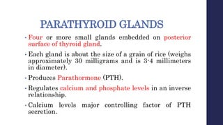
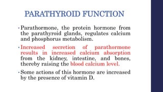
PARULINSTITUTEOFNURSING
PeerGroupteachingPresentation
Prepared by:
Dharti Patel
F. Y. M.Sc. nursing
PARATHYROID GLANDS
• Four or more small glands embedded on posterior
surface of thyroid gland.
• Each gland is about the size of a grain of rice (weighs
approximately 30 milligrams and is 3-4 millimeters
in diameter).
• Produces Parathormone (PTH).
• Regulates calcium and phosphate levels in an inverse
relationship.
• Calcium levels major controlling factor of PTH
secretion.
PARATHYROID FUNCTION
• Parathormone, the protein hormone from
the parathyroid glands, regulates calcium
and phosphorus metabolism.
• Increased secretion of parathormone
results in increased calcium absorption
from the kidney, intestine, and bones,
thereby raising the blood calcium level.
• Some actions of this hormone are increased
by the presence of vitamin D.
CONT...
• Parathormone also tends to lower the blood
phosphorus level.
• Excess parathormone can result in markedly
elevated levels of serum calcium, a potentially
life-threatening situation.
• When the product of serum calcium and
serum phosphorus (calcium and phosphorus)
rises, calcium phosphate may precipitate in
various organs of the body and cause tissue
calcification.
CONT...
• The serum level of ionized calcium
regulates the output of parathormone.
• Increased serum calcium results in
decreased parathormone secretion,
creating a negative feedback system.
To regulate the calcium level in the blood
HYPERPARATHYROIDISM
• When your parathyroid glands create too
much parathyroid hormones in the blood
steam.
OR
• Over activity of one or more of the
parathyroid glands.
• Incidence
Greatest numbers after 60 years
Females males (2:1)
Hyperparathyroidism
• Hyperparathyroidism, which is caused by
overproduction of parathyroid hormone by the
parathyroid glands, is characterized by bone
decalcification and the development of renal
calculi (kidney stones) containing calcium.
Hyperparathyroidism Classifications
• Primary related to secreting tumor of the
parathyroid gland.
Hyperplasia
Adenoma
• Develops when the normal regulatory relationship
between serum calcium levels and parathyroid
hormone secretion is interrupted.
• Primary hyperparathyroidism is related to no
injury, just a parathyroid gone bad.
CONT...
• Increased calcium in the kidneys - kidney stones;
• Increased calcium in the small intestines -
gastrointestinal upset;
• Increased bone resorption occurs, this means there
is increased bone demineralization, bone damage,
osteoporosis.
• These symptoms are part of what is known as the
moans and groans, stones and bones.
Hyperparathyroidism Classifications
•Secondary- occurs when the glands are
hyperplastic because of malfunction of another organ
system. Usually the cause is renal failure or
hyperphosphatemia.
• Compensatory response to chronic hypocalcaemia.
Vitamin D deficiency
Malabsorption
Result of renal failure
Hyperphosphatemia
Hyperparathyroidism Classifications
•Tertiary- Excess production of PTH. It
is characterized by excessive secretion
of PTH after longstanding secondary
hyperparathyroidism.
Pathophysiology
• Normal function of PTH is to increase
bone reabsorption, thereby
maintaining proper balance of calcium
and phosphorus ions in the blood
• Excessive circulating PTH leads to
bone damage, hypercalcemia, and
kidney damage
Clinical Manifestations
• Skeletal disease ( backache, joint pain, bone pain,
pathologic fracture of spine, ribs and long bones).
• Renal involvement (polyuria, polydipsia, gravel, or stones
in the urine, azotaemia, hypertension).
• GI manifestations (thirst, nausea, anorexia, constipation,
abdominal pain, peptic ulcer, GI bleeding, and
pancreatitis).
• Psychiatric manifestations (lethargy, fatigue, depression,
memory loss, confusion, coma, paranoia are sometimes
associated with high calcium levels).
Hyperparathyroidism- Assessment
• Decreased deep tendon reflexes.
• CNS changes-headache, drowsiness,
fatigue
• Muscle weakness & atrophy.
Hyperparathyroidism- Assessment
 Diagnostic Tests
• Increased serum Calcium (>5.2mEq/l)
• Decreased serum Phosphate (<4.5 mg/dl)
• Increased serum PTH levels.
• X-Rays bone demineralization.
• Ultrasound or MRI of glands.
Medical/Surgical Management
To Lower elevated calcium levels
• By Hydration and Calcinuria
• Lasix to promote calcinuria after rehydration has occurred IV
saline solutions both expands volume and acts in the kidney to
inhibit reabsorption of calcium.
• Diet low in calcium and vitamin D
Oral or IV phosphates
Antiresorption Agents
Calcitonin
Mithracin ( Plicamycin)
Glucocorticoids decrease Gl absorption of calcium.
Hyperparathyroidism- Medical
Management
• Goal is to lower severely elevated calcium
levels.
• Long term management of hypercalcemia
with drugs to increase bone reabsorption of
calcium.
Hyperparathyroidism-
Medical/Surgical Management
Secondary Disease
• Calcium supplements
• Vitamin D
Tertiary Disease
• Phosphate Binders
Amphogel, Basaljel
Tums
Surgical Management
PARATHYROIDECTOMY:
•Removal of the parathyroid or
parathyroid tumors.
High risk for injury R/ted to demineralization
of bones resulting in pathologic fractures.-
• Prevent accidents (keep bed in low position,
use side rails, help with ambulation).
• Space activities, plan rest periods.
• Encourage weight bearing 30mins/day to
promote bone rebuilding.
• Avoid high-impact activities or sports.
• Home should be cleared of articles that can
increase risk of falling such as throw
rugs/carpet.
Impaired urinary elimination related to renal
involvement secondary to hypercalcemia and
hypophosphaturia resulting in urolithiasis,
painful urination, haematuria and spasms.
Encourage fluids (2-3000ml /day)
• Prevent urolithiasis ( cranberry juice helps
in making urine more acidic which helps in
preventing renal stone: formation as
calcium is more soluble in acidic urine
than in alkaline urine)
Imbalanced nutrition less than body
requirements related to anorexia
resulting in decreased food intake and
weight loss.
•Low calcium diet to correct hypercalcemia
•Antacids for clients with peptic ulcer
•Diet high in calories without dairy
products
Constipation r/ted adverse effects of
hypercalcemia on GI tract resulting in
decreased frequency of stools and painful
defecation.
• Add fiber to diet
• Increase fluid intake.
• Use stool softeners or laxatives
Knowledge Deficit Related to Dietary Changes
• Low calcium diet
• Limit milk products
Risk For Renal Stones
Increase fluid intake (2-3000ml/day)
Encourage cranberry juice
• Assess for:
flank pain
hematuria
Nursing management of surgical
client
•Monitor for post-op complications:
Haemorrhage, airway obstruction,
recurrent injury to laryngeal nerve.
Eat foods high in calcium.
Encourage ambulation as soon as after
surgery as weightbearing exercises
speeds recalcification
HYPOPARATHYROIDISM
Hypoparathyroidism
• Deficiency of PTH due to hyposecretion of
Parathyroid glands Serum calcium levels
are abnormally low, serum phosphate
levels are abnormally high and tetany may
develop.
Incidence Women are more prone
than men Incidence related to thyroid
surgery.
Hypoparathyroidism- CAUSES
• Iatrogenic i.e.- treatment induced (removal of
viable parathyroid tissue)
Surgery (accidental removal during
thyroidectomy.
Infarction of parathyroid glands because of
an inadequate blood supply to the glands
during surgery.
Trauma.
Idiopathic- autoimmune disorder.
Pathophysiology
Due to the etiological causes
Decrease in the gland function
Resistance to PTH action
Inadequate PTH secretion and increased reabsorption of Ca
in GI tract
Blood calcium fall to low level
Muscular hyper irritability
Uncontrolled spasm, hypo calcaemic tetany
Clinical Manifestations
of hypoparathyroidism
Assessment
• Physical Assessment –
Acute Hypoparathyroidism
Positive CHVOSTEK SIGN (spasms of facial
muscle after a tap over the facial nerve signifying
facial nerve hyper irritability) and TROUSSEAU
SIGNS (Spasm of wrist and hand after
compression of upper arm as by BP cuff.)
Hyperactive Deep Tendon Reflexes
Paraesthesia:- Numbness and tingling of fingers
Hypoparathyroidism- Assessment
• Chronic Hypoparathyroidism
Lethargy, weakness, fatigue
Thin, patchy hair
Brittle nails
Dry scaly skin
Personality changes
Cataracts
Permanent Brain Damage
Hypoparathyroidism- Assessment
•Diagnostic Tests-
Decreased serum Calcium(<4.8mEq/l)
Increased serum Phosphate (>1.75mEq/l)
Low PTH levels
Decreased Urine Calcium
Ophthalmic exam: calcification of lens
Radiograph- increased bone density of Skull
Hypoparathyroidism- Medical
Management
• Active Form of Vit D-to maintain Ca
Rocaltrol
Hytakerol (Dihydortachysterol)
• IV Calcium –
10% Calcium Chloride or Calcium Gluconate Emergency Treatment
• Supplemental Calcium
Oral
Dietary
Address seizures and laryngeal spasms ( Ca gluconate, tracheostomy set )
Parathyroid hormone replacement
Hypoparathyroidism –
Nursing Management
•Knowledge Deficit of Dietary Management/
Medications
High calcium, low phosphate
avoid milk and cheese (high in phosphorus).
Calcium rich food: Beans, Almonds, 7 dark green
leafy vegetables (spinach).
Discuss medications and ensure compliance with
treatment.
CONT…
•High risk for injury: muscle tetany
related to hypocalcaemia
- Assess chvostek and trousseau signs
- Assess for twitching, numbness and tingling,
- Seizures
- Safety precautions for seizures
- Maintain patent airway, keep trach set at
bedside
- Keep ampoule of IV calcium at bedside
hyper_and_hypoparathyrodism.pptx

hyper_and_hypoparathyrodism.pptx

  • 1.
  • 3.
    PARATHYROID GLANDS • Fouror more small glands embedded on posterior surface of thyroid gland. • Each gland is about the size of a grain of rice (weighs approximately 30 milligrams and is 3-4 millimeters in diameter). • Produces Parathormone (PTH). • Regulates calcium and phosphate levels in an inverse relationship. • Calcium levels major controlling factor of PTH secretion.
  • 5.
    PARATHYROID FUNCTION • Parathormone,the protein hormone from the parathyroid glands, regulates calcium and phosphorus metabolism. • Increased secretion of parathormone results in increased calcium absorption from the kidney, intestine, and bones, thereby raising the blood calcium level. • Some actions of this hormone are increased by the presence of vitamin D.
  • 6.
    CONT... • Parathormone alsotends to lower the blood phosphorus level. • Excess parathormone can result in markedly elevated levels of serum calcium, a potentially life-threatening situation. • When the product of serum calcium and serum phosphorus (calcium and phosphorus) rises, calcium phosphate may precipitate in various organs of the body and cause tissue calcification.
  • 7.
    CONT... • The serumlevel of ionized calcium regulates the output of parathormone. • Increased serum calcium results in decreased parathormone secretion, creating a negative feedback system.
  • 8.
    To regulate thecalcium level in the blood
  • 9.
    HYPERPARATHYROIDISM • When yourparathyroid glands create too much parathyroid hormones in the blood steam. OR • Over activity of one or more of the parathyroid glands. • Incidence Greatest numbers after 60 years Females males (2:1)
  • 10.
    Hyperparathyroidism • Hyperparathyroidism, whichis caused by overproduction of parathyroid hormone by the parathyroid glands, is characterized by bone decalcification and the development of renal calculi (kidney stones) containing calcium.
  • 12.
    Hyperparathyroidism Classifications • Primaryrelated to secreting tumor of the parathyroid gland. Hyperplasia Adenoma • Develops when the normal regulatory relationship between serum calcium levels and parathyroid hormone secretion is interrupted. • Primary hyperparathyroidism is related to no injury, just a parathyroid gone bad.
  • 13.
    CONT... • Increased calciumin the kidneys - kidney stones; • Increased calcium in the small intestines - gastrointestinal upset; • Increased bone resorption occurs, this means there is increased bone demineralization, bone damage, osteoporosis. • These symptoms are part of what is known as the moans and groans, stones and bones.
  • 14.
    Hyperparathyroidism Classifications •Secondary- occurswhen the glands are hyperplastic because of malfunction of another organ system. Usually the cause is renal failure or hyperphosphatemia. • Compensatory response to chronic hypocalcaemia. Vitamin D deficiency Malabsorption Result of renal failure Hyperphosphatemia
  • 15.
    Hyperparathyroidism Classifications •Tertiary- Excessproduction of PTH. It is characterized by excessive secretion of PTH after longstanding secondary hyperparathyroidism.
  • 17.
    Pathophysiology • Normal functionof PTH is to increase bone reabsorption, thereby maintaining proper balance of calcium and phosphorus ions in the blood • Excessive circulating PTH leads to bone damage, hypercalcemia, and kidney damage
  • 18.
    Clinical Manifestations • Skeletaldisease ( backache, joint pain, bone pain, pathologic fracture of spine, ribs and long bones). • Renal involvement (polyuria, polydipsia, gravel, or stones in the urine, azotaemia, hypertension). • GI manifestations (thirst, nausea, anorexia, constipation, abdominal pain, peptic ulcer, GI bleeding, and pancreatitis). • Psychiatric manifestations (lethargy, fatigue, depression, memory loss, confusion, coma, paranoia are sometimes associated with high calcium levels).
  • 20.
    Hyperparathyroidism- Assessment • Decreaseddeep tendon reflexes. • CNS changes-headache, drowsiness, fatigue • Muscle weakness & atrophy.
  • 21.
    Hyperparathyroidism- Assessment  DiagnosticTests • Increased serum Calcium (>5.2mEq/l) • Decreased serum Phosphate (<4.5 mg/dl) • Increased serum PTH levels. • X-Rays bone demineralization. • Ultrasound or MRI of glands.
  • 22.
    Medical/Surgical Management To Lowerelevated calcium levels • By Hydration and Calcinuria • Lasix to promote calcinuria after rehydration has occurred IV saline solutions both expands volume and acts in the kidney to inhibit reabsorption of calcium. • Diet low in calcium and vitamin D Oral or IV phosphates Antiresorption Agents Calcitonin Mithracin ( Plicamycin) Glucocorticoids decrease Gl absorption of calcium.
  • 23.
    Hyperparathyroidism- Medical Management • Goalis to lower severely elevated calcium levels. • Long term management of hypercalcemia with drugs to increase bone reabsorption of calcium.
  • 24.
    Hyperparathyroidism- Medical/Surgical Management Secondary Disease •Calcium supplements • Vitamin D Tertiary Disease • Phosphate Binders Amphogel, Basaljel Tums
  • 25.
    Surgical Management PARATHYROIDECTOMY: •Removal ofthe parathyroid or parathyroid tumors.
  • 27.
    High risk forinjury R/ted to demineralization of bones resulting in pathologic fractures.- • Prevent accidents (keep bed in low position, use side rails, help with ambulation). • Space activities, plan rest periods. • Encourage weight bearing 30mins/day to promote bone rebuilding. • Avoid high-impact activities or sports. • Home should be cleared of articles that can increase risk of falling such as throw rugs/carpet.
  • 28.
    Impaired urinary eliminationrelated to renal involvement secondary to hypercalcemia and hypophosphaturia resulting in urolithiasis, painful urination, haematuria and spasms. Encourage fluids (2-3000ml /day) • Prevent urolithiasis ( cranberry juice helps in making urine more acidic which helps in preventing renal stone: formation as calcium is more soluble in acidic urine than in alkaline urine)
  • 29.
    Imbalanced nutrition lessthan body requirements related to anorexia resulting in decreased food intake and weight loss. •Low calcium diet to correct hypercalcemia •Antacids for clients with peptic ulcer •Diet high in calories without dairy products
  • 30.
    Constipation r/ted adverseeffects of hypercalcemia on GI tract resulting in decreased frequency of stools and painful defecation. • Add fiber to diet • Increase fluid intake. • Use stool softeners or laxatives Knowledge Deficit Related to Dietary Changes • Low calcium diet • Limit milk products
  • 31.
    Risk For RenalStones Increase fluid intake (2-3000ml/day) Encourage cranberry juice • Assess for: flank pain hematuria
  • 32.
    Nursing management ofsurgical client •Monitor for post-op complications: Haemorrhage, airway obstruction, recurrent injury to laryngeal nerve. Eat foods high in calcium. Encourage ambulation as soon as after surgery as weightbearing exercises speeds recalcification
  • 33.
  • 34.
    Hypoparathyroidism • Deficiency ofPTH due to hyposecretion of Parathyroid glands Serum calcium levels are abnormally low, serum phosphate levels are abnormally high and tetany may develop. Incidence Women are more prone than men Incidence related to thyroid surgery.
  • 35.
    Hypoparathyroidism- CAUSES • Iatrogenici.e.- treatment induced (removal of viable parathyroid tissue) Surgery (accidental removal during thyroidectomy. Infarction of parathyroid glands because of an inadequate blood supply to the glands during surgery. Trauma. Idiopathic- autoimmune disorder.
  • 36.
    Pathophysiology Due to theetiological causes Decrease in the gland function Resistance to PTH action Inadequate PTH secretion and increased reabsorption of Ca in GI tract Blood calcium fall to low level Muscular hyper irritability Uncontrolled spasm, hypo calcaemic tetany
  • 37.
  • 39.
    Assessment • Physical Assessment– Acute Hypoparathyroidism Positive CHVOSTEK SIGN (spasms of facial muscle after a tap over the facial nerve signifying facial nerve hyper irritability) and TROUSSEAU SIGNS (Spasm of wrist and hand after compression of upper arm as by BP cuff.) Hyperactive Deep Tendon Reflexes Paraesthesia:- Numbness and tingling of fingers
  • 40.
    Hypoparathyroidism- Assessment • ChronicHypoparathyroidism Lethargy, weakness, fatigue Thin, patchy hair Brittle nails Dry scaly skin Personality changes Cataracts Permanent Brain Damage
  • 41.
    Hypoparathyroidism- Assessment •Diagnostic Tests- Decreasedserum Calcium(<4.8mEq/l) Increased serum Phosphate (>1.75mEq/l) Low PTH levels Decreased Urine Calcium Ophthalmic exam: calcification of lens Radiograph- increased bone density of Skull
  • 42.
    Hypoparathyroidism- Medical Management • ActiveForm of Vit D-to maintain Ca Rocaltrol Hytakerol (Dihydortachysterol) • IV Calcium – 10% Calcium Chloride or Calcium Gluconate Emergency Treatment • Supplemental Calcium Oral Dietary Address seizures and laryngeal spasms ( Ca gluconate, tracheostomy set ) Parathyroid hormone replacement
  • 43.
    Hypoparathyroidism – Nursing Management •KnowledgeDeficit of Dietary Management/ Medications High calcium, low phosphate avoid milk and cheese (high in phosphorus). Calcium rich food: Beans, Almonds, 7 dark green leafy vegetables (spinach). Discuss medications and ensure compliance with treatment.
  • 44.
    CONT… •High risk forinjury: muscle tetany related to hypocalcaemia - Assess chvostek and trousseau signs - Assess for twitching, numbness and tingling, - Seizures - Safety precautions for seizures - Maintain patent airway, keep trach set at bedside - Keep ampoule of IV calcium at bedside