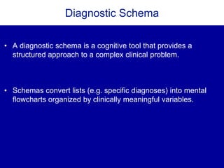
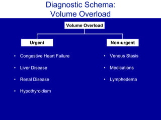
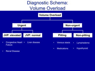
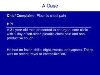
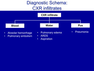
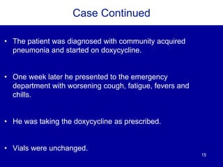
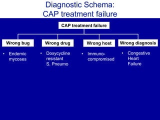
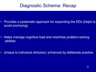
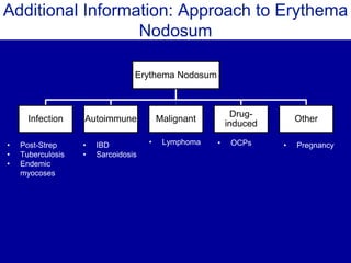
A diagnostic schema is a cognitive tool that allows clinicians to systematically approach a clinical problem by providing an organizing scaffold. A commonly used schema for acute kidney injury (AKI) separates this problem into pre-renal, intrinsic, and post-renal causes. By approaching AKI using these categories, clinicians can systematically access and explore individual illness scripts as potential diagnoses.
Kolkata Call Girls Services 9907093804 @24x7 High Class Babes Here Call Now
Diagnostic Schema
1. When the Script Doesn’t Fit
J Gen Intern Med. 2017 Mar 23
Daniel J Wheeler, MD; Thomas Cascino, MD; Bradley A Sharpe, MD; Denise
M Connor, MD
DIAGNOSTIC SCHEMA
JGIM EXERCISES IN CLINICAL REASONING
Teaching Slides by: Rabih Geha, MD & Denise M. Connor, MD
2. Diagnostic Schema
• A diagnostic schema is a cognitive tool that provides a
structured approach to a complex clinical problem.
• Schemas convert lists (e.g. specific diagnoses) into mental
flowcharts organized by clinically meaningful variables.
6. A Case
Chief Complaint: Pleuritic chest pain
HPI
A 31-year-old man presented to an urgent care clinic
with 1 day of left-sided pleuritic chest pain and non-
productive cough.
He had no fever, chills, night sweats, or dyspnea. There
was no recent travel or immobilization.
8. More HistoryMore History
Medications
• None
Social History
• Lives in California
• One female partner
• Works in office setting
• Modest alcohol use
• No tobacco or recreational
drugs
PMH
• None
PSH
• None
Family History
• No significant FH
9. Physical Exam
T 99F BP 117/90 HR 70 RR 19 Sat 96% RA
• Young man in no acute distress
• Chest: clear to ausculatation, normal percussion
• CV: regular rate and rhythm with no murmurs
• Abdomen: Soft, non-tender, non-distended
• Skin: No rashes
• Remainder of the exam is normal
15. 15
Case Continued
• The patient was diagnosed with community acquired
pneumonia and started on doxycycline.
• One week later he presented to the emergency
department with worsening cough, fatigue, fevers and
chills.
• He was taking the doxycycline as prescribed.
• Vials were unchanged.
23. Imaging
CT chest with contrast
1. Scattered centrilobular nodules in the left lung with
associated ground glass.
2. Consolidation in the lingula, left hilar adenopathy
and a small left-sided pleural effusion.
3. No pulmonary embolism.
24. 24
Pause
How would you incorporate these findings into a updated
problem representation?
26. Diagnostic Schema:
Pulmonary Infiltrates with Eosinophilia
Pulmonary Infiltrates + eosinophilia
Hypersensitivity Idiopathic
• Drug
reactions
Infection
• Parasites • Acute
eosinophilic
pneumonia
Autoimmune Dx
• Eosinophilic
granulomatosis
with polyangiitis
27. 27
Case Continued
• The patient was diagnosed with a non-resolving
pneumonia and started on Levofloxacin.
• Four days later, he returned to the ED with worsening
pleuritic pain, cough, night sweats and multiple new,
painful, red lesions on his right lower extremity.
29. 29
Case Continued: Further History
• After additional history was asked, the patient mentioned
that two weeks prior to symptom onset, he had participated
in a 10-mile outdoor race near San Diego, California that
involved scrambling through an obstacle course of dust, dirt,
and mud.
30. 30
Pause
What is your updated problem representation?
What is your prioritized differential diagnosis?
32. 32
Case Continued
• Coccidioides immunodiffusion was positive with a
complement fixation titer of 1:4
• The patient was started on Fluconazole with resolution
of his symptoms over the next month.
33. 33
Coccidiomycosis
• Coccidiomycosis is caused by C. imminitis and C. posadasii,
dimorphic fungi endemic to the South Western United States.
• Broad spectrum of disease from self-limited flu-like illness to severe
disseminated disease in less than 1% of infections.
• Other manifestations include rash (erythema nodosum, erythema
multiforme) and peripheral eosinophilia.
• A travel history is important in making the diagnosis.
34. 34
Diagnostic Schema: Recap
• Provides a systematic approach for expanding the DDx (helps to
avoid anchoring)
• Helps manage cognitive load and maximize problem-solving
abilities
• Unique to individual clinicians; enhanced by deliberate practice
35. 35
Acknowledgements
• We thank Mark Henderson MD, Jeff Kohlwes MD, and Reza
Manesh MD for their critical review of this material
Teacher’s Guide:
1. Before clicking on the slide, ask learners if they have heard of this concept before
2. If yes, explore what this term means to them and solicit examples
3. Click slide to reveal definition
Teacher’s Guide:
1. Before clicking slide, ask learners if they have an approach to how they think about LE edema – i.e. you may hear organ system based approaches
2. Write learners’ examples/approaches on the board
3. Click slide to show a list of potential causes
Teacher’s Guide:
1. Reveal example of a schema and discuss; if learners gave other examples previously, you can compare/contrast the different approaches
2. Key to make the point that there is no one gold standard diagnostic schema for a given problem, but rather that different clinicians, depending on their prior knowledge, experience and practice patterns develop schema that make sense to them and help them to organize their thinking around complex problems.
Teacher’s guide
Use this more elaborated schema as an example of the development of a provider’s schema over time. With experience, providers incorporate additional branch points to further refine their diagnostic schema
In the urgent causes of volume overload, the jugular venous pressure to further divide the causes into JVP elevated (CHF, renal disease) and JVP negative (liver disease)
Similarly the pitting versus non-pitting distinction can be used to further divide these non-urgent causes of edema.
Teacher’s Guide
Start by drawing out your learners’ schema to this problem and consider emphasizing the important of prioritizing urgent over non-urgent causes.
Depending on the discussion, you can offer your own approach, or add nuance to what the learners discuss
As an example:
There are six causes of potentially lethal chest pain (CP). A schema for CP might have a branch point of urgent/non-urgent, with clinicians always considering the urgent/life-threatening potential causes first:
Cardiac (3)
Myocardial Infarction
Tamponade
Aortic Dissection
Pulmonary (2)
Pulmonary Embolism
Pneumothorax
Esophageal (1)
Perforation
After considering the life-threatening causes, non-urgent causes may be considered using an anatomic approach:
***Note: You do not need to cover all of the possible causes, but rather, help your learners consider which organ systems to consider, with just 1 exemplar in each category to bring it to life:
Skin
Herpes Zoster
Muscle
Muscle Strain
Bone (sternal, ribs)
Fracture
Pulmonary
Pneumonia
Cardiac
Pericarditis
Esophagus/stomach
GERD
Mediastinum
Mediastinitis
Teacher’s Guide
1. Ask learners what they would want to focus on in the physical exam (and why) before revealing next slide
Teacher’s guide
Ask someone (or a team of learners) to interpret the chest-x-ray:
For example: there is an consolidation in the left lower lobe. No cardiomegaly or pleural effusions.
Teacher’s guide
The problem representation is a one sentence summary that highlights the defining features of case (for more please see:: http://www.sgim.org/web-only/clinical-reasoning-exercises/problem-representation-overview)
Compare/contrast a few different problem representations from your learners; a good challenging question is: Why would you include that detail, how does that detail change your thinking about this patient?
An example would be:
31yo previously healthy man with acute pleuritic chest pain, cough, and a unilateral infiltrate on CXR.
Teacher’s guide
1. Scribe learners’ answers on the board
2. Click on to the next slide to show as one helpful approach
Teacher’s guide
Compare and contrast approaches mentioned by learners vs. that shown on the slide.
Make the point that splitting a long list of diagnoses (ARDS, PE, pneumonia, aspiration, etc.) into smaller, categories makes it easier to (1) remember and (2) incorporate new diagnoses into the schema over time.
The categories chosen can vary by schema or provider. For example, an anatomic categorization is one way to approach chest pain (see prior example; but other providers may chose to use organ system based approach). In the schema of CXR infiltrates one set of categories is the nature of the fluid causing the infiltrate.
Teacher’s guide
At this point learners should come up with 1-3 competing diagnosis.
Encourage learners to discuss the “for” and “against” categories for each diagnosis
Consider using a table on the board to visually represent the discussion.
Teacher’s guide
The important piece to incorporate is the failure to improve with antibiotic therapy aimed at community acquired pneumonia.
Example: 31 year-old previously healthy man with ongoing pleuritic chest pain and cough despite completing therapy for CAP.
2. Would gently guide learner’s in that direction before proceeding to the next slide.
Teacher’s guide
1.Before clicking on animation to reveal the question, ask learners what the crux of the problem being solved is at this point (goal – to help students identify CAP treatment failure)
2. Similar to above, have the learners discuss, and write possible causes on the board.
3.Advance to the next slide to see one approach.
Teacher’s guide:
Examples
Wrong “bug”
CAP is the correct diagnosis, but the causative organism is unusual:
gram-negative rod, virus, endemic fungus
Wrong “drug”
CAP is the correct diagnosis, but the causative organism is resistant to treatment:
doxycycline-resistant S. pneumonia
Wrong “host”
CAP is the correct diagnosis, but the patient does not improve due to anatomic obstruction, immunocompromise, or disease severity. (e.g. malignancy, HIV)
Wrong diagnosis-
Patient has an alternative diagnosis, or a complication of CAP:
interstitial lung disease, pulmonary abscess, recurrent aspiration, congestive heart failure
Teacher’s guide
1. Ask a learner to walk you through the labs and how they impact their diagnostic thinking.
2. Consider emphasizing the mild leukocytosis and low grade eosinophilia.
3. Pending time, may ask learners to suggest next diagnostic steps (and why).
Teacher’s guide
1. The read for the CT is on the next slide.
Teacher’s guide
The important features to include in an updated problem representation include:
Eosinophilia
CT with evidence of consolidation
Example: 31yo previously healthy man with ongoing, subacute pleuritic chest pain despite CAP therapy, now with eosinophilia, lingular consolidation, pleural effusion and lymphadenopathy.
Teacher’s guide
As above, consider using the a board to list the diagnoses mentioned by the students and then use the following slide to illustrate an exemplar schema.
As learner’s begin to outline a collective schema, would make the emphasize how their precise problem representation in the previous slide makes it easier to generate a schema.
Teacher’s guide
1. The picture shows lesions characteristic of erythema nodosum: nodular lesions over the anterior shin that are exquisitely tender.
2. For the sake of time, we have not included a discussion of E.Nodosum in this section. However, if additional time is available, please refer to “additional information” slide (slide 36) for a discussion of E.Nodosum.
Teacher’s guide
At this point, important features of the problem representation include:
Development of erythema nodosum
Travel to the San Joaquin Valley
Example: 31yo previously healthy man with pleuritic chest pain and cough after a trip to the San Joaquin Valley found to have eosinophilia and on-going symptoms despite CAP therapy.
Teacher’s guide
If there is time, please use this as a guide for the discussion on slide 28: E.Nodosum
Erythema Nodosum
Definition: A form of panniculitis that presents as an outcropping of painful, erythematous nodules, usually on ventral aspect of the lower extremities.
Causes: In the majority of cases (55%), no clear cause is identified. However, the presence of E.Nodosum should prompt investigation for systemic causes. There can be categorized as:
Infection (post-streptococcal infection,post-enteritis, tuberculosis and endemic mycoses including coccidiomycosis)
Autoimmune disease (inflammatory bowel disease and sarcoidosis)
Drug-induced (oral contraceptives, azathioprine)
Malignancy (lymphoma)
Other (pregnancy)