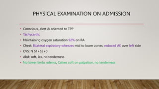
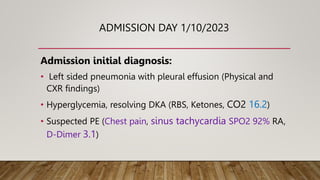
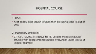
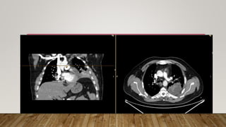
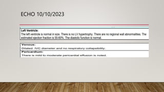
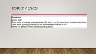
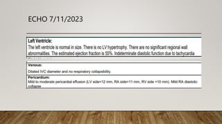
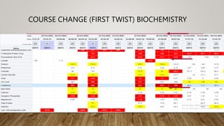
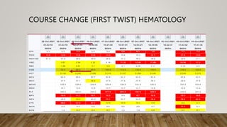
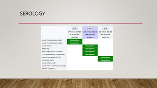
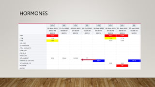
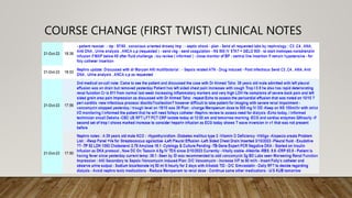
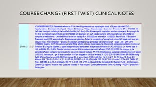
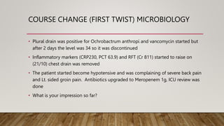
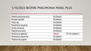
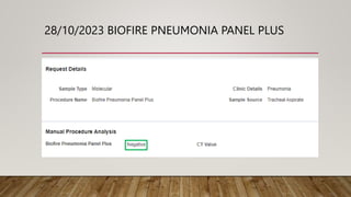
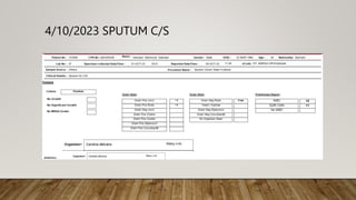
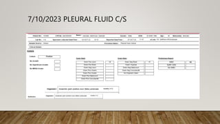
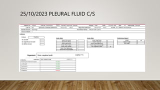
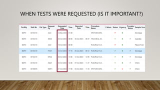
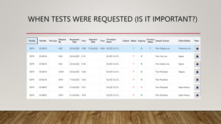
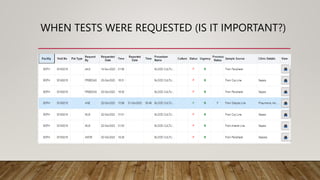
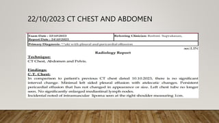
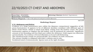
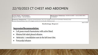
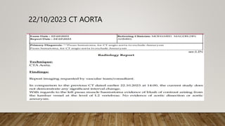
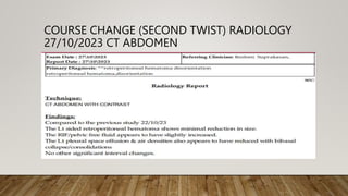
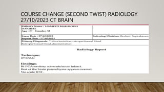
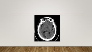
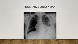
CASE PRESENTATION
DONE BY DR. MOHAMED TAHA
SUPERVISED BY DR. AHMED TAHA
HISTORY AND PRESENTATION
• 39 years old male presented to the ER with left sided
chest pain, radiating to the back/left shoulder for 3/7.
• He describes the pain as spasm-like in nature, it was
preceded by history of vigorous physical exercises 2
days, and it worsens with respiration and cough.
• No history of hemoptysis, fever, dizziness, LOC, leg pain
or swelling and history of sick contact or recent travel.
• K/C/O DM2 (not compliant to his medications), DLP,
Hypothyroidism, Vitiligo & Alopecia areata
• No Past surgical hx
• Ex-smoker
• Non-drinker
PHYSICAL EXAMINATION ON ADMISSION
• Conscious, alert & oriented to TPP
• Tachycardic
• Maintaining oxygen saturation 92% on RA
• Chest: Bilateral expiratory wheezes mid to lower zones, reduced AE over left side
• CVS: N S1+S2+0
• Abd: soft, lax, no tenderness
• No lower limbs edema, Calves soft on palpation, no tenderness
INVESTIGATIONS
• WBC 17.3 (NE% 82.6), Hb 12.5, Plt 301, D-Dimer 3.1
• Na 133.9, K 4.69, Cl 94.9, CO2 16.2, RFT NL, Cardiac enzymes NL, Trop-I 0
(repeated set of Trop-I <0.010)
• Ketones 2.7 (was 4.8), BS 13.7 (15.4)
• VBG PH 7.397 PCO2 33.3 HCO3 20 BE -3.5
• ECG sinus tachycardia
• CXR was done
WHAT DO YOU SEE?
• Do you think this patient require an admission?
• Why?
ADMISSION DAY 1/10/2023
Admission initial diagnosis:
• Left sided pneumonia with pleural effusion (Physical and
CXR findings)
• Hyperglycemia, resolving DKA (RBS, Ketones, CO2 16.2)
• Suspected PE (Chest pain, sinus tachycardia SPO2 92% RA,
D-Dimer 3.1)
HOSPITAL COURSE
1- DKA:-
• Kept on low dose insulin infusion then on sliding scale till out of
DKA.
2- Pulmonary Embolism:-
• CTPA (1/10/2023): Negative for PE. Lt sided moderate pleural
effusion with collapse/consolidation involving Lt lower lobe & Lt
lingular segment
ECHO 10/10/2023
ECHO 21/10/2023
ECHO 7/11/2023
LEFT SIDED PNEUMONIA WITH PLEURAL EFFUSION
• Left Sided Pleural Drain was Kept by Interventional Radiologist on 3/10
until 21/10
• Pleural Fluid Exudative: Chest tube kept draining continuously
• TB-Gene Expert PCR: Negative
• Received Tazocin 1-21/10/2023 and Azithromycin 1-2-10/2023
• Resp panel (1/10): Streptococcus agalactia
• CRP markedly improved
• US chest report (9/10): Scanning both CP angles revealed bilateral
minimal pleural effusion
12 DAYS POST DRAINAGE
COURSE
• High Resolution CT Chest(10/10) : Significant decrease in the previously
seen left pleural effusion volume with mild residual/insisted
appearance.
• On (10/10) ECHO was done showing mild to moderate pericardial
effusion
• The patient management kept the same, draining and monitoring
inflammatory markers and the patient was stable until (20/10)
COURSE CHANGE (FIRST TWIST) CLINICAL
COURSE CHANGE (FIRST TWIST) BIOCHEMISTRY
COURSE CHANGE (FIRST TWIST) HEMATOLOGY
SEROLOGY
HORMONES
COURSE CHANGE (FIRST TWIST) CLINICAL NOTES
COURSE CHANGE (FIRST TWIST) MICROBIOLOGY
• Plural drain was positive for Ochrobactrum anthropi and vancomycin started but
after 2 days the level was 34 so it was discontinued
• Inflammatory markers (CRP230, PCT 63.9) and RFT (Cr 811) started to raise on
(21/10) chest drain was removed
• The patient started become hypotensive and was complaining of severe back pain
and Lt. sided groin pain. Antibiotics upgraded to Meropenem 1g, ICU review was
done
• What is your impression so far?
1/10/2023 BIOFIRE PNEUMONIA PANEL PLUS
28/10/2023 BIOFIRE PNEUMONIA PANEL PLUS
4/10/2023 SPUTUM C/S
7/10/2023 PLEURAL FLUID C/S
25/10/2023 PLEURAL FLUID C/S
WHEN TESTS WERE REQUESTED (IS IT IMPORTANT?)
BLOOD CULTURES
• All blood cultures from admission till discharge came
negative.
RADIOLOGY
22/10/2023 CT CHEST AND ABDOMEN
22/10/2023 CT AORTA
• On (22/10) CT was performed for this pain showing Lt sided retroperitoneal
hematoma
• IR Procedure done (23/10/23): Selective left lumbar angiography shows contrast
extravasation from L2/3 lumbar artery. Embolization done with particles 300-500U
and one coil 4mm. Bleeding stopped. The right CFA access closed with angioseal.
23/10/2023 LUMBAR ARTERY EMBOLIZATIION
COURSE CHANGE (SECOND TWIST) CLINICAL
• Presentation of stroke?
• CT Brain done
COURSE CHANGE (SECOND TWIST) RADIOLOGY
27/10/2023 CT ABDOMEN
COURSE CHANGE (SECOND TWIST) RADIOLOGY
27/10/2023 CT BRAIN
WHAT DO YOU SEE?
• On (6/11) patient was discharged from the ICU
• Kept on Linezolid 27/10 to 15/11/2023 until inflammatory markers and
RFT normalized
• CVP removed on (12/11) - Dialysis Catheter removed on (14/11)
• Patient was discharged on (16/11)
DISCHARGE CHEST X-RAY
THANK YOU

Case PRESENTATION A Taha modifications.pptx

  • 1.
    CASE PRESENTATION DONE BYDR. MOHAMED TAHA SUPERVISED BY DR. AHMED TAHA
  • 2.
    HISTORY AND PRESENTATION •39 years old male presented to the ER with left sided chest pain, radiating to the back/left shoulder for 3/7. • He describes the pain as spasm-like in nature, it was preceded by history of vigorous physical exercises 2 days, and it worsens with respiration and cough. • No history of hemoptysis, fever, dizziness, LOC, leg pain or swelling and history of sick contact or recent travel.
  • 3.
    • K/C/O DM2(not compliant to his medications), DLP, Hypothyroidism, Vitiligo & Alopecia areata • No Past surgical hx • Ex-smoker • Non-drinker
  • 4.
    PHYSICAL EXAMINATION ONADMISSION • Conscious, alert & oriented to TPP • Tachycardic • Maintaining oxygen saturation 92% on RA • Chest: Bilateral expiratory wheezes mid to lower zones, reduced AE over left side • CVS: N S1+S2+0 • Abd: soft, lax, no tenderness • No lower limbs edema, Calves soft on palpation, no tenderness
  • 5.
    INVESTIGATIONS • WBC 17.3(NE% 82.6), Hb 12.5, Plt 301, D-Dimer 3.1 • Na 133.9, K 4.69, Cl 94.9, CO2 16.2, RFT NL, Cardiac enzymes NL, Trop-I 0 (repeated set of Trop-I <0.010) • Ketones 2.7 (was 4.8), BS 13.7 (15.4) • VBG PH 7.397 PCO2 33.3 HCO3 20 BE -3.5 • ECG sinus tachycardia • CXR was done
  • 6.
  • 7.
    • Do youthink this patient require an admission? • Why?
  • 8.
    ADMISSION DAY 1/10/2023 Admissioninitial diagnosis: • Left sided pneumonia with pleural effusion (Physical and CXR findings) • Hyperglycemia, resolving DKA (RBS, Ketones, CO2 16.2) • Suspected PE (Chest pain, sinus tachycardia SPO2 92% RA, D-Dimer 3.1)
  • 9.
    HOSPITAL COURSE 1- DKA:- •Kept on low dose insulin infusion then on sliding scale till out of DKA. 2- Pulmonary Embolism:- • CTPA (1/10/2023): Negative for PE. Lt sided moderate pleural effusion with collapse/consolidation involving Lt lower lobe & Lt lingular segment
  • 11.
  • 12.
  • 13.
  • 14.
    LEFT SIDED PNEUMONIAWITH PLEURAL EFFUSION • Left Sided Pleural Drain was Kept by Interventional Radiologist on 3/10 until 21/10 • Pleural Fluid Exudative: Chest tube kept draining continuously • TB-Gene Expert PCR: Negative • Received Tazocin 1-21/10/2023 and Azithromycin 1-2-10/2023 • Resp panel (1/10): Streptococcus agalactia • CRP markedly improved • US chest report (9/10): Scanning both CP angles revealed bilateral minimal pleural effusion
  • 15.
    12 DAYS POSTDRAINAGE
  • 16.
    COURSE • High ResolutionCT Chest(10/10) : Significant decrease in the previously seen left pleural effusion volume with mild residual/insisted appearance. • On (10/10) ECHO was done showing mild to moderate pericardial effusion • The patient management kept the same, draining and monitoring inflammatory markers and the patient was stable until (20/10)
  • 17.
    COURSE CHANGE (FIRSTTWIST) CLINICAL
  • 18.
    COURSE CHANGE (FIRSTTWIST) BIOCHEMISTRY
  • 19.
    COURSE CHANGE (FIRSTTWIST) HEMATOLOGY
  • 20.
  • 21.
  • 22.
    COURSE CHANGE (FIRSTTWIST) CLINICAL NOTES
  • 23.
    COURSE CHANGE (FIRSTTWIST) CLINICAL NOTES
  • 24.
    COURSE CHANGE (FIRSTTWIST) MICROBIOLOGY • Plural drain was positive for Ochrobactrum anthropi and vancomycin started but after 2 days the level was 34 so it was discontinued • Inflammatory markers (CRP230, PCT 63.9) and RFT (Cr 811) started to raise on (21/10) chest drain was removed • The patient started become hypotensive and was complaining of severe back pain and Lt. sided groin pain. Antibiotics upgraded to Meropenem 1g, ICU review was done • What is your impression so far?
  • 25.
  • 26.
  • 27.
  • 28.
  • 29.
  • 30.
    WHEN TESTS WEREREQUESTED (IS IT IMPORTANT?)
  • 31.
    WHEN TESTS WEREREQUESTED (IS IT IMPORTANT?)
  • 32.
    WHEN TESTS WEREREQUESTED (IS IT IMPORTANT?)
  • 33.
    BLOOD CULTURES • Allblood cultures from admission till discharge came negative.
  • 34.
  • 35.
  • 36.
  • 37.
  • 39.
  • 40.
    • On (22/10)CT was performed for this pain showing Lt sided retroperitoneal hematoma • IR Procedure done (23/10/23): Selective left lumbar angiography shows contrast extravasation from L2/3 lumbar artery. Embolization done with particles 300-500U and one coil 4mm. Bleeding stopped. The right CFA access closed with angioseal.
  • 41.
  • 42.
    COURSE CHANGE (SECONDTWIST) CLINICAL
  • 43.
    COURSE CHANGE (SECONDTWIST) CLINICAL
  • 44.
    • Presentation ofstroke? • CT Brain done
  • 45.
    COURSE CHANGE (SECONDTWIST) RADIOLOGY 27/10/2023 CT ABDOMEN
  • 46.
    COURSE CHANGE (SECONDTWIST) RADIOLOGY 27/10/2023 CT BRAIN
  • 47.
  • 49.
    • On (6/11)patient was discharged from the ICU • Kept on Linezolid 27/10 to 15/11/2023 until inflammatory markers and RFT normalized • CVP removed on (12/11) - Dialysis Catheter removed on (14/11) • Patient was discharged on (16/11)
  • 50.
  • 51.