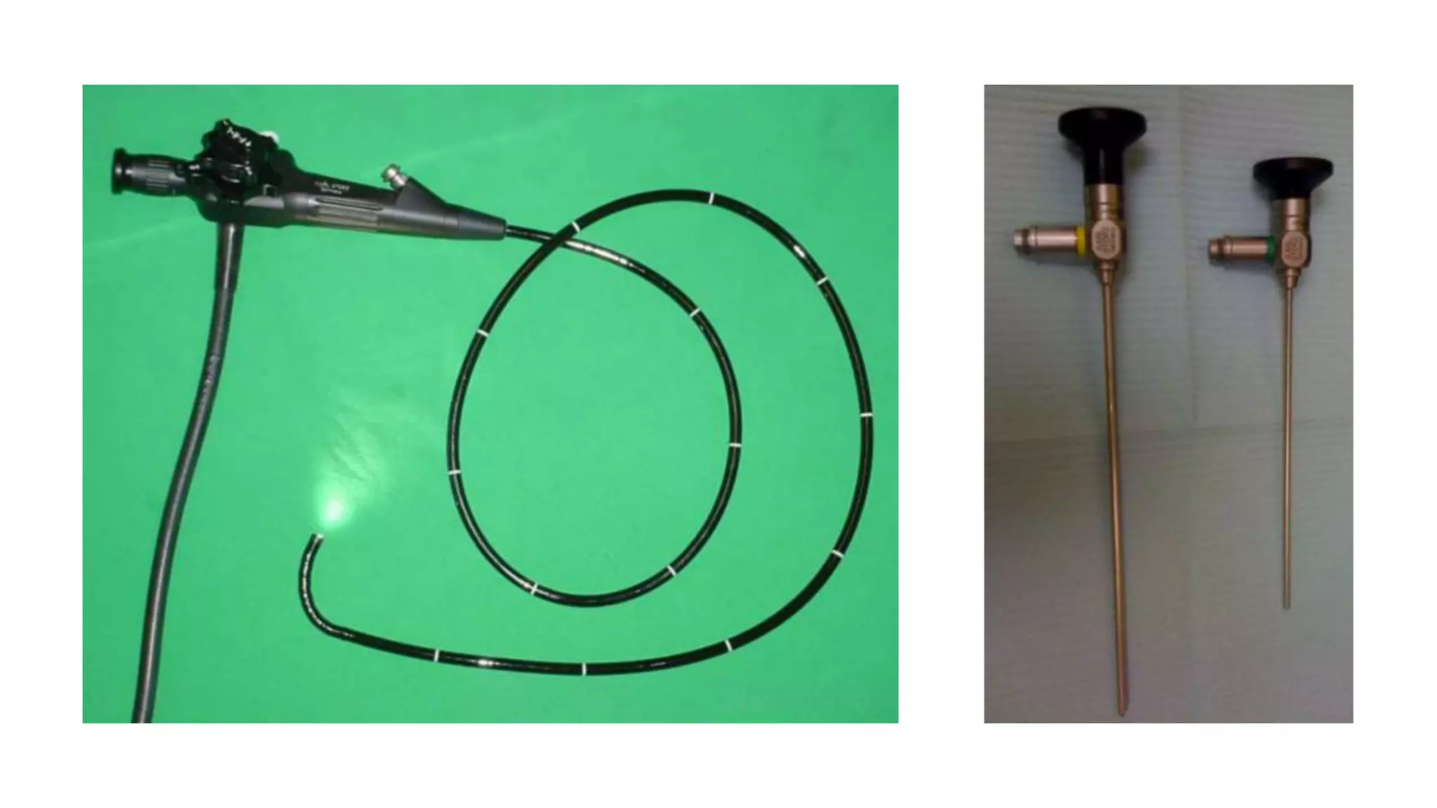
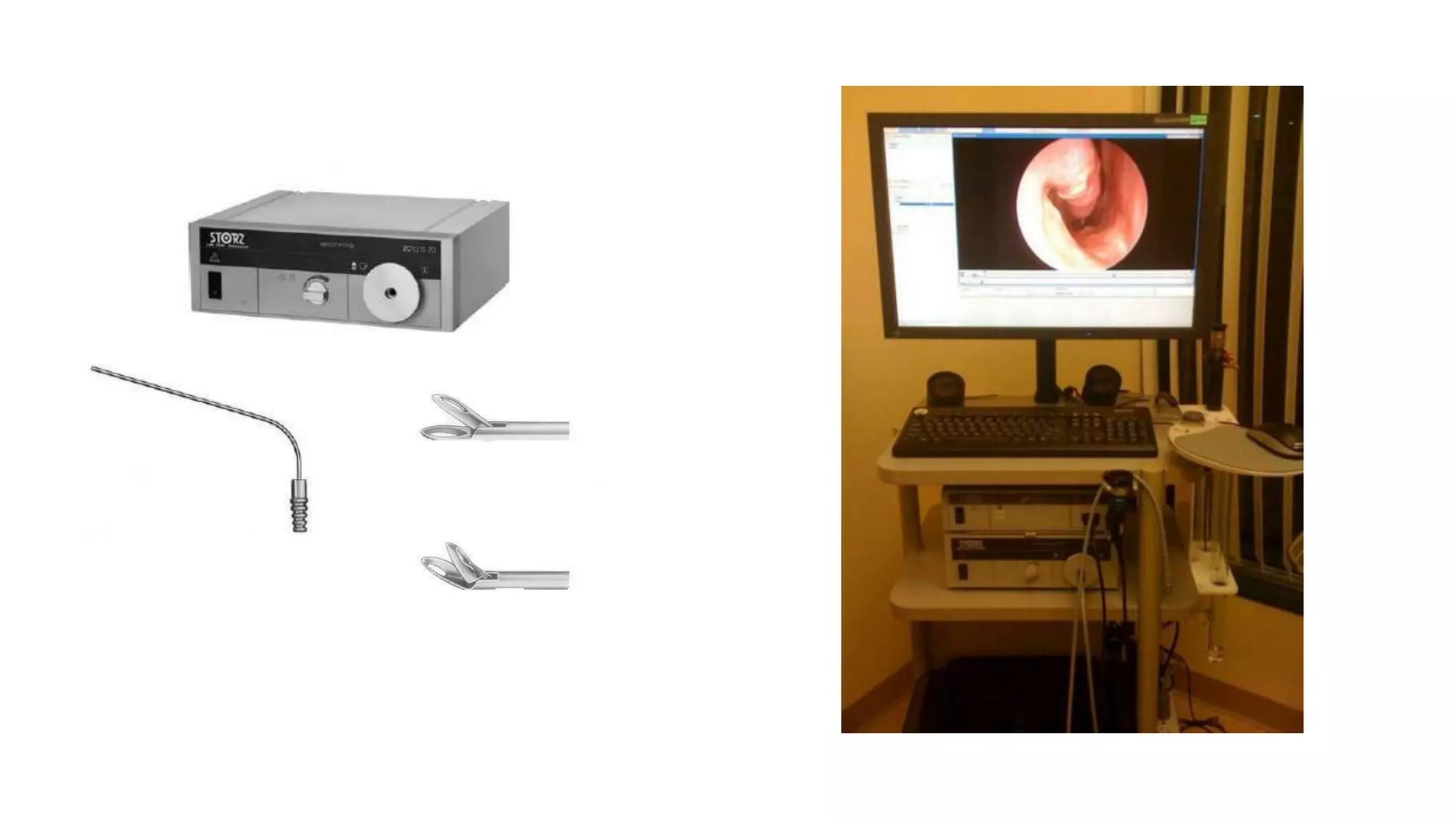
DIAGNOSTIC NASAL
ENDOSCOPY
Dr.Abam Fasal
ENT PG II YEAR
AL-AMEEN MEDICAL COLLEGE
INTRODUCTION
• Nasal endoscopy involves evaluation of the nasal and sinus
passages with direct vision using a magnified high-quality
view.
• It is a commonly performed procedure in the
otolaryngologist’s office and serves as an objective
diagnostic tool in the evaluation of nasal mucosa, sinonasal
anatomy, and nasal pathology.
INTRODUCTION
• Nasal endoscopy may be accomplished with either a flexible
fiberoptic endoscope or a rigid endoscope.
• When performed by experienced practitioners, both flexible
endoscopy and rigid endoscopy are usually well tolerated.
INTRODUCTION
• The fiberoptic telescope has the advantage of being flexible
and generally smaller in diameter, which means that it is
readily manipulated in multiple directions to permit
visualization of tight areas.
• However, flexible endoscopy requires 2 hands for
manipulation of the instrument and is thus a more difficult
procedure.
INTRODUCTION
• Traditionally, flexible endoscopy has provided inferior
visualization, but this drawback has been overcome with the
development of digital flexible endoscopes.
• The rigid endoscope provides superior image clarity,
facilitates culture and tissue sampling, controls epistaxis
better, and affords the endoscopist the ability to perform
surgery.
INTRODUCTION
• Rigid endoscopes for the nose come in diameters of 2.7-4
mm and have tips of different angles (generally 0-70º),
allowing the physician to visualize various sinuses and areas
within the nasal cavity and sinuses.
• In addition to affording superior visualization, nasal
endoscopy provides improved illumination, greater
magnification, and the ability to navigate directly to
pathologic areas.
INTRODUCTION
• In one study, rigid nasal endoscopy identified nasal
pathology in almost 40% of patients who had normal
examinations on anterior rhinoscopy.
• Endoscopy plays an important role in the preoperative,
postoperative, and medical management of patients with
sinonasal complaints.
INDICATIONS
Nasal endoscopy has a clear role in the identification of
sinonasal disease in patients presenting to the
otolaryngologist's office. It should be viewed as an essential
component of a complete examination of the nose and
sinuses.
Indications for nasal endoscopy include, but are not limited to,
the following
INDICATIONS
• Initial identification of disease in patients experiencing
sinonasal symptoms (eg, mucopurulent drainage, facial pain
or pressure, headache, nasal obstruction or congestion, or
decreased sense of smell)
• Evaluation of patient’s response to medical treatment (eg,
resolution of polyps, purulent secretions, or mucosal edema
and inflammation after treatment with topical nasal steroids,
antibiotics, oral steroids, and antihistamines)
INDICATIONS
• Evaluation of unilateral disease.
• Evaluation of patients with complications or impending
complications of sinusitis.
• Obtaining a culture of purulent secretions.
• Debridement and removal of crusting, mucus, and fibrin from
obstructed nasal and sinus cavities after functional endoscopic
sinus surgery.
INDICATIONS
• Evaluation for recurrence of pathology after FESS to (this is
particularly valuable in monitoring for recurrence of intranasal
tumors).
• Evaluation and biopsy of nasal masses or lesions.
• Evaluation of the nasopharynx for lymphoid hyperplasia,
eustachian tube problems, and nasal obstruction.
• Evaluation of cerebrospinal fluid (CSF) leak.
INDICATIONS
• To determine the effects of conditions such as severe allergies,
immune deficiencies and mucociliary disorders (disorders that
affect mucous membranes and cilia).
• Evaluation and treatment of epistaxis.
• Evaluation of hyposmia or anosmia.
• Evaluation and treatment of nasal foreign bodies.
CONTRAINDICATIONS
• No absolute contraindications to nasal endoscopy exist;
however, some patient populations are at increased risk for
complications.
• In patients who have a history of a bleeding disorder or are
receiving anticoagulants, nasal endoscopy should be
performed carefully so as not to provoke bleeding.
CONTRAINDICATIONS
• Additionally, in an anxious patient or a patient with
cardiovascular disease, there is a risk of a vasovagal episode.
TECHNICAL CONSIDERATIONS
• Nasal endoscopy and imaging are the 2 most widely used
objective measures in the diagnosis of chronic rhinosinusitis
(CRS).
• Endoscopy has multiple uses in the management of patients
with sinonasal symptoms and plays an important role in both
the preoperative and postoperative management of
patients.
TECHNICAL CONSIDERATIONS
• The Lund-Kennedy endoscopic scoring system quantifies the
pathologic states of the nose and paranasal sinuses, focusing
on the presence of polyps, discharge, edema, scarring, or
adhesions and crusting.
• Endoscopic staging is performed bilaterally and typically
takes place during the initial evaluation, preoperatively, and
postoperatively at regular intervals (intervals of 3, 6, 12, 24,
and 36 months are recommended).
TECHNICAL CONSIDERATIONS
• Lund-Kennedy scores range from 0 to 20.
• Polyps are graded as absent (0), present in the middle
meatus (1), or present beyond the middle meatus (3).
• Discharge is graded as not present (0), thin (1), or thick and
purulent (2). Edema, scarring, and crusting are each graded
as absent (0), mild (1), or severe (2).
TECHNICAL CONSIDERATIONS
• Interrater agreement studies examining the reliability of the
Lund-Kennedy endoscopic scoring system demonstrate that
in a high proportion of cases, 2 independent observers agree
on the examination findings.
• The addition of nasal endoscopy to the care of patients with
CRS has resulted in improved diagnostic accuracy.
TECHNICAL CONSIDERATIONS
• In combination with established symptom criteria,
endoscopic findings improve the specificity, positive
predictive value, and negative predictive value of assessment
for CRS.
• This development suggests that the use of diagnostic
endoscopy may help decrease the need for computed
tomography (CT) and reduce costs and radiation exposure.
TECHNICAL CONSIDERATIONS
• Ferguson et al in their 2012 study found that the sensitivity
of nasal endoscopy was 24% and the specificity was 100%,
with the finding of mucopurulence only present in those
patients with positive CRS on CT.
• Diagnostic endoscopy has also proven to be an integral
aspect of staging for allergic fungal sinusitis (AFS).
Kupferberg-Kuhn created 4 stages of disease for AFS on the
basis of findings from nasal endoscopy.
TECHNICAL CONSIDERATIONS
• Endoscopy is also considered the criterion standard for tissue
sampling and culture collection when performed properly.
• Traditionally, inferior meatal puncture was the diagnostic
method used to identify pathogens in sinusitis.
TECHNICAL CONSIDERATIONS
• Study results have documented a greater than 90% correlation
between endoscopically obtained cultures and maxillary sinus
aspirates, making endoscopically guided cultures the current criterion
standard.
• 90% of the specimens collected by nasal endoscopy resulted in a
culture with 2 or fewer bacterial isolates, and 55% of these contained
a single isolate.These results are superior to those obtained by means
of a nasopharyngeal swab.
EQUIPMENT
• 4 mm 0 and 30" sinuscopes
• 2.7 mm 0° and 30" sinuscopes in cases of children and narrow nasal
cavity
• Freer's elevator
• Suction cannula
• Biopsy forceps
• Antifog solution/savlon to avoid fogging.
EQUIPMENT
• Rigid endoscopes are made in various angles (eg, 0º, 30º, 45º, and
70º).
• The angled telescopes are used to see around corners and to evaluate
areas not easily examined under direct vision.
• The 4 mm 30º scope has been shown to provide sufficient
illumination and an adequate field of vision and may therefore be the
most useful telescope in an average patient.
EQUIPMENT
• Nasal endoscopes also come in pediatric sizes (2.7 mm),
which are also available in various angles and which may be
used for increased comfort in adults.
• The use of video systems has become increasingly popular in
nasal endoscopy.
EQUIPMENT
• Minimum requirements for basic nasal endoscopy include an
endoscope, a high-quality light source and light cable, and a
suction device to clear secretions.
• In addition, sinus instruments (eg, a Freer elevator, a
Blakesley forceps, or a through-cutting instrument for
obtaining biopsy specimens) can be useful for performing
procedures.
PATIENT PREPARATION
Patient preparation includes adequate anesthesia and appropriate
positioning
Anesthesia
Before nasal endoscopy, nasal cavities are often sprayed with a nasal
decongestant, such as oxymetazoline.
PATIENT PREPARATION
It is sometimes useful to performing endoscopy without decongestion,
however, particularly for patients with presumed turbinate
hypertrophy, in whom it is worthwhile to perform endoscopy before
and after decongestion.
In certain patients, especially those who will be undergoing a surgical
procedure, local anesthesia is employed as well; lidocaine 4% is a
commonly used topical anesthetic.
PATIENT PREPARATION
• When cultures are indicated, however, anesthetics should be
avoided so as to provide the best chances of bacterial growth
on the swab medium.
• Anesthetics are typically applied either with a spray atomizer
or directly on a cotton pledget.
PATIENT PREPARATION
• The topical anesthetic should be applied to the inferolateral
surface of the middle turbinate, to the surface of the inferior
turbinate, and to any other sites where pressure may be exerted
by insertion of the scope.
• About 7 ml of 4% xylocaine is mixed with 10 drops of
xylometazoline.
• Cotton pledgets are dipped in the solution, squeezed dry and
used to pack the nasal cavity.
PATIENT PREPARATION
• Pledgets are packed in the inferior, middle and superior
meati.
• Packs are left in place for full 5 minutes.
PATIENT PREPARATION
Positioning
During the endoscopic examination, patients should be seated upright
in the examination chair.
The patient’s head can be manipulated as needed to afford better
viewing of the nasopharynx or the olfactory cleft area.
Procedure can be done in lying down patient also
TECHNIQUE
• After applying a nasal decongestant and, occasionally, a local
anesthetic to the nasal cavities, the endoscope is introduced.
• Typically, a 3 mm or 4 mm 0º or 30º scope is selected first,
and an antifog solution is applied before its introduction into
the nasal cavity.
• The examiner then performs 3 separate passes of the scope
in each nasal cavity.
TECHNIQUE
• With each pass, the appearance of the nasal mucosa and the
structures of the nasal cavity are examined.
• In particular, the examiner notes the color of the nasal
mucosa (pale vs hyperemic), the presence of inflamed or
hypertrophic mucosa, the presence of nasal polyps or
secretions (purulent, thick, or thin), and any visible anatomic
abnormalities (eg, a septal deviation or spur, concha bullosa,
or accessory ostia).
TECHNIQUE
For thorough and complete examination, the scope is passed
through the standard three paths. The examination is
conducted while inserting and withdrawing the scope.
First pass
Second pass
Third pass
FIRST PASS
The 0 endoscope (or 30 ) is passed along the floor of
nasal cavity between inferior tubinate and septum.
Septum – mucosa , spur or deviations.
Inferior turbinate.
FIRST PASS
Posterior choana.
Posterior wall and roof of nasopharynx.
Eustachian tube & fossa of rosenmullar.
Inferior meatus – nasolacrimal duct opening.
SECOND PASS
Scope is passed along the floor upto posterior choana
and then moved upward medial to the middle turbinate
along the roof of posterior choana.
Superior turbinate and meatus.
Sphenoethmoidal recess.
SECOND PASS
Sphenoid ostium lies 1- 1.5 cm above the roof of
posterior choana.
Below ostium at the roof of posterior choana is mesh of
blood vessels – woodruf’s plexus.
The septal branch of sphenopalatine artery runs across
anterior wall of sphenoid.
THIRD PASS
Examine middle meatus.
Gently retracting middle turbinate with freer’s elevator
or advance scope posteriorly and roll the scope under
inferior border of the middle turbinate to enter posterior
roomy part and withdrawn from posterior to anterior .
THIRD PASS
Uncinate process
Bulge of bulla seen behind uncinate process.
Groove btw these two – hiatus semilunaris.
Palpated with ballpoint goes into infundibulum.
COMPLICATIONS OF PROCEDURE
Overall, rigid nasal endoscopy is a safe and low-risk procedure.
Potential complications associated with the procedure
include an adverse reaction to the topical decongestant or
anesthetic, pain or discomfort, epistaxis, and vasovagal
episodes. Before the topical medications are administered, the
patient’s allergies should be verified.
COMPLICATIONS OF PROCEDURE
In patients at increased risk for bleeding (eg, those with a
family history or personal history of bleeding disorders and
those currently receiving anticoagulants), care must be taken;
nasal hemorrhage secondary to mucosal trauma may occur.
Additionally, nasal biopsies obtained in these patients may
result in a significant degree of hemorrhage.
THANK YOU

Diagnostic nasal endoscopy

  • 1.
    DIAGNOSTIC NASAL ENDOSCOPY Dr.Abam Fasal ENTPG II YEAR AL-AMEEN MEDICAL COLLEGE
  • 2.
    INTRODUCTION • Nasal endoscopyinvolves evaluation of the nasal and sinus passages with direct vision using a magnified high-quality view. • It is a commonly performed procedure in the otolaryngologist’s office and serves as an objective diagnostic tool in the evaluation of nasal mucosa, sinonasal anatomy, and nasal pathology.
  • 3.
    INTRODUCTION • Nasal endoscopymay be accomplished with either a flexible fiberoptic endoscope or a rigid endoscope. • When performed by experienced practitioners, both flexible endoscopy and rigid endoscopy are usually well tolerated.
  • 5.
    INTRODUCTION • The fiberoptictelescope has the advantage of being flexible and generally smaller in diameter, which means that it is readily manipulated in multiple directions to permit visualization of tight areas. • However, flexible endoscopy requires 2 hands for manipulation of the instrument and is thus a more difficult procedure.
  • 6.
    INTRODUCTION • Traditionally, flexibleendoscopy has provided inferior visualization, but this drawback has been overcome with the development of digital flexible endoscopes. • The rigid endoscope provides superior image clarity, facilitates culture and tissue sampling, controls epistaxis better, and affords the endoscopist the ability to perform surgery.
  • 7.
    INTRODUCTION • Rigid endoscopesfor the nose come in diameters of 2.7-4 mm and have tips of different angles (generally 0-70º), allowing the physician to visualize various sinuses and areas within the nasal cavity and sinuses. • In addition to affording superior visualization, nasal endoscopy provides improved illumination, greater magnification, and the ability to navigate directly to pathologic areas.
  • 8.
    INTRODUCTION • In onestudy, rigid nasal endoscopy identified nasal pathology in almost 40% of patients who had normal examinations on anterior rhinoscopy. • Endoscopy plays an important role in the preoperative, postoperative, and medical management of patients with sinonasal complaints.
  • 9.
    INDICATIONS Nasal endoscopy hasa clear role in the identification of sinonasal disease in patients presenting to the otolaryngologist's office. It should be viewed as an essential component of a complete examination of the nose and sinuses. Indications for nasal endoscopy include, but are not limited to, the following
  • 10.
    INDICATIONS • Initial identificationof disease in patients experiencing sinonasal symptoms (eg, mucopurulent drainage, facial pain or pressure, headache, nasal obstruction or congestion, or decreased sense of smell) • Evaluation of patient’s response to medical treatment (eg, resolution of polyps, purulent secretions, or mucosal edema and inflammation after treatment with topical nasal steroids, antibiotics, oral steroids, and antihistamines)
  • 11.
    INDICATIONS • Evaluation ofunilateral disease. • Evaluation of patients with complications or impending complications of sinusitis. • Obtaining a culture of purulent secretions. • Debridement and removal of crusting, mucus, and fibrin from obstructed nasal and sinus cavities after functional endoscopic sinus surgery.
  • 12.
    INDICATIONS • Evaluation forrecurrence of pathology after FESS to (this is particularly valuable in monitoring for recurrence of intranasal tumors). • Evaluation and biopsy of nasal masses or lesions. • Evaluation of the nasopharynx for lymphoid hyperplasia, eustachian tube problems, and nasal obstruction. • Evaluation of cerebrospinal fluid (CSF) leak.
  • 13.
    INDICATIONS • To determinethe effects of conditions such as severe allergies, immune deficiencies and mucociliary disorders (disorders that affect mucous membranes and cilia). • Evaluation and treatment of epistaxis. • Evaluation of hyposmia or anosmia. • Evaluation and treatment of nasal foreign bodies.
  • 14.
    CONTRAINDICATIONS • No absolutecontraindications to nasal endoscopy exist; however, some patient populations are at increased risk for complications. • In patients who have a history of a bleeding disorder or are receiving anticoagulants, nasal endoscopy should be performed carefully so as not to provoke bleeding.
  • 15.
    CONTRAINDICATIONS • Additionally, inan anxious patient or a patient with cardiovascular disease, there is a risk of a vasovagal episode.
  • 16.
    TECHNICAL CONSIDERATIONS • Nasalendoscopy and imaging are the 2 most widely used objective measures in the diagnosis of chronic rhinosinusitis (CRS). • Endoscopy has multiple uses in the management of patients with sinonasal symptoms and plays an important role in both the preoperative and postoperative management of patients.
  • 17.
    TECHNICAL CONSIDERATIONS • TheLund-Kennedy endoscopic scoring system quantifies the pathologic states of the nose and paranasal sinuses, focusing on the presence of polyps, discharge, edema, scarring, or adhesions and crusting. • Endoscopic staging is performed bilaterally and typically takes place during the initial evaluation, preoperatively, and postoperatively at regular intervals (intervals of 3, 6, 12, 24, and 36 months are recommended).
  • 18.
    TECHNICAL CONSIDERATIONS • Lund-Kennedyscores range from 0 to 20. • Polyps are graded as absent (0), present in the middle meatus (1), or present beyond the middle meatus (3). • Discharge is graded as not present (0), thin (1), or thick and purulent (2). Edema, scarring, and crusting are each graded as absent (0), mild (1), or severe (2).
  • 19.
    TECHNICAL CONSIDERATIONS • Interrateragreement studies examining the reliability of the Lund-Kennedy endoscopic scoring system demonstrate that in a high proportion of cases, 2 independent observers agree on the examination findings. • The addition of nasal endoscopy to the care of patients with CRS has resulted in improved diagnostic accuracy.
  • 20.
    TECHNICAL CONSIDERATIONS • Incombination with established symptom criteria, endoscopic findings improve the specificity, positive predictive value, and negative predictive value of assessment for CRS. • This development suggests that the use of diagnostic endoscopy may help decrease the need for computed tomography (CT) and reduce costs and radiation exposure.
  • 21.
    TECHNICAL CONSIDERATIONS • Fergusonet al in their 2012 study found that the sensitivity of nasal endoscopy was 24% and the specificity was 100%, with the finding of mucopurulence only present in those patients with positive CRS on CT. • Diagnostic endoscopy has also proven to be an integral aspect of staging for allergic fungal sinusitis (AFS). Kupferberg-Kuhn created 4 stages of disease for AFS on the basis of findings from nasal endoscopy.
  • 22.
    TECHNICAL CONSIDERATIONS • Endoscopyis also considered the criterion standard for tissue sampling and culture collection when performed properly. • Traditionally, inferior meatal puncture was the diagnostic method used to identify pathogens in sinusitis.
  • 23.
    TECHNICAL CONSIDERATIONS • Studyresults have documented a greater than 90% correlation between endoscopically obtained cultures and maxillary sinus aspirates, making endoscopically guided cultures the current criterion standard. • 90% of the specimens collected by nasal endoscopy resulted in a culture with 2 or fewer bacterial isolates, and 55% of these contained a single isolate.These results are superior to those obtained by means of a nasopharyngeal swab.
  • 24.
    EQUIPMENT • 4 mm0 and 30" sinuscopes • 2.7 mm 0° and 30" sinuscopes in cases of children and narrow nasal cavity • Freer's elevator • Suction cannula • Biopsy forceps • Antifog solution/savlon to avoid fogging.
  • 28.
    EQUIPMENT • Rigid endoscopesare made in various angles (eg, 0º, 30º, 45º, and 70º). • The angled telescopes are used to see around corners and to evaluate areas not easily examined under direct vision. • The 4 mm 30º scope has been shown to provide sufficient illumination and an adequate field of vision and may therefore be the most useful telescope in an average patient.
  • 29.
    EQUIPMENT • Nasal endoscopesalso come in pediatric sizes (2.7 mm), which are also available in various angles and which may be used for increased comfort in adults. • The use of video systems has become increasingly popular in nasal endoscopy.
  • 30.
    EQUIPMENT • Minimum requirementsfor basic nasal endoscopy include an endoscope, a high-quality light source and light cable, and a suction device to clear secretions. • In addition, sinus instruments (eg, a Freer elevator, a Blakesley forceps, or a through-cutting instrument for obtaining biopsy specimens) can be useful for performing procedures.
  • 31.
    PATIENT PREPARATION Patient preparationincludes adequate anesthesia and appropriate positioning Anesthesia Before nasal endoscopy, nasal cavities are often sprayed with a nasal decongestant, such as oxymetazoline.
  • 32.
    PATIENT PREPARATION It issometimes useful to performing endoscopy without decongestion, however, particularly for patients with presumed turbinate hypertrophy, in whom it is worthwhile to perform endoscopy before and after decongestion. In certain patients, especially those who will be undergoing a surgical procedure, local anesthesia is employed as well; lidocaine 4% is a commonly used topical anesthetic.
  • 33.
    PATIENT PREPARATION • Whencultures are indicated, however, anesthetics should be avoided so as to provide the best chances of bacterial growth on the swab medium. • Anesthetics are typically applied either with a spray atomizer or directly on a cotton pledget.
  • 34.
    PATIENT PREPARATION • Thetopical anesthetic should be applied to the inferolateral surface of the middle turbinate, to the surface of the inferior turbinate, and to any other sites where pressure may be exerted by insertion of the scope. • About 7 ml of 4% xylocaine is mixed with 10 drops of xylometazoline. • Cotton pledgets are dipped in the solution, squeezed dry and used to pack the nasal cavity.
  • 35.
    PATIENT PREPARATION • Pledgetsare packed in the inferior, middle and superior meati. • Packs are left in place for full 5 minutes.
  • 36.
    PATIENT PREPARATION Positioning During theendoscopic examination, patients should be seated upright in the examination chair. The patient’s head can be manipulated as needed to afford better viewing of the nasopharynx or the olfactory cleft area. Procedure can be done in lying down patient also
  • 37.
    TECHNIQUE • After applyinga nasal decongestant and, occasionally, a local anesthetic to the nasal cavities, the endoscope is introduced. • Typically, a 3 mm or 4 mm 0º or 30º scope is selected first, and an antifog solution is applied before its introduction into the nasal cavity. • The examiner then performs 3 separate passes of the scope in each nasal cavity.
  • 38.
    TECHNIQUE • With eachpass, the appearance of the nasal mucosa and the structures of the nasal cavity are examined. • In particular, the examiner notes the color of the nasal mucosa (pale vs hyperemic), the presence of inflamed or hypertrophic mucosa, the presence of nasal polyps or secretions (purulent, thick, or thin), and any visible anatomic abnormalities (eg, a septal deviation or spur, concha bullosa, or accessory ostia).
  • 39.
    TECHNIQUE For thorough andcomplete examination, the scope is passed through the standard three paths. The examination is conducted while inserting and withdrawing the scope. First pass Second pass Third pass
  • 40.
    FIRST PASS The 0endoscope (or 30 ) is passed along the floor of nasal cavity between inferior tubinate and septum. Septum – mucosa , spur or deviations. Inferior turbinate.
  • 41.
    FIRST PASS Posterior choana. Posteriorwall and roof of nasopharynx. Eustachian tube & fossa of rosenmullar. Inferior meatus – nasolacrimal duct opening.
  • 44.
    SECOND PASS Scope ispassed along the floor upto posterior choana and then moved upward medial to the middle turbinate along the roof of posterior choana. Superior turbinate and meatus. Sphenoethmoidal recess.
  • 45.
    SECOND PASS Sphenoid ostiumlies 1- 1.5 cm above the roof of posterior choana. Below ostium at the roof of posterior choana is mesh of blood vessels – woodruf’s plexus. The septal branch of sphenopalatine artery runs across anterior wall of sphenoid.
  • 48.
    THIRD PASS Examine middlemeatus. Gently retracting middle turbinate with freer’s elevator or advance scope posteriorly and roll the scope under inferior border of the middle turbinate to enter posterior roomy part and withdrawn from posterior to anterior .
  • 49.
    THIRD PASS Uncinate process Bulgeof bulla seen behind uncinate process. Groove btw these two – hiatus semilunaris. Palpated with ballpoint goes into infundibulum.
  • 52.
    COMPLICATIONS OF PROCEDURE Overall,rigid nasal endoscopy is a safe and low-risk procedure. Potential complications associated with the procedure include an adverse reaction to the topical decongestant or anesthetic, pain or discomfort, epistaxis, and vasovagal episodes. Before the topical medications are administered, the patient’s allergies should be verified.
  • 53.
    COMPLICATIONS OF PROCEDURE Inpatients at increased risk for bleeding (eg, those with a family history or personal history of bleeding disorders and those currently receiving anticoagulants), care must be taken; nasal hemorrhage secondary to mucosal trauma may occur. Additionally, nasal biopsies obtained in these patients may result in a significant degree of hemorrhage.
  • 54.