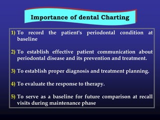
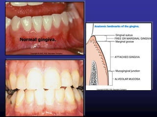
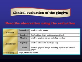
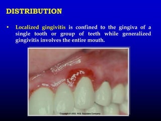
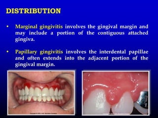
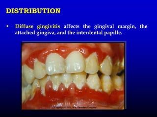
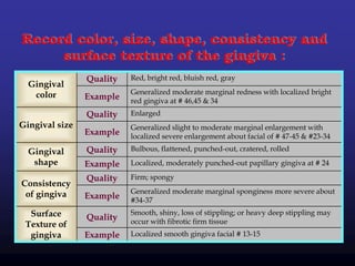
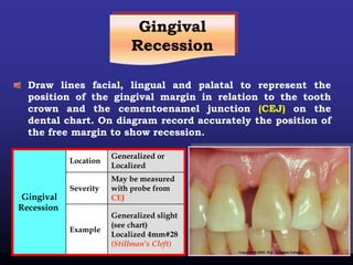
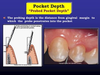
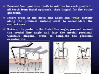
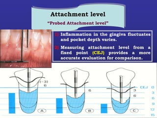
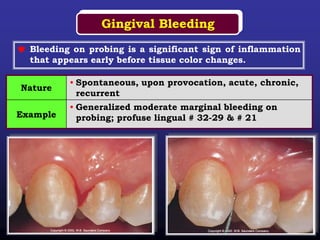
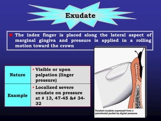
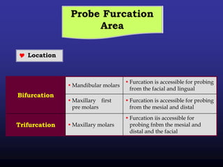
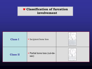
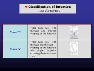
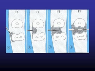
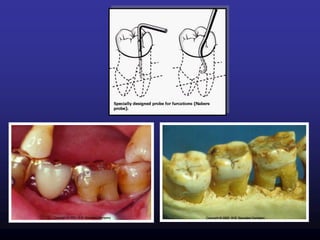
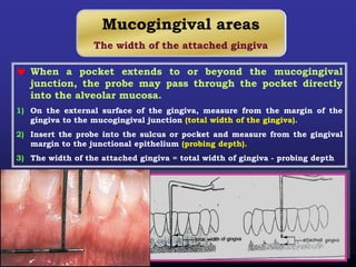
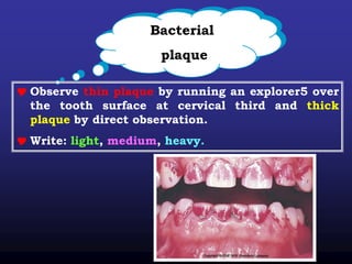
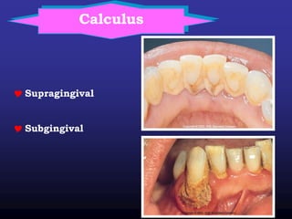
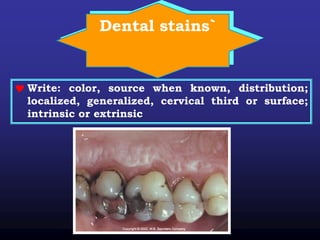
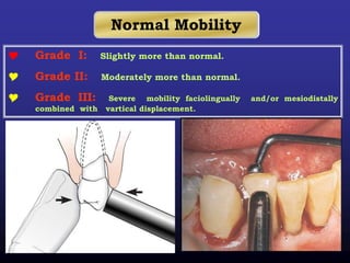
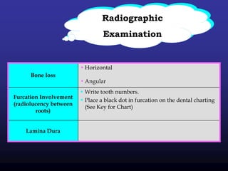
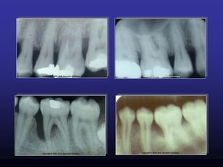
SlideShare a Scribd company logo
1 of 40
Download to read offline
‫الرحيم‬ ‫الرحمن‬ ‫الله‬ ‫بسم‬
‫توكلنا‬ ‫الله‬ ‫على‬ ‫علما‬ ‫شئ‬ ‫كل‬ ‫ربنا‬ ‫وسع‬
‫بالحك‬ ‫لومنا‬ ‫وبين‬ ‫بيننا‬ ‫افتح‬ ‫ربنا‬
‫الفاتحين‬ ‫خير‬ ‫وانت‬
‫العظيم‬ ‫الله‬ ‫صدق‬”‫األعراف‬89“
Prof Dr.Eman Abd El -Sattar Tella
1) To record the patient's periodontal condition at
baseline
2) To establish effective patient communication about
periodontal disease and its prevention and treatment.
3) To establish proper diagnosis and treatment planning.
4) To evaluate the response to therapy.
5) To serve as a baseline for future comparison at recall
visits during maintenance phase
Importance of dental Charting
Involves entire mouthGeneralized
Location
Confined to a single tooth or group of teethLocalized
Involves gingival margin including papillaeMarginal
Distribution
Involves interdental papilla(e) onlyPapillary
Involves gingival margin including papillae and attached
gingiva
Diffuse
Slight, Moderate, SevereSeverity
Clinical evaluation of the gingiva
Describe observation using the evaluation
DISTRIBUTION
• Localized gingivitis is confined to the gingiva of a
single tooth or group of teeth while generalized
gingivitis involves the entire mouth.
DISTRIBUTION
• Marginal gingivitis involves the gingival margin and
may include a portion of the contiguous attached
gingiva.
• Papillary gingivitis involves the interdental papillae
and often extends into the adjacent portion of the
gingival margin.
DISTRIBUTION
• Diffuse gingivitis affects the gingival margin, the
attached gingiva, and the interdental papille.
Record color, size, shape, consistency and
surface texture of the gingiva :
Red, bright red, bluish red, grayQuality
Gingival
color Generalized moderate marginal redness with localized bright
red gingiva at # 46,45 & 34
Example
EnlargedQuality
Gingival size Generalized slight to moderate marginal enlargement with
localized severe enlargement about facial of # 47-45 & #23-34
Example
Bulbous, flattened, punched-out, cratered, rolledQualityGingival
shape Localized, moderately punched-out papillary gingiva at # 24Example
Firm; spongyQuality
Consistency
of gingiva Generalized moderate marginal sponginess more severe about
#34-37
Example
Smooth, shiny, loss of stippling; or heavy deep stippling may
occur with fibrotic firm tissue
QualitySurface
Texture of
gingiva Localized smooth gingiva facial # 13-15Example
Healthy gingiva
Pale pink & stippled. Narrow
distinguishable free gingival
margin. No bleeding on probing
Mild gingivitis
Localized mild erythema & slight
edema. Some stippling is lost.
Minimal bleeding after probing.
Moderate gingivitis
Obvious erythema & edema.
No stippling, bleeding on
probing
Severe gingivitis
Fiery redness, edematous &
hyperplastic swelling, complete
absence of stippling, bleeding on
probing & spontaneous
hemorrhage.
Mild gingivitis in anterior area:
Mild erythema in maxilla. Slight
edematous swelling & erythema.
In mandible, slight edematous
swelling & erythema.
Papilla Bleeding Index: Grade 1
& 2
Stained plaque: Small plaque
accumulations arounds the necks
of the teeth & in interdental areas.
Moderate gingivitis in
anterior teeth :Erythema
& enlargement of gingiva
pronounced in mand than
in maxilla.
Papilla Bleeding Index :
grade 3 & 4
Stained plaque : Moderate plaque
accumulation in maxilla. Heavier plaque in mandible.
Radiographically, no destruction of interdental bony septa.
Gingival
Recession
Draw lines facial, lingual and palatal to represent the
position of the gingival margin in relation to the tooth
crown and the cementoenamel junction (CEJ) on the
dental chart. On diagram record accurately the position of
the free margin to show recession.
Generalized or
Localized
Location
Gingival
Recession
May be measured
with probe from
CEJ
Severity
Generalized slight
(see chart)
Localized 4mm#28
(Stillman's Cleft)
Example
Pocket Depth
“Probed Pocket Depth”
The probing depth is the distance from gingival margin to
which the probe penetrates into the pocket
Proceed from posterior teeth to midline for each quadrant,
all teeth from facial approach, then lingual for the entire
quadrant.
Insert probe at the distal line angle and "walk" distally
along the proximal surface; slant to accomodate the
contact area.
Return, the probe to the distal line angle; proceed around
the mesial line angle and into the mesial proximal.
Carefully diagonal probe to complete the proximal
examination.
Rationale
Attachment level
“Probed Attachment level”
1) Inflammation in the gingiva fluctuates
and pocket depth varies.
2) Measuring attachment level from a
fixed point (CEJ) provides a more
accurate evaluation for comparison.
Gingival Bleeding
 Bleeding on probing is a significant sign of inflammation
that appears early before tissue color changes.
• Spontaneous, upon provocation, acute, chronic,
recurrent
Nature
• Generalized moderate marginal bleeding on
probing; profuse lingual # 32-29 & # 21Example
Exudate
 The index finger is placed along the lateral aspect of
marginal gingiva and pressure is applied in a rolling
motion toward the crown
• Visible or upon
palpation (linger
pressure)
Nature
• Localized severe
exudate on pressure
at # 13, 47-45 &# 34-
32
Example
Probe Furcation
Area
 Location
• Furcation is accessible for probing
from the facial and lingual
• Mandibular molars
Bifurcation
• Furcation is accessible for probing
from the mesial and distal
• Maxillary first
pre molars
• Furcation iis accessible for
probing fnbm the mesial and
distal and the facial
• Maxillary molarsTrifurcation
 Classification of furcation
involvement
 Incipient bone lessClass I
 Partial bone loss (cul-de-
sac)
Class II
 Classification of furcation
involvement
 Total bone loss with
through and through
opening of the furcation
Class III
 Total bone loss with
through-and-through
opening ot the furcation
with gingival recession
exposing the furcation to
view
Class IV
Mucogingival areas
The width of the attached gingiva
 When a pocket extends to or beyond the mucogingival
junction, the probe may pass through the pocket directly
into the alveolar mucosa.
1) On the external surface of the gingiva, measure from the margin of the
gingiva to the mucogingival junction (total width of the gingiva).
2) Insert the probe into the sulcus or pocket and measure from the gingival
margin to the junctional epithelium (probing depth).
3) The width of the attached gingiva = total width of gingiva - probing depth
Bacterial
plaque
 Observe thin plaque by running an explorer5 over
the tooth surface at cervical third and thick
plaque by direct observation.
 Write: light, medium, heavy.
Calculus
 Supragingival
 Subgingival
Dental stains`
 Write: color, source when known, distribution;
localized, generalized, cervical third or surface;
intrinsic or extrinsic
Functional
relations
• Pathologic migration occurs most frequently in
anterior teeth. Distinguish from "mesial drift” which
occurs in posterior teeth with healthy gingival
Pathologic Migration
• Test for open contacts where food impaction can occur
by using dental floss.
• Record on the tooth chart by parallel lines.
Open Contacts
•Record any symptoms such as pain, tenderness sounds
(crepitation) or limitation of movement.
Temporomandibular
Joint Disorder
Parafunctional
 Note tooth wear facets and occlusal and incisal
wear.
 Question patient concerning habits such as
bruxing, clenching, or tapping
o Bruxism = grinding of teeth in directions different from normal
chewing at night
o Clenching = closing of teeth in the chewing position at day &
night
o Tapping = grading of an isolated tooth
Fremitus
 Fremitus is palpable_vibration (or) movement, It is an
important sign during examination % of the occlusion,
and is commonly used as an indicator of the need for
further analysis
NOMINAL SCALE
o N normal
o + vibration felt
o 1 slight movement felt against finger
o 2 clearly palpable, movement visible
o 3 movement very apparent
Percussion
 Percussion is the act of tapping a surface of a tooth
with an instrument. Sensitivity to percussion is a
manifestation of inflammation in the periodontal
ligament.
Mobility
 Position the patient in supine for clear visibility.
 Stabilize the head. Motion of head can interfere with a
true evaluation of tooth movement.
 Begin with most posterior tooth and move
systematically around each arch.
 Use two single-ended metal instruments. Hold in
modified pen grasp. Using wooden tongue depressors
or plastic mirror handles is not good, because of their
flexibility. Testing with fingers without the metal
instruments can be misleading since the soft tissue
moves.
Normal Mobility
 Grade I: Slightly more than normal.
 Grade II: Moderately more than normal.
 Grade III: Severe mobility faciolingually and/or mesiodistally
combined with vartical displacement.
Radiographic
Examination
• Horizontal
• Angular
Bone loss
 Write tooth numbers.
 Place a black dot in furcation on the dental charting
(See Key for Chart)
Furcation Involvement
(radiolucency between
roots)
Lamina Dura
Use of clinical photographs and study casts
1. Clinical Photographs
 Color photographs are useful for recording the
appearance of the tissue before, and after treatment.
2. Casts
 position of the gjngival margins
 position and inclination of the teeth
 proximal contact relationships
 Food impactions areas.
Finally casts also serve as visual aids in discussions with the
patient and are useful for pre and post-treatment comparisons, as
well as for reference at check-up visits.
References
-Caranza’s Clinical Periodontology, 10th ed.
WB Saunders, 2006.
-Color Atlas of Dental Medicine:
Periodontology By Klaus H. Rateitschak,
Edith M.
Dental sharting

More Related Content

What's hot

Periodontal examination ,chart. ppt
Periodontal examination ,chart. pptPeriodontal examination ,chart. ppt
Periodontal examination ,chart. pptArezooJahed1
 
Traumatic Occlusion and Pathologic tooth migration
Traumatic Occlusion and Pathologic tooth migrationTraumatic Occlusion and Pathologic tooth migration
Traumatic Occlusion and Pathologic tooth migrationAyam Chhatkuli
 
Caries diagnosis
Caries diagnosisCaries diagnosis
Caries diagnosisdrkskumar
 
Periodontal examination and diagnosis
Periodontal examination and diagnosisPeriodontal examination and diagnosis
Periodontal examination and diagnosisNael Almasri
 
Icon case reports
Icon case reportsIcon case reports
Icon case reportsterradent
 
Case history & diagnosis in periodontics /certified fixed orthodontic course...
Case history & diagnosis in periodontics  /certified fixed orthodontic course...Case history & diagnosis in periodontics  /certified fixed orthodontic course...
Case history & diagnosis in periodontics /certified fixed orthodontic course...Indian dental academy
 
Role of occlusion in periodontal disease
Role of occlusion in periodontal diseaseRole of occlusion in periodontal disease
Role of occlusion in periodontal diseaseDr. Virshali Gupta
 
Furcation involvement (Dr. Himanshu Shekhar)
Furcation involvement (Dr. Himanshu Shekhar)Furcation involvement (Dr. Himanshu Shekhar)
Furcation involvement (Dr. Himanshu Shekhar)himanshu shekhar
 
Tooth examination
Tooth examinationTooth examination
Tooth examinationDrGhadooRa
 
Interdisciplinary periodontics
Interdisciplinary periodonticsInterdisciplinary periodontics
Interdisciplinary periodonticsDr Sreelakshmi
 
Iatroenic factors in periodontits
Iatroenic factors in periodontitsIatroenic factors in periodontits
Iatroenic factors in periodontitsSwati Gupta
 
Iatrogenic factors in periodontal disease
Iatrogenic factors  in periodontal diseaseIatrogenic factors  in periodontal disease
Iatrogenic factors in periodontal diseaselobna elsaadawy
 
Recent Methodology in Management of Non-carious Lesions
Recent Methodology in Management of Non-carious LesionsRecent Methodology in Management of Non-carious Lesions
Recent Methodology in Management of Non-carious LesionsAlaaDokmak
 
Sequelae of wearing complete dentures/ orthodontics training courses
Sequelae of wearing complete dentures/ orthodontics training coursesSequelae of wearing complete dentures/ orthodontics training courses
Sequelae of wearing complete dentures/ orthodontics training coursesIndian dental academy
 

What's hot (20)

Periodontal examination ,chart. ppt
Periodontal examination ,chart. pptPeriodontal examination ,chart. ppt
Periodontal examination ,chart. ppt
 
Traumatic Occlusion and Pathologic tooth migration
Traumatic Occlusion and Pathologic tooth migrationTraumatic Occlusion and Pathologic tooth migration
Traumatic Occlusion and Pathologic tooth migration
 
Complete dentures 3.history and exam
Complete dentures 3.history and examComplete dentures 3.history and exam
Complete dentures 3.history and exam
 
Coronoplasty
CoronoplastyCoronoplasty
Coronoplasty
 
Trauma from occlusion
Trauma from occlusionTrauma from occlusion
Trauma from occlusion
 
Caries diagnosis
Caries diagnosisCaries diagnosis
Caries diagnosis
 
Periodontal examination and diagnosis
Periodontal examination and diagnosisPeriodontal examination and diagnosis
Periodontal examination and diagnosis
 
Icon case reports
Icon case reportsIcon case reports
Icon case reports
 
Case history & diagnosis in periodontics /certified fixed orthodontic course...
Case history & diagnosis in periodontics  /certified fixed orthodontic course...Case history & diagnosis in periodontics  /certified fixed orthodontic course...
Case history & diagnosis in periodontics /certified fixed orthodontic course...
 
Role of occlusion in periodontal disease
Role of occlusion in periodontal diseaseRole of occlusion in periodontal disease
Role of occlusion in periodontal disease
 
Furcation involvement (Dr. Himanshu Shekhar)
Furcation involvement (Dr. Himanshu Shekhar)Furcation involvement (Dr. Himanshu Shekhar)
Furcation involvement (Dr. Himanshu Shekhar)
 
Tooth examination
Tooth examinationTooth examination
Tooth examination
 
Interdisciplinary periodontics
Interdisciplinary periodonticsInterdisciplinary periodontics
Interdisciplinary periodontics
 
Furcation ppt
Furcation pptFurcation ppt
Furcation ppt
 
Iatroenic factors in periodontits
Iatroenic factors in periodontitsIatroenic factors in periodontits
Iatroenic factors in periodontits
 
Jc hypersensitivity
Jc hypersensitivityJc hypersensitivity
Jc hypersensitivity
 
Iatrogenic factors in periodontal disease
Iatrogenic factors  in periodontal diseaseIatrogenic factors  in periodontal disease
Iatrogenic factors in periodontal disease
 
MANAGEMENT OF OPEN BITE
MANAGEMENT OF OPEN BITEMANAGEMENT OF OPEN BITE
MANAGEMENT OF OPEN BITE
 
Recent Methodology in Management of Non-carious Lesions
Recent Methodology in Management of Non-carious LesionsRecent Methodology in Management of Non-carious Lesions
Recent Methodology in Management of Non-carious Lesions
 
Sequelae of wearing complete dentures/ orthodontics training courses
Sequelae of wearing complete dentures/ orthodontics training coursesSequelae of wearing complete dentures/ orthodontics training courses
Sequelae of wearing complete dentures/ orthodontics training courses
 

Similar to Dental sharting

Dental sharting
Dental shartingDental sharting
Dental shartingeatella
 
intraoral and extraoral examination.pptx
intraoral and extraoral examination.pptxintraoral and extraoral examination.pptx
intraoral and extraoral examination.pptxmisthysrishty
 
Pi,gi Loe and Sliness : ORIGINAL ARTICLE
Pi,gi Loe and Sliness : ORIGINAL ARTICLEPi,gi Loe and Sliness : ORIGINAL ARTICLE
Pi,gi Loe and Sliness : ORIGINAL ARTICLENavneet Randhawa
 
Macroscopic features of gingiva
Macroscopic features of gingivaMacroscopic features of gingiva
Macroscopic features of gingivaDr.Himanshu Singh
 
Examination, Diagnosis, Treatment Planing I
Examination, Diagnosis, Treatment Planing IExamination, Diagnosis, Treatment Planing I
Examination, Diagnosis, Treatment Planing IIAU Dent
 
Clinical diagnosis in periodontology
Clinical diagnosis in periodontologyClinical diagnosis in periodontology
Clinical diagnosis in periodontologyChetan Basnet
 
11 from gingivitis to
11 from gingivitis to11 from gingivitis to
11 from gingivitis toemantella
 
CLINICAL_DIAGNOSIS.pptx
CLINICAL_DIAGNOSIS.pptxCLINICAL_DIAGNOSIS.pptx
CLINICAL_DIAGNOSIS.pptxmalti19
 
Gingivalenlargment HOD SIR.pptx
Gingivalenlargment HOD SIR.pptxGingivalenlargment HOD SIR.pptx
Gingivalenlargment HOD SIR.pptxDentalYoutube
 
Diagnosis of Periodontal Diseases
Diagnosis of Periodontal DiseasesDiagnosis of Periodontal Diseases
Diagnosis of Periodontal Diseasesssuseraf61fb
 
Dental Charting, Color Coding and Symbols: Class Activity
Dental Charting, Color Coding and Symbols: Class ActivityDental Charting, Color Coding and Symbols: Class Activity
Dental Charting, Color Coding and Symbols: Class ActivityHeatherSeghi
 
pulp therapy in pediatric dentistry
pulp therapy in pediatric dentistrypulp therapy in pediatric dentistry
pulp therapy in pediatric dentistryalaa Mohamed
 
Gingival enlrgment
Gingival enlrgmentGingival enlrgment
Gingival enlrgmentImran Bhatt
 
5 prevention of periodontal disease
5 prevention of periodontal disease5 prevention of periodontal disease
5 prevention of periodontal diseaseLama K Banna
 
Sequelae of periodontal diseases
Sequelae of periodontal diseasesSequelae of periodontal diseases
Sequelae of periodontal diseasesDhruvMukherjee3
 
Periodontal Chart presentation
Periodontal Chart presentationPeriodontal Chart presentation
Periodontal Chart presentationEnas Elgendy
 

Similar to Dental sharting (20)

Dental sharting
Dental shartingDental sharting
Dental sharting
 
Dental sharting
Dental shartingDental sharting
Dental sharting
 
intraoral and extraoral examination.pptx
intraoral and extraoral examination.pptxintraoral and extraoral examination.pptx
intraoral and extraoral examination.pptx
 
Pi,gi Loe and Sliness : ORIGINAL ARTICLE
Pi,gi Loe and Sliness : ORIGINAL ARTICLEPi,gi Loe and Sliness : ORIGINAL ARTICLE
Pi,gi Loe and Sliness : ORIGINAL ARTICLE
 
Macroscopic features of gingiva
Macroscopic features of gingivaMacroscopic features of gingiva
Macroscopic features of gingiva
 
Examination, Diagnosis, Treatment Planing I
Examination, Diagnosis, Treatment Planing IExamination, Diagnosis, Treatment Planing I
Examination, Diagnosis, Treatment Planing I
 
Interdental aids
Interdental aidsInterdental aids
Interdental aids
 
Clinical diagnosis in periodontology
Clinical diagnosis in periodontologyClinical diagnosis in periodontology
Clinical diagnosis in periodontology
 
11 from gingivitis to
11 from gingivitis to11 from gingivitis to
11 from gingivitis to
 
CLINICAL_DIAGNOSIS.pptx
CLINICAL_DIAGNOSIS.pptxCLINICAL_DIAGNOSIS.pptx
CLINICAL_DIAGNOSIS.pptx
 
Gingivalenlargment HOD SIR.pptx
Gingivalenlargment HOD SIR.pptxGingivalenlargment HOD SIR.pptx
Gingivalenlargment HOD SIR.pptx
 
From Gingivitis to Periodontitis
From Gingivitis to PeriodontitisFrom Gingivitis to Periodontitis
From Gingivitis to Periodontitis
 
Diagnosis of Periodontal Diseases
Diagnosis of Periodontal DiseasesDiagnosis of Periodontal Diseases
Diagnosis of Periodontal Diseases
 
Dental Charting, Color Coding and Symbols: Class Activity
Dental Charting, Color Coding and Symbols: Class ActivityDental Charting, Color Coding and Symbols: Class Activity
Dental Charting, Color Coding and Symbols: Class Activity
 
pulp therapy in pediatric dentistry
pulp therapy in pediatric dentistrypulp therapy in pediatric dentistry
pulp therapy in pediatric dentistry
 
Gingival enlrgment
Gingival enlrgmentGingival enlrgment
Gingival enlrgment
 
5 prevention of periodontal disease
5 prevention of periodontal disease5 prevention of periodontal disease
5 prevention of periodontal disease
 
Sequelae of periodontal diseases
Sequelae of periodontal diseasesSequelae of periodontal diseases
Sequelae of periodontal diseases
 
Periodontal Chart presentation
Periodontal Chart presentationPeriodontal Chart presentation
Periodontal Chart presentation
 
Oral health for orthodontists by Almuzian
Oral health for orthodontists by AlmuzianOral health for orthodontists by Almuzian
Oral health for orthodontists by Almuzian
 

Dental sharting

  • 1. ‫الرحيم‬ ‫الرحمن‬ ‫الله‬ ‫بسم‬ ‫توكلنا‬ ‫الله‬ ‫على‬ ‫علما‬ ‫شئ‬ ‫كل‬ ‫ربنا‬ ‫وسع‬ ‫بالحك‬ ‫لومنا‬ ‫وبين‬ ‫بيننا‬ ‫افتح‬ ‫ربنا‬ ‫الفاتحين‬ ‫خير‬ ‫وانت‬ ‫العظيم‬ ‫الله‬ ‫صدق‬”‫األعراف‬89“
  • 2. Prof Dr.Eman Abd El -Sattar Tella
  • 3. 1) To record the patient's periodontal condition at baseline 2) To establish effective patient communication about periodontal disease and its prevention and treatment. 3) To establish proper diagnosis and treatment planning. 4) To evaluate the response to therapy. 5) To serve as a baseline for future comparison at recall visits during maintenance phase Importance of dental Charting
  • 4.
  • 5. Involves entire mouthGeneralized Location Confined to a single tooth or group of teethLocalized Involves gingival margin including papillaeMarginal Distribution Involves interdental papilla(e) onlyPapillary Involves gingival margin including papillae and attached gingiva Diffuse Slight, Moderate, SevereSeverity Clinical evaluation of the gingiva Describe observation using the evaluation
  • 6. DISTRIBUTION • Localized gingivitis is confined to the gingiva of a single tooth or group of teeth while generalized gingivitis involves the entire mouth.
  • 7. DISTRIBUTION • Marginal gingivitis involves the gingival margin and may include a portion of the contiguous attached gingiva. • Papillary gingivitis involves the interdental papillae and often extends into the adjacent portion of the gingival margin.
  • 8. DISTRIBUTION • Diffuse gingivitis affects the gingival margin, the attached gingiva, and the interdental papille.
  • 9. Record color, size, shape, consistency and surface texture of the gingiva : Red, bright red, bluish red, grayQuality Gingival color Generalized moderate marginal redness with localized bright red gingiva at # 46,45 & 34 Example EnlargedQuality Gingival size Generalized slight to moderate marginal enlargement with localized severe enlargement about facial of # 47-45 & #23-34 Example Bulbous, flattened, punched-out, cratered, rolledQualityGingival shape Localized, moderately punched-out papillary gingiva at # 24Example Firm; spongyQuality Consistency of gingiva Generalized moderate marginal sponginess more severe about #34-37 Example Smooth, shiny, loss of stippling; or heavy deep stippling may occur with fibrotic firm tissue QualitySurface Texture of gingiva Localized smooth gingiva facial # 13-15Example
  • 10. Healthy gingiva Pale pink & stippled. Narrow distinguishable free gingival margin. No bleeding on probing Mild gingivitis Localized mild erythema & slight edema. Some stippling is lost. Minimal bleeding after probing.
  • 11. Moderate gingivitis Obvious erythema & edema. No stippling, bleeding on probing Severe gingivitis Fiery redness, edematous & hyperplastic swelling, complete absence of stippling, bleeding on probing & spontaneous hemorrhage.
  • 12. Mild gingivitis in anterior area: Mild erythema in maxilla. Slight edematous swelling & erythema. In mandible, slight edematous swelling & erythema. Papilla Bleeding Index: Grade 1 & 2 Stained plaque: Small plaque accumulations arounds the necks of the teeth & in interdental areas.
  • 13. Moderate gingivitis in anterior teeth :Erythema & enlargement of gingiva pronounced in mand than in maxilla. Papilla Bleeding Index : grade 3 & 4 Stained plaque : Moderate plaque accumulation in maxilla. Heavier plaque in mandible. Radiographically, no destruction of interdental bony septa.
  • 14. Gingival Recession Draw lines facial, lingual and palatal to represent the position of the gingival margin in relation to the tooth crown and the cementoenamel junction (CEJ) on the dental chart. On diagram record accurately the position of the free margin to show recession. Generalized or Localized Location Gingival Recession May be measured with probe from CEJ Severity Generalized slight (see chart) Localized 4mm#28 (Stillman's Cleft) Example
  • 15. Pocket Depth “Probed Pocket Depth” The probing depth is the distance from gingival margin to which the probe penetrates into the pocket
  • 16. Proceed from posterior teeth to midline for each quadrant, all teeth from facial approach, then lingual for the entire quadrant. Insert probe at the distal line angle and "walk" distally along the proximal surface; slant to accomodate the contact area. Return, the probe to the distal line angle; proceed around the mesial line angle and into the mesial proximal. Carefully diagonal probe to complete the proximal examination.
  • 17. Rationale Attachment level “Probed Attachment level” 1) Inflammation in the gingiva fluctuates and pocket depth varies. 2) Measuring attachment level from a fixed point (CEJ) provides a more accurate evaluation for comparison.
  • 18. Gingival Bleeding  Bleeding on probing is a significant sign of inflammation that appears early before tissue color changes. • Spontaneous, upon provocation, acute, chronic, recurrent Nature • Generalized moderate marginal bleeding on probing; profuse lingual # 32-29 & # 21Example
  • 19. Exudate  The index finger is placed along the lateral aspect of marginal gingiva and pressure is applied in a rolling motion toward the crown • Visible or upon palpation (linger pressure) Nature • Localized severe exudate on pressure at # 13, 47-45 &# 34- 32 Example
  • 20. Probe Furcation Area  Location • Furcation is accessible for probing from the facial and lingual • Mandibular molars Bifurcation • Furcation is accessible for probing from the mesial and distal • Maxillary first pre molars • Furcation iis accessible for probing fnbm the mesial and distal and the facial • Maxillary molarsTrifurcation
  • 21.  Classification of furcation involvement  Incipient bone lessClass I  Partial bone loss (cul-de- sac) Class II
  • 22.  Classification of furcation involvement  Total bone loss with through and through opening of the furcation Class III  Total bone loss with through-and-through opening ot the furcation with gingival recession exposing the furcation to view Class IV
  • 23.
  • 24.
  • 25. Mucogingival areas The width of the attached gingiva  When a pocket extends to or beyond the mucogingival junction, the probe may pass through the pocket directly into the alveolar mucosa. 1) On the external surface of the gingiva, measure from the margin of the gingiva to the mucogingival junction (total width of the gingiva). 2) Insert the probe into the sulcus or pocket and measure from the gingival margin to the junctional epithelium (probing depth). 3) The width of the attached gingiva = total width of gingiva - probing depth
  • 26. Bacterial plaque  Observe thin plaque by running an explorer5 over the tooth surface at cervical third and thick plaque by direct observation.  Write: light, medium, heavy.
  • 28. Dental stains`  Write: color, source when known, distribution; localized, generalized, cervical third or surface; intrinsic or extrinsic
  • 29. Functional relations • Pathologic migration occurs most frequently in anterior teeth. Distinguish from "mesial drift” which occurs in posterior teeth with healthy gingival Pathologic Migration • Test for open contacts where food impaction can occur by using dental floss. • Record on the tooth chart by parallel lines. Open Contacts •Record any symptoms such as pain, tenderness sounds (crepitation) or limitation of movement. Temporomandibular Joint Disorder
  • 30. Parafunctional  Note tooth wear facets and occlusal and incisal wear.  Question patient concerning habits such as bruxing, clenching, or tapping o Bruxism = grinding of teeth in directions different from normal chewing at night o Clenching = closing of teeth in the chewing position at day & night o Tapping = grading of an isolated tooth
  • 31. Fremitus  Fremitus is palpable_vibration (or) movement, It is an important sign during examination % of the occlusion, and is commonly used as an indicator of the need for further analysis NOMINAL SCALE o N normal o + vibration felt o 1 slight movement felt against finger o 2 clearly palpable, movement visible o 3 movement very apparent
  • 32. Percussion  Percussion is the act of tapping a surface of a tooth with an instrument. Sensitivity to percussion is a manifestation of inflammation in the periodontal ligament.
  • 33. Mobility  Position the patient in supine for clear visibility.  Stabilize the head. Motion of head can interfere with a true evaluation of tooth movement.  Begin with most posterior tooth and move systematically around each arch.  Use two single-ended metal instruments. Hold in modified pen grasp. Using wooden tongue depressors or plastic mirror handles is not good, because of their flexibility. Testing with fingers without the metal instruments can be misleading since the soft tissue moves.
  • 34. Normal Mobility  Grade I: Slightly more than normal.  Grade II: Moderately more than normal.  Grade III: Severe mobility faciolingually and/or mesiodistally combined with vartical displacement.
  • 35. Radiographic Examination • Horizontal • Angular Bone loss  Write tooth numbers.  Place a black dot in furcation on the dental charting (See Key for Chart) Furcation Involvement (radiolucency between roots) Lamina Dura
  • 36.
  • 37. Use of clinical photographs and study casts 1. Clinical Photographs  Color photographs are useful for recording the appearance of the tissue before, and after treatment. 2. Casts  position of the gjngival margins  position and inclination of the teeth  proximal contact relationships  Food impactions areas. Finally casts also serve as visual aids in discussions with the patient and are useful for pre and post-treatment comparisons, as well as for reference at check-up visits.
  • 38.
  • 39. References -Caranza’s Clinical Periodontology, 10th ed. WB Saunders, 2006. -Color Atlas of Dental Medicine: Periodontology By Klaus H. Rateitschak, Edith M.