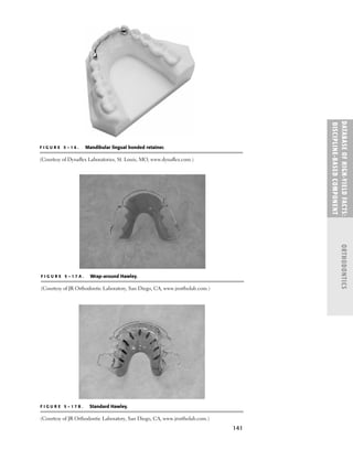
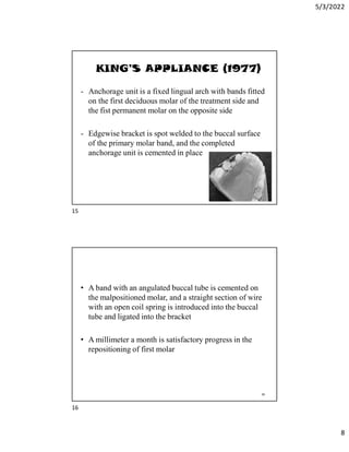
The orthodontic patient examination and diagnosis involves interviewing the patient to understand their concerns and dental history. It also includes assessing their medical history to determine if any conditions could impact treatment. Factors like bleeding disorders, diabetes, immunosuppression, and allergies may require special consideration during orthodontic care. A thorough examination provides information needed to develop an appropriate treatment plan.


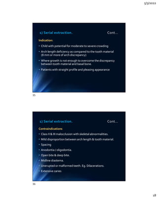
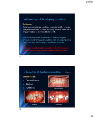
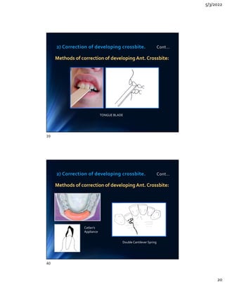

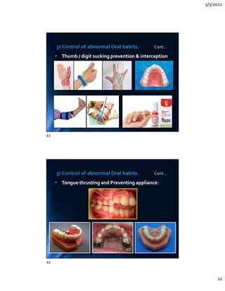


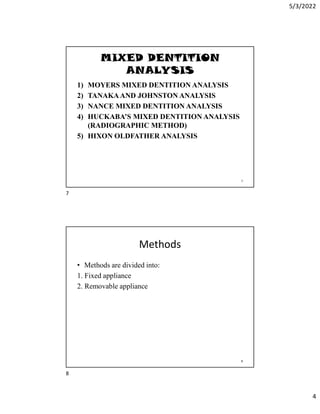
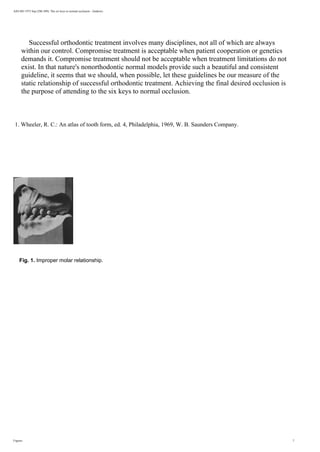
![1
Introduction
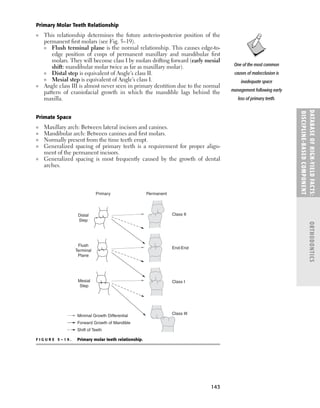
When deciding on a treatment protocol for young children (7- 8 years old) with newly erupted
crooked teeth, a dentist faces questions such as whether treatment should be recommended
or not, and if orthodontic treatment is recommended, what technical protocol should be
suggested to the concerned parents of a young child?
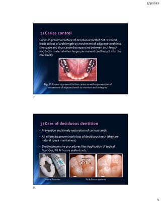
Social aspects should be considered when evaluating the timing of orthodontic treatment. By
age 8, children’s criteria for attractiveness are the same as those of adults, and the
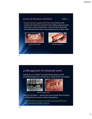
appearance of the smile is considered to be an important criterion when judging facial
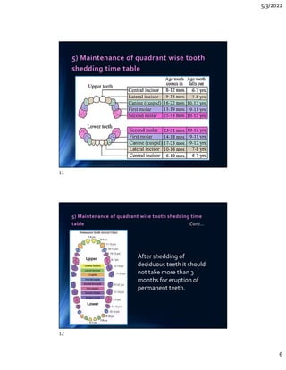
attractiveness [1]. Thus, interceptive treatment, such as the correction of jaw deformities and
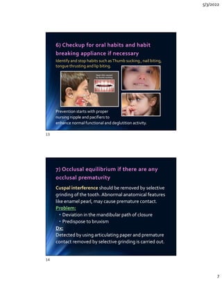
dental irregularities, can help raise a young child’s self-esteem.
While there are some who question the benefits of interceptive treatment [2-6], there are
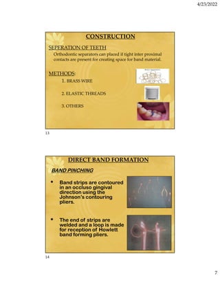
others who have argued in favor of some form of intervention. A survey by College of
Diplomates of the American Board of Orthodontics (CDABO) shows that a majority of the ABO
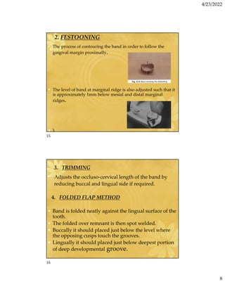
diplomats value interceptive orthodontics and are actively involved in some sort of mixed
dentition treatment [7]. One thing that is clear is there has been minimal progress in the
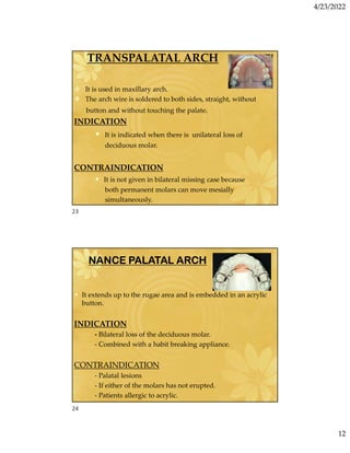
development of appliances and techniques that can efficiently move young children’s teeth [8].
Functional appliances used alone or in combination with fixed appliances have not produced
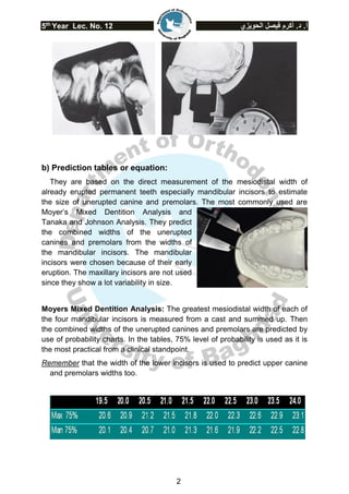
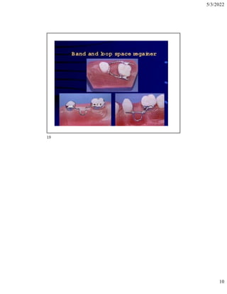
predictable results quickly [9, 10].
This paper is intended for dental and orthodontic professionals, and it presents new
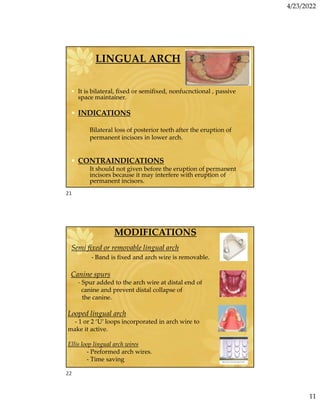
approaches that use deciduous molars and canines as anchors to accelerate treatment of
many mixed dentition cases such as: anterior crowding, open bite, overbite, and crossbite.](https://image.slidesharecdn.com/ilovepdfmerged1-230426002353-eade8add/85/ilovepdf_merged-1-pdf-112-320.jpg)
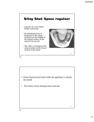
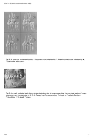
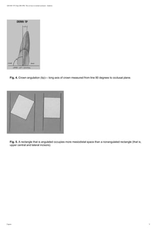
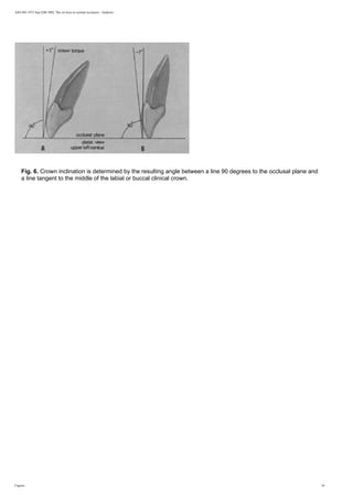
![2
Correcting Crowding: Creating Space through Expansion
The primary way to create space in the mixed dentition protocol proposed in this paper is
through expansion of the transverse dimension. The recommended period to begin this
protocol is at 7-8 years of age. This coincides with the eruption of the permanent first molars
and permanent incisors during the early mixed dentition period. One of the key benefits of this
early expansion is a reduction in the need to remove deciduous teeth in grade school children
and permanent teeth in middle school and high school children.
The protocol follows McNamara’s method [11], with some changes to make it more practical.
These changes include avoiding occlusal coverage for the maxillary expander and using fixed
expansion in the mandible instead of a removable Schwarz.
Early expansion of the maxilla is a stable and effective way to correct arch length
deficiencies [12-15]. Conversely, the effectiveness of expansion in the mandibular arch
has been disputed [16-20]. Disagreement with regard to the effectiveness of the mandibular
arch expansion may be related to the differences in the timing of treatment or the methods
being used.
The expansion appliances used in this protocol for the maxillary and the mandibular arches
take advantage of different growth mechanisms in the corresponding jawbones. In the maxilla,
the increase in the transverse dimension is accomplished through skeletal expansion at the
intermaxillary suture. In the mandible, dentoaveolar expansion of the buccal segments is used
to increase the arch width.
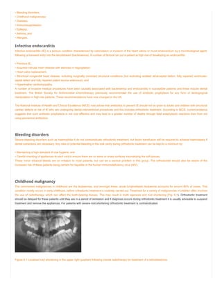
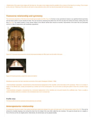
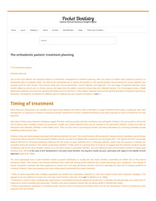
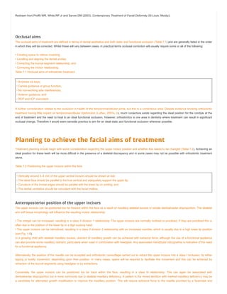
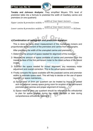
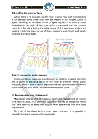
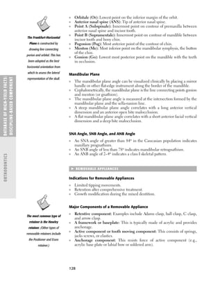
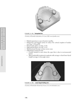
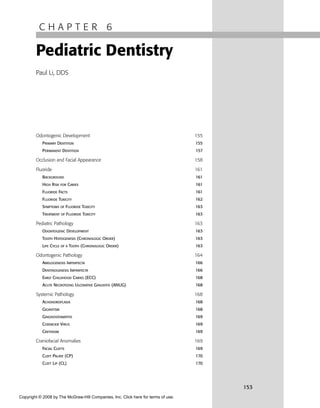
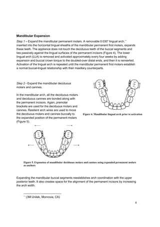
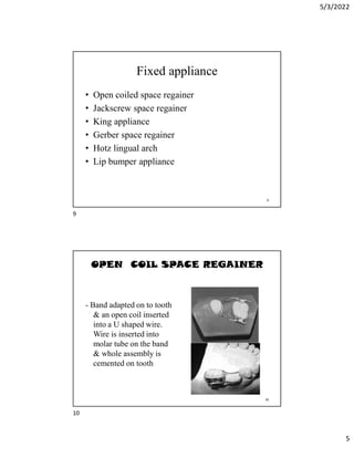
Maxillary Expansion
Expansion of the maxilla is achieved with a 2-banded maxillary expansion appliance (MEA)
attached to the first permanent molars. This produces expansion of the maxilla equivalent to
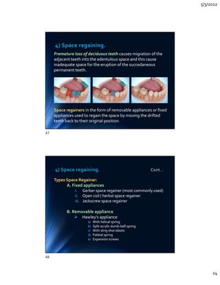
the more traditional 4-banded appliance [21,22].
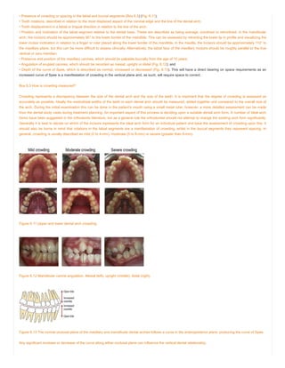
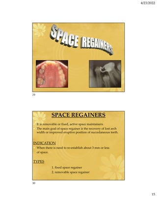
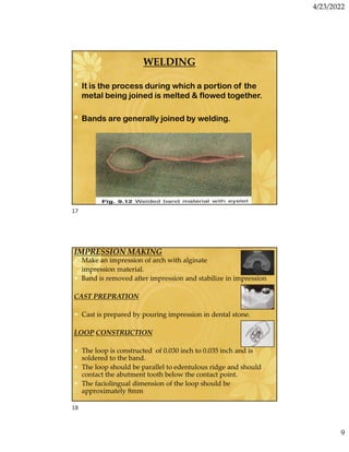
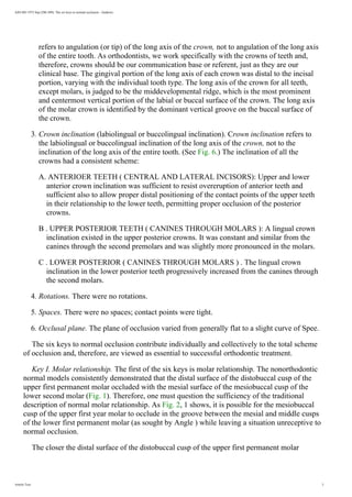
A 12mm expansion screw ∗
is used with additional 0.036” arms extending from the first
permanent molars mesially to the deciduous canines on the palatal side (Figure 1). The
appliance is activated once a day until the
palatal cusps of the maxillary posterior teeth
touch the buccal cusps of the mandibular
posterior teeth. In the maxillary arch,
deciduous molars and canines are expanded
simultaneously with the permanent molars by
the MEA arms.
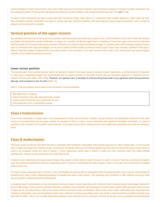
The maxillary deciduous canines are ideal
anchors for crowded maxillary incisors
∗
(Dentaurum, Ispringen, Germany) Figure 1. Maxillary Expansion Appliance prior to activation](https://image.slidesharecdn.com/ilovepdfmerged1-230426002353-eade8add/85/ilovepdf_merged-1-pdf-113-320.jpg)
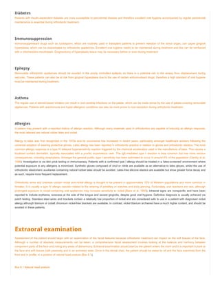
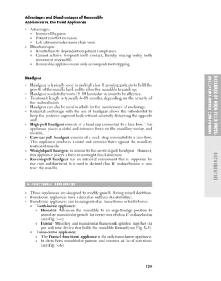
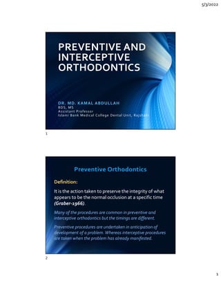
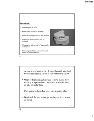
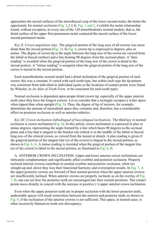
![3
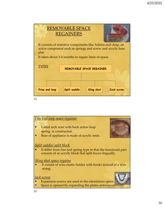
because they are close to the
permanent incisors. Premolar
brackets are used on the
deciduous canines because they
adapt to their buccal surface better
than other brackets [23].
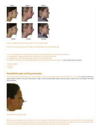
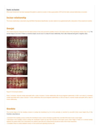
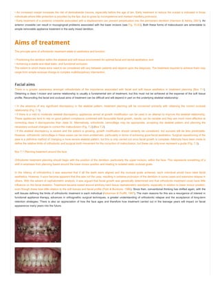
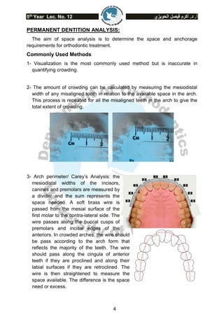
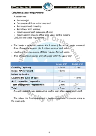
Deciduous canines are bonded at
the same time as the permanent
maxillary incisors. Resilient arch
wires align the incisors and move
them together. The space
developed in the midline is transferred
distally to the lateral incisor and canine
areas (Figure 2).
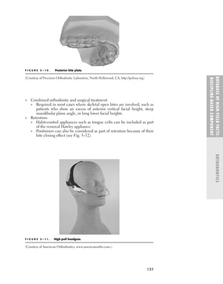
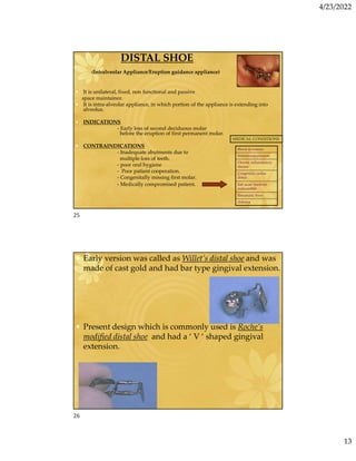
Once the desired amount of expansion is achieved, the MEA is left in place for two months to
allow for skeletal stability. A benefit of this early expansion is a reduction in the incidence of
impaction for maxillary permanent canines [24]. Figure 3A shows an upper left canine at risk
of impaction before expansion. Figure 3B depicts the canine following expansion, with
adequate space to erupt.
Figure 3. Creating adequate space for proper canine eruption
Figure 2. Closure of anterior spaces after activation of Maxillary
Expansion Appliance
A B](https://image.slidesharecdn.com/ilovepdfmerged1-230426002353-eade8add/85/ilovepdf_merged-1-pdf-114-320.jpg)
![5
This additional arch space eliminates the need for extraction of the deciduous canines or
deciduous first molars when aligning the permanent incisors. Furthermore, expansion of the
mandibular deciduous molars and canines can enhance appositional growth of the buccal
alveolar surfaces [25].The resulting appositional growth of the alveolar bone potentially
improves the environment for the periodontal support system of the developing permanent
canines and premolars.
Expanding the mandibular buccal segments allows for further expansion of the maxilla [26].
This is often required in cases of severe crowding.
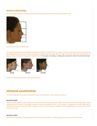
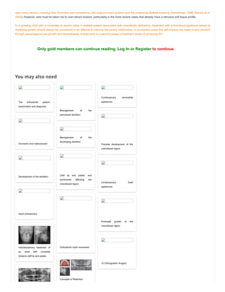
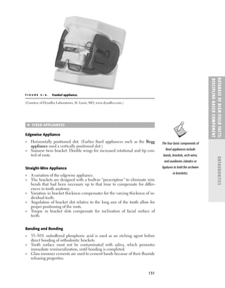
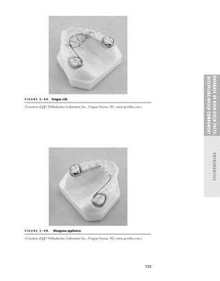
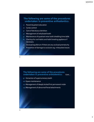
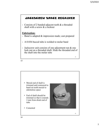
Correcting Open Bite and Overbite
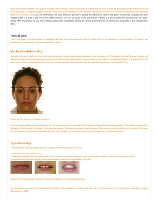
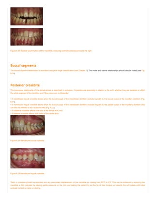
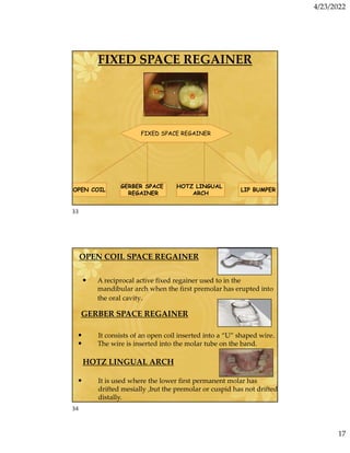
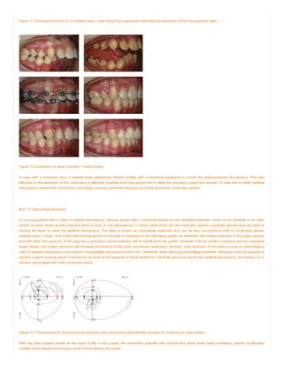
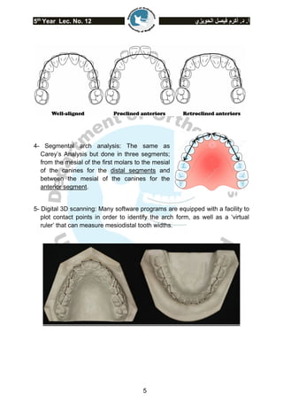
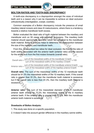
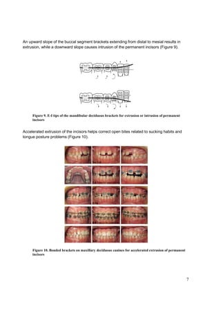
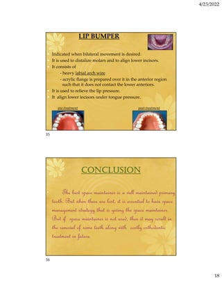
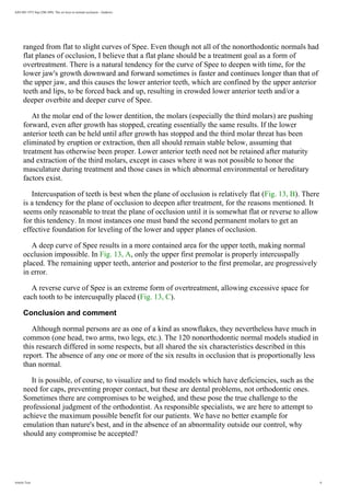
Deciduous teeth can provide temporary anchorage to jump-start extrusion or intrusion of the
incisors in cases where open bite or overbite is caused by under- or over-eruption of the
permanent incisors. This is accomplished by changing the angle of brackets when bonding the
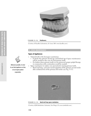
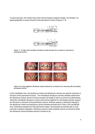
deciduous teeth, producing extrusive or intrusive forces on the permanent incisors (Figure 6).
These angle changes are called E-I tips, where E stands for extrusion and I for intrusion.
In the maxillary arch, the deciduous canines provide anchorage for extrusion or intrusion of
the permanent incisors. Maxillary deciduous canines are usually the last deciduous teeth to
exfoliate and they maintain adequate root lengths until late mixed dentition. They are also
close to the permanent incisors, providing mechanical efficiency for extrusion or intrusion of
the incisors. Maxillary deciduous first or second molars can be used if the deciduous canines
are missing or loose.
Figure 6. Position of deciduous brackets determines the position of
permanent incisors](https://image.slidesharecdn.com/ilovepdfmerged1-230426002353-eade8add/85/ilovepdf_merged-1-pdf-116-320.jpg)

![11
Conclusion
Creating a normal occlusal relationship and a balanced neuromuscular environment at an
early age can help the normal growth of the facial skeleton in an otherwise healthy child [27].
Although some debate still exists regarding interceptive orthodontics, early treatment is
advantageous in correcting certain forms of malocclusion such as crowding, overbite, open
bite, and crossbite [28-32].
The mixed dentition protocol presented in this paper uses expansion in the transverse
dimension as the primary method to create space. An MEA device is used to expand the
maxilla. Early expansion of the maxillary skeletal complex in non-crossbite individuals can
correct maxillary arch length deficiencies [11, 14]. Using maxillary deciduous canines as
anchorage helps align the maxillary permanent incisors. In the mandible, expansion of the
buccal segments, including deciduous molars and canines, can increase the arch width to
accommodate crowded permanent incisors.
The protocol uses E-I tips to carefully position the deciduous brackets and improve the
mechanical efficiency of appliances to accelerate the correction of open bite, overbite, and
crossbite conditions.
Benefits of the protocol include:
1. Accelerates treatment time
2. Reduces the occurrence of impaction of maxillary permanent canines
3. Eliminates the need to extract the deciduous canines or deciduous first molars
4. Reduces the need to remove permanent teeth
5. Raises a young child’s self-esteem
To get more information about mixed dentition orthodontics and to participate in an open
discussion about the subject, please visit www.interceptiveortho.com.](https://image.slidesharecdn.com/ilovepdfmerged1-230426002353-eade8add/85/ilovepdf_merged-1-pdf-122-320.jpg)


![5/3/2022
16
MUSCLE EXERCISE
a. Exercise for the masseter muscle:
• To strengthen the masseter muscle .
• Clenching of teeth by the patient while
counting to ten.
• Repeat the exercise for some duration of
time.
b. Exercise for the lip [circum oral muscles]
I. Upper lip is stretched in the posteroinferior direction by
overlapping the lower lip .such muscular lip allow the
hypotonic lips to form oral seal labially.
II. Hypotonic lips can also be exercised by holding a piece
of paper between the lips.
III. Parent can stretch the lips of the child in the
posteroinferior direction at regular interval.
IV. Swashing of water between the lips until they get tired .
V. Massaging of the lips.
VI. Use of oral screen with a holder-to exercise the lips.
VII. Button pull exercise.
VIII. Tug of war exercise.
31
32](https://image.slidesharecdn.com/ilovepdfmerged1-230426002353-eade8add/85/ilovepdf_merged-1-pdf-185-320.jpg)


![ONFH[AVN HIP] -TRIPLE REGIME -A NOVAL SURGICAL CONCEPT .pptx](https://cdn.slidesharecdn.com/ss_thumbnails/onfhavnhip2026koaconcalicutdrgokuldevdrmashraf-260210064517-213ec005-thumbnail.jpg?width=640&height=640&fit=bounds)


![PERI-PROSTHETIC FRACTURE NAIL-PLATE CONSTRUCT [NPC].pptx](https://cdn.slidesharecdn.com/ss_thumbnails/drarunkumardrmohamedashrafperiprostheticfrasturenail-plateconstructnpc-260209164459-7e9d15a1-thumbnail.jpg?width=640&height=640&fit=bounds)


