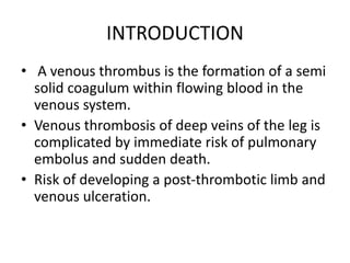
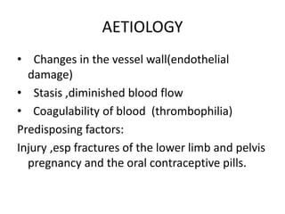
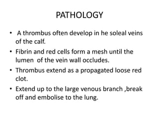
INTRODUCTION
• A venous thrombus is the formation of a semi
solid coagulum within flowing blood in the
venous system.
• Venous thrombosis of deep veins of the leg is
complicated by immediate risk of pulmonary
embolus and sudden death.
• Risk of developing a post-thrombotic limb and
venous ulceration.
AETIOLOGY
• Changes in the vessel wall(endothelial
damage)
• Stasis ,diminished blood flow
• Coagulability of blood (thrombophilia)
Predisposing factors:
Injury ,esp fractures of the lower limb and pelvis
pregnancy and the oral contraceptive pills.
PATHOLOGY
• A thrombus often develop in he soleal veins
of the calf.
• Fibrin and red cells form a mesh until the
lumen of the vein wall occludes.
• Thrombus extend as a propagated loose red
clot.
• Extend up to the large venous branch ,break
off and embolise to the lung.
• Embolus arising from the lower leg veins
becomes detached passes through the large
veins of the limb and venacava.
• Lodges in the pulmonary arteries
• Occlude the pefusion to all or part of one or
both lungs.
• Sudden collapse and death.
DIAGNOSIS
• The most common presentation in DVT is pain
and swelling esp in the calf.
• Many patients have no symptoms of
thrombosis and present with pulmonary
embolism.
• Sometimes leg appears cellulitic.
• Presenting with venous gangrene have
underlying neoplasm.
• Mild pitting edemaof the ankle ,dialated
veins,stiff calf and tenderness over the course
of the deep veins.
• Low grade pyrexia in patients with pulmonary
embolism.
• Signs-cyanosis, dyspnoea, raised neck veins,
split second heart sound, pleural rub if there is
right heart strain.
INVESTIGATION
• Patients with idiopathic thrombosis ususally
have a D-dimer measurement.
• If d-dimer is raised ,a Duplex ultrasound
examination of the deep veins should be
performed.
• Ascending venography
• CTscan
DIFFERENTIAL DIAGNOSIS
• Ruptured baker’s cyst
• calf muscle haematoma
• Ruptured plantaris muscle
• Thrombosed popliteal aneurysm
• Arterial ischaemia
PROPHYLAXIS
Prophylactical methods include:
• Mechanical
• Pharmacological
Mechanical :graduated elastic compression
stocking,external pneumatic compression.
These reduces the incidence of thrombosis.
Pharmacological:low molecular weight heparin
given sc .
TREATMENT
• Patients confirmed to have DVT on duplex
imaging should be started on sc low
molecular weight heparin&rapidly
anticagulated with warfarin.
 Warfarin started at a dose of 10mgon day 1
 10mg on day 2
 5mg on day 3.
• Prothrombin time is taken on day 3.
• Thrombolysis :Tissue plasminogen activator
in most patients is administered directly into
the thrombus ,via the popliteal vein or by
direct puncture in the groin.
• Thrombi can be compressed by stent
grafting,this technique is very good in
patients with ‘iliac vein compression
syndrome’.
Deep vein thrombosis

Deep vein thrombosis

  • 2.
    INTRODUCTION • A venousthrombus is the formation of a semi solid coagulum within flowing blood in the venous system. • Venous thrombosis of deep veins of the leg is complicated by immediate risk of pulmonary embolus and sudden death. • Risk of developing a post-thrombotic limb and venous ulceration.
  • 3.
    AETIOLOGY • Changes inthe vessel wall(endothelial damage) • Stasis ,diminished blood flow • Coagulability of blood (thrombophilia) Predisposing factors: Injury ,esp fractures of the lower limb and pelvis pregnancy and the oral contraceptive pills.
  • 4.
    PATHOLOGY • A thrombusoften develop in he soleal veins of the calf. • Fibrin and red cells form a mesh until the lumen of the vein wall occludes. • Thrombus extend as a propagated loose red clot. • Extend up to the large venous branch ,break off and embolise to the lung.
  • 5.
    • Embolus arisingfrom the lower leg veins becomes detached passes through the large veins of the limb and venacava. • Lodges in the pulmonary arteries • Occlude the pefusion to all or part of one or both lungs. • Sudden collapse and death.
  • 7.
    DIAGNOSIS • The mostcommon presentation in DVT is pain and swelling esp in the calf. • Many patients have no symptoms of thrombosis and present with pulmonary embolism. • Sometimes leg appears cellulitic. • Presenting with venous gangrene have underlying neoplasm.
  • 8.
    • Mild pittingedemaof the ankle ,dialated veins,stiff calf and tenderness over the course of the deep veins. • Low grade pyrexia in patients with pulmonary embolism. • Signs-cyanosis, dyspnoea, raised neck veins, split second heart sound, pleural rub if there is right heart strain.
  • 9.
    INVESTIGATION • Patients withidiopathic thrombosis ususally have a D-dimer measurement. • If d-dimer is raised ,a Duplex ultrasound examination of the deep veins should be performed. • Ascending venography • CTscan
  • 10.
    DIFFERENTIAL DIAGNOSIS • Rupturedbaker’s cyst • calf muscle haematoma • Ruptured plantaris muscle • Thrombosed popliteal aneurysm • Arterial ischaemia
  • 11.
    PROPHYLAXIS Prophylactical methods include: •Mechanical • Pharmacological Mechanical :graduated elastic compression stocking,external pneumatic compression. These reduces the incidence of thrombosis. Pharmacological:low molecular weight heparin given sc .
  • 12.
    TREATMENT • Patients confirmedto have DVT on duplex imaging should be started on sc low molecular weight heparin&rapidly anticagulated with warfarin.  Warfarin started at a dose of 10mgon day 1  10mg on day 2  5mg on day 3. • Prothrombin time is taken on day 3.
  • 13.
    • Thrombolysis :Tissueplasminogen activator in most patients is administered directly into the thrombus ,via the popliteal vein or by direct puncture in the groin. • Thrombi can be compressed by stent grafting,this technique is very good in patients with ‘iliac vein compression syndrome’.