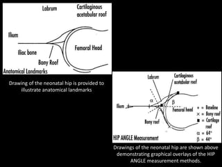
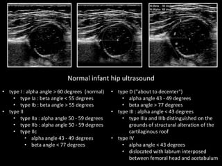
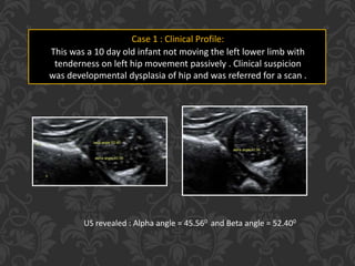
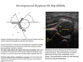
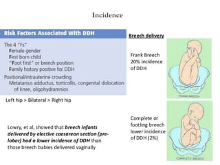
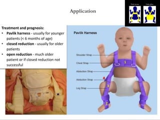
Presenting Author –
Dr. Sanjay M Khaladkar
Co-Authors –
Dr. Dhaval K. Thakkar
Institution –
Dr. D. Y. Patil Medical College,
Pimpri, Pune
Drawing of the neonatal hip is provided to
illustrate anatomical landmarks
Drawings of the neonatal hip are shown above
demonstrating graphical overlays of the HIP
ANGLE measurement methods.
Normal infant hip ultrasound
• type I : alpha angle > 60 degrees (normal)
• type Ia : beta angle < 55 degrees
• type Ib : beta angle > 55 degrees
• type II
• type IIa : alpha angle 50 - 59 degrees
• type IIb : alpha angle 50 - 59 degrees
• type IIc
• alpha angle 43 - 49 degrees
• beta angle < 77 degrees
• type D ("about to decenter")
• alpha angle 43 - 49 degrees
• beta angle > 77 degrees
• type III : alpha angle < 43 degrees
• type IIIa and IIIb distinguished on the
grounds of structural alteration of the
cartilaginous roof
• type IV
• alpha angle < 43 degrees
• dislocated with labrum interposed
between femoral head and acetabulum
Case 1 : Clinical Profile:
This was a 10 day old infant not moving the left lower limb with
tenderness on left hip movement passively . Clinical suspicion
was developmental dysplasia of hip and was referred for a scan .
US revealed : Alpha angle = 45.560 and Beta angle = 52.400
Developmental Dysplasia Of Hip (DDH)
Case 2 : Clinical Profile:
21 years/female came with complaints of left
hip pain since childhood which is slowly
increasing in intensity and aggravates during
winters. Patient is not able to squat for a long
time. No h/o trauma/fall No h/o fever/TB
Hilgenreiner's line
Hilgenreiner's line is drawn horizontally through the
superior aspect of both triradiate cartilages. It should
be horizontal, but is mainly used as a reference for
Perkin's line and measurement of the acetabular
angle
Perkin's line
Perkin's line is drawn perpendicular to Hilgenreiner's
line, intersecting the lateral most aspect of the
acetabular roof. The upper femoral epiphysis should
be seen in the inferomedial quadrant (i.e below
Hilgenreiner's line, and medial to Perkin's line)
Case 2 : Clinical Profile:
21 years/female came with complaints of left
hip pain since childhood which is slowly
increasing in intensity and aggravates during
winters. Patient is not able to squat for a long
time. No h/o trauma/fall No h/o fever/TB
Acetabular angle
The acetabular angle is formed by the
intersection between a line drawn
tangential to the acetabular roof
and Hilgenreiner's line, forming an acute
angle. It should be approximately 30
degrees at birth and progressively
reduce with maturation of the joint.
Case 2 : Clinical Profile:
21 years/female came with complaints of left
hip pain since childhood which is slowly
increasing in intensity and aggravates during
winters. Patient is not able to squat for a long
time. No h/o trauma/fall No h/o fever/TB
Lateral Center Edge Angle Of Wiberg –
Angle formed by a line drawn from the center
of the femoral head to the outer edge of the
acetabular roof, and a verticle line drawn
through the center of the femoral head;
- angle greater than 25 deg are considered
normal;
- less than 20 deg indicates severe dysplasia;
Developmental Dysplasia Of Hip (DDH)
Coronal and Axial CT Scan Images reveals –
Subarticular sclerosis, subarticular geords, shallow acetlabulum, malformed femoral head
 Definition –
 Developmental dysplasia of the hip (DDH) results from an abnormal
relationship of the femoral head to the acetabulum. It usually occurs from
ligamentous laxity and / or abnormal position in utero
 Incidence = 1.5 and 20 per 1000 births
 with the majority (60-80%) of abnormal hips resolving spontaneously within
2-8 weeks (so called immature hip)
Developmental Dysplasia Of Hip (DDH)
Incidence
Secondary Hip Dysplasia
Treatment and prognosis:
• Pavlik harness - usually for younger
patients (< 6 months of age)
• closed reduction - usually for older
patients
• open reduction - much older
patient or if closed reduction not
successful
Application
1. Graf R. The diagnosis of congenital hip-joint dislocation by the ultrasonic Combound treatment. Arch
Orthop Trauma Surg. 1980;97 (2): 117-33.
2. Graf R, Scott S, Lercher K. Hip sonography, diagnosis and management of infant hip dysplasia. Springer
Verlag. (2006) ISBN:3540309578.
3. US Preventive Services Task Force. “Screening for Developmental Dysplasia of the Hip:
Recommendation Statement.” PEDIATRICS 117, no. 3 (3, 2006): 898-902.
4. Roposch Andreas, Nicole M. Moreau, Elizabeth Uleryk and Andrea S. Doria. “Developmental Dysplasia
of the Hip: Quality of Reporting of Diagnostic Accuracy for US1.” Radiology 241, no. 3 (December 2006):
854-860.
5. Taeusch H. William, Roberta A. Ballard, Christine A. Gleason and Mary Ellen Avery. Avery's diseases of
the newborn. Elsevier Health Sciences, 2005.
REFERENCES
Developmental Dysplasia Of Hip (DDH)

Ddh final dt2

  • 1.
    Presenting Author – Dr.Sanjay M Khaladkar Co-Authors – Dr. Dhaval K. Thakkar Institution – Dr. D. Y. Patil Medical College, Pimpri, Pune
  • 2.
    Drawing of theneonatal hip is provided to illustrate anatomical landmarks Drawings of the neonatal hip are shown above demonstrating graphical overlays of the HIP ANGLE measurement methods.
  • 3.
    Normal infant hipultrasound • type I : alpha angle > 60 degrees (normal) • type Ia : beta angle < 55 degrees • type Ib : beta angle > 55 degrees • type II • type IIa : alpha angle 50 - 59 degrees • type IIb : alpha angle 50 - 59 degrees • type IIc • alpha angle 43 - 49 degrees • beta angle < 77 degrees • type D ("about to decenter") • alpha angle 43 - 49 degrees • beta angle > 77 degrees • type III : alpha angle < 43 degrees • type IIIa and IIIb distinguished on the grounds of structural alteration of the cartilaginous roof • type IV • alpha angle < 43 degrees • dislocated with labrum interposed between femoral head and acetabulum
  • 4.
    Case 1 :Clinical Profile: This was a 10 day old infant not moving the left lower limb with tenderness on left hip movement passively . Clinical suspicion was developmental dysplasia of hip and was referred for a scan . US revealed : Alpha angle = 45.560 and Beta angle = 52.400
  • 5.
  • 6.
    Case 2 :Clinical Profile: 21 years/female came with complaints of left hip pain since childhood which is slowly increasing in intensity and aggravates during winters. Patient is not able to squat for a long time. No h/o trauma/fall No h/o fever/TB Hilgenreiner's line Hilgenreiner's line is drawn horizontally through the superior aspect of both triradiate cartilages. It should be horizontal, but is mainly used as a reference for Perkin's line and measurement of the acetabular angle Perkin's line Perkin's line is drawn perpendicular to Hilgenreiner's line, intersecting the lateral most aspect of the acetabular roof. The upper femoral epiphysis should be seen in the inferomedial quadrant (i.e below Hilgenreiner's line, and medial to Perkin's line)
  • 7.
    Case 2 :Clinical Profile: 21 years/female came with complaints of left hip pain since childhood which is slowly increasing in intensity and aggravates during winters. Patient is not able to squat for a long time. No h/o trauma/fall No h/o fever/TB Acetabular angle The acetabular angle is formed by the intersection between a line drawn tangential to the acetabular roof and Hilgenreiner's line, forming an acute angle. It should be approximately 30 degrees at birth and progressively reduce with maturation of the joint.
  • 8.
    Case 2 :Clinical Profile: 21 years/female came with complaints of left hip pain since childhood which is slowly increasing in intensity and aggravates during winters. Patient is not able to squat for a long time. No h/o trauma/fall No h/o fever/TB Lateral Center Edge Angle Of Wiberg – Angle formed by a line drawn from the center of the femoral head to the outer edge of the acetabular roof, and a verticle line drawn through the center of the femoral head; - angle greater than 25 deg are considered normal; - less than 20 deg indicates severe dysplasia;
  • 9.
    Developmental Dysplasia OfHip (DDH) Coronal and Axial CT Scan Images reveals – Subarticular sclerosis, subarticular geords, shallow acetlabulum, malformed femoral head
  • 10.
     Definition – Developmental dysplasia of the hip (DDH) results from an abnormal relationship of the femoral head to the acetabulum. It usually occurs from ligamentous laxity and / or abnormal position in utero  Incidence = 1.5 and 20 per 1000 births  with the majority (60-80%) of abnormal hips resolving spontaneously within 2-8 weeks (so called immature hip) Developmental Dysplasia Of Hip (DDH)
  • 11.
     Definition – Developmental dysplasia of the hip (DDH) results from an abnormal relationship of the femoral head to the acetabulum. It usually occurs from ligamentous laxity and / or abnormal position in utero  Incidence = 1.5 and 20 per 1000 births  with the majority (60-80%) of abnormal hips resolving spontaneously within 2-8 weeks (so called immature hip) Developmental Dysplasia Of Hip (DDH)
  • 12.
  • 13.
  • 14.
    Treatment and prognosis: •Pavlik harness - usually for younger patients (< 6 months of age) • closed reduction - usually for older patients • open reduction - much older patient or if closed reduction not successful Application
  • 15.
    1. Graf R.The diagnosis of congenital hip-joint dislocation by the ultrasonic Combound treatment. Arch Orthop Trauma Surg. 1980;97 (2): 117-33. 2. Graf R, Scott S, Lercher K. Hip sonography, diagnosis and management of infant hip dysplasia. Springer Verlag. (2006) ISBN:3540309578. 3. US Preventive Services Task Force. “Screening for Developmental Dysplasia of the Hip: Recommendation Statement.” PEDIATRICS 117, no. 3 (3, 2006): 898-902. 4. Roposch Andreas, Nicole M. Moreau, Elizabeth Uleryk and Andrea S. Doria. “Developmental Dysplasia of the Hip: Quality of Reporting of Diagnostic Accuracy for US1.” Radiology 241, no. 3 (December 2006): 854-860. 5. Taeusch H. William, Roberta A. Ballard, Christine A. Gleason and Mary Ellen Avery. Avery's diseases of the newborn. Elsevier Health Sciences, 2005. REFERENCES Developmental Dysplasia Of Hip (DDH)