MRI is increasingly used to evaluate developmental dysplasia of the hip (DDH) as it is a noninvasive imaging modality that provides excellent anatomic detail of both ossified and unossified structures of the hip. While ultrasound and radiography were previously the standard modalities depending on patient age, MRI is now widely used for treatment planning, monitoring, and in the postoperative period. The radiologist should be familiar with the critical MRI findings of DDH and the increasing role of MRI in the evaluation and management of this condition.
![1324 AJR:203, December 2014
Imaging Update on Developmental
Dysplasia of the Hip With the Role
of MRI
Vanessa Starr1
Bo Yoon Ha
Starr V, Ha BY
1
Both authors: Department of Radiology, Santa Clara
Valley Medical Center, 751 S Bascom Ave, San Jose, CA
95128. Address correspondence to V. Starr
(vanessaleestarr@gmail.com).
Pediatric Imaging • Review
This article is available for credit.
AJR 2014; 203:1324–1335
0361–803X/14/2036–1324
© American Roentgen Ray Society
Keywords: developmental dysplasia of the hip (DDH),
MRI of the hips, musculoskeletal MRI, musculoskeletal
ultrasound, pediatric imaging
DOI:10.2214/AJR.13.12449
Received December 20, 2013; accepted after revision
March 9, 2014.
Based on a presentation at the ARRS 2013 Annual
Meeting, Washington, DC.
pared with selective ultrasound screening
[6]. Holen et al. [7] conducted a randomized
controlled study comparing the two strate-
gies. Of 15,529 infants after 6–11 years of
follow-up, there were five cases of late-diag-
nosed DDH in the selective group and one
case in the general group. Therefore, if uni-
versal screening is used, a large number of
infants require screening to detect one addi-
tional case of DDH.
Therapy is most effective and tends to be
noninvasive when DDH is detected early [8,
9]. Untreated, DDH can progress to abnor-
mal gait; leg length discrepancies; early osteo-
arthritis; and, rarely, avascular necrosis [10,
11]. Patients younger than 6 months old are
typically braced in Pavlik harnesses [12]. Sur-
gical hip reduction and casting are used for pa-
tients who fail the Pavlik harness or those with
late diagnoses. Iliac and femoral osteotomies
are reserved for severe cases of DDH [13].
Imaging Algorithm
Multiple modalities are used for the initial
diagnosis and further workup of DDH. The
recommended imaging modality for the ini-
tial workup depends primarily on patient age
(Table 1). In infants up to 4–5 months old,
ultrasound is the standard imaging modali-
ty. Radiography is recommended thereafter,
once ossification of the femoral epiphysis be-
gins to obscure visualization of sonographic
landmarks. CT is reserved primarily for
problem solving, typically in the postopera-
tive period. It is currently used infrequently
because of the disadvantage of ionizing radi-
D
evelopmental dysplasia of the
hip (DDH) is a disease that in-
volves abnormal development of
the femoral head and acetabu-
lum. Although the precise mechanism of dis-
ease pathogenesis has yet to be elucidated, a
normal acetabulum stimulates the femoral
head to develop adequately and, conversely,
an appropriately positioned femoral head en-
ables normal acetabular development [1].
The incidence of DDH ranges from 1.5 to 20
per 1000 births. Multiple risk factors have been
described and include breech positioning in
utero, oligohydramnios, family history, female
sex, and first born [2]. Increased joint laxity in
the setting of exposure to maternal estrogens in
the perinatal period may also play a role in the
development of DDH. The left hip is affected
more frequently than the right.
Different screening strategies have been
described, including clinical examination
alone, selective ultrasound screening, and
universal ultrasound screening. Selective ul-
trasound is indicated in patients with associ-
ated risk factors or abnormal clinical exami-
nations [3]. A common method of screening
is serial physical examinations using the
Barlow and Ortolani maneuvers and selec-
tive ultrasound if indicated [4]. The Barlow
maneuver is performed by adducting a flexed
hip and exerting posterior pressure to iden-
tify a dislocatable hip. The Ortolani maneu-
ver is performed by abducting a flexed hip
with anterior force to relocate an already dis-
located hip [5]. Some studies have addressed
the effectiveness of universal screening com-
OBJECTIVE. The purpose of this article is to review developmental dysplasia of the hip
(DDH), a well-described entity previously evaluated with a standard multimodality imag-
ing algorithm, typically consisting of ultrasound and radiography depending on patient age.
CONCLUSION. MRI is increasingly used because it is a noninvasive imaging modal-
ity that offers excellent anatomic detail, enabling the differentiation of ossified and unossified
components of the hip. The radiologist should be aware of the increasing role of MRI and rec-
ognize the critical MRI findings of DDH.
Starr and Ha
Imaging Developmental Dysplasia of the Hip
Pediatric Imaging
Review
Downloaded
from
www.ajronline.org
by
202.138.240.223
on
02/02/23
from
IP
address
202.138.240.223.
Copyright
ARRS.
For
personal
use
only;
all
rights
reserved](https://image.slidesharecdn.com/ajr-230209013909-a665189c/85/ajr-13-12449-pdf-1-320.jpg)
![AJR:203, December 2014 1325
Imaging Developmental Dysplasia of the Hip
ation. MRI is increasingly used for treatment
planning and monitoring. It is now widely
used in the postoperative period.
Ultrasound
Ultrasound is the reference standard
for evaluating the hip in an infant before 6
months, when capital femoral epiphyseal os-
sification usually occurs. It is a nonionizing,
quick, and portable examination that fur-
thermore offers the advantage of dynamic
imaging in addition to standard static views.
The American College of Radiology rec-
ommends that a standard ultrasound ex-
amination be performed in two orthogonal
planes: a coronal view in the standard plane
at rest and a transverse view of the flexed
hip with and without stress [14]. Three ana-
tomic landmarks—ilial line, triradiate car-
tilage, and labrum—are used to measure the
α and β angles. A standard plane includes
a straight iliac line, the femoral head with
maximum diameter, the tip of the echogen-
ic acetabular labrum, and the triradiate car-
tilage. Figure 1 shows the anatomic land-
marks in a normal hip. Meticulous scrutiny
of the α angle measurement is necessary
because false-positive findings can occur if
the imaging plane is suboptimal. When re-
porting the α angle, the largest angle, not
the average angle, should be given.
Femoral Head Position Relative to
the Acetabulum
A normally positioned femoral head is
more than 50% covered by the acetabulum.
DDH results in a shallow acetabulum and de-
creased coverage of the femoral head.
Graf α Angle
The Graf α angle is measured in the coro-
nal plane and is defined as the angle formed
between the vertical cortex of the ilium and
the acetabular roof. An α angle less than 60°
is abnormal and reflects a shallow acetabu-
lum [15]. Figure 2A shows a normal α angle
and Figure 2B shows an α angle in an infant
with DDH. The modified Graf grading clas-
sification is based on the α angle and degree
of acetabular roof coverage (Table 2).
Graf β Angle
The Graf β angle is formed by a line through
the vertical ilium and the cartilaginous acetab-
ular labrum (Fig. 2A). A Graf β angle greater
than 55° is abnormal. With superolateral femo-
ral head displacement, the labrum is elevated,
thereby increasing the β angle [16, 17].
Dynamic Harcke Method
The purpose of dynamic stress imaging
is to determine the position and stability of
the femoral head during stress manipulation.
Coronal and axial images are obtained in
neutral position and hip flexion. The stress
maneuver is similar to the clinical Barlow
examination in which the hip is adducted and
pressure is exerted on the knee to force the
femoral head to dislocate posteriorly [14].
When monitoring is performed in the Pavlik
harness, only static images are obtained [18].
Color Doppler imaging has been used to
evaluate perfusion to the proximal femoral
epiphysis [19, 20], although there is little lit-
erature in the setting of DDH. After place-
ment of the Pavlik harness, serial follow-up
hip ultrasound examinations are performed
to assess response to treatment. The infant is
left in the Pavlik harness and only static im-
ages are obtained [21].
Radiography
After the child is 4–5 months old, the ossi-
fication of the femoral epiphysis begins to ob-
scure sonographic landmarks and radiography
becomes more reliable for detection of DDH.
This is the standard tool to diagnose DDH af-
ter 6 months [22]. An anteroposterior radio-
graph of the hips in neutral position is used to
assess the morphology of the acetabulum, os-
sification of the femoral head, and position of
the femoral head relative to the acetabulum.
In early infancy, a normal acetabulum is rela-
tively steeper and straighter. The morphology
of the acetabulum changes with age, with the
acetabulum becoming more curved inferiorly
along the medial and lateral margins. Figure 3
shows the spectrum of normal hips in antero-
posterior radiographs in a 6-month-old child
and a 2-year-old child, respectively. In DDH,
there is delayed ossification of the femoral
head and an abnormally shallow acetabulum,
thereby predisposing to subluxation and dislo-
cation. Additionally, late complications, such
as osteoarthritis and avascular necrosis, can
occur. A frog-leg lateral view is sometimes
used to determine whether a subluxed hip re-
duces. Several lines and angles are used to di-
agnose and further characterize DDH (Fig.
3B and Table 3): The first is the Hilgenreiner
line, which is a line crossing through both tri-
radiate cartilages. The second is the acetabu-
lar angle, which is formed by the Hilgenreiner
line and a line drawn through the acetabular
roof. A neonate should normally have an ac-
TABLE 1: Multimodality Imaging Algorithm
Modality Age or Indication Advantages and Disadvantages
Ultrasound Up to 4–5 mo Unossified femoral head, bony, and
nonbony landmarks well evaluated
Radiography After 5–6 mo Once femoral head ossifies, bony
landmarks evaluated
CT Problem solving, mostly postoperative
evaluation
Used for problem solving in past;
however, has disadvantage of
unnecessary ionizing radiation
MRI Treatment planning and monitoring,
including postoperative evaluation
Treatment planning and monitoring,
including postoperative evaluation
TABLE 2: Modified Graf Classification Scale
Graf Type Description α and β Angle
Type 1 Normal, mature hip with more than 50%
acetabular roof coverage
α angle ≥ 60°, β angle < 55°
Type 2a Physiologic immaturity at younger than 3 mo α angle 50–59°
Type 2b Immature at age 3 mo or older α angle 50–59°
Type 2c Extremely deficient bony acetabulum;
femoral head is concentric but not stable
α angle 43–49°, β angle < 77°
Type 2d Femoral head is grossly subluxed and labrum is
everted, increasing β angle
α angle difficult to measure but is
approximately 43–49°; β angle > 77°
Type 3 Dislocated femoral head with shallow
acetabulum
α angle < 43°
Type 4 Dislocated femoral head with severely shallow,
dysplastic acetabulum and inverted labrum
Downloaded
from
www.ajronline.org
by
202.138.240.223
on
02/02/23
from
IP
address
202.138.240.223.
Copyright
ARRS.
For
personal
use
only;
all
rights
reserved](https://image.slidesharecdn.com/ajr-230209013909-a665189c/85/ajr-13-12449-pdf-2-320.jpg)
![1326 AJR:203, December 2014
Starr and Ha
etabular angle of less than 30°. The acetabular
angle should be less than 22° at and beyond 1
year of age [23]. Acetabular morphology and
the degree of femoral head ossification chang-
es with age (Fig. 3). The third is the Perkins
line, which is a vertical line drawn perpendic-
ular to the Hilgenreiner line and intersecting
the lateral rim of the acetabular roof. A nor-
mally situated femoral head is in the inferi-
or medial quadrant. The fourth is the Shen-
ton line, which is a C-shaped line drawn
along the inferior border of the superior pu-
bic ramus and the inferomedial border of the
femoral neck. A normal Shenton line should
form a smooth arc [2] (Fig. 3B). The fifth is
the anterior center-edge angle, which is an an-
gle subtended by a craniocaudal line through
the center of the ossified femoral head and a
line from the center of the femoral head to the
lateral margin of the acetabular roof (Fig. 3B).
A center edge angle less than 20° is indicative
of dysplasia [24].
Serial radiography can be used to track
disease progression and response to treat-
ment. Figure 4 shows temporal evolution in
an infant with mild DDH. Figure 5A shows
severe DDH in a 3 year 9 month old child
with a late diagnosis. Figure 5B shows the
postoperative radiograph in the same patient.
Arthrography
Arthrography is typically performed in-
traoperatively by the orthopedic surgeon at
the time of reduction. Obstacles to success-
ful reduction, such as limbus eversion, can
be identified. Arthrography during recon-
structive osteotomy helps obtain concentric
reduction of the hip [25] (Fig. 6).
CT
CT is generally reserved for problem solv-
ing in difficult cases and involves a low-
dose technique, often in the setting of pre-
or postoperative evaluation (Fig. 7). The CT
technique at our institution is weight based
(Table 4). CT is more commonly used post-
operatively after the patient has been placed
in a cast to define the success of reduction
[26]. Postoperatively, concentric reduction of
the femoral head can be confirmed (Fig. 7).
Preoperative assessment includes evaluation
of bony acetabular morphology and the ossi-
fied femoral epiphysis as well as the femoral
head position relative to the acetabulum.
A recent study compared the use of CT ver-
sus MRI to evaluate hip reduction in patients
with DDH and found that both modalities of-
fer excellent sensitivity and specificity [27].
CT had sensitivity of 100% and specificity of
96% for the postoperative nonsubluxed hip,
whereas MRI showed sensitivity of 100% and
a specificity of 100%. Compared with MRI,
CT requires shorter imaging time and less, if
any, postoperative anesthesia. It is also a use-
ful modality for patients with surgical hard-
ware. However, the primary disadvantage of
CT is the exposure to ionizing radiation.
MRI
MRI Indications
MRI, like CT, is often reserved for more
difficult cases; however, the major advantage
of MRI is the ability to delineate soft-tissue
structures as well as osseous structures with-
out ionizing radiation. Many MRI studies
are ordered in the postoperative period, usu-
ally after reduction and spica cast placement.
In fact, spica cast placement is one of the
most common indications for MRI in the set-
ting of DDH. After open reduction, the hip is
held in 90° flexion and partial abduction, and
the femoral head is held in position by a plas-
ter spica cast. The degree of abduction must
be carefully controlled because too little re-
sults in redislocation and too much can in-
crease the risk of avascular necrosis. Neither
hip should be abducted more than 55–60°
[28]. Surgeons have varying thresholds and
criteria for ordering MRI after spica casting;
however, inability to clinically confirm fem-
oral head reduction or abnormal radiography
after casting are common indications [29].
MRI Technique
One drawback of MRI is the relatively
lengthy time of the examination compared
with CT or radiography. Protocols differ from
one institution to another and the length of
MRI examinations has ranged in the litera-
ture from as little as 3 minutes to 45 minutes
[30–32]. Conroy et al. [29] reviewed the ef-
ficiency and accuracy of MRI in confirming
femoral head location after closed reduction
and spica cast application and concluded that,
in their experience, axial STIR MRI was suf-
ficient for confirmation of concentric femoral
head reduction. All of the scans in their study
were obtained in less than 5 minutes and none
TABLE 3: Summary of Radiographic Lines and Measurements
Line or Angle Definition Normal Measurement
Hilgenreiner line Horizontal line through both triradiate cartilages
Acetabular angle Angle subtended by Hilgenreiner line and line through
acetabular roof
Normal acetabular angle in a neonate is < 30° and < 22° at and
beyond 1 year old
Perkins line Vertical line intersecting lateral rim of acetabular roof
perpendicular to Hilgenreiner line
Normal femoral head should lay in inferior medial quadrant of
acetabulum
Shenton line C-shaped line drawn along inferior border of superior pubic
ramus and inferomedial border of femoral neck
Normal Shenton line should form a smooth arc
Anterior center edge angle Angle subtended by vertical line through center of ossified
femoral head and line from center to lateral margin of
acetabular roof
Normal center edge angle should be > 25°; angle < 20°
indicates dysplasia
TABLE 4: Weight-Based 64-MDCT Protocol
Weight Division (kg) Kilovoltage (kV) Current (mA) Slice Thickness (mm) Slice Spacing (ms) Gantry Rotation Speed (s) Pitch
< 15 120 40 0.6 0.3 0.5 1.4
15–24 120 65 0.6 0.3 0.5 1.4
25–34 120 80 0.6 0.3 0.5 1.4
Downloaded
from
www.ajronline.org
by
202.138.240.223
on
02/02/23
from
IP
address
202.138.240.223.
Copyright
ARRS.
For
personal
use
only;
all
rights
reserved](https://image.slidesharecdn.com/ajr-230209013909-a665189c/85/ajr-13-12449-pdf-3-320.jpg)
![AJR:203, December 2014 1327
Imaging Developmental Dysplasia of the Hip
required sedation. Laor et al. [30] also eval-
uated the utility of limited MRI after surgi-
cal reduction for DDH and reported a mean
imaging time of 3 minutes for two sequenc-
es. Gould et al. [33] found that T2-weighted
fast spin-echo sequences were superior with
regard to diagnostic performance and were
performed in less than 3 minutes. They ad-
vised orthopedic surgeons to request axial
and coronal T2 fast spin-echo sequences to
obtain a diagnostic study in less than 15 min-
utes, eliminating the need for sedation. At
our institution, axial and coronal fast spin-
echo sequences using conventional fast spin-
echo or fat-suppressed equivalent T1-weight-
ed and T2-weighted sequences (IDEAL, GE
Healthcare) are routinely obtained. Ultra-
fast spin-echo sequences (single-shot fast
spin-echo) are sometimes used to decrease
scanning time. MRI after spica casting is
typically performed in the immediate post-
operative period while patients are still un-
der sedation. Gadolinium is not routinely
administered. However, if there is concern
for avascular necrosis of the femoral head,
gadolinium is used to evaluate for femoral
head enhancement abnormalities [34]. Table
5 provides the MRI protocol specifications.
MRI Findings of the Normal Hip
Familiarity with the normal appearance of
the pediatric hip on MRI is critical to detect
pathology (Fig. 8). The ossified and unossi-
fied femoral heads, cartilage, and ligaments
are clearly depicted. The infantile acetab-
ulum can be categorized into three basic
components: bony, cartilaginous, and liga-
mentous or soft tissue [35]. The bony ace-
tabulum is seen on radiography and is com-
posed of the acetabular parts of the ilium,
ischium, and pubis, all of which are held to-
gether by the triradiate cartilage. The carti-
laginous acetabulum consists of the hyaline
cartilage at the articular surface, which is U-
shaped and is bridged by the transverse ac-
etabular ligament, and the supporting vascu-
larized growth cartilage, which includes the
triradiate cartilage [36]. The labrum, trans-
verse acetabular ligament, and the ligamen-
tum teres are the primary ligamentous struc-
tures. The labrum is of low to intermediate
signal intensity and appears as a small trian-
gular structure along the edge of the acetab-
ulum on axial images. The labrum’s intrinsic
signal intensity typically increases slightly
from T1- to T2-weighted images [32]. It is
important to evaluate for normal morphol-
ogy and position of the labrum when eval-
uating dysplastic hips. The transverse ace-
tabular ligament is located inferiorly, where
there is a deficiency of cartilaginous acetab-
ulum. The ligamentum teres originates from
the transverse ligament and inserts on the
femoral head fovea. The iliopsoas tendon is
a low-signal-intensity structure that is seen
just anteromedial to the anterior labrum on
the axial plane. The intraarticular fat pad, or
pulvinar, lies in the central portion of the ac-
etabulum and has the highest signal intensi-
ty of all the structures in the hip, paralleling
that of subcutaneous fat [36]. It is important
to assess for pulvinar hypertrophic changes,
which can serve as an obstacle to successful
reduction. The pulvinar in the affected hip
can be compared with the contralateral side
to determine any relative size asymmetry.
The ossified femoral epiphysis appears as
a low-signal-intensity structure within the
high-signal-intensity unossified hyaline car-
tilage. Symmetry between the two ossified
femoral heads should be noted. When eval-
uating for concentric femoral head position-
ing, a line can be drawn through both trira-
diate cartilages. After successful reduction,
the ossified portion of the femoral epiphyses
should lie anterior to this line [28]. The os-
sified portions of the anterior and posterior
columns are low to intermediate in signal
intensity, with an interposed band of high-
signal-intensity triradiate cartilage. Depend-
ing on the degree of acetabular dysplasia, the
unossified parts of the anterior and posteri-
or columns affect acetabular depth. The fi-
brous joint capsule attaches to the acetabu-
lar margin peripheral to the labrum. At birth,
the femoral attachment is near the metaphy-
sis and migrates inferiorly as the hip devel-
ops. By 12 months of age, the capsule is part-
ly fused to the femoral neck periosteum and
runs up the femoral neck, attaching to the
edge of the cartilaginous femoral head [36].
Normal acetabular development is depen-
dent on concentric positioning of the femoral
head within the acetabulum.
MRI Findings of Developmental Dysplasia of
the Hip
When characterizing DDH using MRI, the
dysplastic acetabulum should be evaluated
for retroversion and degree of femoral head
coverage. There may be associated cartilagi-
nous defects or delamination. Delayed ossifi-
cation of the femoral head can be determined
by comparing the ossific nucleus of the femo-
ral head in the affected hip with the contra-
lateral side. A major advantage of MRI is the
ability to visualize the cartilaginous acetabu-
lum and determine its contribution to femoral
head coverage. MRI depicts the unossified ac-
etabular epiphysis in the ilium and underlying
labrum, therefore showing greater and more
accurate acetabular coverage than that seen
on radiography alone [37].
Recent orthopedic articles [37–40] have
described the utility of bony and cartilagi-
nous acetabular indexes on MRI in the evalu-
ation of DDH. The bony acetabular index can
be measured by MRI using an anteroposterior
coronal view and is similar to the acetabular
index measured on radiography. To obtain the
bony acetabular index, the Hilgenreiner line
and Perkins line are drawn using the same
TABLE 5: MRI Protocol Parameters
Protocol
TR Range
(ms)
TE Range
(ms)
Echo-Train
Length
Flip Angle
(°)
No. of Signals
Acquired Matrix
Slice
Thickness
(mm)
Slice
Spacing
(ms)
FOV
(cm)
Conventional FSE T1-weighted 1000–1100 15–20 3 90 1–2 192 × 192–320 × 256 3 3.0 18–24
Conventional FSE T2-weighted 3500–4000 65–75 15–18 90 1–2 192 × 192–320 × 256 3 3.0 18–24
Fat-suppressed equivalent
T1-weighted (IDEAL)
680–800 10–13 2–3 90 2–6 192 × 192–256 × 224 3 3.5 18–24
Fat-suppressed equivalent
T2-weighted (IDEAL)
4000–4600 90–100 24 90 2–6 192 × 192–256 × 224 3 3.5 18–24
Note—All studies performed on a 3-T scanner using either a multichannel torso array coil or multichannel neurovascular array coil in axial and coronal planes for each
sequence. FSE = fast spin-echo. IDEAL manufactured by GE Healthcare.
Downloaded
from
www.ajronline.org
by
202.138.240.223
on
02/02/23
from
IP
address
202.138.240.223.
Copyright
ARRS.
For
personal
use
only;
all
rights
reserved](https://image.slidesharecdn.com/ajr-230209013909-a665189c/85/ajr-13-12449-pdf-4-320.jpg)
![1328 AJR:203, December 2014
Starr and Ha
landmarks as used on radiography. The bony
acetabular index line is drawn from the Hil-
genreiner line at the lateral part of the trira-
diate cartilage to the Perkins line at the lat-
eral aspect of the bony acetabulum. The angle
subtended by the bony acetabular index line
and the Hilgenreiner line is the bony acetabu-
lar index angle (Fig. 9I). The cartilaginous ac-
etabular index is measured by drawing a line
from the lateral part of the triradiate cartilage
at the Hilgenreiner line to the lateral acetab-
ular cartilaginous margin (the cartilaginous
acetabular index line). The cartilaginous ac-
etabular index angle is formed by the carti-
laginous acetabular index line and the Hilgen-
reiner line [38] (Fig. 9J).
Pirpiris et al. [38] compared MRI and radi-
ography in 14 hips with a diagnosis of DDH
and no prior surgery to determine the corre-
lation between the bony acetabular index on
MRI and the acetabular index on radiography.
There was a significant correlation between the
bony acetabular index measured on MRI and
the radiographic acetabular index. The bony
acetabular index and cartilaginous acetabular
index also correlated with each other; howev-
er, the cartilaginous acetabular index measured
significantly less than the bony acetabular in-
dex (6.8° ± 3.3°). Therefore, if bony angle is
desired, those authors argue that radiography
provides sufficient information; however, MRI
provides significant additional information
about the true cartilaginous coverage of the
femoral head. Li et al. [40] evaluated the bony
acetabular index and cartilaginous acetabu-
lar index in 81 children with DDH and com-
pared them with 241 healthy control children.
In contrast to the study by Pirpiris et al., which
showed a significant correlation between the
bony acetabular index and cartilaginous ace-
tabular index, Li et al. found that the normal
cartilaginous acetabular index decreased rap-
idly within the first 2 years of life and then
remained constant at a mean (SD) of 8.25°
(1.65°) until adolescence. A notable difference
in the level of dislocation was present between
the Tonnis grade in the bony acetabular index
and cartilaginous acetabular index. Therefore,
bony acetabular development does not always
represent cartilaginous development.
MRI enables direct and accurate evalua-
tion of the cartilage and important character-
ization of the cartilaginous acetabular angle
[40]. After successful reduction, the femoral
head should be concentrically located in the
acetabulum. The angle of abduction can be
measured between the main axis of the femur
and the midsagittal plane of the subject [31].
This is important to note because too much
abduction can lead to avascular necrosis. If
contrast material has been administered, the
enhancement of the femoral head should be
noted. Jaramillo et al. [31] evaluated 23 dys-
plastic hips immediately after spica casting
with contrast-enhanced MRI. They classified
the degree of femoral epiphyseal enhancement
with a 5-point grading scale, with 1 indicating
normal enhancement and 5 indicating glob-
ally decreased or absent enhancement. They
found a significant correlation between great-
er abduction and more severe femoral head
enhancement abnormalities. In their series,
only two of the 14 femoral heads abducted
less than 55° showed enhancement abnormal-
ities, and of the hips abducted less than 50°,
none had enhancement defects.
Ray et al. [39] treated late-presented DDH
with nonoperative graduated traction and gen-
tle manipulation. They evaluated 12 hips treat-
ed as such to confirm concentric reduction. In
all 12 hips, there was excellent soft-tissue re-
modeling around the hip and confirmation of
concentric reduction as evidenced by the car-
tilaginous acetabular extension. Radiography
would not have shown the extensive soft-tissue
remodeling, and therefore MRI was particular-
ly useful to confirm successful reduction.
MRI is particularly useful for determin-
ing ligamentous and soft-tissue abnormali-
ties that may serve as obstacles to successful
reduction [41]. The fibrofatty pulvinar in the
acetabular fossa can become hypertrophied,
preventing adequate femoral head reduction
(Figs. 10B and 10C). The labrum should be
evaluated closely for hypertrophy and abnor-
mal position, such as eversion and inversion
(Fig. 10B). Similarly, the transverse liga-
ment or ligamentum teres can be hypertro-
phied and should be routinely evaluated [41].
Rarely, the iliopsoas tendon may be inter-
posed between the femoral head and acetab-
ulum. Table 6 contains a checklist of stan-
dard structures to evaluate for DDH.
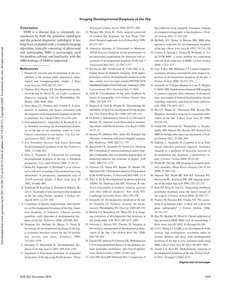
MRI Examples in Two Patients
Patient A is shown as an example of MRI
after spica casting (Figs. 10A–10C). Patient
B underwent MRI for spica casting first and
another MRI later to guide further clinical
management (Figs. 9A–9J). On occasion,
MRI may show discrepant findings com-
pared with radiography. In patient B, there
was persistent subluxation of the affected hip
on follow-up radiography, prompting a sec-
ond MRI using fat-suppressed equivalent
T1- and T2-weighted sequences in antici-
pation of a possible reoperation. Compared
with follow-up radiography, the degree of
dysplasia and hip subluxation was not as se-
vere on MRI because the cartilaginous por-
tions of the hip were clearly shown. This case
clearly illustrates the utility of MRI because
the cartilaginous acetabular index measured
17° whereas the bony acetabular index mea-
sured 39°, which was concordant with the
34° acetabular angle measured radiographi-
cally. The MRI findings led the surgeon to
elect less-aggressive management. Besides
MRI after spica casting, another indication
for MRI in the setting of DDH is preoper-
ative identification of potential obstacles to
successful femoral head reduction, such as
labral inversion, pulvinar fibrofatty prolifer-
ation, and transverse ligament and ligamen-
tum teres hypertrophy [41].
TABLE 6: Checklist of Anatomic Structures to Evaluate in Developmental Dysplasia of Hip (DDH)
Anatomic Structures MRI Findings in DDH
Acetabular morphology Shallow, dysmorphic acetabulum; need to evaluate for retroversion and inadequate femoral head coverage
Symmetry of femoral heads Delayed ossification of femoral head
Femoral head position relative to acetabulum Femoral head subluxation or dislocation
Labrum Labral hypertrophy; may see mucoid degeneration or tear
Pulvinar Pulvinar hypertrophy appears as fibrofatty proliferation
Ligamentum teres or transverse ligament Hypertrophy
Femoral head perfusion Avascular necrosis
Downloaded
from
www.ajronline.org
by
202.138.240.223
on
02/02/23
from
IP
address
202.138.240.223.
Copyright
ARRS.
For
personal
use
only;
all
rights
reserved](https://image.slidesharecdn.com/ajr-230209013909-a665189c/85/ajr-13-12449-pdf-5-320.jpg)


![1334 AJR:203, December 2014
Starr and Ha
G H
I J
Fig. 9 (continued)—11-month-old girl with hip click
(patient B).
E and F, Follow-up MRI was performed to assess
whether second operation was indicated. Axial
(E) and coronal (F) fat-suppressed equivalent T1-
weighted images show hypertrophic acetabular
cartilage and good morphology of cartilage portion of
right femoral head, overall improved since prior MRI.
G and H, Coronal non–fat-suppressed (G) and fat-
suppressed (H) equivalent T1-weighted images
show mild right pulvinar fat hypertrophy (arrow)
with improved position of femoral head relative to
acetabulum since prior MRI.
I and J, Coronal T1-weighted images with fat
saturation show superimposed bony acetabular
index angle (I) and cartilaginous acetabular index
angle (J). Bony acetabular index measures 39.6°,
which is fairly concordant with 34° acetabular angle
measured on radiographs. Hypertrophic acetabular
cartilage contributes to 15° cartilaginous acetabular
index, which is still abnormal but relatively closer
to normal range (mean cartilaginous acetabular
index in 2-year-old is 8.2 ± 1.9 [40]) compared with
measured bony acetabular index. This examination
served as guide for further orthopedic management.
Compared with radiographs, femoral head appears
more concentrically located in acetabulum. Surgeon
subsequently elected to treat more conservatively.
E F
Downloaded
from
www.ajronline.org
by
202.138.240.223
on
02/02/23
from
IP
address
202.138.240.223.
Copyright
ARRS.
For
personal
use
only;
all
rights
reserved](https://image.slidesharecdn.com/ajr-230209013909-a665189c/85/ajr-13-12449-pdf-11-320.jpg)