SlideShare a Scribd company logo
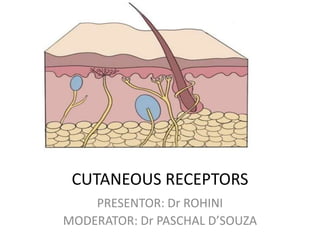
CUTANEOUS RECEPTORS
PRESENTOR: Dr ROHINI
MODERATOR: Dr PASCHAL D’SOUZA
SHERRINGTON CLASSIFICATION OF
RECEPTORS
Depending upon the
source of stimulus
• Exteroreceptors
• Interoreceptors
• Proprioreceptors
Exteroreceptors
• Receptors responsible for sensing stimulus
coming from external environment.
• Present at or near the surface of the body.
• Includes:
1. Touch
2. Temperature
3. Nociception
Interoreceptors
• Respond to stretch ,volume and pressure in
the wall of viscera, excessive muscle
contraction and to overstretching in the
visceral organs.
• Essential in regulating blood flow ,pressure in
the CVS ,maintaining respiration etc.
Proprioceptors
• Responds to stimulus arising in muscles ,tendons,
joints etc.
• Essential for coordination of movements and
maintenance of posture.
• Includes
1. Neuromuscular
2. Neurotendinous spindles
Depending upon type of stimulus
energy
• Mechanoreceptors
1. Cutaneous receptors
2. Muscle and joint receptors
3. Hair cells
4. Baroreceptors of carotid sinus and aortic arch
• Thermoreceptors
• Nociceptors: receptors responding to pain
• Photorecepetors: rods and cones
• Chemoreceptors
Cutaneous receptors
• Sensory receptors
• Located in epidermis as well as dermis of skin
• Exteroreceptors :informs us about touch , temperature
,pain and pressure.
• Densly present over face ,finger tips.
• Includes:
1. Mechanoreceptors
2. Thermoreceptors
3. Nociceptors
Mechanoreceptors
• Touch ,pressure , softness, texture of the
stimulus.
• Includes
1. Merkels disc and Meissners corpuscles
2. Pacinian corpuscles
3. Ruffini ‘s end organs
4. Krause end bulbs
5. Free nerve endings
Meissner's corpuscles
• Meissner's corpuscles localize in the dermis
between epidermal ridges.
• They contain an unmyelinated nerve ending
surrounded by Schwann cells.
• The center of the capsule contains one or
more afferent nerve fibers that generate rapidly
adapting action potentials following minimal skin
depression.
• Present in glabrous (smooth, hairless) skin (the
fingertips)
• Their afferent fibers account for about 40% of
the sensory innervation of the human hand.
• Efficient in transducing information about the
relatively low-frequency vibrations (30–50 Hz)
that occur when textured objects are moved
across the skin.
Pacinian corpuscles
• Large encapsulated endings located in the subcutaneous
tissue .
• Has an onion like capsule in which the inner core of
membrane lamellae is separated from an outer lamella by a
fluid-filled space.
• one or more rapidly adapting afferent axons lie at the
center of this structure.
• The capsule again acts as a filter, allowing only transient
disturbances at high frequencies (250–350 Hz) to activate
the nerve endings.
• Rapidly adapting
• Involved in the
discrimination of fine
surface textures or high-
frequency vibrations.
• Provide information
primarily about the
dynamic qualities of
mechanical stimuli.
Merkel's disks
• Located in the epidermis, where they are
precisely aligned with the papillae that lie
beneath the dermal ridges.
• Account for about 25% of the mechanoreceptors
of the hand and are particularly dense in the
fingertips, lips, and external genitalia.
• The slowly adapting nerve fiber associated with
each Merkel's disk enlarges into a saucer-shaped
ending that is closely applied to another
specialized cell containing vesicles that
apparently release peptides that modulate the
nerve terminal.
• Selective stimulation of these receptors in
humans produces a sensation of light pressure.
IGGO DOME RECEPTORS
• Merkel's discs are often grouped together in a receptor organ
called the Iggo dome receptor, which projects upward against
the underside of the epithelium of the skin.
• This causes the epithelium at this point to protrude outward,
thus creating a dome and constituting an extremely sensitive
receptor.
RUFFINI CORPUSCLES
• These elongated, spindle-shaped capsular
specializations are located deep in the skin, as
well as in ligaments and tendons.
• The long axis of the corpuscle is usually oriented
parallel to the stretch lines in skin.
• Particularly sensitive to the cutaneous stretching
produced by digit or limb movements.
Ruffini corpuscle from original slide sent by
Ruffini to Sir Charles Sherrington
HAIR FOLLICLE RECEPTOR
• Unencapsulated
• Primary afferent spiral around hair follicle
base.
• Runs parallel to the hair shaft to form a lattice
like pattern.
• Signals the direction and velocity of
movement.
• Rapidly adaptating
Krause end bulb
• Type of meissners receptor
• Mainly present in glabrous skin such as skin of
genitalia, papillae of lips,conjunctiva etc
• Afferent fibres belong to A delta
group
• Detects fine touch and pressure
Adaptation in cutaneous receptors
• Rapid adaptation means that there is no response to
sustained pressure, only to changes in pressure (either
an increase or a decrease).
• Slow adaptation means that the receptor continues to
respond to pressure for as long as it is sustained, within
some reasonable time frame.
ADAPTATION
RAPIDLY SLOWLY
RAPIDLY
ADAPTING
PACINIAN CORPUSCELS
MEISSNER'S CORPUSCLES
High frequency vibration , pressure
Light touch
SLOWLY ADAPTING
MERKELS DISC
RUFFINI
Low frequency vibration and texture
Skin stretching
Receptive fields of cutaneous
receptors
• Portion of the skin which, when stimulated,
affects the activity or state of the receptor.
• Receptive Fields — the typically oval areas of skin along
which a mechanical stimulus (e.g., touch) leads to a
change in that neurons action potential firing rates.
• In particular, Pacinian corpuscles have fairly large
receptive fields, making them more sensitive (if you
consider the goal of a mechanoreceptor to sense touch
anywhere on the body)
• Meissner’s corpuscles have fairly small receptive
fields, making them more specific
The A-beta fibers are large, fast, myelinated afferents. The Meisnner's
corpuscles and Pacinian corpuscles belong to this class. These are
rapidly adapting touch afferents.
Pathways from Skin to Cortex
• Two major pathways in the spinal cord: –
• Medial lemniscal pathway consists of large fibers that
carry proprioceptive and touch information .
• Spinothalamic pathway consists of smaller fibers that
carry temperature and pain information .
• These cross over to the opposite side of the body and
synapse in the thalamus, and then on to the
Somatosensory cortex
• Signals travel from the thalamus to the somatosensory
receiving area (S1) and the secondary receiving area (S2) in
the parietal lobe .
• Body map (homunculus) on the cortex shows more cortical
space allocated to parts of the body that are responsible for
detail.
THERMORECEPTORS
• Thermoreceptors are free dendrite endings in skin, and
thus are primary sensory organs. They have no
specialized epithelial cells or supporting cells.
• The nerves of skin branch from musculocutaneous
nerves that arise segmentally from spinal nerves.
• The pattern of nerve fibers in skin is similar to the
vascular patterns—nerve fibers form a deep plexus,
then ascend to a superficial, subpapillary plexus.
• The penicillate fibers are the primary nerve fibers found
subepidermally in haired skin.
• Rapidly adapting receptors that function in the perception
of touch, temperature, pain, and itch.
• Overlapping innervation,leads discrimination to be
generalized.
FREE NERVE ENDING
Penicillate Papillary
• Papillary nerve endings are found at the
orifice of a follicle and are thought to be
particularly receptive to cold sensation.
• Hair follicles also contain , slow-adapting
receptors that respond to the bending or
movement of hairs.
• A very high concentration of thermosensitive
TRP (Transient Receptor Potential )ion
channels are found in keratinocytes in the
epidermis.
• Each TRP has a unique temperature threshold
of firing.
THERMORECEPTIVE NEURONS
C-FIBRES
1.Heat sense neurons
2.Unmyelinated
3.Less velocity of transduction
4.Innervated epidermis
5.Fewer dendrites per neuron
,small receptive fied
A DELTA FIBRES
1.Cold sense neuron
2.Myelinated
3.More velocity of transduction
4.Innervated layers between
epidermis and dermis
5.High receptive field
• Cell bodies of the afferent fibers of cutaneous
thermoreceptors reside in the dorsal root
ganglion (DRG) or the trigeminal ganglion on
the dorsal horn of the spinal cord.
• The trigeminal neuron is particularily sensitive
to cold due to its high expression of cold-
activated TRP ion channels.
NOCICEPTORS
• Mediate pain
• Terminal branches of thin myelinated A delta and
unmyleniated C fibres
1. Somatic nociceptors: free nerve endings in skin
2. Visceral nociceptors: not well known
• Nociceptors are generally electrically silent and
transmit all-or-none action potentials only when
stimulated.
• Nociceptive fibers have been classified on the
basis of their conduction velocity and sensitivity
and threshold to noxious mechanical (M), heat (H),
and cold (C) .
• C fibres:(C-MH, C-MC, C-MHC)
• A-fiber nociceptors are predominately heat- and
or mechanosensitive (A-MH, A-H, A-M)
• Excitatory neurons and release glutamate as
their primary neurotransmitter as well as
other components including peptides (e.g.,
substance P, calcitonin gene-related peptide
[CGRP], somatostatin.)

More Related Content

What's hot

Receptors classification
Receptors classificationReceptors classification
Receptors classification
Dr Sara Sadiq
 
Sensory systems 1
Sensory systems 1Sensory systems 1
Sensory systems 1whisper119
 
Receptors
ReceptorsReceptors
Receptors
Maylord Demol
 
Sweat gland & sebaceous gland dermatology revision notes
Sweat gland & sebaceous gland dermatology  revision notes Sweat gland & sebaceous gland dermatology  revision notes
Sweat gland & sebaceous gland dermatology revision notes
TONY SCARIA
 
Physiology of sensory systems
Physiology of sensory systemsPhysiology of sensory systems
Physiology of sensory systems
Serge Eddy Mba
 
Taste and smell
Taste and smellTaste and smell
Taste and smellvajira54
 
Ch 15 sensory pathways
Ch 15 sensory pathwaysCh 15 sensory pathways
Ch 15 sensory pathways
Ajaindu Shrivastava
 
Tousif, role of receptors
Tousif, role of receptorsTousif, role of receptors
Tousif, role of receptorsfarhan_aq91
 
Sweat gland - anatomy and function
Sweat gland - anatomy and functionSweat gland - anatomy and function
Sweat gland - anatomy and function
mamta rai
 
Olfaction mbbs
Olfaction mbbsOlfaction mbbs
Olfaction mbbs
Dr Lekshmi Priya
 
Synaptic Transmission
Synaptic TransmissionSynaptic Transmission
Synaptic Transmissionvacagodx
 
The sense of touch, pain & temperature
The sense of touch, pain & temperatureThe sense of touch, pain & temperature
The sense of touch, pain & temperature
Hashim Ali
 
Anatomy & Physiology Of Olfactory System
Anatomy & Physiology Of Olfactory SystemAnatomy & Physiology Of Olfactory System
Anatomy & Physiology Of Olfactory System
Prasanna Datta
 
Taste buds
Taste budsTaste buds
Taste buds
Sukhbir Brar
 
Auditory pathways
Auditory pathwaysAuditory pathways
Auditory pathways
“Karishma R.Pandey”
 
Development of integumentary system
Development of integumentary systemDevelopment of integumentary system
Development of integumentary system
Dr. Piyush Karn
 
SYNAPSE
SYNAPSESYNAPSE
Physioloy of olfaction and olfactory dysfunctions
Physioloy of olfaction and olfactory dysfunctionsPhysioloy of olfaction and olfactory dysfunctions
Physioloy of olfaction and olfactory dysfunctions
Yousuf Choudhury
 

What's hot (20)

Receptors classification
Receptors classificationReceptors classification
Receptors classification
 
Sensory systems 1
Sensory systems 1Sensory systems 1
Sensory systems 1
 
Skin histology
Skin histologySkin histology
Skin histology
 
Receptors
ReceptorsReceptors
Receptors
 
Sweat gland & sebaceous gland dermatology revision notes
Sweat gland & sebaceous gland dermatology  revision notes Sweat gland & sebaceous gland dermatology  revision notes
Sweat gland & sebaceous gland dermatology revision notes
 
Physiology of sensory systems
Physiology of sensory systemsPhysiology of sensory systems
Physiology of sensory systems
 
Taste and smell
Taste and smellTaste and smell
Taste and smell
 
Ch 15 sensory pathways
Ch 15 sensory pathwaysCh 15 sensory pathways
Ch 15 sensory pathways
 
Tousif, role of receptors
Tousif, role of receptorsTousif, role of receptors
Tousif, role of receptors
 
Sweat gland - anatomy and function
Sweat gland - anatomy and functionSweat gland - anatomy and function
Sweat gland - anatomy and function
 
Olfaction mbbs
Olfaction mbbsOlfaction mbbs
Olfaction mbbs
 
Synaptic Transmission
Synaptic TransmissionSynaptic Transmission
Synaptic Transmission
 
Physiology of olfaction
Physiology of olfactionPhysiology of olfaction
Physiology of olfaction
 
The sense of touch, pain & temperature
The sense of touch, pain & temperatureThe sense of touch, pain & temperature
The sense of touch, pain & temperature
 
Anatomy & Physiology Of Olfactory System
Anatomy & Physiology Of Olfactory SystemAnatomy & Physiology Of Olfactory System
Anatomy & Physiology Of Olfactory System
 
Taste buds
Taste budsTaste buds
Taste buds
 
Auditory pathways
Auditory pathwaysAuditory pathways
Auditory pathways
 
Development of integumentary system
Development of integumentary systemDevelopment of integumentary system
Development of integumentary system
 
SYNAPSE
SYNAPSESYNAPSE
SYNAPSE
 
Physioloy of olfaction and olfactory dysfunctions
Physioloy of olfaction and olfactory dysfunctionsPhysioloy of olfaction and olfactory dysfunctions
Physioloy of olfaction and olfactory dysfunctions
 

Similar to Cutaneous receptors

Tracts of the spinalcord
Tracts of the spinalcordTracts of the spinalcord
Tracts of the spinalcorddrnaveent
 
Anatomical type of receptors
Anatomical type of receptorsAnatomical type of receptors
Anatomical type of receptors
ABDUL QADEER MEMON
 
Y2 s1 sensory system
Y2 s1 sensory systemY2 s1 sensory system
Y2 s1 sensory systemvajira54
 
Physiology of sensory & pain.pdf
Physiology of sensory & pain.pdfPhysiology of sensory & pain.pdf
Physiology of sensory & pain.pdf
qahtanali2
 
Somatic sensory system
Somatic sensory systemSomatic sensory system
Somatic sensory system
AmbrishGupta6
 
lecture 2 physio ppt.pptx
lecture 2 physio ppt.pptxlecture 2 physio ppt.pptx
lecture 2 physio ppt.pptx
Superior college
 
Neuroanatomy of the pain structures in the spinal
Neuroanatomy of the pain structures in the spinalNeuroanatomy of the pain structures in the spinal
Neuroanatomy of the pain structures in the spinal
Tural Abdullayev
 
Sensory Receptors 2023 (2).pdf
Sensory Receptors 2023 (2).pdfSensory Receptors 2023 (2).pdf
Sensory Receptors 2023 (2).pdf
Anaaya Batool
 
2nd year small group discussion Synapse and sensory receptors.pptx
2nd year small group discussion Synapse and sensory receptors.pptx2nd year small group discussion Synapse and sensory receptors.pptx
2nd year small group discussion Synapse and sensory receptors.pptx
bobehe4189
 
Ascending pathways
Ascending pathwaysAscending pathways
Ascending pathways
ANKITARANI20
 
SSEP BY FZ.pptx
SSEP BY FZ.pptxSSEP BY FZ.pptx
SSEP BY FZ.pptx
Faizan Abdullah
 
Neurophysiology lecture
Neurophysiology lectureNeurophysiology lecture
Neurophysiology lecture
RASHA
 
Neurophysiology lecture
Neurophysiology lectureNeurophysiology lecture
Neurophysiology lecture
RASHA
 
physiology of Sensory nervous system, updated 2021
physiology of Sensory nervous system,  updated 2021physiology of Sensory nervous system,  updated 2021
physiology of Sensory nervous system, updated 2021
dina merzeban
 
Sensation and perception
Sensation and perceptionSensation and perception
Sensation and perception
Binibining Kalawakan
 
Psych b sensory system
Psych b sensory systemPsych b sensory system
Psych b sensory systemvajira54
 
Sensory system review
Sensory system reviewSensory system review
Sensory system review
dina merzeban
 
Somatic mechanoceptive sensation.hussein f. sakr
Somatic mechanoceptive sensation.hussein f. sakrSomatic mechanoceptive sensation.hussein f. sakr
Somatic mechanoceptive sensation.hussein f. sakr
Hussein Sakr
 
Ha presentation
Ha presentationHa presentation
Ha presentation
Dennis Dujon
 

Similar to Cutaneous receptors (20)

Tracts of the spinalcord
Tracts of the spinalcordTracts of the spinalcord
Tracts of the spinalcord
 
Anatomical type of receptors
Anatomical type of receptorsAnatomical type of receptors
Anatomical type of receptors
 
Y2 s1 sensory system
Y2 s1 sensory systemY2 s1 sensory system
Y2 s1 sensory system
 
Physiology of sensory & pain.pdf
Physiology of sensory & pain.pdfPhysiology of sensory & pain.pdf
Physiology of sensory & pain.pdf
 
Somatic sensory system
Somatic sensory systemSomatic sensory system
Somatic sensory system
 
lecture 2 physio ppt.pptx
lecture 2 physio ppt.pptxlecture 2 physio ppt.pptx
lecture 2 physio ppt.pptx
 
Neuroanatomy of the pain structures in the spinal
Neuroanatomy of the pain structures in the spinalNeuroanatomy of the pain structures in the spinal
Neuroanatomy of the pain structures in the spinal
 
Sensory Receptors 2023 (2).pdf
Sensory Receptors 2023 (2).pdfSensory Receptors 2023 (2).pdf
Sensory Receptors 2023 (2).pdf
 
2nd year small group discussion Synapse and sensory receptors.pptx
2nd year small group discussion Synapse and sensory receptors.pptx2nd year small group discussion Synapse and sensory receptors.pptx
2nd year small group discussion Synapse and sensory receptors.pptx
 
Ascending pathways
Ascending pathwaysAscending pathways
Ascending pathways
 
SSEP BY FZ.pptx
SSEP BY FZ.pptxSSEP BY FZ.pptx
SSEP BY FZ.pptx
 
Neurophysiology lecture
Neurophysiology lectureNeurophysiology lecture
Neurophysiology lecture
 
Neurophysiology lecture
Neurophysiology lectureNeurophysiology lecture
Neurophysiology lecture
 
physiology of Sensory nervous system, updated 2021
physiology of Sensory nervous system,  updated 2021physiology of Sensory nervous system,  updated 2021
physiology of Sensory nervous system, updated 2021
 
Sensation and perception
Sensation and perceptionSensation and perception
Sensation and perception
 
Psych b sensory system
Psych b sensory systemPsych b sensory system
Psych b sensory system
 
Sensory system review
Sensory system reviewSensory system review
Sensory system review
 
Somatic mechanoceptive sensation.hussein f. sakr
Somatic mechanoceptive sensation.hussein f. sakrSomatic mechanoceptive sensation.hussein f. sakr
Somatic mechanoceptive sensation.hussein f. sakr
 
Ha presentation
Ha presentationHa presentation
Ha presentation
 
Receptor
ReceptorReceptor
Receptor
 

Recently uploaded

Deep Software Variability and Frictionless Reproducibility
Deep Software Variability and Frictionless ReproducibilityDeep Software Variability and Frictionless Reproducibility
Deep Software Variability and Frictionless Reproducibility
University of Rennes, INSA Rennes, Inria/IRISA, CNRS
 
(May 29th, 2024) Advancements in Intravital Microscopy- Insights for Preclini...
(May 29th, 2024) Advancements in Intravital Microscopy- Insights for Preclini...(May 29th, 2024) Advancements in Intravital Microscopy- Insights for Preclini...
(May 29th, 2024) Advancements in Intravital Microscopy- Insights for Preclini...
Scintica Instrumentation
 
Body fluids_tonicity_dehydration_hypovolemia_hypervolemia.pptx
Body fluids_tonicity_dehydration_hypovolemia_hypervolemia.pptxBody fluids_tonicity_dehydration_hypovolemia_hypervolemia.pptx
Body fluids_tonicity_dehydration_hypovolemia_hypervolemia.pptx
muralinath2
 
role of pramana in research.pptx in science
role of pramana in research.pptx in sciencerole of pramana in research.pptx in science
role of pramana in research.pptx in science
sonaliswain16
 
Observation of Io’s Resurfacing via Plume Deposition Using Ground-based Adapt...
Observation of Io’s Resurfacing via Plume Deposition Using Ground-based Adapt...Observation of Io’s Resurfacing via Plume Deposition Using Ground-based Adapt...
Observation of Io’s Resurfacing via Plume Deposition Using Ground-based Adapt...
Sérgio Sacani
 
BLOOD AND BLOOD COMPONENT- introduction to blood physiology
BLOOD AND BLOOD COMPONENT- introduction to blood physiologyBLOOD AND BLOOD COMPONENT- introduction to blood physiology
BLOOD AND BLOOD COMPONENT- introduction to blood physiology
NoelManyise1
 
bordetella pertussis.................................ppt
bordetella pertussis.................................pptbordetella pertussis.................................ppt
bordetella pertussis.................................ppt
kejapriya1
 
Deep Behavioral Phenotyping in Systems Neuroscience for Functional Atlasing a...
Deep Behavioral Phenotyping in Systems Neuroscience for Functional Atlasing a...Deep Behavioral Phenotyping in Systems Neuroscience for Functional Atlasing a...
Deep Behavioral Phenotyping in Systems Neuroscience for Functional Atlasing a...
Ana Luísa Pinho
 
GBSN- Microbiology (Lab 3) Gram Staining
GBSN- Microbiology (Lab 3) Gram StainingGBSN- Microbiology (Lab 3) Gram Staining
GBSN- Microbiology (Lab 3) Gram Staining
Areesha Ahmad
 
GBSN - Biochemistry (Unit 5) Chemistry of Lipids
GBSN - Biochemistry (Unit 5) Chemistry of LipidsGBSN - Biochemistry (Unit 5) Chemistry of Lipids
GBSN - Biochemistry (Unit 5) Chemistry of Lipids
Areesha Ahmad
 
DERIVATION OF MODIFIED BERNOULLI EQUATION WITH VISCOUS EFFECTS AND TERMINAL V...
DERIVATION OF MODIFIED BERNOULLI EQUATION WITH VISCOUS EFFECTS AND TERMINAL V...DERIVATION OF MODIFIED BERNOULLI EQUATION WITH VISCOUS EFFECTS AND TERMINAL V...
DERIVATION OF MODIFIED BERNOULLI EQUATION WITH VISCOUS EFFECTS AND TERMINAL V...
Wasswaderrick3
 
Leaf Initiation, Growth and Differentiation.pdf
Leaf Initiation, Growth and Differentiation.pdfLeaf Initiation, Growth and Differentiation.pdf
Leaf Initiation, Growth and Differentiation.pdf
RenuJangid3
 
Orion Air Quality Monitoring Systems - CWS
Orion Air Quality Monitoring Systems - CWSOrion Air Quality Monitoring Systems - CWS
Orion Air Quality Monitoring Systems - CWS
Columbia Weather Systems
 
Chapter 12 - climate change and the energy crisis
Chapter 12 - climate change and the energy crisisChapter 12 - climate change and the energy crisis
Chapter 12 - climate change and the energy crisis
tonzsalvador2222
 
Earliest Galaxies in the JADES Origins Field: Luminosity Function and Cosmic ...
Earliest Galaxies in the JADES Origins Field: Luminosity Function and Cosmic ...Earliest Galaxies in the JADES Origins Field: Luminosity Function and Cosmic ...
Earliest Galaxies in the JADES Origins Field: Luminosity Function and Cosmic ...
Sérgio Sacani
 
NuGOweek 2024 Ghent - programme - final version
NuGOweek 2024 Ghent - programme - final versionNuGOweek 2024 Ghent - programme - final version
NuGOweek 2024 Ghent - programme - final version
pablovgd
 
原版制作(carleton毕业证书)卡尔顿大学毕业证硕士文凭原版一模一样
原版制作(carleton毕业证书)卡尔顿大学毕业证硕士文凭原版一模一样原版制作(carleton毕业证书)卡尔顿大学毕业证硕士文凭原版一模一样
原版制作(carleton毕业证书)卡尔顿大学毕业证硕士文凭原版一模一样
yqqaatn0
 
extra-chromosomal-inheritance[1].pptx.pdfpdf
extra-chromosomal-inheritance[1].pptx.pdfpdfextra-chromosomal-inheritance[1].pptx.pdfpdf
extra-chromosomal-inheritance[1].pptx.pdfpdf
DiyaBiswas10
 
Salas, V. (2024) "John of St. Thomas (Poinsot) on the Science of Sacred Theol...
Salas, V. (2024) "John of St. Thomas (Poinsot) on the Science of Sacred Theol...Salas, V. (2024) "John of St. Thomas (Poinsot) on the Science of Sacred Theol...
Salas, V. (2024) "John of St. Thomas (Poinsot) on the Science of Sacred Theol...
Studia Poinsotiana
 
erythropoiesis-I_mechanism& clinical significance.pptx
erythropoiesis-I_mechanism& clinical significance.pptxerythropoiesis-I_mechanism& clinical significance.pptx
erythropoiesis-I_mechanism& clinical significance.pptx
muralinath2
 

Recently uploaded (20)

Deep Software Variability and Frictionless Reproducibility
Deep Software Variability and Frictionless ReproducibilityDeep Software Variability and Frictionless Reproducibility
Deep Software Variability and Frictionless Reproducibility
 
(May 29th, 2024) Advancements in Intravital Microscopy- Insights for Preclini...
(May 29th, 2024) Advancements in Intravital Microscopy- Insights for Preclini...(May 29th, 2024) Advancements in Intravital Microscopy- Insights for Preclini...
(May 29th, 2024) Advancements in Intravital Microscopy- Insights for Preclini...
 
Body fluids_tonicity_dehydration_hypovolemia_hypervolemia.pptx
Body fluids_tonicity_dehydration_hypovolemia_hypervolemia.pptxBody fluids_tonicity_dehydration_hypovolemia_hypervolemia.pptx
Body fluids_tonicity_dehydration_hypovolemia_hypervolemia.pptx
 
role of pramana in research.pptx in science
role of pramana in research.pptx in sciencerole of pramana in research.pptx in science
role of pramana in research.pptx in science
 
Observation of Io’s Resurfacing via Plume Deposition Using Ground-based Adapt...
Observation of Io’s Resurfacing via Plume Deposition Using Ground-based Adapt...Observation of Io’s Resurfacing via Plume Deposition Using Ground-based Adapt...
Observation of Io’s Resurfacing via Plume Deposition Using Ground-based Adapt...
 
BLOOD AND BLOOD COMPONENT- introduction to blood physiology
BLOOD AND BLOOD COMPONENT- introduction to blood physiologyBLOOD AND BLOOD COMPONENT- introduction to blood physiology
BLOOD AND BLOOD COMPONENT- introduction to blood physiology
 
bordetella pertussis.................................ppt
bordetella pertussis.................................pptbordetella pertussis.................................ppt
bordetella pertussis.................................ppt
 
Deep Behavioral Phenotyping in Systems Neuroscience for Functional Atlasing a...
Deep Behavioral Phenotyping in Systems Neuroscience for Functional Atlasing a...Deep Behavioral Phenotyping in Systems Neuroscience for Functional Atlasing a...
Deep Behavioral Phenotyping in Systems Neuroscience for Functional Atlasing a...
 
GBSN- Microbiology (Lab 3) Gram Staining
GBSN- Microbiology (Lab 3) Gram StainingGBSN- Microbiology (Lab 3) Gram Staining
GBSN- Microbiology (Lab 3) Gram Staining
 
GBSN - Biochemistry (Unit 5) Chemistry of Lipids
GBSN - Biochemistry (Unit 5) Chemistry of LipidsGBSN - Biochemistry (Unit 5) Chemistry of Lipids
GBSN - Biochemistry (Unit 5) Chemistry of Lipids
 
DERIVATION OF MODIFIED BERNOULLI EQUATION WITH VISCOUS EFFECTS AND TERMINAL V...
DERIVATION OF MODIFIED BERNOULLI EQUATION WITH VISCOUS EFFECTS AND TERMINAL V...DERIVATION OF MODIFIED BERNOULLI EQUATION WITH VISCOUS EFFECTS AND TERMINAL V...
DERIVATION OF MODIFIED BERNOULLI EQUATION WITH VISCOUS EFFECTS AND TERMINAL V...
 
Leaf Initiation, Growth and Differentiation.pdf
Leaf Initiation, Growth and Differentiation.pdfLeaf Initiation, Growth and Differentiation.pdf
Leaf Initiation, Growth and Differentiation.pdf
 
Orion Air Quality Monitoring Systems - CWS
Orion Air Quality Monitoring Systems - CWSOrion Air Quality Monitoring Systems - CWS
Orion Air Quality Monitoring Systems - CWS
 
Chapter 12 - climate change and the energy crisis
Chapter 12 - climate change and the energy crisisChapter 12 - climate change and the energy crisis
Chapter 12 - climate change and the energy crisis
 
Earliest Galaxies in the JADES Origins Field: Luminosity Function and Cosmic ...
Earliest Galaxies in the JADES Origins Field: Luminosity Function and Cosmic ...Earliest Galaxies in the JADES Origins Field: Luminosity Function and Cosmic ...
Earliest Galaxies in the JADES Origins Field: Luminosity Function and Cosmic ...
 
NuGOweek 2024 Ghent - programme - final version
NuGOweek 2024 Ghent - programme - final versionNuGOweek 2024 Ghent - programme - final version
NuGOweek 2024 Ghent - programme - final version
 
原版制作(carleton毕业证书)卡尔顿大学毕业证硕士文凭原版一模一样
原版制作(carleton毕业证书)卡尔顿大学毕业证硕士文凭原版一模一样原版制作(carleton毕业证书)卡尔顿大学毕业证硕士文凭原版一模一样
原版制作(carleton毕业证书)卡尔顿大学毕业证硕士文凭原版一模一样
 
extra-chromosomal-inheritance[1].pptx.pdfpdf
extra-chromosomal-inheritance[1].pptx.pdfpdfextra-chromosomal-inheritance[1].pptx.pdfpdf
extra-chromosomal-inheritance[1].pptx.pdfpdf
 
Salas, V. (2024) "John of St. Thomas (Poinsot) on the Science of Sacred Theol...
Salas, V. (2024) "John of St. Thomas (Poinsot) on the Science of Sacred Theol...Salas, V. (2024) "John of St. Thomas (Poinsot) on the Science of Sacred Theol...
Salas, V. (2024) "John of St. Thomas (Poinsot) on the Science of Sacred Theol...
 
erythropoiesis-I_mechanism& clinical significance.pptx
erythropoiesis-I_mechanism& clinical significance.pptxerythropoiesis-I_mechanism& clinical significance.pptx
erythropoiesis-I_mechanism& clinical significance.pptx
 

Cutaneous receptors

  • 1. CUTANEOUS RECEPTORS PRESENTOR: Dr ROHINI MODERATOR: Dr PASCHAL D’SOUZA
  • 2. SHERRINGTON CLASSIFICATION OF RECEPTORS Depending upon the source of stimulus • Exteroreceptors • Interoreceptors • Proprioreceptors
  • 3. Exteroreceptors • Receptors responsible for sensing stimulus coming from external environment. • Present at or near the surface of the body. • Includes: 1. Touch 2. Temperature 3. Nociception
  • 4. Interoreceptors • Respond to stretch ,volume and pressure in the wall of viscera, excessive muscle contraction and to overstretching in the visceral organs. • Essential in regulating blood flow ,pressure in the CVS ,maintaining respiration etc.
  • 5. Proprioceptors • Responds to stimulus arising in muscles ,tendons, joints etc. • Essential for coordination of movements and maintenance of posture. • Includes 1. Neuromuscular 2. Neurotendinous spindles
  • 6. Depending upon type of stimulus energy • Mechanoreceptors 1. Cutaneous receptors 2. Muscle and joint receptors 3. Hair cells 4. Baroreceptors of carotid sinus and aortic arch • Thermoreceptors • Nociceptors: receptors responding to pain • Photorecepetors: rods and cones • Chemoreceptors
  • 7. Cutaneous receptors • Sensory receptors • Located in epidermis as well as dermis of skin • Exteroreceptors :informs us about touch , temperature ,pain and pressure. • Densly present over face ,finger tips. • Includes: 1. Mechanoreceptors 2. Thermoreceptors 3. Nociceptors
  • 8. Mechanoreceptors • Touch ,pressure , softness, texture of the stimulus. • Includes 1. Merkels disc and Meissners corpuscles 2. Pacinian corpuscles 3. Ruffini ‘s end organs 4. Krause end bulbs 5. Free nerve endings
  • 9.
  • 10. Meissner's corpuscles • Meissner's corpuscles localize in the dermis between epidermal ridges. • They contain an unmyelinated nerve ending surrounded by Schwann cells. • The center of the capsule contains one or more afferent nerve fibers that generate rapidly adapting action potentials following minimal skin depression.
  • 11. • Present in glabrous (smooth, hairless) skin (the fingertips) • Their afferent fibers account for about 40% of the sensory innervation of the human hand. • Efficient in transducing information about the relatively low-frequency vibrations (30–50 Hz) that occur when textured objects are moved across the skin.
  • 12.
  • 13. Pacinian corpuscles • Large encapsulated endings located in the subcutaneous tissue . • Has an onion like capsule in which the inner core of membrane lamellae is separated from an outer lamella by a fluid-filled space. • one or more rapidly adapting afferent axons lie at the center of this structure. • The capsule again acts as a filter, allowing only transient disturbances at high frequencies (250–350 Hz) to activate the nerve endings.
  • 14. • Rapidly adapting • Involved in the discrimination of fine surface textures or high- frequency vibrations. • Provide information primarily about the dynamic qualities of mechanical stimuli.
  • 15.
  • 16. Merkel's disks • Located in the epidermis, where they are precisely aligned with the papillae that lie beneath the dermal ridges. • Account for about 25% of the mechanoreceptors of the hand and are particularly dense in the fingertips, lips, and external genitalia.
  • 17. • The slowly adapting nerve fiber associated with each Merkel's disk enlarges into a saucer-shaped ending that is closely applied to another specialized cell containing vesicles that apparently release peptides that modulate the nerve terminal. • Selective stimulation of these receptors in humans produces a sensation of light pressure.
  • 18.
  • 19. IGGO DOME RECEPTORS • Merkel's discs are often grouped together in a receptor organ called the Iggo dome receptor, which projects upward against the underside of the epithelium of the skin. • This causes the epithelium at this point to protrude outward, thus creating a dome and constituting an extremely sensitive receptor.
  • 20. RUFFINI CORPUSCLES • These elongated, spindle-shaped capsular specializations are located deep in the skin, as well as in ligaments and tendons. • The long axis of the corpuscle is usually oriented parallel to the stretch lines in skin. • Particularly sensitive to the cutaneous stretching produced by digit or limb movements.
  • 21. Ruffini corpuscle from original slide sent by Ruffini to Sir Charles Sherrington
  • 22. HAIR FOLLICLE RECEPTOR • Unencapsulated • Primary afferent spiral around hair follicle base. • Runs parallel to the hair shaft to form a lattice like pattern. • Signals the direction and velocity of movement. • Rapidly adaptating
  • 23.
  • 24. Krause end bulb • Type of meissners receptor • Mainly present in glabrous skin such as skin of genitalia, papillae of lips,conjunctiva etc • Afferent fibres belong to A delta group • Detects fine touch and pressure
  • 25.
  • 26. Adaptation in cutaneous receptors • Rapid adaptation means that there is no response to sustained pressure, only to changes in pressure (either an increase or a decrease). • Slow adaptation means that the receptor continues to respond to pressure for as long as it is sustained, within some reasonable time frame. ADAPTATION RAPIDLY SLOWLY
  • 27.
  • 28. RAPIDLY ADAPTING PACINIAN CORPUSCELS MEISSNER'S CORPUSCLES High frequency vibration , pressure Light touch SLOWLY ADAPTING MERKELS DISC RUFFINI Low frequency vibration and texture Skin stretching
  • 29. Receptive fields of cutaneous receptors • Portion of the skin which, when stimulated, affects the activity or state of the receptor.
  • 30. • Receptive Fields — the typically oval areas of skin along which a mechanical stimulus (e.g., touch) leads to a change in that neurons action potential firing rates. • In particular, Pacinian corpuscles have fairly large receptive fields, making them more sensitive (if you consider the goal of a mechanoreceptor to sense touch anywhere on the body) • Meissner’s corpuscles have fairly small receptive fields, making them more specific
  • 31.
  • 32. The A-beta fibers are large, fast, myelinated afferents. The Meisnner's corpuscles and Pacinian corpuscles belong to this class. These are rapidly adapting touch afferents.
  • 33. Pathways from Skin to Cortex • Two major pathways in the spinal cord: – • Medial lemniscal pathway consists of large fibers that carry proprioceptive and touch information . • Spinothalamic pathway consists of smaller fibers that carry temperature and pain information . • These cross over to the opposite side of the body and synapse in the thalamus, and then on to the Somatosensory cortex
  • 34.
  • 35. • Signals travel from the thalamus to the somatosensory receiving area (S1) and the secondary receiving area (S2) in the parietal lobe . • Body map (homunculus) on the cortex shows more cortical space allocated to parts of the body that are responsible for detail.
  • 36. THERMORECEPTORS • Thermoreceptors are free dendrite endings in skin, and thus are primary sensory organs. They have no specialized epithelial cells or supporting cells. • The nerves of skin branch from musculocutaneous nerves that arise segmentally from spinal nerves. • The pattern of nerve fibers in skin is similar to the vascular patterns—nerve fibers form a deep plexus, then ascend to a superficial, subpapillary plexus.
  • 37. • The penicillate fibers are the primary nerve fibers found subepidermally in haired skin. • Rapidly adapting receptors that function in the perception of touch, temperature, pain, and itch. • Overlapping innervation,leads discrimination to be generalized. FREE NERVE ENDING Penicillate Papillary
  • 38. • Papillary nerve endings are found at the orifice of a follicle and are thought to be particularly receptive to cold sensation. • Hair follicles also contain , slow-adapting receptors that respond to the bending or movement of hairs.
  • 39.
  • 40. • A very high concentration of thermosensitive TRP (Transient Receptor Potential )ion channels are found in keratinocytes in the epidermis. • Each TRP has a unique temperature threshold of firing.
  • 41.
  • 42. THERMORECEPTIVE NEURONS C-FIBRES 1.Heat sense neurons 2.Unmyelinated 3.Less velocity of transduction 4.Innervated epidermis 5.Fewer dendrites per neuron ,small receptive fied A DELTA FIBRES 1.Cold sense neuron 2.Myelinated 3.More velocity of transduction 4.Innervated layers between epidermis and dermis 5.High receptive field
  • 43.
  • 44. • Cell bodies of the afferent fibers of cutaneous thermoreceptors reside in the dorsal root ganglion (DRG) or the trigeminal ganglion on the dorsal horn of the spinal cord. • The trigeminal neuron is particularily sensitive to cold due to its high expression of cold- activated TRP ion channels.
  • 45.
  • 46. NOCICEPTORS • Mediate pain • Terminal branches of thin myelinated A delta and unmyleniated C fibres 1. Somatic nociceptors: free nerve endings in skin 2. Visceral nociceptors: not well known • Nociceptors are generally electrically silent and transmit all-or-none action potentials only when stimulated.
  • 47. • Nociceptive fibers have been classified on the basis of their conduction velocity and sensitivity and threshold to noxious mechanical (M), heat (H), and cold (C) . • C fibres:(C-MH, C-MC, C-MHC) • A-fiber nociceptors are predominately heat- and or mechanosensitive (A-MH, A-H, A-M)
  • 48. • Excitatory neurons and release glutamate as their primary neurotransmitter as well as other components including peptides (e.g., substance P, calcitonin gene-related peptide [CGRP], somatostatin.)