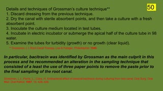
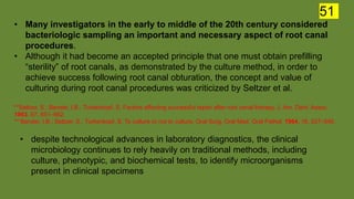
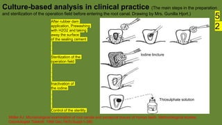
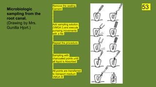
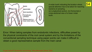
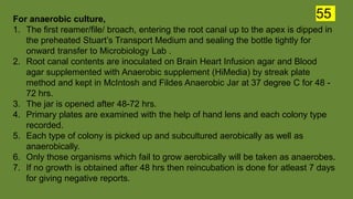
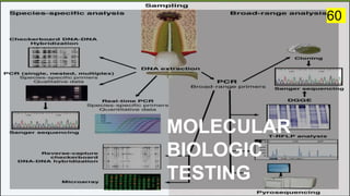
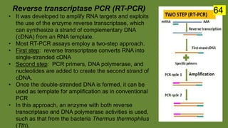
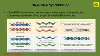
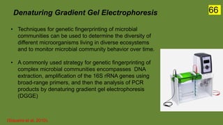
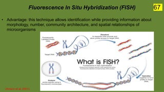
The document discusses the historical and contemporary perspectives on the microbiological aspects of endodontics. It describes some of the early culture-based techniques used to sample and identify microbes in root canals, including Grossman's culture technique from 1940. It also discusses limitations of culture-based analysis and the development of molecular biological testing methods like PCR, DNA-DNA hybridization, and fluorescence in situ hybridization that have improved identification of endodontic pathogens. Finally, it reviews recent studies on chairside culture tests and ATP bioluminescence assays that allow for rapid detection of microbes in root canals.
50. Details and techniques of Grossmanās culture technique**
1. Discard dressing from the previous technique.
2. Dry the canal with sterile absorbent points, and then take a culture with a fresh
absorbent point.
3. Inoculate the culture medium located in test tubes.
4. Incubate in electric incubator or submerge the apical half of the culture tube in 98
water.
5. Examine the tubes for turbidity (growth) or no growth (clear liquid).
** Grossman, L l. Root Canal Therapy. Lea & Febiger; Philadelphia: 1940.
In particular, bacitracin was identified by Grossman as the main culprit in this
process and he recommended an alteration in the sampling technique that
consisted of a least the use of three paper points to remove the paste prior to
the final sampling of the root canal.
Grossman, L.I.; Parris, L.; Cobe, H. Antibacterial effect of residual bacitracin during culturing from root canal. Oral Surg. Oral
Med. Oral Pathol. 1957, 10, 426ā429.
50
51. ā¢ Many investigators in the early to middle of the 20th century considered
bacteriologic sampling an important and necessary aspect of root canal
procedures.
ā¢ Although it had become an accepted principle that one must obtain prefilling
āsterilityā of root canals, as demonstrated by the culture method, in order to
achieve success following root canal obturation, the concept and value of
culturing during root canal procedures was criticized by Seltzer et al.
**Seltzer, S.; Bender, I.B.; Turkenkopf, S. Factors affecting successful repair after root canal therapy. J. Am. Dent. Assoc.
1963, 67, 651ā662.
** Bender, I.B.; Seltzer, S.; Turkenkopf, S. To culture or not to culture. Oral Surg. Oral Med. Oral Pathol. 1964, 18, 527ā540.
ā¢ despite technological advances in laboratory diagnostics, the clinical
microbiology continues to rely heavily on traditional methods, including
culture, phenotypic, and biochemical tests, to identify microorganisms
present in clinical specimens
51
52. Mƶller AJ. Microbiological examination of root canals and periapical tissues of human teeth. Methodological studies.
Odontologisk Tidskrift. 1966 Dec;74(5):Suppl:1-380.
Culture-based analysis in clinical practice (The main steps in the preparation
and sterilization of the operation field before entering the root canal. Drawing by Mrs. Gunilla Hjort.)
Iodine tincture
Thiosulphate solution
After rubber dam
application, Prewashing
with H2O2 and taking
away the surface
of the sealing cement
Sterilization of the
operation field
Inactivation of
the iodine
Control of the sterility
5
2
53. Remove the sealing
Cement
Add sampling solution
(VMGA I) and execute
pumping movements
with a file
Repeat the procedure
Sampling with
charcoaled points until
all liquid is Absorbed
All points are transferred
to transport medium
VMGA III
Microbiologic
sampling from the
root canal.
(Drawing by Mrs.
Gunilla Hjort.)
53
54. A molar tooth indicating the location where
special attention has to be taken for reaching
bacteria at sampling.
(a) Longitudinal section. (b) Horizontal or
cross-sectional view of a lower molar
tooth.
Fouad, Ashraf F Endodontic microbiology. Wiley-Blackwell, Oxford, 2009
Error: When taking samples from endodontic infections, difficulties posed by
the physical constraints of the root canal system and by the limitations of the
conventional sampling technique using paper points can make it difficult to
obtain a good representative sample from the main canal
54
55. For anaerobic culture,
1. The first reamer/file/ broach, entering the root canal up to the apex is dipped in
the preheated Stuartās Transport Medium and sealing the bottle tightly for
onward transfer to Microbiology Lab .
2. Root canal contents are inoculated on Brain Heart Infusion agar and Blood
agar supplemented with Anaerobic supplement (HiMedia) by streak plate
method and kept in McIntosh and Fildes Anaerobic Jar at 37 degree C for 48 -
72 hrs.
3. The jar is opened after 48-72 hrs.
4. Primary plates are examined with the help of hand lens and each colony type
recorded.
5. Each type of colony is picked up and subcultured aerobically as well as
anaerobically.
6. Only those organisms which fail to grow aerobically will be taken as anaerobes.
7. If no growth is obtained after 48 hrs then reincubation is done for atleast 7 days
for giving negative reports.
55
56. For aerobic culture,
1. next reamer or file containing root canal contents will be put in a sterile test
tube, sealed and transferred to the laboratory.
2. Root canal contents will be inoculated on Blood agar and MaConkey agar
and placed in an incubator at 37 degree C for 18 ā 24hrs.
3. Colony characteristics will be noted in case of any growth and identification
of microorganisms is done by Grams staining and by standard biochemical
reactions.
Rani A. and Chopra A. Isolation and identification of root canal bacteria from symptomatic nonvital teeth with periapical pathosis.
Journal of Endodontology. 2007. vol 23(4). Page 45-6
56
57. In 1970 Morse presented a critical evaluation of the culturing technique. His
stated support for this concept was:
ā¢ A culture is a dependable indicator of the microbiological status of the root
canal
ā¢ Use of the culture acts as a check on an aseptic technique
ā¢ Culturing is useful for teaching purposes
ā¢ The use of the culture technique impresses the patient as being āscientificā
ā¢ The culture provides a check on the thoroughness of the canal preparation
ā¢ A negative culture allows for filling at the next visit
Morse, D.R. The endodontic culture technique: A critical evaluation. Oral Surg. Oral Med. Oral Pathol. 1970,
30, 540ā544
ADVANTAGES
57
58. Limitations for this concept were as follows :
ā¢ Sampling for bacteria was significantly hindered by inaccessible areas of the
root canal system
ā¢ Some critical species could be lost in transfers, allowing for overgrowth of
opportunistic bacteria and contamination of the sample
ā¢ Lack of adequacy and appropriateness of the culture media, and failure to
provide the necessary growth factors to enable bacteria to survive once they
were obtained from the root canal
ā¢ Species could be uncultivable based on the techniques and materials used
ā¢ Bacterial identification was expensive, time-consuming, and for many species
their taxonomy could not be defined
Theilade, E. The microbiology of the necrotic pulp. In Textbook of Endodontology; Bergenholtz, G., HĆørsted-Bindslev, P.,
Reit, C., Eds.; Blackwell Munksgaard: Oxford, UK, 2003; pp. 111ā129.
DISADVANTAGES 58
59. CULTURE REVERSAL
ā¢ A culture reversal is defined for analysis as the number of negative
cultures at initial sampling, which subsequently turn positive at the āgold
standardā sampling, and vice versa.
ā¢ In other words, the higher the number of culture reversals, the lower the
accuracy of the Microbiological Root canal Sampling (MRS)
ā¢ Culture reversal means there is a change in bacterial status between the
first (test results) and second visit (gold standard) where canals have been
left empty or filled with nutrient medium between visits.
Sathorn C et al. How Useful Is Root Canal Culturing in Predicting Treatment Outcome?- A review article. JOE ā Volume 33,
Number 3, March 2007
59
61. PCR (Polymerase Chain Reaction)
ā¢ The PCR process was conceived by Kary Mullis in 1983
ā¢ Numerous derivatives in PCR technology have been developed since its inception.
ā¢ Briefly, the method consists of three steps that are repeated in several cycles of
amplification:
61
62. The 16S rRNA gene (rDNA). Areas in orange correspond to variable regions, which
contain information about the genus and the species. Primers designed on these regions
are used in species-specific assays. Red areas correspond to conserved regions of the
gene. Primers designed on these areas are used in broad-range assays.
Species-specific PCR
ā¢ One of the simplest approaches to detect a target species in a sample is to use a
species-specific PCR assay.
ā¢ By this method, primers designed to anneal to signature genomic DNA sequences
of a given species are used to detect this species directly in clinical samples even
in the presence of a background of nontargeted species and without the need for
cultivation.
ā¢ Most assays use the
variable regions of the
16S rRNA gene to design
primers specific for
bacterial species
62
63. Multiplex PCR
ā¢ Multiplex PCR is a process where multiple primer pairs
are used to simultaneously amplify several sequences
in a single reaction
ā¢ As more than one unique target sequence in a clinical
specimen can be amplified at the same time, multiplex PCR
assays permit the concomitant detection of different
species.
ā¢ Multiplex PCR assays have been used to minimize the time
and expenditure needed for detection approaches.
ā¢ Primers used in multiplex assays must be designed
carefully to have similar annealing temperatures and avoid
complementarity among them
(Chamberlain et al. 1988).
63
64. Reverse transcriptase PCR (RT-PCR)
ā¢ It was developed to amplify RNA targets and exploits
the use of the enzyme reverse transcriptase, which
can synthesize a strand of complementary DNA
(cDNA) from an RNA template.
ā¢ Most RT-PCR assays employ a two-step approach.
ā¢ First step: reverse transcriptase converts RNA into
single-stranded cDNA
ā¢ Second step: PCR primers, DNA polymerase, and
nucleotides are added to create the second strand of
cDNA.
ā¢ Once the double-stranded DNA is formed, it can be
used as template for amplification as in conventional
PCR
ā¢ In this approach, an enzyme with both reverse
transcriptase and DNA polymerase activities is used,
such as that from the bacteria Thermus thermophilus
(Tth).
64
65. DNAāDNA hybridization
ā¢ DNAāDNA hybridization methodology is the process of annealing the
complementary bases of two single stranded DNA molecules.
(Li and Hanna 2004).
65
66. Denaturing Gradient Gel Electrophoresis
ā¢ Techniques for genetic fingerprinting of microbial
communities can be used to determine the diversity of
different microorganisms living in diverse ecosystems
and to monitor microbial community behavior over time.
ā¢ A commonly used strategy for genetic fingerprinting of
complex microbial communities encompasses DNA
extraction, amplification of the 16S rRNA genes using
broad-range primers, and then the analysis of PCR
products by denaturing gradient gel electrophoresis
(DGGE)
(Siqueira et al. 2010).
66
67. Fluorescence In Situ Hybridization (FISH)
ā¢ Advantage: this technique allows identification while providing information about
morphology, number, community architecture, and spatial relationships of
microorganisms
(Amann et al. 2001).
67
68. Application of a Chairside Anaerobic Culture
Test for Endodontic Treatment
Yoneda, Masahiro et al. āApplication of a chairside anaerobic culture test for endodontic treatment.ā International journal of
dentistry vol. 2010
Anaerobic culture tests have been used in many endodontic cases, and they
have brought about good treatment outcomes.
These tests, however, require specific apparatuses and bacteriological
techniques.
Here, they reported a chairside anaerobic culture test that does not require
any specialized apparatuses or techniques.
Result: this simple chairside anaerobic culture test is effective for evaluating
periapical lesion treatment procedures.
68
69. Result of a chairside anaerobic culture test
at the fourth visit showing scattered
bacterial colonies.
Result of a chairside anaerobic culture
test at the second visit showing thin and
dotted bacterial lines.
Anaerobic jar (left) and anaerobic
gas-producing pouch (right). An
opened pouch and blood agar plate
are inside the jar.
69
70. Bioluminescence-based ATP assay
Adenosine triphosphate (ATP) is an indicator of viable cells.
The bioluminescence-based ATP assay is easy to perform, and results can
be obtained in a clinically relevant time frame of 5 minutes.
AIMS: to evaluate the sensitivity of the ATP detection method and the
specificity of this assay for viable cells and to compare the ATP and culture
methods from root canal samples of patients undergoing endodontic
treatment.
RESULTS:
The sensitivity of the ATP assay was determined to be between 10 and 100
bacterial cells. This method of detection also correlated well with the
presence of viable bacteria. The ATP readings obtained allowed clear
segregation of anaerobic culture-positive and -negative samples obtained
from infected root canals of patients.
Tan, Kai Soo et al . Rapid Method for the Detection of Root Canal Bacteria in Endodontic Therapy. Journal of Endodontics
April 2015, Volume 41, Issue 4, 447 - 450
70
71. CONCLUSION
ā¢ The biggest challenge in identification of microbes remains the root
anatomy itself, as it can harbor bacterial species, especially within
biofilms, allowing them to grow, expand and infect the periapical
tissues, often times without immediate patient signs or symptoms.
ā¢ Many of these species may go undetected due to sampling
procedures or protection within the community of biofilms.
71
72. REFERENCES
ā¢ Fouad, Ashraf F Endodontic microbiology. Wiley-Blackwell, Oxford, 2009.
ā¢ Rotstein I, Ingle JI. Ingleās Endodontics, 7/e. Raleigh (North Carolina) :
PMPH USA Ltd ; 2019
ā¢ Grossmanās Endodontic Practice, 13th edition. New Delhi: Wolters Kluwer
(India)Pvt. Ltd ; 2014
ā¢ Hargreaves, Kenneth M., and Louis H. Berman. Cohen's pathways of the
pulp . 2016.
72