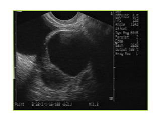
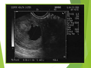
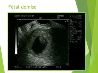
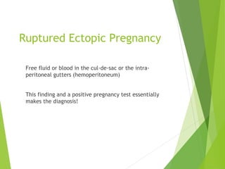
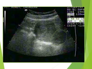
Sonographic Imaging of
the Female Patient with
Pelvic Pain/ Bleeding
Michael Kyeremeh Owusu
REGISTERED MEDICAL
SONOGRAPHER
Ghana
Case Presentation
 24 yo female presents with missed period, cramping,
midline abdominal pain and spotting
 VS: BP 120/80 HR 110
 Pelvic:
 Cervical os is closed with minimal bleeding
 No CMT, adenexa symmetric
 Urine hCG is +
Case presentation
 A bedside ultrasound
is performed
 The US reveals an IUP
 The patient is
discharged to home
with threatened
abortion precautions
 LOS = 30 minutes
 Applies to 60% of pts
Role of Bedside Sonography
 Identify an IUP
 Establish fetal viability
Secondary Indications
 Hemodynamic instability in a female pt
 Trauma and pregnancy
 Localization of IUD/foreign body
 Identify sources of pelvic pain in non-pregnant patients
Imaging: Transabdominal
 Uses a lower frequency transducer: 3.5 –5 mHz
 Better penetration, larger field of view
 It should be the initial imaging window to assess
for
 Advanced IUP
 Fibroids/masses
 Pelvic fluid
 The bladder should be full to provide an
acoustic window
Endovaginal
 Uses a higher frequency transducer: 6.0-
7.5mHz
 Provides optimal imaging of:
 Endometrium
 Myometrium
 Cul-de-sac
 Ovaries
 A full bladder is not necessary for this
approach
 Is usually better tolerated by patients
Scanning Protocol: Transabdominal
 Image the patient before obtaining a urine sample
 Can fill the bladder via foley and instill 300 cc NS but…
 If the bladder is empty, go directly to TV imaging after the pelvic
exam
Probe Selection
 “Workhorse”probe
 3.5 to 5.0 MHz
 Multi-frequency probe
 Good for most cardiac/abdominal
applications
Uterus
 An oval organ located
superior to the full
bladder
 The maximum size of the
non-gravid uterus is 5-7
cm x 4-5 cm
 The endometrial stripe is
the opposed surfaces of
the endometrial cavity
Transabdominal / Transverse
view
Right Left
Cul-de-sac
 Located posterior to the
uterus and upper vagina
 A small amount of fluid
may be seen in mid cycle
 A small amount of fluid in
the posterior cul-de-sac
may be the only
sonographic finding in EP
Bladder
uterus
Probe Selection
 Endovaginal Probe
 5 to 8 mHz variable frequency probe
 Up to 180 degree angle of view
Endovaginal Examination
 Best performed immediately following
the pelvic exam
 An empty bladder is required for an
optimal endovaginal (EV) exam
 A full bladder:
 Displaces the anatomy beyond the focal
length of the transducer
 Will create artifacts that will compromise
imaging
Before Performing a TV Exam:
 Explain that the EV exam is better for seeing ovaries
and early pregnancy
 Show the patient the probe
 Allow her the option of inserting it herself
 Inform her that it is usually more comfortable than the
TA exam which requires a full bladder
 The transducer probe should be covered
with a coupling gel followed by a
protective probe cover
 Non-medicated/ non-lubricated condoms
are recommended as a probe cover
 Patients with latex allergies will require an
alternative barrier
 Air bubbles within the sheath may increase
artifacts and compromise imaging
Longitudinal view
Coronal view
The Uterus
 Early in the menstrual cycle
 endometrium measures 4-8mm
 Secretory phase
 endometrium measures 7-14 mm
 Post-menopausal patient
 endometrial stripe usually less than 9 mm
Endometrial Stripe (ES)
Measurements
 In the post-partum patient, a thickened ES is
suggestive of retained products of conception
 In the pregnant patient, an ES measurement
of < 8 mm in the absence of an IUP is
suggestive of EP
 Thickening of the endometrial stripe in the
post-menopausal patient with vaginal
bleeding should raise suspicions for
endometrial carcinoma
Ovaries
 Lie posterior/lateral to
the uterus
 Anterior to the internal
iliac vessels and medial
to the external iliac
vessels
 Identified by a ring of
follicles in the periphery
Ovaries
 After ovulation a corpus luteal cyst
may be present
 Observed in approximately 50% of ovulating females
 Should not be seen beyond 72 hours into the next cycle
 Small amount of fluid in the
rectouterine pouch may be seen
during ovulation
Ovarian Cysts
 Follicular cyst (2.5 –10 cm)
 Thin, round, unilocular
 Functional corpus luteum cyst
 Normal up to 16 weeks GA
 Appears as a unilateral, unilocular 5-11 cm cyst
 Appearance can be highly variable
 Hemorrhage inside the cyst not uncommon
Assessment of the Pregnant Patient
 Identify gestational sac
 Demonstrate a myometrial mantle in the transverse
view
 Identify yolk sac and/or fetal pole
 Note if there is fluid in the cul-de-sac
Gestational Sac
 Anechoic area within the uterus surrounded by two
bright echogenic rings
 Decidua vera (the outer ring)
 Decidua capsularis (the inner ring)
 This is referred to as the double decidual sac sign
(DDSS)
Yolk Sac
 First embryonic structure that can be detected
sonographically
 Visualized approximately 5-6 weeks after the last
menstrual period
 Bright, ring like structure within the GS
 Should be readily seen when the GS sac is greater than
10 mm (using EVS)
Fetal Pole
 Can be first seen on EV when the fetus is approximately
2 mm in size
 A thickened area adjacent to the yolk sac
 The CRL is the most accurate sonographic measurement
that can be obtained during pregnancy
A Fetal Heart Beat
 An important prognostic indicator
 The rate of spontaneous abortion is extremely low (2-
4%) after the detection of normal embryonic cardiac
activity
 The normal fetal heart rate in early pregnancy is 112-
136
Definite IUP
 A gestational sac
with a sonolucent
center (greater
than 5 mm
diameter)
 Surrounded by a
thick, concentric,
echogenic ring
 GS contains a fetal
pole or yolk sac, or
both
Abnormal IUP
 A GS larger than 10-13 mm diameter(TV) or 20mm (TA)
without a yolk sac
 A GS larger than 18 mm (TV) or 25mm (TA) without a
fetal pole
 A definite fetal pole without cardiac activity after 7 wks
GA
Empty gestational sac
Fetal demise
Sonographic Spectrum of EP
 Ruptured ectopic pregnancy
 Definite ectopic pregnancy
 Extrauterine empty gestational sac
 Adenexal mass
 Pseudogestational sac
 Empty uterus
Definite Ectopic Pregnancy
A thick, brightly echogenic, ring-like structure located
outside the uterus with a gestational sac containing an
obvious fetal pole, yolk sac or both.
Ruptured Ectopic Pregnancy
Free fluid or blood in the cul-de-sac or the intra-
peritoneal gutters (hemoperitoneum)
This finding and a positive pregnancy test essentially
makes the diagnosis!
Clot/fluid
clot
Extrauterine Gestational Sac
 Extra-uterine mass
containing a thick,
brightly echogenic
ring surrounding an
anechoic area
 Brightly echogenic
appearance may be
helpful
 Tubal ring
Adenexal Mass
Pseudogestational Sac
 Stimulation of the endometrium
 Decidual breakdown results in a central anechoic area
 Can be confused with “early IUP”
 Does not have double decidual sac sign
 Correlation with ß hCG helpful
Pseudogestational sac
Ectopic
Interstitial Ectopic Pregnancy
 Implantation near the insertion of the fallopian tubes
 Highly vascular area
 Suspect when GS is not centrally located
 Demonstration of endometrial mantle is critical to the
diagnosis
Empty Uterus
 Correlation with
ßhCG critical
 ßhCG
>discriminatory
zone and empty
uterus is EP until
proven otherwise
Discriminatory HCG Zone
 5 weeks since last
normal LMP
 ß hCG value = 1800 mIU
 TAS landmarks
 5 to 8-mm GS
 TVS landmarks
 5 to 8-mm GS
 With or w/o yolk sac
Discriminatory HCG Zone
 6 weeks since last
normal LMP
 ß hCG = 7200
 TAS landmarks
 Yolk sac
 TVS landmarks
 Yolk sac and embryo
 Possibly FHM
Discriminatory HCG Zone
 7 weeks since last
normal LMP
 ß hCG = 21,000
 TAS landmarks
 5 to 10-mm embryo with FHM
 TVS landmarks
 5 to 10 mm embryo with FHM
Rule - in IUP Protocol
Clinically stable females with:Clinically stable females with:
(1)(1) Lower abdominal painLower abdominal pain
(2)(2) Vaginal bleedingVaginal bleeding
(3)(3) OrthostasisOrthostasis
(4)(4) Or risk factors for EPOr risk factors for EP
Positive urine preg Ultrasound
Rule - in IUP Protocol
Ultrasound
Definite IUP
Can DC to home
with f/u
Definite EP
OB consultation
Rule - in IUP Protocol
Ultrasound
No IUP but…
+ Adenexal tenderness or CMT
Free fluid in the cul de sac
And/or hCG > discriminatory zone
OB
Consultation
Rule - in IUP Protocol
Ultrasound
No IUP
Benign exam
ßhCG > discriminatory zone
DC to home
F/u exam and
ßhCG w/in 48 hrs
Rule-In IUP Protocol
 Sixty percent of patients will have IUP
“Rules out” ectopic pregnancy
by “ruling in” IUP
 What about heterotopic pregnancy?
Increased in patients
undergoing ovulation induction
consult OB
Risk is 1/30,000 in non-induced
pregancy
Pitfalls
 Diagnosing intrauterine fluid collections as “early” IUP
 Low hCG does not mean “low risk” for EP
 Failure to determine the exact location of a gestational sac
 Cul-de-sac fluid may be the only sonographic finding of extrauterine
pregnancy

CASE PRESENTATION

  • 1.
    Sonographic Imaging of theFemale Patient with Pelvic Pain/ Bleeding Michael Kyeremeh Owusu REGISTERED MEDICAL SONOGRAPHER Ghana
  • 2.
    Case Presentation  24yo female presents with missed period, cramping, midline abdominal pain and spotting  VS: BP 120/80 HR 110  Pelvic:  Cervical os is closed with minimal bleeding  No CMT, adenexa symmetric  Urine hCG is +
  • 3.
    Case presentation  Abedside ultrasound is performed  The US reveals an IUP  The patient is discharged to home with threatened abortion precautions  LOS = 30 minutes  Applies to 60% of pts
  • 4.
    Role of BedsideSonography  Identify an IUP  Establish fetal viability
  • 5.
    Secondary Indications  Hemodynamicinstability in a female pt  Trauma and pregnancy  Localization of IUD/foreign body  Identify sources of pelvic pain in non-pregnant patients
  • 6.
    Imaging: Transabdominal  Usesa lower frequency transducer: 3.5 –5 mHz  Better penetration, larger field of view  It should be the initial imaging window to assess for  Advanced IUP  Fibroids/masses  Pelvic fluid  The bladder should be full to provide an acoustic window
  • 7.
    Endovaginal  Uses ahigher frequency transducer: 6.0- 7.5mHz  Provides optimal imaging of:  Endometrium  Myometrium  Cul-de-sac  Ovaries  A full bladder is not necessary for this approach  Is usually better tolerated by patients
  • 8.
    Scanning Protocol: Transabdominal Image the patient before obtaining a urine sample  Can fill the bladder via foley and instill 300 cc NS but…  If the bladder is empty, go directly to TV imaging after the pelvic exam
  • 9.
    Probe Selection  “Workhorse”probe 3.5 to 5.0 MHz  Multi-frequency probe  Good for most cardiac/abdominal applications
  • 10.
    Uterus  An ovalorgan located superior to the full bladder  The maximum size of the non-gravid uterus is 5-7 cm x 4-5 cm  The endometrial stripe is the opposed surfaces of the endometrial cavity
  • 11.
  • 12.
    Cul-de-sac  Located posteriorto the uterus and upper vagina  A small amount of fluid may be seen in mid cycle  A small amount of fluid in the posterior cul-de-sac may be the only sonographic finding in EP
  • 13.
  • 14.
    Probe Selection  EndovaginalProbe  5 to 8 mHz variable frequency probe  Up to 180 degree angle of view
  • 15.
    Endovaginal Examination  Bestperformed immediately following the pelvic exam  An empty bladder is required for an optimal endovaginal (EV) exam  A full bladder:  Displaces the anatomy beyond the focal length of the transducer  Will create artifacts that will compromise imaging
  • 16.
    Before Performing aTV Exam:  Explain that the EV exam is better for seeing ovaries and early pregnancy  Show the patient the probe  Allow her the option of inserting it herself  Inform her that it is usually more comfortable than the TA exam which requires a full bladder
  • 17.
     The transducerprobe should be covered with a coupling gel followed by a protective probe cover  Non-medicated/ non-lubricated condoms are recommended as a probe cover  Patients with latex allergies will require an alternative barrier  Air bubbles within the sheath may increase artifacts and compromise imaging
  • 19.
  • 21.
  • 22.
    The Uterus  Earlyin the menstrual cycle  endometrium measures 4-8mm  Secretory phase  endometrium measures 7-14 mm  Post-menopausal patient  endometrial stripe usually less than 9 mm
  • 23.
    Endometrial Stripe (ES) Measurements In the post-partum patient, a thickened ES is suggestive of retained products of conception  In the pregnant patient, an ES measurement of < 8 mm in the absence of an IUP is suggestive of EP  Thickening of the endometrial stripe in the post-menopausal patient with vaginal bleeding should raise suspicions for endometrial carcinoma
  • 26.
    Ovaries  Lie posterior/lateralto the uterus  Anterior to the internal iliac vessels and medial to the external iliac vessels  Identified by a ring of follicles in the periphery
  • 27.
    Ovaries  After ovulationa corpus luteal cyst may be present  Observed in approximately 50% of ovulating females  Should not be seen beyond 72 hours into the next cycle  Small amount of fluid in the rectouterine pouch may be seen during ovulation
  • 28.
    Ovarian Cysts  Follicularcyst (2.5 –10 cm)  Thin, round, unilocular  Functional corpus luteum cyst  Normal up to 16 weeks GA  Appears as a unilateral, unilocular 5-11 cm cyst  Appearance can be highly variable  Hemorrhage inside the cyst not uncommon
  • 30.
    Assessment of thePregnant Patient  Identify gestational sac  Demonstrate a myometrial mantle in the transverse view  Identify yolk sac and/or fetal pole  Note if there is fluid in the cul-de-sac
  • 31.
    Gestational Sac  Anechoicarea within the uterus surrounded by two bright echogenic rings  Decidua vera (the outer ring)  Decidua capsularis (the inner ring)  This is referred to as the double decidual sac sign (DDSS)
  • 33.
    Yolk Sac  Firstembryonic structure that can be detected sonographically  Visualized approximately 5-6 weeks after the last menstrual period  Bright, ring like structure within the GS  Should be readily seen when the GS sac is greater than 10 mm (using EVS)
  • 36.
    Fetal Pole  Canbe first seen on EV when the fetus is approximately 2 mm in size  A thickened area adjacent to the yolk sac  The CRL is the most accurate sonographic measurement that can be obtained during pregnancy
  • 40.
    A Fetal HeartBeat  An important prognostic indicator  The rate of spontaneous abortion is extremely low (2- 4%) after the detection of normal embryonic cardiac activity  The normal fetal heart rate in early pregnancy is 112- 136
  • 41.
    Definite IUP  Agestational sac with a sonolucent center (greater than 5 mm diameter)  Surrounded by a thick, concentric, echogenic ring  GS contains a fetal pole or yolk sac, or both
  • 42.
    Abnormal IUP  AGS larger than 10-13 mm diameter(TV) or 20mm (TA) without a yolk sac  A GS larger than 18 mm (TV) or 25mm (TA) without a fetal pole  A definite fetal pole without cardiac activity after 7 wks GA
  • 44.
  • 45.
  • 46.
    Sonographic Spectrum ofEP  Ruptured ectopic pregnancy  Definite ectopic pregnancy  Extrauterine empty gestational sac  Adenexal mass  Pseudogestational sac  Empty uterus
  • 47.
    Definite Ectopic Pregnancy Athick, brightly echogenic, ring-like structure located outside the uterus with a gestational sac containing an obvious fetal pole, yolk sac or both.
  • 49.
    Ruptured Ectopic Pregnancy Freefluid or blood in the cul-de-sac or the intra- peritoneal gutters (hemoperitoneum) This finding and a positive pregnancy test essentially makes the diagnosis!
  • 50.
  • 51.
    Extrauterine Gestational Sac Extra-uterine mass containing a thick, brightly echogenic ring surrounding an anechoic area  Brightly echogenic appearance may be helpful  Tubal ring
  • 52.
  • 53.
    Pseudogestational Sac  Stimulationof the endometrium  Decidual breakdown results in a central anechoic area  Can be confused with “early IUP”  Does not have double decidual sac sign  Correlation with ß hCG helpful
  • 54.
  • 55.
    Interstitial Ectopic Pregnancy Implantation near the insertion of the fallopian tubes  Highly vascular area  Suspect when GS is not centrally located  Demonstration of endometrial mantle is critical to the diagnosis
  • 57.
    Empty Uterus  Correlationwith ßhCG critical  ßhCG >discriminatory zone and empty uterus is EP until proven otherwise
  • 58.
    Discriminatory HCG Zone 5 weeks since last normal LMP  ß hCG value = 1800 mIU  TAS landmarks  5 to 8-mm GS  TVS landmarks  5 to 8-mm GS  With or w/o yolk sac
  • 59.
    Discriminatory HCG Zone 6 weeks since last normal LMP  ß hCG = 7200  TAS landmarks  Yolk sac  TVS landmarks  Yolk sac and embryo  Possibly FHM
  • 60.
    Discriminatory HCG Zone 7 weeks since last normal LMP  ß hCG = 21,000  TAS landmarks  5 to 10-mm embryo with FHM  TVS landmarks  5 to 10 mm embryo with FHM
  • 61.
    Rule - inIUP Protocol Clinically stable females with:Clinically stable females with: (1)(1) Lower abdominal painLower abdominal pain (2)(2) Vaginal bleedingVaginal bleeding (3)(3) OrthostasisOrthostasis (4)(4) Or risk factors for EPOr risk factors for EP Positive urine preg Ultrasound
  • 62.
    Rule - inIUP Protocol Ultrasound Definite IUP Can DC to home with f/u Definite EP OB consultation
  • 63.
    Rule - inIUP Protocol Ultrasound No IUP but… + Adenexal tenderness or CMT Free fluid in the cul de sac And/or hCG > discriminatory zone OB Consultation
  • 64.
    Rule - inIUP Protocol Ultrasound No IUP Benign exam ßhCG > discriminatory zone DC to home F/u exam and ßhCG w/in 48 hrs
  • 65.
    Rule-In IUP Protocol Sixty percent of patients will have IUP “Rules out” ectopic pregnancy by “ruling in” IUP  What about heterotopic pregnancy? Increased in patients undergoing ovulation induction consult OB Risk is 1/30,000 in non-induced pregancy
  • 66.
    Pitfalls  Diagnosing intrauterinefluid collections as “early” IUP  Low hCG does not mean “low risk” for EP  Failure to determine the exact location of a gestational sac  Cul-de-sac fluid may be the only sonographic finding of extrauterine pregnancy