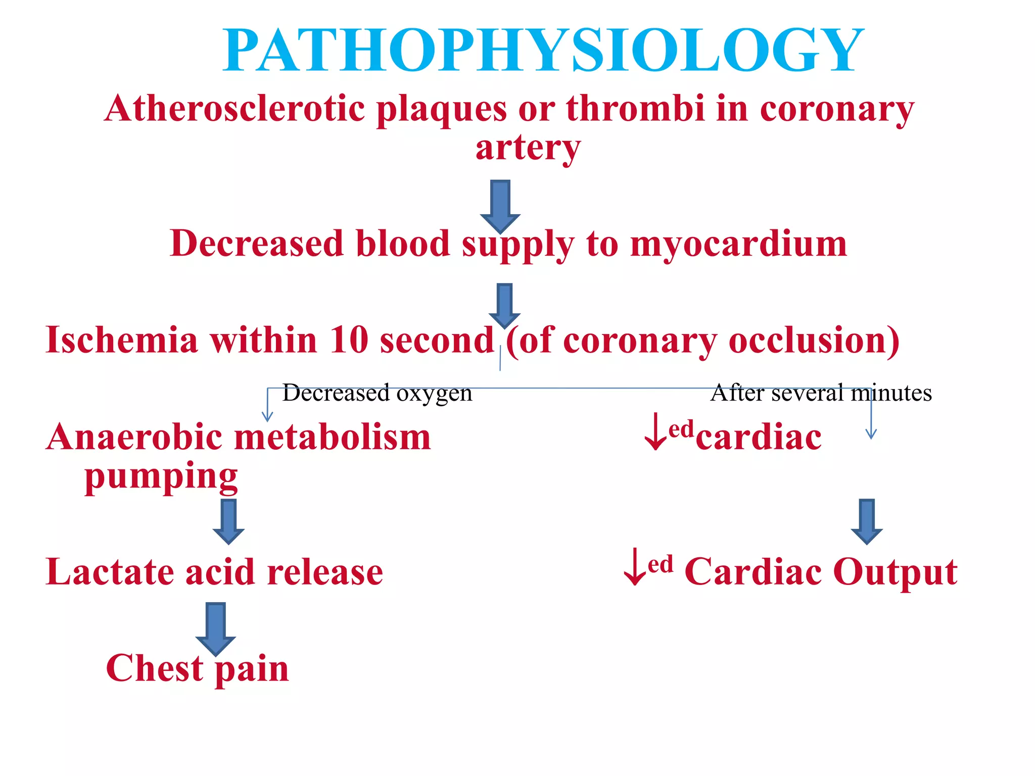
Coronary Artery Disease
Prepare by
Chanak Trikhatri
B.Sc.N, MA (Sociology), MN (Advance Adult Health/Medical
Surgical ), RN
Department of Medical Surgical Nursing
NMCTH
INTRODUCTION
• Encompasses acute Myocardial Infarction,
Angina Pectoris, atherosclerotic cardiovascular
disease & acute or chronic ischemic disease.
• Is a generic designation for many different
conditions that involve obstructed blood flow
through the coronary arteries.
DEFINITION
• CAD is a type of blood vessel disorder that is
included in the general category of
atherosclerosis.
• Atherosclerosis is often referred to the
hardening of the arteries.
• Atherosclerosis, an abnormal accumulation of
lipid, or fatty substances & fibrous tissue in the
lining of arterial blood vessel walls.
ETIOLOGY & RISK
FACTORS
• Modifiable risk factors
• Non- modifiable risk factors
• Contributing factors-
DM: FBS>110 mg/dl, incidence of 2-4 times
greater among the person with DM
Psychological states- stressful
Homocystine level- leads to damage to blood
vessels
Metabolic syndrome( multiple metabolic
disease)
NON
MODIFIABLE
MODIFIABLE
• Age
• Gender (M>F until 60
yrs)
• Ethnicity(white>Africans
)
• Genetic predisposition
• Family history
• Serum lipids: fasting
triglycerides ≥ 150 mg/dl,
cholesterol ≥ 200mg/dl,
elevated LDL &  HDL
• HTN: ≥ 140/90 mm of Hg
• Tobacco use 2-6 times higher
• Physical inactivity: obesity,
BMI> 30kg/m2
PATHOPHYSIOLOGY
Lipid filled in smooth muscle cells
Appearance of yellow tinge
Changes in endothelium
Thickening of arterial wall
Endothelial wall injury
Cholesterol & lipoprotein adheres into arterial intima
Fibrous plaque
Occlusion of blood flow continuous inflammatory response
Instability of plaque, ulceration, rupture
accumulation of platelets at sites
Thrombosis
Occlusion
CLINICAL FEATURES
• CAD produces symptoms & complications
according to the location & degree of
narrowing of arterial lumen.
• Ischemic: deprivation of cardiac muscle
cells of oxygen needed for their survival.
• Angina pectoris
• Shortness of breath
• Dyspnea
• Nausea
• weakness
DIAGNOSTIC
FINDINGS
• History collection
• Physical examination
• Electrocardiogram
• B- mode ultrasonography
• Doppler flow studies
• Intravascular ultrasound
• Electron bean computed tomography
• Echocardiograph stress tests
MANAGEMENT
• Identification of high risk persons- screening
family & personal history, environmental
factors, psychological history
• Management of high risk persons- life style
modification
• Physical activity
• Promoting cessation of tobacco use
• Nutritional therapy
• Medication
MEDICATIONS
• HMG- CoA reductase inhibitor(statins)- blocks synthesis
of cholesterol. Eg: atrovastatin, Lovastatin
• Niacin- inhibits synthesis & secretion of LDL. Eg:
Niacin, Nicotinic acid
• Fibric Acid Derivatives- reduces triglyceride. Eg:
Fenofibrate, Gemfibrozil
• Bile acid sequestrants-removal of LDL & cholesterol.
Eg: Colestipol, Cholestyramine
• Cholesterol Absorption inhibitor- reduces reabsorption
of cholesterol. Eg: Ezetimibe
• Omega 3 Acid Ethyl Esters- fish oil capsules
• Antiplatelet therapy- aspirin, clopidogrel
SURGICAL MANAGEMENT
1. PERCUTANEOUS CORONARY
INTERVENTIONS
a. PTCA
b. Intracoronary stent implantation
c. Atherectomy- removal of atheroma
d. Brachytherapy- delivery of gamma or beta
radiation
2. CORONARY ARTERY REVASCULARIZATION
 Coronary artery Bypass Graft
PTCA (PERCUTANEOUS TRANSLUMINAL
CORONARY ANGIOPLASTY):
 A balloon tipped catheter is used to open
blocked coronary vessels
Hollow catheters called sheaths are inserted
usually in femoral artery
Catheter then threaded up through the aorta &
then coronary arteries.
When catheter is properly positioned the
balloon is inflated with high pressure for
several seconds & then deflated.
INTRACORONARY STENT IMPLANTATION/
CORONARY ARTERY STENT:
– Stent is metal mesh that provides structurally support
to a vessel at a risk of acute closure.
– The stent is positioned over the angioplasty balloon
– When the balloon is inflated, the mesh expands &
presses against the vessel wall, left permanently in
the place after the balloon is withdrawn.
CORONARY ARTERY BYPASS GRAFT
• General anesthesia
• Cardiopulmonary bypass
• Commonly used- greater & lesser Saphenous
vein & internal mammary artery
• Hypothermia maintained (28-32 degree
Celsius)
NURSING DIAGNOSIS
• Ineffective cardiac tissue perfusion related to reduced
coronary blood flow as evidenced by decreased
cardiac output
• Decreased cardiac output related to decreased
coronary blood flow as evidenced by increased heart
rate
• Acute pain related to reduced coronary blood flow as
evidenced by facial expression
• Fear & anxiety related to cardiac event as evidenced
by verbalization
• Risk for impaired gas exchange related to reduced
blood flow
POST OP
• Decreased cardiac output related to blood loss &
compromised myocardial function as evidenced by
decreased pulse rate.
• Impaired gas exchange related to chest surgery as
evidenced by respiratory rate, rhythm
• Acute pain related to surgical trauma & pleural
irritation by chest tubes as evidenced by pain scale
score
• Ineffective thermoregulation related to infection as
evidenced by Vital signs
• Deficient knowledge related to treatment regimen as
evidenced by asking frequent doubts.
ANGINA PECTORIS
INTRODUCTION
• It is literally translated as pain(angina) in the
chest(pectoris)
• Is transient chest pain cased by myocardial ischemia.
• Usually lasts for few minutes(3-5minutes) commonly
subsides when precipitating factor(usual exertion) is
relieved.
• Typical exertional angina should not persist longer
than 20minutes after rest & administration of NTG.
• The cause is insufficiency coronary blood flow
resulting in adequate supply f oxygen to meet the
myocardial demand.
DEFINITION
• Is a clinical syndrome characterized by paroxysms of
pain or a feeling of pressure in the anterior chest.
- BT BASAVANTHAPPA
• Angina Pectoris is a chest pain resulting
from myocardial ischemia(inadequate blood
supply to the myocardium).
- JOYCE M BLACK
• Angina or chest pain is the clinical
manifestation of reversible myocardial
ischemia. Either an ed demand for oxygen
or as a decreased supply of oxygen can lead
to myocardial ischemia.
ETIOLOGY
• Associated with atherosclerotic lesions
• Chronic or acute blockage of a coronary artery or by
coronary artery spasm.
• Activities that increases the myocardial oxygen
demand like physical activity, emotion, exposure to
cold.
• Consumption of heavy meal increases the work load
of heart.
• Tobacco use
• Sexual activity
• Stimulants like cocaine, amphetamine
PATHOPHYSIOLOGY
Atherosclerotic plaques or thrombi in coronary
artery
Decreased blood supply to myocardium
Ischemia within 10 second (of coronary occlusion)
Decreased oxygen After several minutes
Anaerobic metabolism edcardiac
pumping
Lactate acid release ed Cardiac Output
Chest pain
CLINICAL
FEATURES
CLINICAL FEATURES
• Characterized by discomfort in the chest, jaw,
shoulder, back, or arm.
CHARACTERSTICS
 ONSET- can develop quickly or slowly
 LOCATION- 90% of client experience pain at
retrosternal or slightly to the left of the sternum.
 RADIATION- radiates to left shoulder & upper arm
& may travel down the inner aspect of the left arm to
the elbow, wrist & 4th & 5th fingers. May also radiate
to right shoulder, neck, jaw or epigastric region.
Sometimes pain may be felt only in the area of
radiation & not in the chest.
 DURATION- lasts less than 5mts. Sometimes may
longer than 15-20minutes.
 SENSTATION- squeezing, burning, pressing,
choking, aching or bursting pressure. Client often
say gas, heartburn or indigestion.
 SEVERITY- often called “discomfort” not “pain”.
 ASSOCIATED CHARACTERISTICS- dyspnea,
pallor, sweating, faintness, palpitations, dizziness &
digestive disturbances.
 ATYPICAL PRESENTATION- in women,
dyspnea or back pain, fatigue
 RELIEVING & AGGRAVATING FACTORS-
activity & subsides by administration of NTG
PATTERNS OF ANGINA
1. STABLE ANGINA- paroxysmal chest pain or
discomfort triggered by predictable degree of
exertion or emotion. Usually relieved with rest or
NTG or both.
2. UNSTABLE ANGINA- preinfarction angina,
crescendo angina or intermittent coronary syndrome
is paroxysmal chest pain triggered by unpredictable
degree of exertion or emotion, which may occur at
night. Immediate treatment as Medical emergency
3. NOCTURANALANGINA-associated
with rapid eye movement , sleep during
dreaming
4. SILENT ISCHEMIA- refers to the
ischemia that occurs in the absence of any
subjective symptoms.
DIAGNOSTIC TESTS
• History collection related to
P- Precipating events
Q- Quality of pain
R- Radiation of pain
S- Severity of pain
T- Timing
• ECG -12lead ECG (ST elevation, flat or
inverted T)
• Exercise ECG
• Electron Beam Computed Tomography(EBCT)
• Coronary Angiography
• Chest X-ray
MANAGEMENT
AIM
To reduce manifestations & ultimately to reduce the risk
of mortality & morbid events.
Goals are accomplished by:
1. Antiaginal pharmacologic interventions
2. Education & risk factor modification
3. Revascularization through interventional cardiology
or CABG treatment.
A- Antiplatelet, Antianginal, ACE inhibitor
B- Beta adrenergic blocker, Blood pressure
C- Cigarette smoking, Cholesterol
D- Diet, Diabetes
E- Education, Exercise
PHARMACOLOGIC THERAPY
• Antiplatelet agent:- aspirin
• Adenosine Diphosphate Receptor Antagonists:-
inhibits platelet aggregation- Clopidogrel
• Nitrates:- promotes peripheral vasodilation,
decreasing preload & afterload- sulingual NTG,
Isosorbide dinitrate
• Beta adrenergic blockers:- reduces both HR &
contractility- atenolol, propranolol
• Calcium channel blockers:- Nicardipine, nifedepine
• AEC inhibitor:–Captopril
Contd….
• Unfractionated Heparin- Heparin
• Low molecular weight heparin- Dalteparin
• Opoid analgesics – morphine
• Fibrinolytic therapy- Recombinant
plasminogen activator
NURSING MANAGEMENT
• Cardiac monitoring
• Obtain 12 lead ECG
• To relieve chest pain – MONA
M- Morphine Sulphate
O- Oxygen therapy
N- nitrates
A- Aspirin
• Administer sublingual NTG or spray
• Comfortable environment
• Keep patient in complete bed rest.
• Make patient comfortable and relaxed.
• Administer oxygen.
• Advice patient to avoid anxiety and
exertion.
• Monitor vital signs.
• Monitor for arrythmias.
MYOCARDIAL
INFARCTION
DEFINITION
• Acute MI is also known as heart attack,
coronary occlusion which is a life threatening
condition characterized by the formation of
localized necrotic tissue within the
myocardium.
• AMI results of sustained ischemia, causing
irreversible myocardial cell death.
ETIOLOGY
• Complete or nearly complete occlusion of a
coronary artery
• Atherosclerotic plaque
• Strenuous physical activity
• Severe emotional stress such as anger
Increases sympathetic activity increases
myocardial oxygen demand.
RISK FACTORS
NON MODIFIABLE RISK FACTORS
• Hereditary
• Increasing age- above 65years
MODIFIABLE RISK FACTORS
• Smoking
• HTN
• Elevated serum cholesterol
• DM
• Physical inactivity
• Obesity
CONTRUBUITING RISK FACTORS
• Stress, Homocysteine levels
PATHOPHYSIOLOGY
Atherosclerotic plaque
Decreased blood supply to myocardium
Ischemia
Necrosis Anaerobic metabolism
Transluminal infarction
PH imbalance
Changes in the architecture
Heart Dysarrythmia
HF
CLINICAL FEATURES
• Chest pain- usually described as Heaviness in the
chest, Pressure in the chest, Tightness, Burning
sensation, Constriction or crushing
• Pain may radiate to neck, jaw & arms or to the back
• May occur when patient is active or at rest.
• Shortness of breath
• Some experience only discomfort, weakness, or
shortness of breath
• Dizziness
• Confusion, nausea and vomiting
• Pulmonary edema
• Profuse sweating
• Fever(due to inflammation or immune system reaction of
blood vessels)
• Tachycardia
• Hypotension
• Decreased urine output
• Jugular vein distension
• Hepatic enlargement
• Peripheral edema
DIAGNOSTIC FINDINGS
• History collection
• physical examination
LAB FINDINGS:
• creatine kinase (ck)- MB enzyme, increases 3-6
hrs after attack, peaks 12-18 hrs normal after3-4 days
• Myoglobin: rapidly released & detected within 2hrs
of AMI
• Trophonin- 3-6 days , normalizes 14-21 days
• ECG changes- Q wave changes, ST elevation
• Echo cardiogram
MANAGEMENT
GOAL:
Initiating prompt care
Reducing pain
Delivering successful treatment
Preventing complication
Rehabilitating & educating
FIBRINOLYTIC
THERAPY
• Is aimed at stopping the infarction process by
dissolving the thrombus in the coronary artery
& reperfusing the myocardium
• Ideally given within the first 6hours after onset
of symptoms
INCLUSION CRITERIA:
Within 6hrs of onset of symptoms
12 lead ECG suggesting acute MI
ABSOLUTE
CONTRAINDICATION
• Active internal bleeding
• Known history of aneurysm or AV
malformation
• Any known neoplasm
• Previous cerebral hemorrhage
• Suspected aortic dissection
DRUG THERAPY:-
 Nitroglycerin – to reduce anginal pain &
improves blood supply
Morphine sulfate- to reduce chest pain
ACE –inhibitors- to prevent ventricular
remodeling & slow the progression of
HF. Eg: Captopril
Antidysrhythmia drugs- to treat
arrhythmias
Cholesterol lowering drugs- statins
Stool softners
cont……
• Heparin - the complex than binds to clotting factor
• Beta –adrenergic blockers:-to decrease myocardial
oxygen demand by reducing heart rate , BP and
contractility. atenolol
SURGICAL MANAGEMENT
• Coronary artery bypass graft surgery
• Transmyocardial laser revascularization-
involves use of high energy laser to improve
the blood flow to ischemic area
NURSING MANAGEMENT
• Pain management
• Monitoring
• Rest & comfort

Coronary Artery Disease.ppt

  • 1.
    Coronary Artery Disease Prepareby Chanak Trikhatri B.Sc.N, MA (Sociology), MN (Advance Adult Health/Medical Surgical ), RN Department of Medical Surgical Nursing NMCTH
  • 2.
    INTRODUCTION • Encompasses acuteMyocardial Infarction, Angina Pectoris, atherosclerotic cardiovascular disease & acute or chronic ischemic disease. • Is a generic designation for many different conditions that involve obstructed blood flow through the coronary arteries.
  • 4.
    DEFINITION • CAD isa type of blood vessel disorder that is included in the general category of atherosclerosis. • Atherosclerosis is often referred to the hardening of the arteries. • Atherosclerosis, an abnormal accumulation of lipid, or fatty substances & fibrous tissue in the lining of arterial blood vessel walls.
  • 6.
    ETIOLOGY & RISK FACTORS •Modifiable risk factors • Non- modifiable risk factors • Contributing factors- DM: FBS>110 mg/dl, incidence of 2-4 times greater among the person with DM Psychological states- stressful Homocystine level- leads to damage to blood vessels Metabolic syndrome( multiple metabolic disease)
  • 7.
    NON MODIFIABLE MODIFIABLE • Age • Gender(M>F until 60 yrs) • Ethnicity(white>Africans ) • Genetic predisposition • Family history • Serum lipids: fasting triglycerides ≥ 150 mg/dl, cholesterol ≥ 200mg/dl, elevated LDL &  HDL • HTN: ≥ 140/90 mm of Hg • Tobacco use 2-6 times higher • Physical inactivity: obesity, BMI> 30kg/m2
  • 9.
  • 12.
    Lipid filled insmooth muscle cells Appearance of yellow tinge Changes in endothelium Thickening of arterial wall Endothelial wall injury
  • 13.
    Cholesterol & lipoproteinadheres into arterial intima Fibrous plaque Occlusion of blood flow continuous inflammatory response Instability of plaque, ulceration, rupture accumulation of platelets at sites Thrombosis Occlusion
  • 14.
    CLINICAL FEATURES • CADproduces symptoms & complications according to the location & degree of narrowing of arterial lumen. • Ischemic: deprivation of cardiac muscle cells of oxygen needed for their survival. • Angina pectoris • Shortness of breath • Dyspnea • Nausea • weakness
  • 16.
    DIAGNOSTIC FINDINGS • History collection •Physical examination • Electrocardiogram • B- mode ultrasonography • Doppler flow studies • Intravascular ultrasound • Electron bean computed tomography • Echocardiograph stress tests
  • 17.
    MANAGEMENT • Identification ofhigh risk persons- screening family & personal history, environmental factors, psychological history • Management of high risk persons- life style modification • Physical activity • Promoting cessation of tobacco use • Nutritional therapy • Medication
  • 18.
    MEDICATIONS • HMG- CoAreductase inhibitor(statins)- blocks synthesis of cholesterol. Eg: atrovastatin, Lovastatin • Niacin- inhibits synthesis & secretion of LDL. Eg: Niacin, Nicotinic acid • Fibric Acid Derivatives- reduces triglyceride. Eg: Fenofibrate, Gemfibrozil • Bile acid sequestrants-removal of LDL & cholesterol. Eg: Colestipol, Cholestyramine • Cholesterol Absorption inhibitor- reduces reabsorption of cholesterol. Eg: Ezetimibe • Omega 3 Acid Ethyl Esters- fish oil capsules • Antiplatelet therapy- aspirin, clopidogrel
  • 19.
    SURGICAL MANAGEMENT 1. PERCUTANEOUSCORONARY INTERVENTIONS a. PTCA b. Intracoronary stent implantation c. Atherectomy- removal of atheroma d. Brachytherapy- delivery of gamma or beta radiation 2. CORONARY ARTERY REVASCULARIZATION  Coronary artery Bypass Graft
  • 20.
    PTCA (PERCUTANEOUS TRANSLUMINAL CORONARYANGIOPLASTY):  A balloon tipped catheter is used to open blocked coronary vessels Hollow catheters called sheaths are inserted usually in femoral artery Catheter then threaded up through the aorta & then coronary arteries. When catheter is properly positioned the balloon is inflated with high pressure for several seconds & then deflated.
  • 23.
    INTRACORONARY STENT IMPLANTATION/ CORONARYARTERY STENT: – Stent is metal mesh that provides structurally support to a vessel at a risk of acute closure. – The stent is positioned over the angioplasty balloon – When the balloon is inflated, the mesh expands & presses against the vessel wall, left permanently in the place after the balloon is withdrawn.
  • 25.
    CORONARY ARTERY BYPASSGRAFT • General anesthesia • Cardiopulmonary bypass • Commonly used- greater & lesser Saphenous vein & internal mammary artery • Hypothermia maintained (28-32 degree Celsius)
  • 29.
    NURSING DIAGNOSIS • Ineffectivecardiac tissue perfusion related to reduced coronary blood flow as evidenced by decreased cardiac output • Decreased cardiac output related to decreased coronary blood flow as evidenced by increased heart rate • Acute pain related to reduced coronary blood flow as evidenced by facial expression • Fear & anxiety related to cardiac event as evidenced by verbalization • Risk for impaired gas exchange related to reduced blood flow
  • 30.
    POST OP • Decreasedcardiac output related to blood loss & compromised myocardial function as evidenced by decreased pulse rate. • Impaired gas exchange related to chest surgery as evidenced by respiratory rate, rhythm • Acute pain related to surgical trauma & pleural irritation by chest tubes as evidenced by pain scale score • Ineffective thermoregulation related to infection as evidenced by Vital signs • Deficient knowledge related to treatment regimen as evidenced by asking frequent doubts.
  • 31.
  • 32.
    INTRODUCTION • It isliterally translated as pain(angina) in the chest(pectoris) • Is transient chest pain cased by myocardial ischemia. • Usually lasts for few minutes(3-5minutes) commonly subsides when precipitating factor(usual exertion) is relieved. • Typical exertional angina should not persist longer than 20minutes after rest & administration of NTG. • The cause is insufficiency coronary blood flow resulting in adequate supply f oxygen to meet the myocardial demand.
  • 34.
    DEFINITION • Is aclinical syndrome characterized by paroxysms of pain or a feeling of pressure in the anterior chest. - BT BASAVANTHAPPA
  • 35.
    • Angina Pectorisis a chest pain resulting from myocardial ischemia(inadequate blood supply to the myocardium). - JOYCE M BLACK • Angina or chest pain is the clinical manifestation of reversible myocardial ischemia. Either an ed demand for oxygen or as a decreased supply of oxygen can lead to myocardial ischemia.
  • 36.
    ETIOLOGY • Associated withatherosclerotic lesions • Chronic or acute blockage of a coronary artery or by coronary artery spasm. • Activities that increases the myocardial oxygen demand like physical activity, emotion, exposure to cold. • Consumption of heavy meal increases the work load of heart. • Tobacco use • Sexual activity • Stimulants like cocaine, amphetamine
  • 38.
    PATHOPHYSIOLOGY Atherosclerotic plaques orthrombi in coronary artery Decreased blood supply to myocardium Ischemia within 10 second (of coronary occlusion) Decreased oxygen After several minutes Anaerobic metabolism edcardiac pumping Lactate acid release ed Cardiac Output Chest pain
  • 39.
  • 40.
    CLINICAL FEATURES • Characterizedby discomfort in the chest, jaw, shoulder, back, or arm. CHARACTERSTICS  ONSET- can develop quickly or slowly  LOCATION- 90% of client experience pain at retrosternal or slightly to the left of the sternum.  RADIATION- radiates to left shoulder & upper arm & may travel down the inner aspect of the left arm to the elbow, wrist & 4th & 5th fingers. May also radiate to right shoulder, neck, jaw or epigastric region. Sometimes pain may be felt only in the area of radiation & not in the chest.
  • 43.
     DURATION- lastsless than 5mts. Sometimes may longer than 15-20minutes.  SENSTATION- squeezing, burning, pressing, choking, aching or bursting pressure. Client often say gas, heartburn or indigestion.  SEVERITY- often called “discomfort” not “pain”.  ASSOCIATED CHARACTERISTICS- dyspnea, pallor, sweating, faintness, palpitations, dizziness & digestive disturbances.  ATYPICAL PRESENTATION- in women, dyspnea or back pain, fatigue  RELIEVING & AGGRAVATING FACTORS- activity & subsides by administration of NTG
  • 45.
    PATTERNS OF ANGINA 1.STABLE ANGINA- paroxysmal chest pain or discomfort triggered by predictable degree of exertion or emotion. Usually relieved with rest or NTG or both. 2. UNSTABLE ANGINA- preinfarction angina, crescendo angina or intermittent coronary syndrome is paroxysmal chest pain triggered by unpredictable degree of exertion or emotion, which may occur at night. Immediate treatment as Medical emergency
  • 46.
    3. NOCTURANALANGINA-associated with rapideye movement , sleep during dreaming 4. SILENT ISCHEMIA- refers to the ischemia that occurs in the absence of any subjective symptoms.
  • 47.
    DIAGNOSTIC TESTS • Historycollection related to P- Precipating events Q- Quality of pain R- Radiation of pain S- Severity of pain T- Timing • ECG -12lead ECG (ST elevation, flat or inverted T) • Exercise ECG • Electron Beam Computed Tomography(EBCT) • Coronary Angiography • Chest X-ray
  • 50.
    MANAGEMENT AIM To reduce manifestations& ultimately to reduce the risk of mortality & morbid events. Goals are accomplished by: 1. Antiaginal pharmacologic interventions 2. Education & risk factor modification 3. Revascularization through interventional cardiology or CABG treatment.
  • 51.
    A- Antiplatelet, Antianginal,ACE inhibitor B- Beta adrenergic blocker, Blood pressure C- Cigarette smoking, Cholesterol D- Diet, Diabetes E- Education, Exercise
  • 52.
    PHARMACOLOGIC THERAPY • Antiplateletagent:- aspirin • Adenosine Diphosphate Receptor Antagonists:- inhibits platelet aggregation- Clopidogrel • Nitrates:- promotes peripheral vasodilation, decreasing preload & afterload- sulingual NTG, Isosorbide dinitrate • Beta adrenergic blockers:- reduces both HR & contractility- atenolol, propranolol • Calcium channel blockers:- Nicardipine, nifedepine • AEC inhibitor:–Captopril
  • 53.
    Contd…. • Unfractionated Heparin-Heparin • Low molecular weight heparin- Dalteparin • Opoid analgesics – morphine • Fibrinolytic therapy- Recombinant plasminogen activator
  • 54.
    NURSING MANAGEMENT • Cardiacmonitoring • Obtain 12 lead ECG • To relieve chest pain – MONA M- Morphine Sulphate O- Oxygen therapy N- nitrates A- Aspirin • Administer sublingual NTG or spray • Comfortable environment
  • 56.
    • Keep patientin complete bed rest. • Make patient comfortable and relaxed. • Administer oxygen. • Advice patient to avoid anxiety and exertion. • Monitor vital signs. • Monitor for arrythmias.
  • 57.
  • 58.
    DEFINITION • Acute MIis also known as heart attack, coronary occlusion which is a life threatening condition characterized by the formation of localized necrotic tissue within the myocardium. • AMI results of sustained ischemia, causing irreversible myocardial cell death.
  • 59.
    ETIOLOGY • Complete ornearly complete occlusion of a coronary artery • Atherosclerotic plaque • Strenuous physical activity • Severe emotional stress such as anger Increases sympathetic activity increases myocardial oxygen demand.
  • 60.
    RISK FACTORS NON MODIFIABLERISK FACTORS • Hereditary • Increasing age- above 65years MODIFIABLE RISK FACTORS • Smoking • HTN • Elevated serum cholesterol • DM • Physical inactivity • Obesity CONTRUBUITING RISK FACTORS • Stress, Homocysteine levels
  • 61.
  • 62.
    Atherosclerotic plaque Decreased bloodsupply to myocardium Ischemia Necrosis Anaerobic metabolism Transluminal infarction PH imbalance Changes in the architecture Heart Dysarrythmia HF
  • 63.
    CLINICAL FEATURES • Chestpain- usually described as Heaviness in the chest, Pressure in the chest, Tightness, Burning sensation, Constriction or crushing • Pain may radiate to neck, jaw & arms or to the back • May occur when patient is active or at rest. • Shortness of breath • Some experience only discomfort, weakness, or shortness of breath
  • 64.
    • Dizziness • Confusion,nausea and vomiting • Pulmonary edema • Profuse sweating • Fever(due to inflammation or immune system reaction of blood vessels) • Tachycardia • Hypotension • Decreased urine output • Jugular vein distension • Hepatic enlargement • Peripheral edema
  • 65.
    DIAGNOSTIC FINDINGS • Historycollection • physical examination LAB FINDINGS: • creatine kinase (ck)- MB enzyme, increases 3-6 hrs after attack, peaks 12-18 hrs normal after3-4 days • Myoglobin: rapidly released & detected within 2hrs of AMI • Trophonin- 3-6 days , normalizes 14-21 days • ECG changes- Q wave changes, ST elevation • Echo cardiogram
  • 66.
    MANAGEMENT GOAL: Initiating prompt care Reducingpain Delivering successful treatment Preventing complication Rehabilitating & educating
  • 67.
    FIBRINOLYTIC THERAPY • Is aimedat stopping the infarction process by dissolving the thrombus in the coronary artery & reperfusing the myocardium • Ideally given within the first 6hours after onset of symptoms INCLUSION CRITERIA: Within 6hrs of onset of symptoms 12 lead ECG suggesting acute MI
  • 68.
    ABSOLUTE CONTRAINDICATION • Active internalbleeding • Known history of aneurysm or AV malformation • Any known neoplasm • Previous cerebral hemorrhage • Suspected aortic dissection
  • 69.
    DRUG THERAPY:-  Nitroglycerin– to reduce anginal pain & improves blood supply Morphine sulfate- to reduce chest pain ACE –inhibitors- to prevent ventricular remodeling & slow the progression of HF. Eg: Captopril Antidysrhythmia drugs- to treat arrhythmias Cholesterol lowering drugs- statins Stool softners
  • 70.
    cont…… • Heparin -the complex than binds to clotting factor • Beta –adrenergic blockers:-to decrease myocardial oxygen demand by reducing heart rate , BP and contractility. atenolol
  • 71.
    SURGICAL MANAGEMENT • Coronaryartery bypass graft surgery • Transmyocardial laser revascularization- involves use of high energy laser to improve the blood flow to ischemic area
  • 72.
    NURSING MANAGEMENT • Painmanagement • Monitoring • Rest & comfort