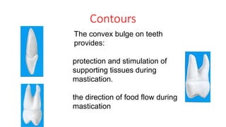
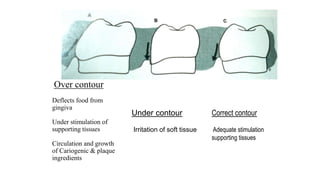
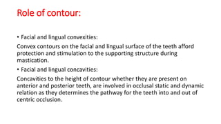
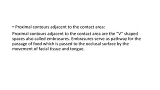
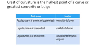
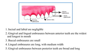
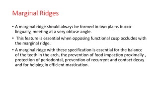
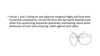
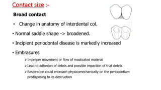
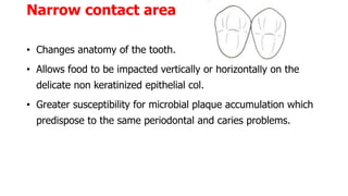
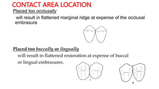
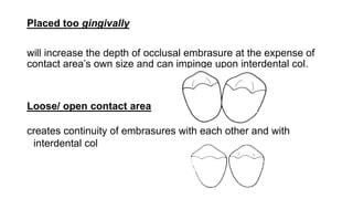
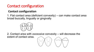
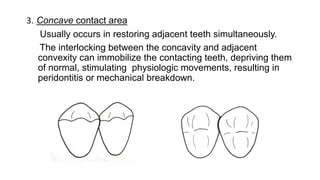
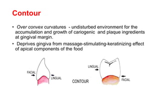
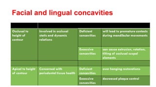
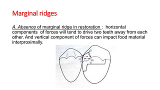
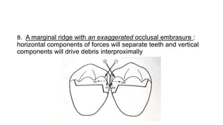
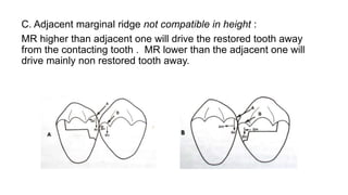
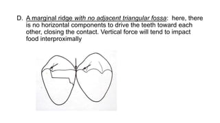
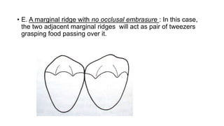
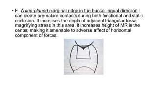
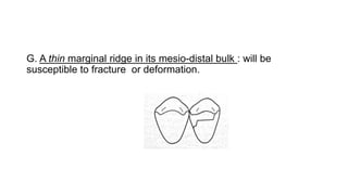
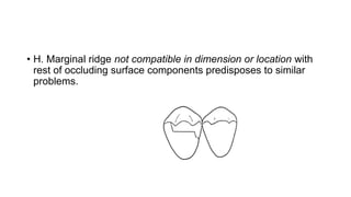
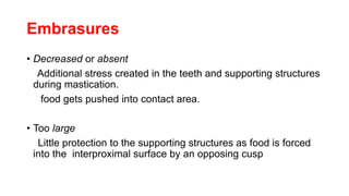
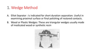
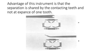
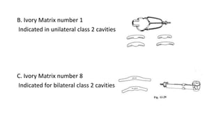
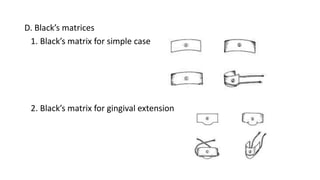
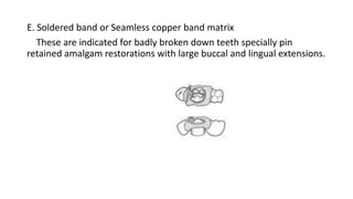
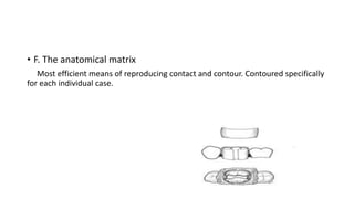
CONTACTS AND
CONTOURS
GUIDED BY : PRESENTED BY:
DR. BOBBIN GILL DR. RITU KHICHAR
DR.NIRMALA BISHNOI
Contents
• Introduction
• Contours
• Contacts
• Marginal ridges
• Facial and lingual contours
• Hazards of faulty reproduction of physio anatomical features of teeth
• Formulation of contacts and contours
• Conclusion
Introduction
• Restorative Dentistry requires an understanding of intra-arch contour
relationship.
• Anatomical factors such as crests of facial and lingual coronal contour
and contact areas must be evaluated for a restoration to possess
contour in harmony with adjacent teeth.
Contours
The convex bulge on teeth
provides:
protection and stimulation of
supporting tissues during
mastication.
the direction of food flow during
mastication
Over contour
Deflects food from
gingiva
Under stimulation of
supporting tissues
Circulation and growth
of Cariogenic & plaque
ingredients
Under contour
Irritation of soft tissue
Correct contour
Adequate stimulation
supporting tissues
Role of contour:
• Facial and lingual convexities:
Convex contours on the facial and lingual surface of the teeth afford
protection and stimulation to the supporting structure during
mastication.
• Facial and lingual concavities:
Concavities to the height of contour whether they are present on
anterior and posterior teeth, are involved in occlusal static and dynamic
relation as they determines the pathway for the teeth into and out of
centric occlusion.
• Proximal contours adjacent to the contact area:
Proximal contours adjacent to the contact area are the “V” shaped
spaces also called embrasures. Embrasures serve as pathway for the
passage of food which is passed to the occlusal surface by the
movement of facial tissue and tongue.
Crest of curvature is the highest point of a curve or
greatest convexity or bulge
Contact Areas
• Are crest of curvature on proximal surface of tooth crowns where
tooth touches the adjacent tooth in same arch where teeth are in
proper alignment.
• On different teeth, contact areas may be in incisal third, middle third
or at junction of incisor or middle third.
• The proximal contact area is located in the incisal third of the
approximating surfaces of the maxillary and mandibular central
incisors, it is positioned slightly facial to the center of the proximal
surface faciolingually.
• Proceeding posteriorly from the incisor region through all the
remaining teeth, the contact area is located near the junction of the
incisal (or occlusal) and middle thirds or in the middle third.
Role of contact:
• Human teeth are designed in such a way that they contribute
significantly to their own support as well as supporting the arch.
• Properly located contact area allows normal healthy interdental
papilla to fill the interproximal spaces.
• The proximal contact of all teeth in same dental arch and their
interdigitation through occlusal contact with the opposing teeth
stabilizes and maintains the integrity of dental arches.
• It prevents the food from packing in between the teeth and thereby
preventing the impingement to the gingival tissue.
Distal contacts are located more cervically than mesial contacts except
• mandibular first premolar (where distal contact is occlusally located)
• mandibular central incisor (located at same level)
Interproximal space/ Embrasure
• It is triangular space between adjacent
tooth in relation to the contact.
• Side of the triangle are proximal surface of adjacent
teeth and apex of the triangle is the area of contact of two teeth.
• Occupied in periodontally healthy persons by interdental papilla.
• When teeth contacts there are four continuous space that surrounds
the contact area:
* Facial * Lingual * Occlusal/Incisal* *Gingival
Functions
• Maintain tissues against trauma during mastication and
invasion by bacteria
• Provides space for bulk of bone thus better anchorage and
support.
• Allows vascular support to nourish inter dental bone and
papillary tissue.
• It makes a spillway for escape of food during mastication
• Permit slight amount of stimulation to the gingiva by frictional
massage of food
1. Incisal and labial are negligible
2. Gingival and lingual embrasures between anterior teeth are the widest
and longest in mouth
3. Buccal embrasures are small
4. Lingual embrasures are long, with medium width
5. Gingival embrasures between posterior teeth are broad and long
Marginal Ridges
• A marginal ridge should always be formed in two plains bucco-
lingually, meeting at a very obtuse angle.
• This feature is essential when opposing functional cusp occludes with
the marginal ridge.
• A marginal ridge with these specification is essential for the balance
of the teeth in the arch, the prevention of food impaction proximally ,
protection of periodontal, prevention of recurrent and contact decay
and for helping in efficient mastication.
• Forces 1 and 2 acting on two adjacent marginal ridges will have their
horizontal components, 1H and 2H drive the two teeth towards each
other thus preventing impaction proximally maintaining mesio distal
dimension of arch and anchoring teeth against each other
• With age the dimensions of marginal ridges and occlusal embrasuras
are reduced due to vertical occlusal attrition and proximal flattening
of the contact areas.
Facial and Lingual Contours and Related
Structures
• In a vertical direction all tooth crowns will exibit convex curvatures
occlusal to cervical line , this curvature is called cervical ridge.
• Curvatures on labial , buccal and lingual surface of all maxillary teeth
and on buccal surface of mandibular posterior teeth is about .5mm or
less.
• Mandibular posterior teeth will have lingual curvature of approx. 1
mm.
• Mandibular anterior teeth will have less than .5 mm curvature
• Canines may show little more curvature than central and lateral
incisors.
• Incisal 1/2 to 2/3 of lingual surface of a anterior teeth display some
concavities.
• Proper mesio-distal contour at different levels and locations of facial
and lingual surfaces is vital for the health of investing periodontiom.
Hazards of faulty reproduction
• Contact area
Size
location
Configuration
• Contours
Buccal and lingual convexities
Buccal and lingual concavities
• Marginal ridges
• Embrasures
Contact size :-
Broad contact
• Change in anatomy of interdental col.
• Normal saddle shape -> broadened.
• Incipient periodontal disease is markedly increased
• Embrasures
Improper movement or flow of masticated material
Lead to adhesion of debris and possible impaction of that debris
Restoration could encroach physicomechanically on the periodontium
predisposing to its destruction
Narrow contact area
• Changes anatomy of the tooth.
• Allows food to be impacted vertically or horizontally on the
delicate non keratinized epithelial col.
• Greater susceptibility for microbial plaque accumulation which
predispose to the same periodontal and caries problems.
CONTACT AREA LOCATION
Placed too occlusally
will result in flattened marginal ridge at expense of the occlusal
embrasure
Placed too buccally or lingually
will result in flattened restoration at expense of buccal
or lingual embrasures.
Placed too gingivally
will increase the depth of occlusal embrasure at the expense of
contact area’s own size and can impinge upon interdental col.
Loose/ open contact area
creates continuity of embrasures with each other and with
interdental col
Contact configuration
1. Flat contact area (deficient convexity) – can make contact area
broad buccally, lingually or gingivally
2. Contact area with excessive convexity – will decrease the
extent of contact area.
3. Concave contact area
Usually occurs in restoring adjacent teeth simultaneously.
The interlocking between the concavity and adjacent
convexity can immobilize the contacting teeth, depriving them
of normal, stimulating physiologic movements, resulting in
peridontitis or mechanical breakdown.
Contour
• Over convex curvatures - undisturbed environment for the
accumulation and growth of cariogenic and plaque ingredients
at gingival margin.
• Deprives gingiva from massage-stimulating-keratinizing effect
of apical components of the food
Facial and lingual concavities
Areas of proximal contour adjacent to contact area
• Include area occlusal, buccal, lingual, and gingival to contact area.
• Fault in restoration of these areas can lead to restoration overhangs
or underhangs and impingement upon adjacent periodontal
structures.
Marginal ridges
A. Absence of marginal ridge in restoration : horizontal
components of forces will tend to drive two teeth away from each
other. And vertical component of forces can impact food material
interproximally.
B. A marginal ridge with an exaggerated occlusal embrasure :
horizontal components of forces will separate teeth and vertical
components will drive debris interproximally
C. Adjacent marginal ridge not compatible in height :
MR higher than adjacent one will drive the restored tooth away
from the contacting tooth . MR lower than the adjacent one will
drive mainly non restored tooth away.
D. A marginal ridge with no adjacent triangular fossa: here, there
is no horizontal components to drive the teeth toward each
other, closing the contact. Vertical force will tend to impact
food interproximally
• E. A marginal ridge with no occlusal embrasure : In this case,
the two adjacent marginal ridges will act as pair of tweezers
grasping food passing over it.
• F. A one-planed marginal ridge in the bucco-lingual direction :
can create premature contacts during both functional and static
occlusion. It increases the depth of adjacent triangular fossa
magnifying stress in this area. It increases height of MR in the
center, making it amenable to adverse affect of horizontal
component of forces.
G. A thin marginal ridge in its mesio-distal bulk : will be
susceptible to fracture or deformation.
• H. Marginal ridge not compatible in dimension or location with
rest of occluding surface components predisposes to similar
problems.
Embrasures
• Decreased or absent
Additional stress created in the teeth and supporting structures
during mastication.
food gets pushed into contact area.
• Too large
Little protection to the supporting structures as food is forced
into the interproximal surface by an opposing cusp
Procedures for formulation of proper contacts
and contours
1. Tooth Movement
2. Matricing
Tooth
movement
Rapid or
immediate
Wedge
Elliot separator,
wood or plastic
wedges
Traction
Non-interfering true
separator, Ferrier
double bow separator
Slow or
delayed
Separating ligature wires,
oversized temporaries,
ortho. Appliance, rubber
dam sheet, separating
bands
1. Wedge Method
A. Elliot Seprator : Is indicated for short duration separation. Useful in
examining proximal surface or final polishing of restored contacts.
B. Wood or Plastic Wedges: These are triangular wedges usually made
of medicated wood or synthetic resin
Functions of wedges
• Help in rapid separation of teeth
• Prevent gingival overhang of restoration
• Provide space to compensate for thickness of matrix band
• Help in stabilization of retainer and matrix during restorative
procedures
• Atraumatically retract the rubberdam and gingival from the gingival
margins of proximal tooth preparations
C. For instantaneous sepration of teeth during operative procedures in
anterior teeth wedging the nail of thumb or first finger between the
teeth will make rapid sepration.
2. Traction Method
Mechanical devices which engage proximal surfaces of teeth to be
separated by means of holding arms .these are mechanically moved
apart creating separation between the clamped teeth.
a)Non interfering true separator
b)Ferrier double bow separator
Advantage of this instrument is that the
separation is shared by the contacting teeth and
not at expance of one tooth.
Slow or delayed tooth movement
• When teeth have drifted or tilted considerably rapid movement of
teeth will endanger periodontal ligaments .therefore slow tooth
movement over a period of weeks will allow proper repositioning of
teeth in physiologic manner.
• A) Separating wires
• B)Oversized temporaries
• C)Orthodontic appliances
Matricing
• Matricing is the procedure by which a temporary wall is built opposite
to the axial wall, surrounding the tooth structure which has been lost
during the tooth preparation. Matrix is an instrument which is used to
hold the restoration within the tooth while it is setting.
• Ideal Requirements of a Matrix
• Ease of application
• Not be cumbersome
• Ease of removal
• Rigidity
• Provide proper proximal contact and contour
• Positive proximal pressure
• Non-reactive
• Inexpensive
Types of matrices
• Matrices for class 1 cavity preparation(double banded tofflemire )
Procedure
• Tighten the vice screw to lock the band in the vice.
• Guide the looped end of band gently over the tooth. Size of loop may
be adjusted using vice moving knob.
• With the band in position around tooth tighten the vice moving knob.
• An additional small piece of band is then contoured to the facial or
lingual axial configuration of contemplated restoration and inserted
between tooth and previously positioned retainer matrix in the area
of facial or lingual extension of cavity preparation.
• Cover the wedge with softened compound and insert it between two
bands and cool to harden.
• Check stability of each component.
• Matrices for class 2 cavity preparations
• A) single banded tofflemire
Procedure
• Repaeat the basic steps as in double baded arrangement
• If there is difference between height of interproximanal gingiva on
mesial and distal sides of tooth matrix band should be trimmed so
that it is narrowe(Occluso-apically) on side where gingiva is more
occlusally located.
• In preparations with sub gingival margins edges of the band
occasionally encounter the gingival margin and become bent inward,
preventing further seating of the band.
• For this reason there should be unprepared exposed tooth surface
apical to the gingival margin to support the band in its apical path.
• This may necessitate gingival retraction or cutting
• Also band edges should be guided in apical path by placing a flat
bladed blunt instrument
• It is preferable to put retainer in buccal vestibule , parallel to adjacent
teeth, sometimes due to shallow sulcus or sizable buckle involvement
of tooth in cavity preparation , retainer is placed on the lingual.
• Junction between retainer and the band should always be located
next to unprepared intact tooth to ensure its stability and prevent
unnecessary accumulation of excess restorative material.
A wedge is chosen and tried .
• Usually one wedge from opposite side of retainer is sufficient.
• More than one wedge could be necessary in gingival recession wide
proximal preparations , gingival margin is located in proximal surface
concavity.
• After insertion and initial hardening of restorative material wedges
are removed, retainer is loosened and disengaged.
• Band is bent against adjacent tooth surface and removed from
between the teeth in occlusal direction while being pressed againsrt
adjacent tooth.
B. Ivory Matrix number 1
Indicated in unilateral class 2 cavities
C. Ivory Matrix number 8
Indicated for bilateral class 2 cavities
D. Black’s matrices
1. Black’s matrix for simple case
2. Black’s matrix for gingival extension
E. Soldered band or Seamless copper band matrix
These are indicated for badly broken down teeth specially pin
retained amalgam restorations with large buccal and lingual extensions.
• F. The anatomical matrix
Most efficient means of reproducing contact and contour. Contoured specifically
for each individual case.
Procedure
• A piece of stainless steel band 18” width is drawn betwwen a handle
of pair of festooning scissors .
• Matrix is then cut to proper length, proximo-buccal and proximo-
lingual cusps are used as guide.
• Band is then trimmed so that matrix will extent below the gingival
margin and at least 2 mm beyond buccal and lingual margins of cavity.
• Wedge is forced into position.
• Two small cones of compound are warmed in hot water. The tips are
heated in a flame.
• These cones are forced in buccal and lingual embrasures using thumb
and finger pressure.
• A wire staple is constructed from a metal paper clip . Length of staple
slightly shorter than the crown of tooth
• Staple is heated in a flame and forced into the compound in buccal
and lingual embrasures
• The matrix is burnished lightly against the contacting tooth
• After initial hardening of restorative material compound is cracked at
its occlusal junctions using chisel or knife.
• The wedges are removed using haemostat and the band is curled
backwards against the adjacent tooth and withdrawn bucco-lingually
• G. Roll-in band matrix (automatrix) : band is self retained by holding
one end of band and rolling other end over itself.
• H. S- shaped matrix band :
• I. T- shaped matrix band
3.Matrices for amalgam restoration on distal
of cuspid
• A. S- shaped matrix
• B. Cavity preparation with incisal access on distal of the cuspid regular
tofflemire could be used
4. Matrices for class 3 direct tooth coloured
restorations
These are usually transparent plastic strips.
For silicate cements celluloid strips and
for resins they are cellophane strips.
• Mylar strips may be used for either material
A. Matrix for class 3 preparation with teeth in
normal alignment
B. Matrix for class 3 preparation in teeth with irregular alignment
C. Matrix for two small proximal preparations in contact with each
other
5. Matrices for class 4 preparations for direct
tooth colored material
A. The plastic strip for inciso-proximal cavities procedure
B. Aluminium foil incisal corner matrix
C. Transparent crown form matrices
D. Anatomic matrix
E. Modified S shaped matrix
6. Matrices for class 5 amalgam restorations
A. Window matrix
B. S- shaped matrix
7. Matrices for class 5 preparation for direct
tooth colour restoration
A. Anatomic matrix for non light cured direct tooth colored materials
B. Aluminium or copper collar
C. Anatomic matrix for light and non light cured
direct tooth colored materials
Extraoral formulation of contacts and contour
A. Wax pattern :- is usually built in a slightly over contoured condition
specially at a contact area to allow for finishing and polishing
surface losses.
B. Cast adjustment:- These are usually done with rubbery stones
incrementally removing cast material surface wise to obtain the
exact dimension of contact and contour.
Conclusion
most neglected aspect in restorative dentistry should be given priority
so that the restoration can serve to its fullest purpose in harmony with
neighboring teeth and supporting tissues.

contacts and contours seminar.pptx endodontics

  • 1.
    CONTACTS AND CONTOURS GUIDED BY: PRESENTED BY: DR. BOBBIN GILL DR. RITU KHICHAR DR.NIRMALA BISHNOI
  • 2.
    Contents • Introduction • Contours •Contacts • Marginal ridges • Facial and lingual contours • Hazards of faulty reproduction of physio anatomical features of teeth • Formulation of contacts and contours • Conclusion
  • 3.
    Introduction • Restorative Dentistryrequires an understanding of intra-arch contour relationship. • Anatomical factors such as crests of facial and lingual coronal contour and contact areas must be evaluated for a restoration to possess contour in harmony with adjacent teeth.
  • 4.
    Contours The convex bulgeon teeth provides: protection and stimulation of supporting tissues during mastication. the direction of food flow during mastication
  • 5.
    Over contour Deflects foodfrom gingiva Under stimulation of supporting tissues Circulation and growth of Cariogenic & plaque ingredients Under contour Irritation of soft tissue Correct contour Adequate stimulation supporting tissues
  • 6.
    Role of contour: •Facial and lingual convexities: Convex contours on the facial and lingual surface of the teeth afford protection and stimulation to the supporting structure during mastication. • Facial and lingual concavities: Concavities to the height of contour whether they are present on anterior and posterior teeth, are involved in occlusal static and dynamic relation as they determines the pathway for the teeth into and out of centric occlusion.
  • 7.
    • Proximal contoursadjacent to the contact area: Proximal contours adjacent to the contact area are the “V” shaped spaces also called embrasures. Embrasures serve as pathway for the passage of food which is passed to the occlusal surface by the movement of facial tissue and tongue.
  • 8.
    Crest of curvatureis the highest point of a curve or greatest convexity or bulge
  • 9.
    Contact Areas • Arecrest of curvature on proximal surface of tooth crowns where tooth touches the adjacent tooth in same arch where teeth are in proper alignment. • On different teeth, contact areas may be in incisal third, middle third or at junction of incisor or middle third.
  • 11.
    • The proximalcontact area is located in the incisal third of the approximating surfaces of the maxillary and mandibular central incisors, it is positioned slightly facial to the center of the proximal surface faciolingually. • Proceeding posteriorly from the incisor region through all the remaining teeth, the contact area is located near the junction of the incisal (or occlusal) and middle thirds or in the middle third.
  • 12.
    Role of contact: •Human teeth are designed in such a way that they contribute significantly to their own support as well as supporting the arch. • Properly located contact area allows normal healthy interdental papilla to fill the interproximal spaces.
  • 13.
    • The proximalcontact of all teeth in same dental arch and their interdigitation through occlusal contact with the opposing teeth stabilizes and maintains the integrity of dental arches. • It prevents the food from packing in between the teeth and thereby preventing the impingement to the gingival tissue.
  • 15.
    Distal contacts arelocated more cervically than mesial contacts except • mandibular first premolar (where distal contact is occlusally located) • mandibular central incisor (located at same level)
  • 16.
    Interproximal space/ Embrasure •It is triangular space between adjacent tooth in relation to the contact. • Side of the triangle are proximal surface of adjacent teeth and apex of the triangle is the area of contact of two teeth. • Occupied in periodontally healthy persons by interdental papilla. • When teeth contacts there are four continuous space that surrounds the contact area: * Facial * Lingual * Occlusal/Incisal* *Gingival
  • 17.
    Functions • Maintain tissuesagainst trauma during mastication and invasion by bacteria • Provides space for bulk of bone thus better anchorage and support. • Allows vascular support to nourish inter dental bone and papillary tissue. • It makes a spillway for escape of food during mastication • Permit slight amount of stimulation to the gingiva by frictional massage of food
  • 18.
    1. Incisal andlabial are negligible 2. Gingival and lingual embrasures between anterior teeth are the widest and longest in mouth 3. Buccal embrasures are small 4. Lingual embrasures are long, with medium width 5. Gingival embrasures between posterior teeth are broad and long
  • 19.
    Marginal Ridges • Amarginal ridge should always be formed in two plains bucco- lingually, meeting at a very obtuse angle. • This feature is essential when opposing functional cusp occludes with the marginal ridge. • A marginal ridge with these specification is essential for the balance of the teeth in the arch, the prevention of food impaction proximally , protection of periodontal, prevention of recurrent and contact decay and for helping in efficient mastication.
  • 20.
    • Forces 1and 2 acting on two adjacent marginal ridges will have their horizontal components, 1H and 2H drive the two teeth towards each other thus preventing impaction proximally maintaining mesio distal dimension of arch and anchoring teeth against each other
  • 21.
    • With agethe dimensions of marginal ridges and occlusal embrasuras are reduced due to vertical occlusal attrition and proximal flattening of the contact areas.
  • 22.
    Facial and LingualContours and Related Structures • In a vertical direction all tooth crowns will exibit convex curvatures occlusal to cervical line , this curvature is called cervical ridge. • Curvatures on labial , buccal and lingual surface of all maxillary teeth and on buccal surface of mandibular posterior teeth is about .5mm or less. • Mandibular posterior teeth will have lingual curvature of approx. 1 mm.
  • 23.
    • Mandibular anteriorteeth will have less than .5 mm curvature • Canines may show little more curvature than central and lateral incisors. • Incisal 1/2 to 2/3 of lingual surface of a anterior teeth display some concavities. • Proper mesio-distal contour at different levels and locations of facial and lingual surfaces is vital for the health of investing periodontiom.
  • 24.
    Hazards of faultyreproduction • Contact area Size location Configuration • Contours Buccal and lingual convexities Buccal and lingual concavities • Marginal ridges • Embrasures
  • 25.
    Contact size :- Broadcontact • Change in anatomy of interdental col. • Normal saddle shape -> broadened. • Incipient periodontal disease is markedly increased • Embrasures Improper movement or flow of masticated material Lead to adhesion of debris and possible impaction of that debris Restoration could encroach physicomechanically on the periodontium predisposing to its destruction
  • 26.
    Narrow contact area •Changes anatomy of the tooth. • Allows food to be impacted vertically or horizontally on the delicate non keratinized epithelial col. • Greater susceptibility for microbial plaque accumulation which predispose to the same periodontal and caries problems.
  • 27.
    CONTACT AREA LOCATION Placedtoo occlusally will result in flattened marginal ridge at expense of the occlusal embrasure Placed too buccally or lingually will result in flattened restoration at expense of buccal or lingual embrasures.
  • 28.
    Placed too gingivally willincrease the depth of occlusal embrasure at the expense of contact area’s own size and can impinge upon interdental col. Loose/ open contact area creates continuity of embrasures with each other and with interdental col
  • 29.
    Contact configuration Contact configuration 1.Flat contact area (deficient convexity) – can make contact area broad buccally, lingually or gingivally 2. Contact area with excessive convexity – will decrease the extent of contact area.
  • 30.
    3. Concave contactarea Usually occurs in restoring adjacent teeth simultaneously. The interlocking between the concavity and adjacent convexity can immobilize the contacting teeth, depriving them of normal, stimulating physiologic movements, resulting in peridontitis or mechanical breakdown.
  • 31.
    Contour • Over convexcurvatures - undisturbed environment for the accumulation and growth of cariogenic and plaque ingredients at gingival margin. • Deprives gingiva from massage-stimulating-keratinizing effect of apical components of the food
  • 32.
    Facial and lingualconcavities
  • 33.
    Areas of proximalcontour adjacent to contact area • Include area occlusal, buccal, lingual, and gingival to contact area. • Fault in restoration of these areas can lead to restoration overhangs or underhangs and impingement upon adjacent periodontal structures.
  • 34.
    Marginal ridges A. Absenceof marginal ridge in restoration : horizontal components of forces will tend to drive two teeth away from each other. And vertical component of forces can impact food material interproximally.
  • 35.
    B. A marginalridge with an exaggerated occlusal embrasure : horizontal components of forces will separate teeth and vertical components will drive debris interproximally
  • 36.
    C. Adjacent marginalridge not compatible in height : MR higher than adjacent one will drive the restored tooth away from the contacting tooth . MR lower than the adjacent one will drive mainly non restored tooth away.
  • 37.
    D. A marginalridge with no adjacent triangular fossa: here, there is no horizontal components to drive the teeth toward each other, closing the contact. Vertical force will tend to impact food interproximally
  • 38.
    • E. Amarginal ridge with no occlusal embrasure : In this case, the two adjacent marginal ridges will act as pair of tweezers grasping food passing over it.
  • 39.
    • F. Aone-planed marginal ridge in the bucco-lingual direction : can create premature contacts during both functional and static occlusion. It increases the depth of adjacent triangular fossa magnifying stress in this area. It increases height of MR in the center, making it amenable to adverse affect of horizontal component of forces.
  • 40.
    G. A thinmarginal ridge in its mesio-distal bulk : will be susceptible to fracture or deformation.
  • 41.
    • H. Marginalridge not compatible in dimension or location with rest of occluding surface components predisposes to similar problems.
  • 42.
    Embrasures • Decreased orabsent Additional stress created in the teeth and supporting structures during mastication. food gets pushed into contact area. • Too large Little protection to the supporting structures as food is forced into the interproximal surface by an opposing cusp
  • 43.
    Procedures for formulationof proper contacts and contours 1. Tooth Movement 2. Matricing
  • 44.
    Tooth movement Rapid or immediate Wedge Elliot separator, woodor plastic wedges Traction Non-interfering true separator, Ferrier double bow separator Slow or delayed Separating ligature wires, oversized temporaries, ortho. Appliance, rubber dam sheet, separating bands
  • 45.
    1. Wedge Method A.Elliot Seprator : Is indicated for short duration separation. Useful in examining proximal surface or final polishing of restored contacts. B. Wood or Plastic Wedges: These are triangular wedges usually made of medicated wood or synthetic resin
  • 46.
    Functions of wedges •Help in rapid separation of teeth • Prevent gingival overhang of restoration • Provide space to compensate for thickness of matrix band • Help in stabilization of retainer and matrix during restorative procedures • Atraumatically retract the rubberdam and gingival from the gingival margins of proximal tooth preparations
  • 47.
    C. For instantaneoussepration of teeth during operative procedures in anterior teeth wedging the nail of thumb or first finger between the teeth will make rapid sepration.
  • 48.
    2. Traction Method Mechanicaldevices which engage proximal surfaces of teeth to be separated by means of holding arms .these are mechanically moved apart creating separation between the clamped teeth. a)Non interfering true separator b)Ferrier double bow separator
  • 49.
    Advantage of thisinstrument is that the separation is shared by the contacting teeth and not at expance of one tooth.
  • 50.
    Slow or delayedtooth movement • When teeth have drifted or tilted considerably rapid movement of teeth will endanger periodontal ligaments .therefore slow tooth movement over a period of weeks will allow proper repositioning of teeth in physiologic manner. • A) Separating wires • B)Oversized temporaries • C)Orthodontic appliances
  • 51.
    Matricing • Matricing isthe procedure by which a temporary wall is built opposite to the axial wall, surrounding the tooth structure which has been lost during the tooth preparation. Matrix is an instrument which is used to hold the restoration within the tooth while it is setting.
  • 52.
    • Ideal Requirementsof a Matrix • Ease of application • Not be cumbersome • Ease of removal • Rigidity • Provide proper proximal contact and contour • Positive proximal pressure • Non-reactive • Inexpensive
  • 53.
    Types of matrices •Matrices for class 1 cavity preparation(double banded tofflemire )
  • 54.
    Procedure • Tighten thevice screw to lock the band in the vice. • Guide the looped end of band gently over the tooth. Size of loop may be adjusted using vice moving knob. • With the band in position around tooth tighten the vice moving knob. • An additional small piece of band is then contoured to the facial or lingual axial configuration of contemplated restoration and inserted between tooth and previously positioned retainer matrix in the area of facial or lingual extension of cavity preparation.
  • 55.
    • Cover thewedge with softened compound and insert it between two bands and cool to harden. • Check stability of each component.
  • 56.
    • Matrices forclass 2 cavity preparations • A) single banded tofflemire
  • 57.
    Procedure • Repaeat thebasic steps as in double baded arrangement • If there is difference between height of interproximanal gingiva on mesial and distal sides of tooth matrix band should be trimmed so that it is narrowe(Occluso-apically) on side where gingiva is more occlusally located. • In preparations with sub gingival margins edges of the band occasionally encounter the gingival margin and become bent inward, preventing further seating of the band.
  • 58.
    • For thisreason there should be unprepared exposed tooth surface apical to the gingival margin to support the band in its apical path. • This may necessitate gingival retraction or cutting • Also band edges should be guided in apical path by placing a flat bladed blunt instrument
  • 59.
    • It ispreferable to put retainer in buccal vestibule , parallel to adjacent teeth, sometimes due to shallow sulcus or sizable buckle involvement of tooth in cavity preparation , retainer is placed on the lingual. • Junction between retainer and the band should always be located next to unprepared intact tooth to ensure its stability and prevent unnecessary accumulation of excess restorative material.
  • 60.
    A wedge ischosen and tried . • Usually one wedge from opposite side of retainer is sufficient. • More than one wedge could be necessary in gingival recession wide proximal preparations , gingival margin is located in proximal surface concavity. • After insertion and initial hardening of restorative material wedges are removed, retainer is loosened and disengaged.
  • 61.
    • Band isbent against adjacent tooth surface and removed from between the teeth in occlusal direction while being pressed againsrt adjacent tooth.
  • 62.
    B. Ivory Matrixnumber 1 Indicated in unilateral class 2 cavities C. Ivory Matrix number 8 Indicated for bilateral class 2 cavities
  • 63.
    D. Black’s matrices 1.Black’s matrix for simple case 2. Black’s matrix for gingival extension
  • 64.
    E. Soldered bandor Seamless copper band matrix These are indicated for badly broken down teeth specially pin retained amalgam restorations with large buccal and lingual extensions.
  • 65.
    • F. Theanatomical matrix Most efficient means of reproducing contact and contour. Contoured specifically for each individual case.
  • 66.
    Procedure • A pieceof stainless steel band 18” width is drawn betwwen a handle of pair of festooning scissors . • Matrix is then cut to proper length, proximo-buccal and proximo- lingual cusps are used as guide. • Band is then trimmed so that matrix will extent below the gingival margin and at least 2 mm beyond buccal and lingual margins of cavity.
  • 67.
    • Wedge isforced into position. • Two small cones of compound are warmed in hot water. The tips are heated in a flame. • These cones are forced in buccal and lingual embrasures using thumb and finger pressure. • A wire staple is constructed from a metal paper clip . Length of staple slightly shorter than the crown of tooth
  • 68.
    • Staple isheated in a flame and forced into the compound in buccal and lingual embrasures • The matrix is burnished lightly against the contacting tooth • After initial hardening of restorative material compound is cracked at its occlusal junctions using chisel or knife. • The wedges are removed using haemostat and the band is curled backwards against the adjacent tooth and withdrawn bucco-lingually
  • 69.
    • G. Roll-inband matrix (automatrix) : band is self retained by holding one end of band and rolling other end over itself.
  • 70.
    • H. S-shaped matrix band :
  • 71.
    • I. T-shaped matrix band
  • 72.
    3.Matrices for amalgamrestoration on distal of cuspid • A. S- shaped matrix • B. Cavity preparation with incisal access on distal of the cuspid regular tofflemire could be used
  • 73.
    4. Matrices forclass 3 direct tooth coloured restorations These are usually transparent plastic strips. For silicate cements celluloid strips and for resins they are cellophane strips. • Mylar strips may be used for either material A. Matrix for class 3 preparation with teeth in normal alignment
  • 74.
    B. Matrix forclass 3 preparation in teeth with irregular alignment C. Matrix for two small proximal preparations in contact with each other
  • 75.
    5. Matrices forclass 4 preparations for direct tooth colored material A. The plastic strip for inciso-proximal cavities procedure B. Aluminium foil incisal corner matrix C. Transparent crown form matrices D. Anatomic matrix E. Modified S shaped matrix
  • 76.
    6. Matrices forclass 5 amalgam restorations A. Window matrix B. S- shaped matrix
  • 77.
    7. Matrices forclass 5 preparation for direct tooth colour restoration A. Anatomic matrix for non light cured direct tooth colored materials B. Aluminium or copper collar C. Anatomic matrix for light and non light cured direct tooth colored materials
  • 78.
    Extraoral formulation ofcontacts and contour A. Wax pattern :- is usually built in a slightly over contoured condition specially at a contact area to allow for finishing and polishing surface losses. B. Cast adjustment:- These are usually done with rubbery stones incrementally removing cast material surface wise to obtain the exact dimension of contact and contour.
  • 79.
    Conclusion most neglected aspectin restorative dentistry should be given priority so that the restoration can serve to its fullest purpose in harmony with neighboring teeth and supporting tissues.