
Recommended
More Related Content
Similar to tmj ana,phy,dys.pptx
Similar to tmj ana,phy,dys.pptx (20)
Recently uploaded
Recently uploaded (20)
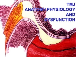
tmj ana,phy,dys.pptx
- 2. Introduction • No specialty in dentistry can be effectively practiced at the highest level of competence without an understanding of how the teeth relate to the rest of the masticatory system including the TMJs • It is impossible to comprehend the fine points of occlusion without the depth of awareness of the anatomy, physiology and biomechanics of the tempromandibular joint
- 3. Temporomandibular Joint • The temporomandibular joint (TMJ) is a diarthrodial joint that connects the condyle of the mandible (lower jaw) to the temporal bone at the side of a skull. As a modified hinge joint, • TMJ enable the jaw to rotate open and closed, it also enables the jaw to translate forward and backward. The condyle can also move laterally and medially.
- 5. TEMPOROMANDIBULAR JOINT (articular disk) (inferior joint cavity) (superi or joint cavity) (lateral disk ligamen t) (capsul ar ligamen t) (medial disk ligament) (capsular ligament) •Ginglymoarthroidal (Hinging and gliding). •Condyle fits into fossa. •Articular disc separates. •Compound joint.
- 6. Articulation • The condyle articulates with the temporal bone in the glenoid fossa. The glenoid fossa is a concave depression in the squamous portion of the temporal bone. • However, these two bones are actually separated by an articular disc, a concept of structure unknown throughout the rest of the body.
- 7. Components There are six main components of the TMJ. • Mandibular condyles • Articular surface of the temporal bone • Capsule • Articular disc • Ligaments • Lateral pterygoid
- 8. Mandibuar fossa • Oval or oblong depression in the temporal bone just ant to auditory cannal • Shape of the fossa conform some extend to posterior and superior surface of the condylar process
- 9. 9 Mandibular Fossa • Dense cortical bony surface of temporal bone Posterior to articular eminence • Posterior nonarticular fossa is formed by tempanic plate • Thin at the roof of the fossa and tympanic plate
- 12. Classification of articular eminence shapes: • Articular eminence morphology was classified • into four types, according to the criteria set by Kurita • et al.13 (2000): • (A) box, • (B) sigmoid, • (C) flattened, • (D) deformed A B C D
- 13. Mandibular Condyle • Convex on all bearing surfaces • Knoblike form is wider lateromedially than anterioposteriorly • Condyle is perpendicular to ascending ramus of the mandible • The long axis are in lateral plane
- 14. Mandibular Condyle • Modified barrel shape approx. 20 x 10 mm (ML x AP) • Dense cortical bone covered with dense fibrous connective tissue with irregular cartilage like cell
- 15. Capsule • The capsule is a fibrous membrane that surrounds the joint and incorporates the articular eminance. It attaches to the articular eminance, the articular disc and the neck of the mandibular condyle.
- 16. Articular Capsule • Ligamentous capsule surrounds the joint • Attached to the neck of the condyle and around the border of the articular surface of the temporal bone • anterolateral aspect of the capsule may thicken form the Temporomandibular ligament function as stabilising structure • The TMJ capsule does not entirely encase the joint. It is incomplete in the anterior aspect, allowing for condylar motion in an anterior direction during wider opening movement and excursive motion
- 17. 17 Synovial tissue • Synovial cell and connective tissue covering the lower and upper-joint spaces • Synovial fluid, a proteoglycan-hyaluronic acid complex acts as a lubricant and may participated in nutritional and metabolic interchange for central part.
- 18. synovial fluid • Synovial fluid - dialysate of plasma secreted by synovial membrane (specialised connective tissue) watery lubricant - viscous, elastic, maintains articular cartilage cells few cells - mainly immune system pH buffer • Leakage prevented by fibrous capsule which forms a cuff
- 19. 19 Articular Disk (Meniscus) • Biconcave oval structure interposed between the condyle and the temporal bone • 1 mm in the middle and 2-3 mm at periphery • Dense collagenous connective tissue • Centre area is avascular, and devoid of nerve
- 20. Articular Disk (Meniscus) • Fuse to a strong ligament at lateral side connect to the neck of the condyle • The other borders are attached to capsule ligaments or synovial membranes separate between two joint spaces.
- 21. Articular disc • The anterior portion of the disc splits coincident with the insertion of the superior head of the lateral pterygoid. • The posterior portion also splits and the area between the split continues posteriorly and is referred to as the retrodiscal tissue. • this piece of connective tissue is vascular and innervated, and in some cases of anterior disc displacement, the pain felt during movement of the mandible is due to the condyle pressing on this area.
- 22. Functions as a nonossified bone. Bulk is dense fibrous tissue. Three sections: A. ANTERIOR - thick B. CENTRAL (intermediate zone) - thin C. POSTERIOR - thickest Shape determined by morphology of condyle & fossa. ARTICULAR DISC (Articular Disc)
- 23. 35 anterior anterior capsular capsular ligament ligament articular articular surface surface superior superior joint joint cavity cavity superior superior retrodiscal retrodiscal lamina lamina inferior inferior joint joint cavity cavity retrodiscal retrodiscal tissue tissue inferior inferior retrodiscal retrodiscal lamina lamina anterior anterior capsular capsular ligament ligament inferior inferior lateral lateral pterygoid pterygoid muscles muscles superior superior lateral lateral pterygoid pterygoid muscle muscle ATTACHMENTS OF THE ARTICULAR DISC Posterior - retrodiscal tissue Superior retrodiscal lamina Inferior retrodiscal lamina Venous plexus Capsular ligaments - superior & inferior Superior lateral pterygoid muscle
- 24. TWO CAVITIES: A. UPPER IS BOUNDED BY MANDIBULAR FOSSA & DISC. B. LOWER IS BOUNDED BY CONDYLE & DISC. LUBRICATED BY SYNNOVIAL FLUID. A. BOUNDARY LUBRICATION B. WEEPING LUBRICATION ARTICULAR DISC (continued) TMJ (continued)
- 25. Classification of disc configuration: • In order to assess disc configuration, • the criterion of Murakami et al.18 (1993) • (A) biconcave, • (B)biplanar, • (C) biconvex, • (D) hemiconvex, • (E) folded. A B C D E
- 27. LIGAMENTS OF THE TMJ: 1. PROTECT THE STRUCTURES. 2. DO NOT STRETCH. 3. ARE NOT ACTIVE IN JOINT FUNCTION. 4. LIMIT & RESTRICT BORDER MOVEMENTS. FUNCTIONAL LIGAMENTS: COLLATERAL CAPSULAR TM LIGAMANT ACCESSARY LIGAMENTS: SPHENOMANDIBULAR STYLOMANDIBULAR
- 28. • There are three ligaments associated with the TMJ: one major and two minor ligaments. • The major ligament, the temporomandibular ligament, is really the thickened lateral portion of the capsule, and it has two parts: an outer oblique portion (OOP) and an inner horizontal portion (IHP). • The minor ligaments, the stylomandibular and sphenomandibular ligaments are accesory and are not directly attached to any part of the joint. – The stylomandibular ligament separates the infratemporal region (anterior) from the parotid region (posterior), and runs from the styloid process to the angle of the mandible. – The sphenomandibular ligament runs from the spine of the sphenoid bone to the lingula of the mandible. IHP OOP
- 31. The nerves of the temporomandibular joint are derived from the auriculotemporal and masseteric branches of V3 INNERVATED BY TRIGEMINAL NERVE (CN V). TMJ (continued)
- 32. TMJ (continued) VASCULARIZED BY Its arterial blood supply is from the superficial temporal branch of the external carotid artery MAJOR: SUPERFICIAL TEMPORAL ART. MIDDLE MENINGEAL ART. INTERNAL MAXILLARY ART. MINOR: DEEP AURICULAR ART. ANTERIOR TYMPANIC ART. ASCENDING PHARYNGEAL ART.
- 35. ORIGIN Upper head: infratemporal surface of sphenoid bone. Lower head: lateral surface of lateral pterygoid plate INSERTION Pterygoid fovea below condyloid process of mandible and intra-articular cartilage of temporomandibular joint ACTION Depresses and protracts mandible to open mouth. Pulls forward cartilage of joint during opening of mouth. Aids in chewing. NERVE Nerves to lateral pterygoid (anterior division of mandibular nerve (V)) LATERAL PTERYGOID
- 37. ORIGIN Temporal fossa between inferior temporal line and infratemporal crest INSERTION Medial and anterior aspect of coronoid process of mandible ACTION Elevates mandible and posterior fibers retract NERVE Deep temporal branches from anterior division of mandibular nerve (V) TEMPORALIS
- 39. ORIGIN Deep head medial side of lateral pterygoid plate and fossa between medial and lateral plates . Superficial head : Tuberosity of maxilla and pyramidal process of palatine bone INSERTION Medial aspect of angle of mandible ACTION Elevates the mandible and assist in closing the jaw. Assists the lateral pterygoids in moving the jaw side-to-side. NERVE Nerve to medial pterygoid (main trunk of mandibular nerve (V)) MEDIAL PTERYGOID
- 41. ORIGIN Anterior two thirds of zygomatic arch and zygomatic process of maxilla INSERTION Lateral surface of angle and lower ramus of mandible ACTION Elevates mandible (enables forced closure of mouth) NERVE Anterior division of mandibular nerve (V) MASSETER
- 43. ORIGIN Anterior belly: digatric fossa on posterior surface of symphysis menti. posterior belly: base of medial aspect of mastoid process INSERTION Fibrous loop to lesser cornu of hyoid bone ACTION Elevates hyoid bone. Aids swallowing and depresses mandible NERVE Anterior belly: mylohyoid nerve (V). posterior belly: facial nerve (VII) DIGASTRIC
- 44. ORIGIN Anterior belly: digastric fossa on posterior surface of symphysis menti. posterior belly : base of medial aspect of mastoid process INSERTION Fibrous loop to lesser cornu of hyoid bone ACTION Elevates hyoid bone: Aids swallowing and depresses mandible NERVE Anterior belly: mylohyoid nerve (V). posterior belly: facial nerve (VII) DIGASTRIC (ANTERIOR VIEW))
- 45. Non Masticatory Muscle • Digastric muscle • Mylohyoid muscle • Geniohyoid muscle • Orbicularis Oris
- 48. • Articulation and jaw movement • Mastication, speech, deglutition • Disc: 1. Stabilization 2. Destabilization 3. Lubrication 4. Nutrition 5. Load dissipation
- 49. Understanding condyle-disk alignment Medial lateral ligaments Posterior ligament Superior elastic stratum Superior lat.pterygoid muscles
- 50. How muscle controls disk alignment
- 51. Articular disc divides the TMJ into two distinct compartments. The inferior compartment allows for pure rotation of the condylar head, which corresponds to the first 20 mm or so of the opening of the mouth. After the mouth is open to this extent, the mouth can no longer open without the superior compartment of the TMJ becoming active.
- 56. MANDIBULAR FUNCTION MOVEMENT PATTERN • Purpose: • To identify incoordination of craniomandibular function. • To determine weakness/inhibition of the depressors of the mandible. • To determine if lateral pterygoids, masseters, temporalis or medial pterygoids are overactive. • To screen for functional pathology related to activity limitations with chewing, swallowing, speaking, respiration, and emotional expression
- 57. Centric relation • The jaw to jaw relationship in which the condyles articulate with the thinnest avascular portion of their respective discs with the complex in the anterior superior position against the slopes of the articular eminences, regardless of any tooth to tooth relationship. • This position is clinically discernable when the mandible is directed superiorly and anteriorly and restricted to a purely rotary movement about a transverse horizontal axis. • In centric relation, the condyle-disc assemblies are braced medially. Thus, centric relation is also the midmost position of the mandible. • If a healthy joint is correctly positioned and aligned in centric relation, it can resist maximum loading in function with no sign of tension or tenderness. GPT
- 58. • The definition of CR has evolved over the past half-century from being a posterior and superior position of the condyle in relation to the glenoid fossa to an anterior-superior position. Before 1987, CR was considered a retruded (posterior-superior) condylar position. The latest edition of the Glossary of Prosthodontic Terms (GPT) defines CR as "a maxillomandibular relationship in which the condyles articulate with the thinnest avascular portion of their respective disks with the complex in the anterior-superior position against the slopes of the articular eminences.”
- 59. Understanding Centric relation • Centric relation is the relationship of the mandible to the maxilla when the properly aligned condyle-disk assemblies are in the most superior position against the eminentaie irrespective of vertical dimension or tooth position • At the most superior position,the condyle-disk assemblies are braced medially,thus centric relation is also the midmost position • A properly aligned condyle-disk assembly in centric relation can resist maximum loading by the elevator muscles with no sign of discomfort
- 60. • To short through the confusion, let’s dissect the definition of centric relation and look at it bit by bit in light of what is known about the anatomy and biomechanics of mandibular function
- 61. The relationship of the mandible to the maxilla • The casts mounted in centric relation enable the dentist to accurately determine the what must be done to bring the teeth into harmony with correct max-mandi relationship • The goal is to achieve maximum intercuspation without requiring displacement of the TMJs.With the condylar axis recorded, all treatment options can be explored including occlusal equilibration,orhodontics,restoration,or surgery
- 62. Properly aligned condyle-disk assemblies • If the disk is not aligned, the condyle is not in centric • When the disk and condyle are properly aligned all the loading force directed through the avascular,noninnervated structure that were designed to accept load
- 63. Against eminentiae Contraction of elevator muscles which are distal to posterior teeth and between the teeth and TMJ,keep the condyle-disk assemblies loaded throughout the functional movements, condyle will be pulled tightly against eminentiae.
- 64. Irrespective of tooth position and vertical dimension Rotates on fixed point when aligned with the rotational center of the condylar axis at the medial poles Condyle can rotate on a Fixed axis to an opening of about 20mm
- 65. Most superior position There are many important facets to this Understanding and every one has high clinical value ? uppermost postion ? unloading the joints ? decompress ? support
- 66. Medial pole bracing in line with internal Pterygoid muscle contraction establishes the midmost position at centric relation Midmost position This medial pole stop also prevents the lower posterior teeth from moving horizontally towards the midline, an essential anatomic design That makes a normal curve of occlusion possible. It also explain why an immediate side shit is not possible from the fully seated position of the condyles in centric relation
- 67. Internal pterygoid muscle contraction establishs the medial position at centric relation The axial alignment of all posterior teeth is nearly parallel with strong inward pull of the internal pterygoid muscles.
- 68. • The purpose of the lateral pterygoids is to work together to pull the mandible forward ("advance" it), and to translate the condyles (open the mouth). (Although one lateral pterygoid can contract without the other, the result is a swinging of the mandible to the opposite side, and serves no functional purpose.) • The lateral pterygoid muscles are always involved in any deviation from centric relation Mao et al found that a significient proportion of fibres within the lat.pterygoid are predominatly anarobic and are fast acting and fatigue- susceptable.The fatigue-susceptible characteristics of these fibres involve in muscle discomfort from burxing and clenching The inferior lat. Pterygoid muscle was implicated in the development of isometric horizontal forces toward the end of the intercuspal phase of chewing cycle
- 69. The medial poles of the condyles are the only rotation points that would permit a fixed axis of rotation because the condyles are not parallel to the horizontal axis. Rotation around fixed angle is probable because the angulation of the condyle in relation to the horizontal axis 90º angulation with plane of the ramus that place their alignment at an abtuse angle each other. Medial pole is the only logical common rotation point that permit a true rotation to occur on a fixed axis The extremly important signifigantce of TMJ socket Design The term centric is an adjective that mean centered The medial poles are centered in the middle of the Medial third of the fossae which stops condyles Upward movement on heavily buttressed bony point FIXED AXIS
- 70. The purpose of lateral pole translation during Condylar rotation is to provide a sort of windshield wiper effect spreading synovial fluid back and forth over the entire surface Irregularites in the surface of the condyle aid the flow of synovial Fluid by providing indentation for the fluid to travel in
- 71. MANDIBULAR POSTURAL REST POSITION (MPRP) • The normal MPRP is in equilibrium between the downward pull of gravity and the myotatic reflex contraction of the mandibular elevators. MPRP depends on muscle tonus of the anterior and posterior cervical muscles, head posture, and the inherent elasticity of muscles. • Significance of MPRP is that, under normal conditions, it is a time for rest and repair of the TMJ system.
- 72. Review of TMJ Normal Function. A) The movement of the condyle and disc occurs together. B) The unfolding of the the Retrodiscal lamina. C) At maximum opening the position of the condyle to the eminence. A B C
- 73. Major function of the Masticatary system 1. Mastication 2. Swallowing 3. Speech Secondary function Respration & expression of emotions Occlusion plays important role in function of the masticatory system
- 74. Mastication • Defined as the act of chewing foods • It is complex function that uses the muscles,teeth,and periodontal supportive structures, as well as the lips,checks,tongue,plate,and salivary glands • It is a functional activity • During function (chewing), there is no purpose in the attempt to translate the condyle.
- 75. • In normal chewing function, the mandible opens, and then, while initiating closing, there is a shift slightly to the side of the bolus, due to the orientation of the masseter and medial pterygoid. There is no "canine rise" during normal chewing fucntion. Canine rise is mechanism to combat parafunction. (From Lindeen and Gibbs: Advances in occlusion, Boston, 1982, John Wright PSG, p.19)
- 76. The moving yellow dot in the jaw tracing to the left shows the tracking of the gingival margin of a lower incisor during the left "working" movement of chewing. The red lines are jaw movements with the teeth in contact (excursive grinding to each side). The outter most line is the envelop of maximum movements. (From Lindeen and Gibbs: Advances in occlusion, Boston, 1982, John Wright PSG, p.19)
- 77. The animation to the right demonstrate how the condyle immediately translates upon opening during masticatory function, due to the contraction of the lateral pterygoid. As closing ensues, the LPs relax, and the elevators contract. The tension of the posterior segment of the temporalis "un-translates" the condyle. During last portion of closing, the anterior temporalis and masseters brace the condyles against the eminance (seating to "CR"), referred to as "rotation".
- 78. Swallowing • Swallowing is series of coordinated muscular contraction that moves a bolus of food from the oral cavity through the esophagus to the stomach • In normal adult swallow the mandible is stabilized by tooth contacts.the swallowing last for 683sec. • The force applied during swallowing appx.66.5pounds that is 7.8pounds more than force applied during mastication • The mandible is braced,it thought to a somewhat posterior or retruded position
- 79. Speech • It occurs when volume of air is forced from the lungs by the diaphragm through the larynx and oral cavity
- 80. Normal unilateral activity and effect of a Lateral Pterygoid muscle • Although Lateral Pterygoids are intended to work together to depress the mandible, a voluntary unilateral activity results in an excursive movement to the contralateral side. • In the example to the right, the contraction of the left inferior belly of the LP advances/translates its condyle and then pulls it still further medially. Following the cessation of its contraction, the reciprocal activity of the temporalis' then elevate the mandible (the superior belly of the LP contracts simultaneously with the temporalis, thought to thereby stabilize the disc assembly).
- 81. Parafunctional Clenching Potential of the Temporalis • Following the translation(s) of the condyle(s), the temporalis' role is to elevate the mandible, with the intent of grasping/biting the object/food that has been positioned between the teeth. The occluding of the teeth is the LPs signal to re-open the mandible. However, in the event the temporalis contraction(s) persist, re-opening may not be possible, depending on the intensity of the continuing temporalis activity.
- 82. Coordinated muscle function at maximum intercuspation Disharmony between the Occlusion and the TMJs Incoordinated muscle function Coordinated muscle function during jaw opening Coordinated muscle function during jaw closing
- 83. This is the scenario that always present in Occluso-muscle pain … the most common type of TMD
- 84. Occlusal interferences that require displacement of the TMJs to achieve maximum intercuspation of the teeth can cause in coordination of all the masticatory Neuromusclature. This called occluso-muscle pain, so called TMJ pain
- 85. Fulcrum:The pressure point of support on which lever rotates Force: exertion of power that starts or stops movement Loading: the pressure structure bears from a compressive force Tension: a pulling force against resistance Distortion: Distortion or change of shape as a result of compressive or tensive force Important terms to understand as they relate to muscle
- 86. Normal function versus Parafunction • Normal reciprocal functioning of the Lateral Pterygoids and Masseters/Med.Pteygoids/Temporalis'. • The Lateral Pterygoids advance the condyles, thereby opening the mouth (depressing the mandible), with the assistance of the Digastric • The oblique orientation of the Masseters and Medial Pterygoids create a sling. The non-working side Medial Pterygoid contacts simultaneously with the opposide side working Masseter. • This oblique orientation of the Med.Pterygoids and Masseters that create the functional "shift" of the mandible, not an unilateral contraction of a Lateral Pterygoid.
- 87. • The degree of frequency, duration and intensity of the contractions of a Lateral Pterygoid is a function of the resistance provided by the parafunction ipsilateral and/or contralateral Temporalis. For example, in the animation to the left, as a Lateral Pterygoid attempts to translate its condyle, it is met with resistance provided by the contralateral Temporalis, thereby causing the Lateral Pterygoid to pull its condyle in a medial direction toward the contralateral contact. Protusive Clenching" depicting the progression of bruxism
- 88. IMPORTANCE OF OCCLUSAL HARMONY • THE CLOSING MUSCLES PULL THE CONDYLE disk assembly up until it is stopped by bone at the medial pole. • The lateral pterygoid muscles are capable of holding the condyles during protrusive function but in the presence of an occusal interference they can never be relived of this function. • The mechanoreceptor system is designed like a glove of periodontal receptors capable of evaluating the direction and intensity of stresses on the teeth and designed to program the lateral ptergoid muscles to position the jaw so that the elevator muscles can close directly into maximum occlusal contact. • The lateral pterygoid muscles are always involved in any deviation from centric relation. • The muscles can not relax the protective bracing contraction as long as the occusal interference is present.
- 89. Disorder of the muscles of mastication, the TMJ and associated structures
- 90. The TMJ Controversies Burt Reynolds‘ illustrates the two kinds of controversy surrounding TMJ: its symptoms can be confused with a lot of other illnesses, and its cure is far from agreed upon. The reason for the difficulty diagnosing TMJ disorders is that pain in one part of your body can be referred to other parts through the connecting nerves and muscles. The reason why the treatment of TMJ disorders is controversial is that the causes are still not fully understood.
- 91. • Temporomandibular disorders (TMD) is a collective term embracing all the problems relating to the TMJ and related musculoskeletal structures • Internal Derangement(ID) / OA(osteoarthritis) is considered to be the most common cause of serious TMJ pain and dysfunction and therefore the most likely to be treated surgically.
- 92. • In dysfunctional joints, non-symmetrical movement or loss of range of motion may be displayed due muscular imbalance or to a derangement of one or both discs. If the disc has become displaced anteriorly or torsioned on the condyle, the patient may experience a popping or clicking sound or jerking or zigzag movement when opening or closing. Range of motion may be reduce significantly and pain may or may not accompany the symptoms. The disc may also be completely or partially destroyed or the condylar head may be eroded by osteoarthritis.
- 93. Causative Factors of TMJ Dysfunction • Some of the most common factors include: • ❑ Intra joint dysfunction - adhesions, scarring, displacement • or destruction of disc(s), arthrosis, deposition of calcium • ❑ Musculoskeletal - hypertonic muscles, referred pain from • trigger points, postural distortions, skeletal misalignments • ❑ Occlusal factors - loss of vertical dimension, premature • contacts, other malocclusal conditions • ❑ Psychological - stress load, emotional distress, depression, • neurosis • ❑ Biochemical - hormonal, neurostimulants, allergies/food • sensitivities (both known and hidden), chemical exposures, • drugs (prescription, recreational), mineral and • vitamin deficiencies, biochemical imbalances or toxicity
- 94. TMD pain or Dysfunction can be separated into three broad categories I. Masticatory muscles disorders II. Structural intercapsular disorders III. Condition that mimic TMDs
- 95. Occluso-muscle disorder • Discomfort or dysfunction resulting from hyperactive, incordinated muscle function that is triggered by deflective occlusal interferences to physiologic jaw movements and noxious habits.
- 96. • Determine the healthy TMJs • Occluso-muscle pain relationship • Occlusal contact-condyle position
- 98. Muscles Deformation at weakest link(s) Muscles Signs •Reduced ROM •Deviation POM •Tender to palpation •Hypertrophy •incoordination Sympt oms •Pain •hyperactivity •Headaches Signs •Disk derangement •Click •Structural deformation Symp toms •Pain •Discomfort Signs •Occlusaldisease •Hypermobility •Excessive wear •Tooth migration •Abfraction •Cusp fracture Sympt oms •Hypersensitivity •Pain on biting Joints Bite/ teeth Tension compression Centric relation mechanoreception CNS
- 99. How to determine healthy TMJs • Screening history • Load test • Range and path of movement tests • Dopller analysis • Radiography / imaging • Anterior bite place for muscle deprogramming
- 100. MASTICATORY MUSCLE RESPONSE Internal pterygoid • It has a direct correlation with the direction of displacement of the same side condyle Masseter muscle • Tenderness and restricted inopening in the morning are almost certain indications of nighttime bruxing Temporalis muscle • A source of sharp pain behind the eye • Temporal headache,pain Inferior lat. Pterygoid • Any deflective occlusal incline that requires displacement of the condyle to achieve maximum intercuspation is a direct causative factor in lateral pterygoid hyperactivity and tenderness Superior lat.Pterygoid • This is the muscle responsible for keeping the disk aligned during function • A reciprocal click is indicative of hyperactivity spasm of this muscle.
- 101. • Hyoid area • SCM(sternocleiodomastoid muscle) • Occipital area • Trapezius muscle
- 102. Nocturnal parafunctional activities • These occur at night when the patient is sleeping and seem to occur as single episodes (clenching) and rhythmic contractions (bruxing). • Bruxism seems to occur during the transition from lighter to deeper sleep. • The individual seems to exert as much as 60% of his maximum biting force during these bruxing events and this is a significant amount of force. • Also, the forces, which occur during functional activity, are parallel to the long axis of the teeth, which are well tolerated, but the forces applied during non-functional movement are in horizontal directions which are deleterious to the periodontium.
- 103. • Bruxism can not be casually described as "hyperactivity of the lateral pterygoid". Each of the graphics below displays identical degreess of LP "hyperactivity": • Only the graphic to the far left can be considered to NOT be bruxism, although there IS hyperactivity of the LPs. The definitive component of bruxism is the degree of parafunctional elevation, that is, the clenching component. An accurated definition of bruxism is: Jaw clenching, with or without forcible excursive movements, where the intensity of the clenching dictates the severity (or lack of) grinding (neither the graphic at the far left or the far right would display tooth wear, yet the graphic at the far right is the most extreme form of bruxism).
- 104. • Functional forces occur near the centric position, which is musculoskeletal stable position whereas parafunctional activity occurs at eccentric positions, which place more strain on the masticatory system. • Parafunctional activities cause sustained contraction of the muscle, which inhibits normal blood flow and ischemia, fatigue and pain in the muscle. • Protective reflexes, which are present during functional activities, seem to be absent during parafunctional activities. • Therefore, this is more likely to cause breakdown of the masticatory system and TMDs than functional activities.
- 105. Cause of bruxing events • It was earlier believed that occlusal interferences were the main cause of bruxism but new studies, show that this is not so. • Instead, the factor that seems to have definite effect of bruxism seems to be emotional stress. It has been observed that in people who encounter a stressful event, the nocturnal masseter activity increases.
- 106. Boyd Classification of Parafunction Primary Clenching Unilateral Post. Resistance Contralateral joint strain/load Unilateral Post. Resistance Ipsilateraljoint strain/load
- 107. Boyd Classification of Parafunction Unilateral Canine Clenching/Resistance Contralateral joint strain/load Protrusive IncisalResistance
- 108. Masticatory muscle hyperactivity in temporomandibular disorders: is it an extrapyramidally expressed disorder? • Masticatory muscle hyperactivity appears to have an important role in temporomandibular disorders. A pathophysiological model for masticatory muscle hyperactivity is proposed that is centrally mediated, yet maintains support for present peripheral causes and therapies. • In this hypothesis, masticatory muscle hyperactivity represents a mild extrapyramidal disorder distantly related to orofacial dyskinesias. • Experimental evidence suggests a neurotransmitter imbalance in the basal ganglia, involving dopaminergic preponderance, or cholinergic and GABA-nergic hypofunction as the underlying cause.
- 110. STAGES OF INTERCAPSULAR DISORDER • Alignment of the disk • Cause of derangement • Condition of the disk and its attachments • Condition of bony structures • Level of discomfort and dysfunction • Potential for further pathosis and its consequences • Potential for correction of the problem through adaptive changes
- 111. • The seven structural elements are • Disk alignment • Disk shape • Ligament • Joint shape • Muscle • Bone surfaces • Pain
- 114. • Neither the joint nor the occlusion is correctly related, so both must be reoriented to achieve the necessary harmony in the system. • The stages of disk derangement usually occur in a fairly consistent sequence that starts with a beginning partial derangement at the lateral pole. • From this stage it is progressive and the degree of damage in the joint is related to time and the intensive of load that the deranged joint must bear because of incoordinated muscle hypercontraction.
- 115. • The disk becomes misaligned in stages, the stages result from forces that progressively stretch or tear the ligaments that binds the disk to the condyle. • In coordinated muscle maintains the contacted force against the joint for prolonged periods of time through clenching or bruxing. • Prolonged loading that caused the adaptive changes to occur in both the hard and soft tissue • most derangements start at the lateral pole.
- 116. In a deformed TMJ the type and degree of adaption must be determined before addressing the relationship between the occlusion and the TMJ ACP Is the manageably stable relationship of the mandible to the maxilla that is achived when deformed TMJs have adopted to a degree that can comfortably Accept firm loading when completely seated at most superior position against the eminentiae ADAPTED CENTRIC POSTURE
- 117. Intercapsular disorders of the TMJs • Any disease,deformation or disorder that involves the tissues within the capsule of the TMJ One of the three things must occur to allow the disk displacement: 1. Stretched 2. Torn 3. migrate
- 118. BEGINNING OF LATERAL POLE DEARANGEMENT The earliest stage of disk derangement starts with slight anterior derangement of the lateral part of the disk A lateral blow to the mandible can traumatize the lateral pole on the opposite side injuring.
- 119. LATERAL POLE DEARANGEMENT cont, Passible casues 1. Muscle incoordination 2. Distalization of the working side condyle in lat excursions 3. Distalization of the condyle during max.intercuspation 4. Trauma 5. Heavy bruxing or clenching signs and symptoms 1. Loud,staccato click or pop when chewing 2. Muscle pain 3. Tooth wear instability or sensivity 4. lat pole displacement can’t occur without help from incoordinated muscles contraction
- 120. Complete anterior disc displacement • Disc completely displaced forward of the condyle Cause • Progressive stage of derangement • Sudden accidental injury • Suddenly by yawn or routine dental procedure • Reduce resistance to muscle hyper contraction • Hormonal imbalance, emotional stress, allergies and reaction to drugs or chemicals
- 121. Medial pole displacement Disc displaced medially on medial pole Cause Reciprocal click click,creptius on rotation and translation If the disk displaces medially there is an increased potential for avascluar necrosis,
- 122. Posterior disc displacement Posterior disc displacement of the TMJ is rare but has probably been overlooked in the past because of a lack of well-defined imaging characteristics Usually no noise - Can't close fully - Teeth don't meet in posterior - May "lock" half open - Painful to open and close fully
- 123. Classification of Intercapsular Disorders PIPER’S CLASSIFICATION
- 124. CLASSIFICATION IF INTRACAPSULAR DISORDER • Stage I – structurally intact TMJ • DIAGNOSIS • History of click negative • Load testing negative • Doppler negative • Imaging Normal • Range and path of motion Normal • MUSCLE PALPATION:- THE MEDIAL PTERYGOID muscle will almost always be tender to some degree when palpated if the same side condyle has to displace from centric relation in order to achieve maximum intercuspation • DISTINGUISHING CHARACTERISTICS OF STAGE 1. No laxity of ligaments and no alteration of bone surfaces therefore the disk can not displace so in patients who do not have retrodiskal edema form trauma the treatment is focused on eliminating the causes of masticatory muscle of any deflective inclines that can activate the lateral pterygoid muscle.
- 125. STAGE ii – INTERMITTEN CLICK • This stage is characterized by beginning laxity of the lateral diskal ligament in combination with lateral pterygoid muscle hyperactivity . disk replacement is reversible if muscle coordination is re-established • DIAGNOSIS • History of click intermitten • Load testing negative • Doppler possible mild crepitus on translation only • Imaging Normal • Range and path of motion Variable • Pain Source Muscle
- 126. STAGE III a – LATERAL POLE CLICK • DIAGNOSIS • History of click Yes (reciprocal) • Load testing negative if muscle contraction is released, • Doppler quite on rotation click crepitus on translation • Imaging (transcranial) Normal • Range and path of motion Variable • Pain Source Muscle • A sure sign of muscle tension in the disk and elongation of the posterior ligament at the lateral pole.Occlusal correction usually effecting in stopping the click in most III a disk derangements. Since there are patients often have also other sign and symptoms including muscle pain and/or sign of tooth wear instability or sensitivity a careful examination of all masticatory. System structures is in order. While some of these patients may go for years without progressive damage of the TMJ .many go on to closed locks and an escalation of symptoms.
- 127. STAGE III b – LATERAL-POLE LOCK DIAGNOSIS • History of click had a click that disappear • Load testing negative when condyles are completely seated • Doppler quite on rotation click and crepitus on translation • Imaging (transcranial) Normal • MRI Disk displaced off lateral pole only • Range and path of motion may vary from normal to abnormal of opening • Pain Source Mostly Muscle,some retrodiskal compression Possible IMPLICATION • Occlusal muscle pain can still be treated successfully.this is the last stage of disk dearangement that is treatable with fairly predictable long term stability of the TMJs. If the occlusion is perfected while the disk still covers the medial pole and adapted centric posture can be verified, we have seen almost no progression to stage IV . at this stage surgery(including arthoscopy) is not needed.
- 128. STAGE IV a – Medial pole clik DIAGNOSIS • History of click Reciprocal Clik • Load testing Pain if not reduced,if reduced, can accept loading • Doppler Click, crepitus on rotation and translation • Imaging (transcranial) Normal if disk is reduced,space closed if disk displaced • Range and path of motion Variable from normal to restricted and deviated Pain Source compression of retrodiskal tissue, muscle pain IMPLICATIONS • This stage is almost always progressive if not treated. Since the disk is still reducible on the medial pole, occlusal correction is sometimes all that is needed to prevent incoordinated muscle contraction(superior lateral pterygoid) from displacing the disk . if the shape of the disk is not too badly deformed yet, an acceptable result can often be achieve. • The key to conservative treatment hinges on whether adapted centric posture can be determined and then maintained . a full occlusal splint, permissive to ACP, may be effective. • MRI may be justified as a diagnostic step for this stage of intracapsular deformation.
- 129. STAGE IV b – MEDIAL-POLE LOCK DIAGNOSIS • History of click Had click that disappeared • Noise may be none present • Load testing Tender to gentle loading in early stages • Doppler crepitus on all movements • Imaging (transcranial) space closed above and behind condyle • MRI disk displaced off both poles • Pain Source compression of retrodiskal tissue, muscle pain IMPLICATIONS • At this stage,it is very doubtful that the disk can be re captured and maintained on the medial pole. Progression is a certainty that typically leads to painful loading of the retrodiskal tissue , perforation, osseous changes – loss of condyle height – excessive posterior tooth wear. If the disk displaces medially there ia an increased potential for avascluar necrosis, thus an MRI analysis and surgical consultation are indicated.conservative splint therapy may lead to pseudodisk formation and improvement of symptoms, but care in diagnosis is particularly important at this stage.
- 131. Symptom Condition Headaches Sinusitis Temporal arteritis Tension, migraine, and cluster headaches Pain Postherpetic neuralgia Reflex sympathetic dystrophy or traumatic neuroma after head or neck surgery Toothache Trigeminal neuralgia Pain accompanied by hearing problems Obstruction of the ear canals or eustachian tube Otitis media Pain in the head, neck, and other areas of the body Pain, numbness Fibromyalgia Generalized myofascial pain Intracranial aneurysm Metastatic tumors Pain that radiates to the temporomandibular joint region Whiplash injuries affecting muscle or cervical spine Pain that worsens when the patient swallows or turns the head Cervical spine or muscle disorders Eagle syndrome (calcified styloid process) Glossopharyngeal neuralgia Subacute thyroiditis Trismus Depressed fracture of the zygomatic arch Infection Osteochondroma of the coronoid process Pericoronitis
- 132. Predisposing factors for TMD • Gender • Age • Posture • Occlusion • Sleeping disorders
- 133. Major Categories of TMDS • Muscle Disorders • Muscle pain • Protective muscle splinting (trismus, bracing, guarding) • Myospasm • Myositis • Myofascial pain (trigger point activity) • Contracture • Hypertrophy • Dyskinesia (muscle incoordination) • Dstonia • Bruxism
- 134. Major Categories of TMDS Temporomandibular joint disorders • Disc displacement or internal derangement • Arthralgia • Capsulitis or synovitis • Arthritis disease (systemic) • Degenerative disease (osteoarthritis) • Traumatic articular disease (sprain or strain, fracture) • Disc displacement without reduction (closed lock) • Disc displacement with reduction • Subluxation of the condyle • Dislocation of the condyle
- 135. Major Categories of TMDS • Inflammatory disorders • Continuous pain that is increased with function
- 136. Major Categories of TMDS • Disorders of mandibular mobility (hyper- or hypo-mobility) • Hypermobility • Adhesion • Ankylosis • Coronoid process elongation • Fibrosis of muscle (contracture)
- 137. Major Categories of TMDS • Growth disorders • Hypoplasias • Hyperplasias • Neoplasias
- 138. TM CLINICAL EXAMINATION 1. History 2. ROM 3. Mandibular tracking 4. Auscultation 5. Palpation 6. Provocations (joint/muscle challenges)
- 139. A large majority of the population report joint sounds in the TMJ; however, it is of greater concern when it is symptomatic or painful. Temporomandibular joint tenderness is evaluated by palpation of the pre-auricular and intra-auricular spaces
- 140. • After TMJ evaluation, the muscles of mastication and associated cervical muscles are examined for tenderness Extraoral examination of the temporalis muscle. Intra-orally, the temporalis insertion, masseter origin, lateral and medial pterygoids are evaluated bilaterally. In order to palpate the temporalis insertion, the patient opens the mouth and a finger is placed on the anterior border of the ramus of the mandible (just lateral and distal to the third molar area). The finger is then moved superiorly until the most superior portion of the anterior border of the ramus is palpated. This is the coronoid process where the temporalis muscle insertion exists.
- 141. Extraoral examination of the masseter muscle In addition, the masseter origin can be palpated by next moving the finger from the lateral pterygoid position in an anterior and superior direction. The masseter originates as a thick tendon from the zygomatic process of the maxilla and from the inferior border of the zygomatic arch.
- 142. • Immediately after palpating the temporalis insertion with the finger in the same location, the patient is asked to move the mandible to the ipsilateral side. After the patient moves the mandible laterally, the finger is moved just lateral and superior-distally, and the lateral pterygoid muscle can be palpated
- 143. • The medial pterygoid muscle is palpated by having the patient open the mouth, and the examiner places finger pressure in the posterior, floor of the mouth (lateral to the tongue and medial to the mandibular posterior teeth). Intra-oral palpation of the lateral and medial pterygoid muscles is difficult due to limited access. Tenderness elicited with these two specific muscle groups should be interpreted with caution, as an unacceptable rate of false positives can occur.
- 144. • TMJ Doppler • Doppler auscultation is a valuable adjunct to clinical and radiographic assessment of TMJ disorders in our office. Listening carefully to the joint sounds provided by the DOPPLER yields highly reliable information about the status of the condyle-disc assembly (jaw joint).
- 145. Different modalities for TMJ disorder evaluation • Magnetic resonance imaging (MRI) • TMJ arthroscopy • In panoramic imaging • Tomography • computed tomography (CT) • Cone Beam Computed Tomography (CBCT).
- 146. PANORAMIC RADIOGRAPHY (OPG) • Used to detect gross osseous abnormalities and dental disease. • Limited value for diagnosis of specific conditions causing temporomandibular joint dysfunction • because mild degenerative disease is seen equally in symptomatic and asymptomatic people. • Not recommended as a routine investigation in all patients who present with TMJ symptoms. • Only patients with clinical evidence of significant TMJ disease or a lack of response to • conservative management should have an OPG.
- 147. MAGNETIC RESONANCE IMAGING • Largely replaced orthography as the imaging modality used to assess the location and morphology of the intra-articular disc. • Superior to radiography and computed tomography for soft tissue definition, and considered the modality of choice for assessing both soft and hard tissues of the TMJ. • MRI can detect anterior displacement of the intra-articular disc with a sensitivity and specificity of 86% and 63% respectively. For sideways and rotational disc displacement the sensitivity and specificity is 81% and 87% respectively • Most studies are limited because surgery is not optimal as a gold standard due to the small surgical incision, and difficulties in observing medial and lateral disc displacement. A study of the accuracy of MRI for TMJ autopsy specimens revealed an accuracy of 95% for disc morphology and position, and 93% for osseous conditions.
- 148. COMPUTED TOMOGRAPHY • Limited accuracy to detect intra-articular disc morphology and position, however studies using multi- detector CT have not been published. • For anterior displacement of the intra-articular disc CT has a sensitivity and specificity of 66% and 68% respectively. For sideways and rotational disc displacement the sensitivity and specificity is 64% and 83% respectively. • Not recommended as a first line investigation for TMJ disorders. • Good accuracy for diagnosing osseous abnormalities, including advanced degenerative joint disease and ankylosis.
- 159. Current Thoughts on TMJ and Its Relationship to Grinding and Clenching of the Teeth • TMJ Disorder is a complex abnormality involving disk displacement and deformity, inflammation, alteration of the synovial fluid, degenerative changes in the articular surfaces, and arthritis. • Sleeping abnormalities such as clenching and grinding of the teeth results in adverse escessive loading of the TMJ causing breakdown of the lubrication system resulting in degenerative changes in the articular surfaces and disk displacement.
- 160. General Signs & Symptoms of TMD • Pain on opening • Trismus (limitation of mouth opening) or deviated jaw mobility • Joint noises (clicking, popping, grinding) • Pain on chewing • Tenderness or pain felt in the jaw joint or muscles, or both • Pain felt in the area of the ear, temples, or cheeks
- 161. General Signs & Symptoms of TMD, con’t • Ear “fullness” • Subjective hearing loss • Change in occlusion • Abnormal wear of the teeth • Headache (most common presentation are frontotemporal & suboccipital) • Muscle hypertonicity • Hypertrophy of the jaw muscles
- 162. General Signs & Symptoms of TMD, con’t • Tinnitis • Dizziness • Neck pain • Upper trapezial pain • Upper extremity pain & paresthesia • Difficulty swallowing
- 163. Treatment of TMD •Bruxing guard •Bite adjusted (deprogrammed) bruxing guard •Prosthetic solution (crowns and bridges) •Orthodontic solution (braces) •The use of drugs in TMJ •The all natural cures •The treatment of severe TMJ and internal joint deterioration •Arthroscopy •Open surgical procedures •Implants and joint replacement •Images showing results of whole joint replacement surgery
- 164. • Reassurance • Once the specialist has explained the nature of the condition and the symptoms usually resolve after a period of time, many patients do not seek further treatment. • Soft diet and limitation of mouth opening • This allows the muscles to recover from their period of overactivity. • Jaw exercises • These are designed to reprogramme the chewing muscles. • Bite raising appliances • These are similar to sports mouthguards. They are worn over the teeth to provide a cushion which in turn reduces muscle activity and muscle spasm. It can also help to move a displaced disc back into position.
- 165. Bruxing guard
- 166. Occlusal Splints Types of Occlusal Splints: Permissive occlusal splints Directive Occlusal splints
- 167. Permissive occlusal splints • Have smooth surface on one side that allows the muscles to move the mandible without interference from deflective tools inclines so the condyle can slide back and up the eminance to complete seating in CR
- 168. Directive Occlusal splints • Direct the lower arch into a specific occlusal relationship that in turn directs the condyle to predetermined position. • Very limited use only for specific condition involving intercapsular TMDs
- 169. B (Bruxism) Splints • The B Splint is useful for rapid harmonization of occluso-muscle disorders. Its effectiveness is due to decreased muscle activity of the Lateral Pterygoids, Medial Pterygoids, Masseters, and Temporalis muscles created by malocclusion and/or parafunction (bruxism). Clinical EMG studies consistently show up to an 80% reduction in elevator muscle activity due to clenching
- 172. The basic principle behind the NTI-tss When considering an abnormal Trigeminal system where the Sensory Nucleus is hypersensitive, it is not unusual for the Motor Division to be also hyperactive. A hyperactive Trigeminal Motor Root results in excessive jaw muscle contraction, during certain stages of sleep, resulting in intense jaw clenching and/or vigorous teeth grinding. These two activities produce a significant bombardment of noxious input (nociception) to the Sensory Nucleus, while also being the known cause of "TMD" (temporomandibular disorders), thereby becoming a self-perpetuation of chronic headache and/or migraine
- 173. • "NTI" refers to the nocturnal inhibition of trigeminal nociception. In order for jaw clenching and teeth grinding to achieve pathologic intensity, the molars and/or canine teeth must be touching each other, or another object (like a traditional mouthpiece). By keeping the molars and canines from touching anything during sleep, Nociception to the Trigeminal is Inhibited. Minimizing jaw muscle intensity (that is, Trigeminal Motor Hyperactvity and the resultant nociception) therefore requires providing for incisor (front teeth) contact only during sleep.
- 174. • The most distinguishing characteristic of the NTI device is the discluding element, or "DE", which creates the exclusive contact between the incisors. In addition to preventing any molar or canine contact during sleep, the practitioner must also ensure that the device's design does not overly "open" the patient's mouth. Excessive opening while the patient is clenching on an object can create a strain of the TMJ (jaw joint), which would result in another noxious sensory input, thereby defeating the purpose of the NTI device.
- 176. • Medication • These are many and varied and may help symptoms by providing pain relief and muscle Relaxation • Physiotherapy • Ultrasound and deep heat treatments may relieve symptoms. • Surgery • Although rarely necessary for TMJ dysfunction, a closer and more detailed look at the joint using an arthroscope (camera put into the joint) may be necessary. If all else fails open surgery of the joint may be indicated.
- 177. • Jaw Exercises • There are two main exercises which may be useful. • a.Range of Movement, • b.Progressive Resisted Range of Motion • It is important to warm up your facial muscles for a few minutes with a hot water bottle before starting your exercises. Opening Lateral Deviation Protrusion / Retrusion Lateral Deviation
- 178. Summary • Remember! • TMJ dysfunction is not a disease, but a temporary malfunction of the jaw joint and its muscles. • Many patients get better without medical treatment. • Almost all remaining patients get better with simple non-surgical methods of treatment. • It is common for patients to present to their family dentist with facial pain that requires evaluation for a possible TMD. It is important for dentist to have current knowledge of temporomandibular disorders and be able to perform a basic assessment of patients presenting with possible TMD for proper treatment and / or referral.
- 179. Reference • Temporomandibular disorder – a practioner guide ;Anni kaisberg • Management of Tempromandibular dysfunction and Occlusion – Jeffrey P Okeson • Functional Occlusion From TMJ • To Smile design – Peter E.Dawson,DDS • Ash and Ramfjord. Occlusion 4th edition. W.B. Saunders Company, 1995 • Mohl, Zarb, Carlsson and Rugh. A textbook of Occlusion. Quintessence Publishing Co., 1998 • Sicher and DuBrul. Oral Anatomy 6th edition. The C.V. Mosby company, 1975 • Kraus, Jordan and Abrams. Dental anatomy and Occlusion. The Williams and Wilkins company, 1969