Downloaded 92 times
![Introduction
Lung cancer is the leading cause of cancer deaths.
Most patients diagnosed with lung cancer already have advanced disease
40% are stage IV and 30% are III
The current five-year survival rate is only 16%
Defective nodules are detected at an early stage
The survival rate can be increased
(a) male (b) female
Trends in death rates for selected cancers, United States, 1930-2008 [1]](https://image.slidesharecdn.com/choiwjbriefpresentationcomputeraideddetectionofpulmonarynodulesinctscans-141001234545-phpapp02/75/computer-aided-detection-of-pulmonary-nodules-in-ct-scans-2-2048.jpg)
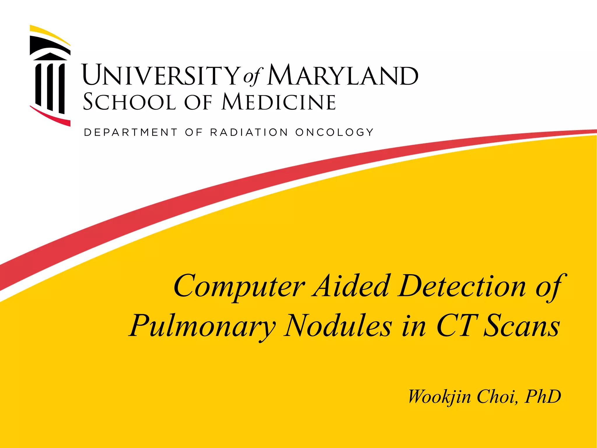
![Pulmonary Nodule Detection CAD system
CAD systems
Lung segmentation
Nodule Candidate Detection
False Positive Reduction
Suzuki et al.(2003)[3]
Thresholding
Multiple thresholding
MTANN
Rubin et al.(2005)[4]
Thresholding
Surface normal overlap
Lantern transform and rule- based classifier
Dehmeshki et al.(2007)[5]
Adaptive thresholding
Shape-based GATM
Rule-based filtering
Suarez-Cuenca et al.(2009)[6]
Thresholding and 3-D connected component labeling
3-D iris filtering
Multiple rule-based LDA classifier
Golosio et al.(2009)[7]
Isosurface-triangulation
Multiple thresholding
Neural network
Ye et al.(2009)[8]
3-D adaptive fuzzy segmentation
Shape based detection
Rule-based filtering and weighted SVM classifier
Sousa et al.(2010)[9]
Region growing
Structure extraction
SVM classifier
Messay et al.(2010)[10]
Thresholding and 3-D connected component labeling
Multiple thresholding and morphological opening
Fisher linear discriminant and quadratic classifier
Riccardi et al.(2011)[11]
Iterative thresholding
3-D fast radial filtering and scale space analysis
Zernike MIP classification based on SVM
Cascio et al.(2012)[12]
Region growing
Mass-spring model
Double-threshold cut and neural network](https://image.slidesharecdn.com/choiwjbriefpresentationcomputeraideddetectionofpulmonarynodulesinctscans-141001234545-phpapp02/75/computer-aided-detection-of-pulmonary-nodules-in-ct-scans-4-2048.jpg)
![Experimental Data Set
Lung Image Database Consortium (LIDC) database [2] is applied to evaluate the performance of the proposed method.
LIDC database, National Cancer Institute (NCI), United States
The LIDC is developing a publicly available database of thoracic computed tomography (CT) scans as a medical imaging research resource to promote the development of computer-aided detection or characterization of pulmonary nodules.
The database consists of 84 CT scans (up to 2007) [2]
100-400 Digital Imaging and Communication (DICOM) images
An XML data file containing the physician annotations of nodules
148 nodules
The pixel size in the database ranged from 0.5 to 0.76 mm
The reconstruction interval ranged from 1 to 3mm](https://image.slidesharecdn.com/choiwjbriefpresentationcomputeraideddetectionofpulmonarynodulesinctscans-141001234545-phpapp02/75/computer-aided-detection-of-pulmonary-nodules-in-ct-scans-5-2048.jpg)
![Comparative Analysis
CAD systems
Nodule size
FPs per case
Sensitivity
Suzuki et al.(2003)[3]
8 - 20 mm
16.1
80.3%
Rubin et al.(2005)[4]
>3 mm
3
76%
Dehmeshki et al.(2007)[5]
3 - 20 mm
14.6
90%
Suarez-Cuenca et al.(2009)[6]
4 - 27 mm
7.7
80%
Golosio et al.(2009)[7]
3 - 30 mm
4.0
79%
Ye et al.(2009)[8]
3 - 20 mm
8.2
90.2%
Sousa et al.(2010)[9]
3 - 40.93 mm
-
84.84%
Messay et al.(2010)[10]
3-30 mm
3
82.66%
Riccardi et al.(2011)[11]
>3 mm
6.5
71.%
Cascio et al.(2012)[12]
3-30 mm
6.1
97.66%
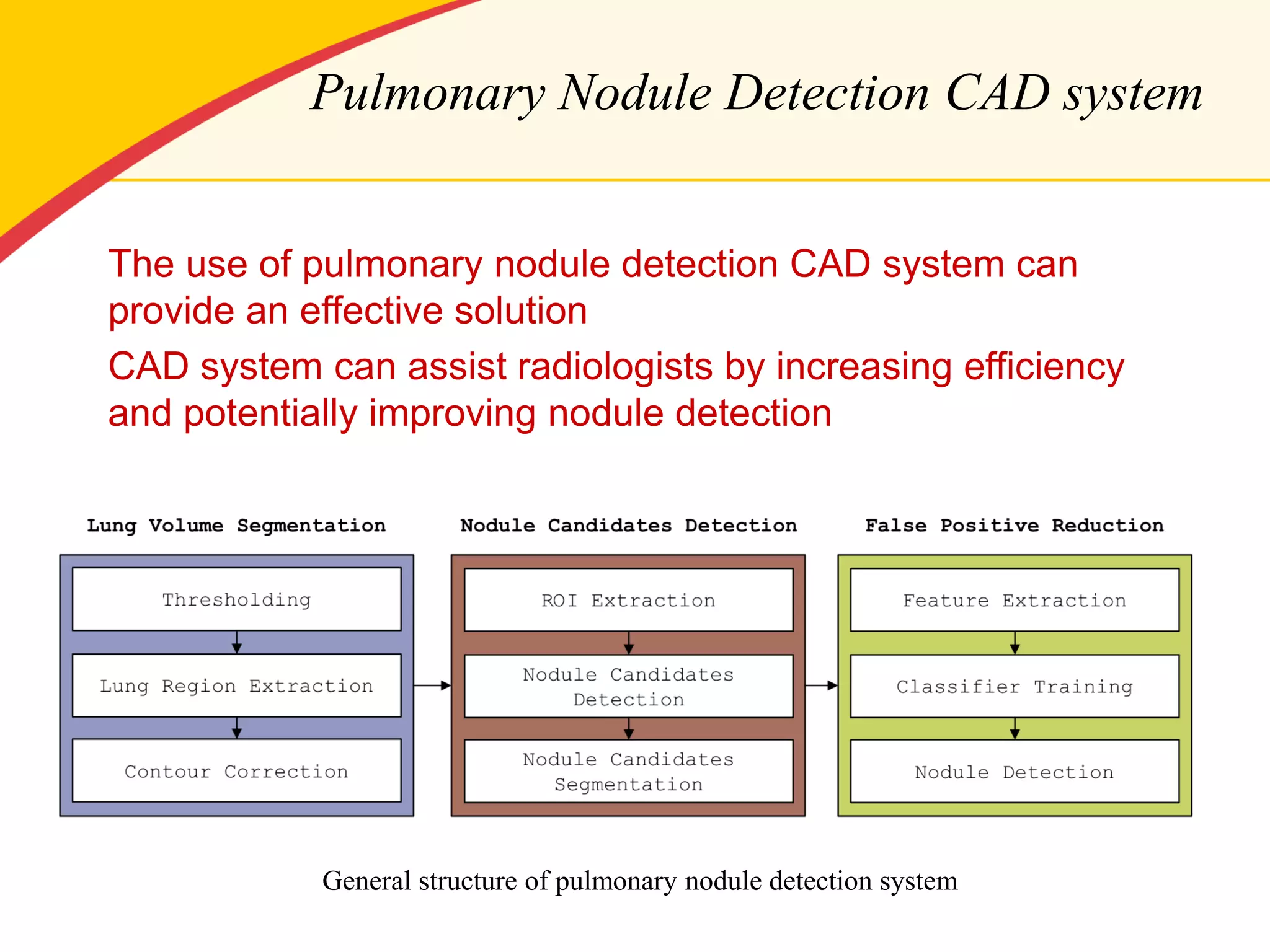
Genetic Programming
3-30 mm
5.45
90.9%
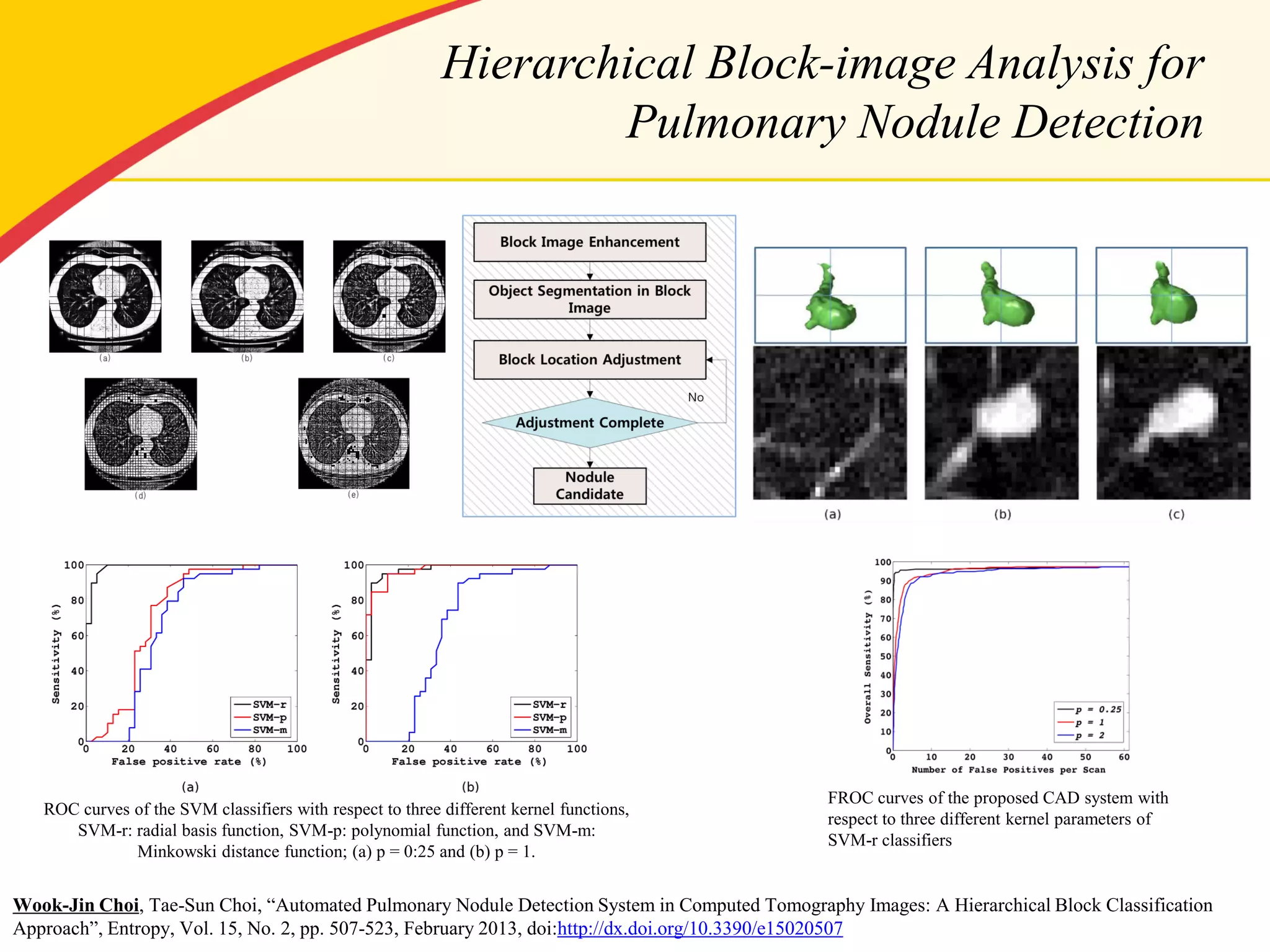
Hierarchical Block Analysis
3-30 mm
2.27
95.2%
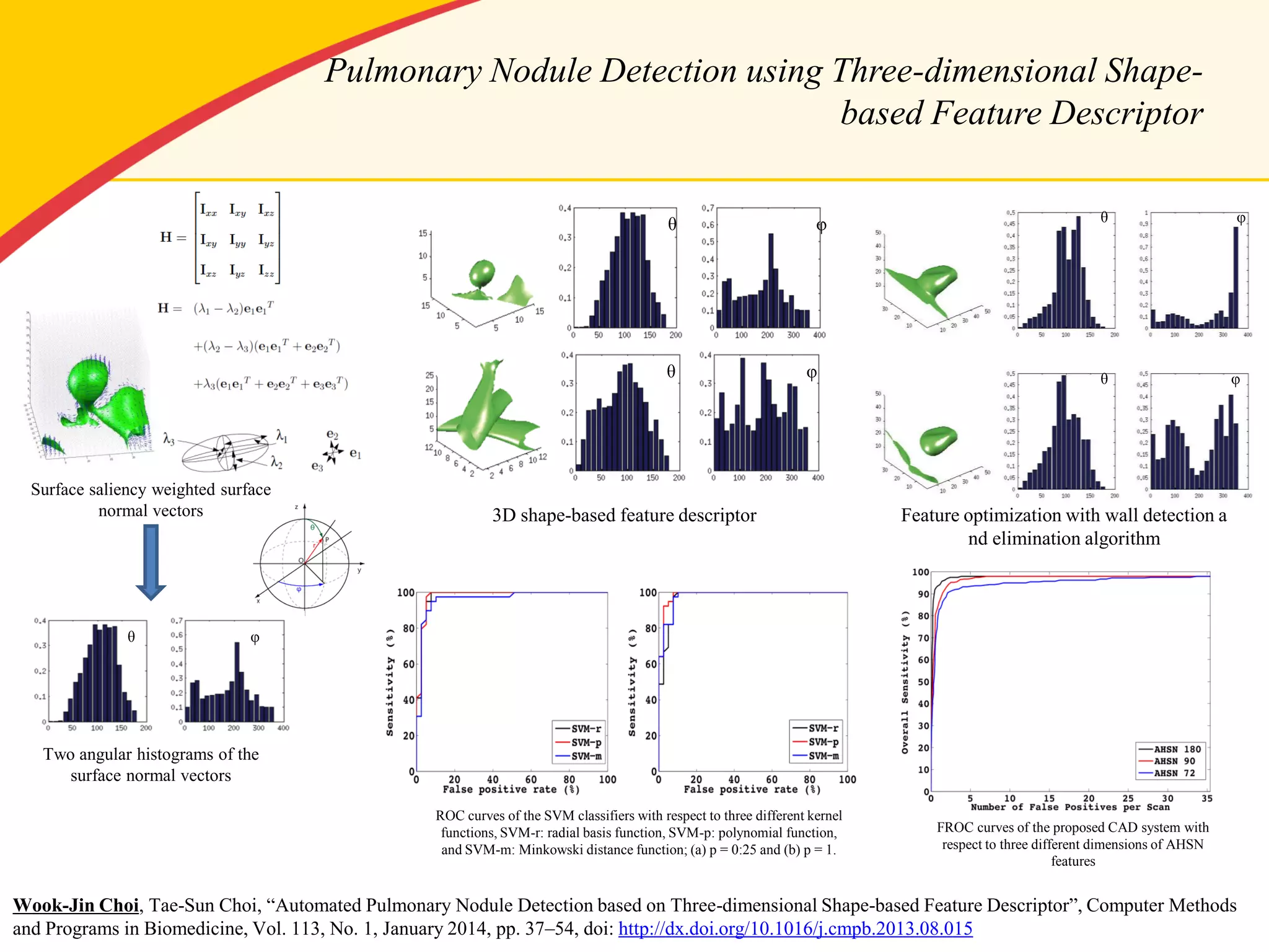
Shape-based Feature
3-30 mm
2.43
95.4%](https://image.slidesharecdn.com/choiwjbriefpresentationcomputeraideddetectionofpulmonarynodulesinctscans-141001234545-phpapp02/75/computer-aided-detection-of-pulmonary-nodules-in-ct-scans-9-2048.jpg)

![References
[1] Rebecca Siegel, Deepa Naishadham, and Ahmedin Jemal, “Cancer statistics, 2012,” CA: A Cancer Journal for Clinicians, vol. 62, no. 1, pp. 10–29, 2012.
[2] M. F. McNitt-Gray, S. G. Armato, C. R. Meyer, A. P. Reeves, G. McLennan, R. C. Pais, J. Freymann, M. S. Brown, R. M. Engelmann, P. H. Bland, G. E. Laderach, C. Piker, J. Guo, Z. Towfic, D. P.-Y. Qing, D. F. Yankelevitz, D. R. Aberle, E. J. R. van Beek, H. MacMahon, E. A. Kazerooni, B. Y. Croft, L. P. Clarke, The Lung Image Database Consortium (LIDC) data collection process for nodule detection and annotation, Acad Radiol 14 (2007) 1464 – 1474.
[3] K Suzuki, SG Armato III, F Li, S Sone, and K Doi, “Massive training artificial neural network (MTANN) for reduction of false positives in computerized detection of lung nodules in low-dose computed tomography,” Medical Physics, vol. 30, pp. 1602 – 1617, 2003.
[4] G.D. Rubin, J.K. Lyo, D.S. Paik, A.J. Sherbondy, L.C. Chow, A.N. Leung, R. Mindelzun, P.K. Schraedley-Desmond, S.E. Zinck, D.P. Naidich, et al., “Pulmonary Nodules on Multi – Detector Row CT Scans: Performance Comparison of Radiologists and Computer-aided Detection,” Radiology, vol. 234, no. 1, pp. 274, 2005.
[6] Jamshid Dehmeshki, Xujiong Ye, Xinyu Lin, Manlio Valdivieso, and Hamdan Amin, “Automated detection of lung nodules in CT images using shape-based genetic algorithm,” Computerized Medical Imaging and Graphics, vol. 31, no. 6, pp. 408 – 417, Sep 2007.
[6] J.J. Suárez-Cuenca, P.G. Tahoces, M. Souto, M.J. Lado, M. Remy-Jardin, J. Remy, and J. José Vidal, “Application of the iris filter for automatic detection of pulmonary nodules on computed tomography images,” Computers in Biology and Medicine, vol. 39, no. 10, pp. 921 – 933, 2009.](https://image.slidesharecdn.com/choiwjbriefpresentationcomputeraideddetectionofpulmonarynodulesinctscans-141001234545-phpapp02/75/computer-aided-detection-of-pulmonary-nodules-in-ct-scans-12-2048.jpg)
![References
[7] Bruno Golosio, Giovanni Luca Masala, Alessio Piccioli, Piernicola Oliva, Massimo Carpinelli, Rosella Cataldo, Piergiorgio Cerello, Francesco De Carlo, Fabio Falaschi, Maria Evelina Fantacci, et al., “A novel multithreshold method for nodule detection in lung ct,” Medical physics, vol. 36, pp. 3607, 2009.
[8] X. Ye, X. Lin, J. Dehmeshki, G. Slabaugh, and G. Beddoe, “Shape-based computer-aided detection of lung nodules in thoracic CT images,” IEEE Transactions on Biomedical Engineering, vol. 56, no. 7, pp. 1810 – 1820, 2009.
[9] João Rodrigo Ferreira da Silva Sousa, Aristófanes Correa Silva, Anselmo Cardoso de Paiva, and Rodolfo Acatauassú Nunes, “Methodology for automatic detection of lung nodules in computerized tomography images.,” Computer methods and programs in biomedicine, vol. 98, no. 1, pp. 1–14, Apr. 2010.
[10] T. Messay, R.C. Hardie, and S.K. Rogers, “A new computationally efficient CAD system for pulmonary nodule detection in CT imagery,” Medical Image Analysis, vol. 14, no. 3, pp. 390 – 406, 2010.
[11] A Riccardi, TS Petkov, G Ferri, M Masotti, and R Campanini, “Computer-aided detection of lung nodules via 3D fast radial transform, scale space representation, and Zernike MIP classification,” Medical Physics, vol. 38, no. 4, pp. 1962–1971, 2011.
[12] D. Cascio, R. Magro, F. Fauci, M. Iacomi, and G. Raso, “Automatic detection of lung nodules in ct datasets based on stable 3d mass-pring models,” Computers in Biology and Medicine, vol. 42, no. 11, pp. 1098 – 1109, 2012.](https://image.slidesharecdn.com/choiwjbriefpresentationcomputeraideddetectionofpulmonarynodulesinctscans-141001234545-phpapp02/75/computer-aided-detection-of-pulmonary-nodules-in-ct-scans-13-2048.jpg)

The document discusses computer aided detection of pulmonary nodules in CT scans. It introduces lung cancer as a major health problem and describes how detecting nodules early can improve survival rates. It then provides an overview of pulmonary nodule detection CAD systems, describing their general structure and evaluating various approaches in the literature. Key contributions are genetic programming and shape-based classifiers and a hierarchical block analysis method that achieved high performance on a publicly available lung image database.