SlideShare a Scribd company logo
1 of 32
Pheretima
1. Introduction to Pheretima
Earthworms are common and very well known to most of us. The common earth-
worm of our country is known as Metaphire sp. It is also commonly found in Sri
Lanka, Japan, Australia and South East Asia. It is represented by 13 species in the
Indian soil. However, in this text we will describe Pheretima posthuma.
2. Habitand Habitat of Pheretima:
Pheretima is a terrestrial earthworm, living in burrows made in moist soil. It
prefers to live in burrow during daytime and at night and rainy seasonthey come
out of their shelter. It is thus nocturnal in habit. They are, however, absent in
regions where the soil is sandy and deficient in humus.
The castings of earthworm are small rounded pellets or balls that lie at the opening
of the burrow. These castings are formed when it goes deep into the hard and
closely packed soil. The soil upon which it feeds, passes through the bodyand are
deposited as castings. While burrowing, the earthworm makes the soil loose and
porous.
The bodywastes of earthworm increases the fertility of the soil. So, earthworms
are often said to be the natural tillers of land. Vermiform culture or vermiculture is
of much practice as earthworms are capable of converting kitchen wastes and other
human wastes into fertilizers.
3. Structure of Pheretima
The body of earthworm is elongated, narrow and cylindrical (Fig. 1.93) measuring
about20 cm in length and 3 to 5 mm in width. The anterior end is more pointed
than the posterior end. Thedorsal side of the body is brown in colour and can be
distinguished from the ventral side which is lighter in colour.
The brown colour is due to the pigment porphyrin which is present in the body
wall and it protects the bodyfrom bright and strong light. The impression of the
dorsalblood vessel can be seen on the dorsal side as a dark median line extending
throughout the length of the body.
The softand naked bodyis made up of distinct segments or metameres separated
from each other by inter-segmental ring-like grooves or annuli. The bodyis made
up of a series of 100-120 similar segments. This external segmentation corresponds
to internal segmentation and is referred to as metameric segmentation.
The segments 14 to 16 from the anterior end are encased in a thick glandular tissue
sheet called the clitellum (saddle) or cingulum (belt) (Fig. 1.93). Considering the
clitellum as the index, the bodymay be divided into three regions, namely the pre-
clitellar, clitellar and post-clitellar regions. Some of the anterior segments bear
superficial furrows and may appear to be subdivided, but these are merely external
subdivisions.
A distinct ‘head’ is absent in Pheretima. The first bodysegment is called
peristomium (Greek : peri, around; stoma, mouth) which bears the mouth aperture
on the ventral surface. The peristomium is prolonged anteriorly into a small, fleshy
lobe, the prostomium (Greek: pro, anterior).
The prostomium is thus considered as a projecting part of the first segment and not
a segment by itself . The last segment, at its posterior end bears the anus.
Digestive System of Earthworm
Digestive system of earthworm consists of alimentary canal and glands along with
physiology of digestion.
Alimentary Canal
Alimentary canal is long and straight, extending from mouth to anus. It consists of
following parts:
1. Mouth : 1st segment
2. Buccal Cavity : 2nd-3rd segment or middle of 3rd segment
3. Pharynx : 3rd-4th segment
4. Oesophagus : 5th -7th segment
5. Gizzard : 8th or 8th-9th segment
6. Stomach : 9th or 10th-14th segment
7. Intestine : 15th up to last segment except anus
8. Anus : last segment
1. Mouth
It is crescentic aperture situated in the 1st segment below the prostomium.
Mouth leads into a buccal cavity.Ingestion of food takes place through it.
2. BuccalCavity
It is a short, wider, thin-walled tube extending from 2nd up to 3rd or middle of
3rd segment. It consists of two kinds of muscle. They are: protractile muscle
and retractile muscle.
Buccal cavity protrude out through mouth with the help of special muscle
for holding the food particles during feeding.Buccal cavity leads into
spacious organ called pharynx.
3. Pharynx
It is small, swollen, wider, thick-walled pear-shaped chamber, which extends up
to 4th segment.It is wider than buccalcavity.It is distinguished from buccal
cavity by means of constriction.It has pharyngeal gland, located in the dorsal
salivary chamber. Pharyngeal gland is composed ofmany chromaphil cells,
which producesaliva containing proteolytic enzyme; protease and mucin. Mucin
makes the food softand protease converts protein into amino acid.
4. Oesophagus
It is narrow thin-walled tubular structure extending from 5th to 7th segment.
It has no gland.It passes the food particles from pharynx to gizzard.
It leads into gizzard.
5. Gizzard
It is oval, thick-walled and highly muscular organ lying in the 8th or 8th-
9th segment.
It is the hardest part of alimentary canal due to the presence of inner lining of
cuticle. It also possess chitinuous teeth like projection. It helps in grinding or
crushing food so act as grinder during feeding.
6. Stomach
Gizzard leads to short, narrow, thin-walled, highly vascular tubular structure
called stomach, which extends from 9th or 10th to 14th segment. It is wider than
oesophagus.Ithas calciferous gland which helps in neutralization of food by
calcification process.Stomachleads to intestine.The glandular cell of stomach
produceproteolytic enzymes for the digestion of protein.
7. Intestine
It is long, wide and thin-walled tube which extends from 15th to last segment
except anus. Its inner lining is ciliated, vascular, folded and glandular. Its
intestinal lining is folded to form villi. One of the villi becomes larger and well
developed than other called typhlosole which runs mid-dorsally from 27th to last
25th segment.Typhlosole divides the intestine into 3 regions. They are:
i.Pre-typhlosolarregion
It extends from 15th segment to 26th segment so it is the first part of the
intestine.It consists of villi but no typhlosole.In 26th segment, there is a pair of
short and conical lateral outgrowth called intestinal caeca which extends upward
up to 23rd segment. Intestinal caeca produceamylase which helps in
starch(carbohydrate) digestion.
ii.Typhlosolarregion
It is the 2nd or middle part of the intestine which extends from 27th segment to
last from 25thsegment. It has both villi and typhlosole.The typhlosole is highly
vascular and glandular fold that increases the absorptive surface area of the
intestine.
iii.Post-typhlosolarregion
It is the last part of the intestine lying in the last 23rd-25th segment in front of
anus. It is also called rectum.It lacks villi and typhlosole.It contains small pellets
of mud which are thrown out through the anus to form casting.
8. Anus
It is a circular opening in the last segment called anal segment.
Undigested food materials release out through anus in the form of worm casting.
Digestive glands
There are different types of digestive glands associated with alimentary canal of
earthworm.
1. Pharyngeal gland
2. Gastric gland
3. Intestinal glands
4. Intestinal caeca
Physiology of digestion
Digestion is the bio-chemical process inwhich complex organic food is brokendown
into simple, soluble and diffusible form in the presence of respective enzymes.
Earthworms feed uponall kinds oforganic humus and debris suchas decaying leaves
and seeds, protozoan, etc. present in soil. They also feed directly on leaves, grasses
and other vegetation.
During feeding the buccal cavity is protrude out with the help of protractile and
retractile muscle. Then the food is drawn into the mouth. The ingested food enters
into pharynx through buccal cavity. The dorsal chamber of pharynx consists of
pharyngeal gland which is composed of chromaphil cell which produce saliva
containing mucin and protease. Mucin lubricates the food and protease converts
protein into amino acids. The foods then pass through oesophagus into gizzard,
where grinding or crushing of food material takes place into fine state due to the
contraction of circular muscles of gizzard. The grinded food material enters into the
stomach where the neutralization of food takes place by calcification process. Also
there occurs the complete digestion of protein byproteolytic enzymes. Now the food
material enters into intestine. In intestine, intestinal caeca produce amylase which
converts starch into glucose.
In intestine several enzymes are secreted and acts on the substrate as follows:
 Protease: converts protein into amino acid.
 Amylase: converts starch into two molecules of glucose i.e. maltose.
 Cellulase: converts cellulose into glucose
 Chitinase: digest chitin of exoskeleton of insects.
 Lipase: converts fats into fatty acids and glycerol.
Digestion occurs mostly in the intestine and the digested food is absorbed by villi.
Then pass into blood stream through capillaries. Undigested food and the soil are
released out in the form of casting through anus.
T.S. of Pharynx
Pharynx is small, wide, thick-walled, pear-shaped chamber. Two chambers are
found in pharynx. They are: dorsal chamber and ventral chamber.
The dorsalchamber is also called salivary chamber which contains pharyngeal gland
that is composed of many chromaphil cells. Dorsally pharyngeal gland is lined by
villi. The chromaphil cells produce saliva containing proteolytic enzymes and
mucin. Mucin makes the food soft and protease converts protein into amino acid.
The ventral chamber is a conducting chamber which conducts digested orundigested
food material from pharynx to oesophagus. Pharynx acts as sucking and pumping
organ during feeding.
Circulatory System of Earthworm
Introduction
The blood vascular system of Pheretima is of closed type. It consists of the blood
vessels, hearts, loops, capillaries and the blood glands.
Blood:
The blood of Pheretima is red coloured due to the presence of a respiratory
pigment haemoglobin in it. The haemoglobin is not contained in the corpuscles like
the vertebrates but it is found dissolved in the plasma. The plasma also contains
many corpuscles which are colourless and nucleated.
Blood Vessels:
The blood vessels are of two types collecting blood vessels and distributing blood
vessels which are closed tubes with definite walls and they break up into capillaries
to ramify in the different parts of the body. The arrangement of blood vessels in the
anterior thirteen segments is somewhat different from that behind the thirteen
segment, i.e., in the region of intestine.
:
Therefore, for convenience the blood vessels canbe described under the
following two heads:
A. Bloodvessels and their arrangementin the segments behind 13th, i.e.,
intestinal region.
B. Bloodvessels and their arrangement in the anterior thirteen segments.
A. BloodVessels and their Arrangement in the Segments behind
13th, i.e., Intestinal Region:
The blood vessels ofthis regioninclude:
1. Median longitudinal blood vessels;
2. The intestinal blood plexus;
3. The commissural vessel;
4. The integumentary vessel; and
5. The nephridial vessels.
1. Median Longitudinal Blood Vessels:
(i) DorsalVessel:
It runs mid-dorsally above the alimentary canal from the posterior to the anterior
end of the body. It is the thickest vessel with contractile muscular walls visible
from outside as a dark line through the thin and semitransparent bodywall.
The direction of flow of blood in this vessel remains from behind to forward (from
posterior to anterior). It is contractile and pulsates rhythmically to force the blood
from posterior to anterior side. In each segment it has a pair of valves internally
which check the backward flow of blood. It is the main collecting vessel behind the
14th segment, but in front it distributes the blood.
From the posterior segment up to the 14th segment it receives two pairs of dorso
intestinal vessels from the intestine in each segment and a pair of commissural
vessels from the sub-neural vessel.
The commissural vessels form a loop behind each septum and they receive blood
from the bodywall, nephridia and prostate glands. The commissural vessels also
give out blood in each segment through a septo intestinal branch to the intestine.
(ii) Ventral Vessel:
It is also a long vessel and runs ventrally below the alimentary canal and above the
ventral nerve cord from second segment up to the last segment of the body.
It is thin- walled without muscles and valves. The direction of flow of blood in this
vessel remains from anterior to the posterior side or from in front to backwards. It
is a distributing vessel. It gives out a pair of ventro in tegumentary vessels, one on
each side in front of the septum in all segments.
The ventrointegumentary vessels run upwards along the bodywall and supply
blood to the bodywall, integumentary nephridia, septal nephridia, gonads, seminal
vesicles and spermathecae.
The ventral vessel also gives out a ventrointestinal vessel in each segment behind
the 13th segment, these take blood to the lower part of the intestine. The branches
in intestine form blood plexuses consisting of two networks in the intestinal wall.
(iii) Sub-neural Vessel:
It is also a long but thin vessel extending from anterior 14th segment up to the last
segment situated mid-ventrally below the ventral nerve cord. It is without muscular
walls and internal valves. The direction of flow of blood in this vessel remains
from anterior to posterior side and it is mainly a collecting vessel.
It receives a pair of slender branches in each segment which bring blood from the
ventral bodywall and the nerve cord. It gives a pair of commissural vessels in each
segment which join the dorsal vessel, as already mentioned while describing the
dorsalvessel. Thus, it collects blood from the ventral bodywall and supplies some
blood to the intestine.
2. Intestinal Blood Plexus:
The intestine of Pheretima is richly supplied with blood capillaries which form a
close network. The intestinal blood plexus consists of a close network of capillaries
in the wall of intestine.
In fact, there are two capillary networks in the intestine:
(i) The external and
(ii) The internal.
The capillary network which is present at the outer surface of intestine is known as
external plexus which receives blood from the ventral vessel through
ventrointestinal and passes it on to the internal plexus.
The capillary network which is present between the circular muscle layer of
intestine and its internal epithelial lining is known as internal plexus which serves
to absorb the nutrients from the gut and is connected with dorsalblood vessel
through the dorsointestinals.
3. Commissural Vessels:
These vessels connect the dorsaland sub-neural vessels. These vessels receive
blood from nephridia, bodywall and reproductive organs through capillaries and
then they send it to dorsalblood vessel.
4. IntegumentaryVessels:
These vessels coming from ventral vessels supply the blood to integument for
aeration and the aerated blood is collected by numerous capillaries of commissural
vessel in each segment. Thus, there is a close parallelism between venous and
arterial capillaries throughout the bodywall.
5. Nephridial Vessels:
These vessels originate from the ventrotegumentary vessels of ventral vessel and
supply the blood to the nephridia.
B. Blood Vessels and their Arrangement in the Anterior 13 Segments:
The blood vascular system in the first thirteen segments is modified considerably
and differs markedly from that of the intestinal region.
It consists ofthe following:
1. Median longitudinal vessels;
2. Hearts and anterior loops;
3. Blood vessels of the gut.
The function of collecting blood from the anterior region of the gut is taken over
by a new vessel supra-oesophageal, while the blood from the peripheral structures
is collected by the right and left lateral oesophageal.
1. Median Longitudinal Blood Vessels:
(i) Dorsalvessel:
This blood vessel becomes the distributing vessel in these segments instead of
collecting vessel. Structurally, it retains its original identity as it was in the
posterior segments. But is has neither dorsointestinals nor commissural vessels
opening into it.
It sends out all the collected blood from the posterior region of the body into hearts
and the anterior region of the gut where it divides into three branches distributed
over the pharyngeal bulb and the roof of the buccal chamber. However, it supplies
to stomach, gizzard, oesophagus, pharynx and other related parts.
(ii) Ventral vessel:
This blood vessel remains distributing in these segments also but extends only up
to the second segment. The ventrointestinals are absent, hence, it does not supply
to the alimentary canal in this region. However, the ventrotegumentary vessels, a
pair in each segment, supply blood to the integument, nephridia, septa and
reproductive organs.
(iii) Supra-oesophageal vessel:
It is the shortestlongitudinal vessel extending from 9th to 13th segment situated
above the stomach. It receives blood from the lateral oesophageals by two pairs of
anterior loops that encircle the stomach in the 10th and 11th segments. It sends its
collected blood by the latero-oesophageal hearts in segments 12th and 13th to the
ventral vessel.
(iv)Lateral oesophageals:
In fact, the sub-neural vessel bifurcates in the 14th segment to form two lateral
oesophageals. These vessels are considerably thick and situated along the ventro-
lateral margins of alimentary canal in the anterior thirteen segments. These vessels
are closely attached to the wall of the stomach from 10th to 13th segments and
communicate with the ring vessels.
But in the region of gizzard and further forwards, they remain free from the wall of
the alimentary canal even though they receive branches from it in each segment.
These vessels receive a pair of branches in each segment bringing blood from the
bodywall and the septum. They also collect blood from the reproductive organs
and nephridia, thus, functioning like the sub-neural and commissural vessels of the
posterior region, i.e., these are collecting vessels.
2. Hearts and Anterior Loops:
In the posterior segments behind 13th the dorsaland ventral blood vessels have no
direct connections but anteriorly both these vessels are connected together by 4
pairs of pulsatile hearts which are neurogenic, i.e., the heart beat originates in the
nerve cells of the hearts. The hearts are contractile and encircle the alimentary
canal, they are in the segments 7th, 9th, 12th and 13th.
The hearts of segments 12th and 13th are joined above to both the dorsal and the
oesophageal vessels, these are called latero-oesophageal hearts. These hearts have
thick muscular walls and a pair of valves at each junction with the dorsal vessels
and supra-oesophagealvessel, and another pair of valves at the ventral end.
These valves allow blood to flow downwards only. The other hearts of the segment
7th and 9th are called lateral hearts. These connect the dorsalvessels to the ventral
vessel. They have four pairs of valves which allow blood to flow only downwards.
Besides four pairs of hearts there are two pairs of loop-like vessels in the 10th and
11th segments which connect the supra-oesophagealwith the lateral oesophageals.
These vessels are neither muscular nor pulsatile and are called anterior loops.
These are devoid of valves.
The blood from lateral oesophageals flows through these loops into supra-
oesophageal which sends all its collected blood into ventral vessel through the
hearts of 12th and 13th segments.
3. Blood Vessels of the Gut:
On either side of stomach are situated ring-like vessels which connectthe supra-
oesophageal and lateral-oesophageal vessels. Through these vessels blood flows
upwards from the lateral- oesophageals into the supra-oesophageal. Buccal cavity,
pharynx and gizzard receive their blood supply from dorsal blood vessel directly.
Circulation of Blood:
The blood collected by the dorsalvessel through the dorsointestinals, blood
plexuses of intestine, and commissurals is given out partly to the anterior
alimentary canal, but mainly through the hearts to the ventral vessel.
In the ventral vessel the blood flows forwards to the anterior region in front of the
hearts, but the main portion of blood flows backwards, this is distributed through
ventrotegumentaries to the bodywall and the organs in the coelom, and through
the ventrointestinal vessels to the alimentary canal.
In other words all parts receive blood from the ventral vessel. From the ventral
bodywall blood is collected by the sub-neural which also receives some blood
through the lateral-oesophageal from the anterior region.
This blood passes from the sub-neural to the dorsalvessel through the
commissurals. The lateral-oesophageals also send blood through the anterior loops
to the supra- oesophageal vessel which then passes it through the latero-
oesophageal hearts to the ventral vessel.
The courseof circulation of blood in Pheretima has been shown in Fig. 66.20.
Functions:
The blood distributes digested food to various bodyregions, and it collects waste
substances like nitrogenous waste and C02 which are given up to nephridia, skin
and the coeiomic fluid. Respiration in almost all aquatic and terrestrial
oligochaetes takes place by diffusion of gases through the integument which in
larger forms contains a capillary network in the outer epidermal layer.
In terrestrial species the film of moisture necessary for diffusion of gases is
supplied by mucous glands, coeiomic fluid, and nephridial excretions. The
haemoglobin of plasma extracts O2 from the capillaries of the skin, but there must
be a moist skin where O2 can combine with haemoglobin to be transported by the
blood.
Haemoglobin is an efficient pigment and it can take up O2 either from the
surrounding air or from an environment comparatively deficient in oxygen. Hence,
earthworms can live in well aerated water and are not drowned. They can also live
for several hours without O2, in this condition they probably carry on anaerobic
respiration.
Blood Glands:
In the segments 4th, 5th and 6th above the pharyngeal mass are several groups of
small rounded follicles of red colour, which are called blood glands. The follicles
have a syncytial wall enclosing a capsule containing a mass of loose cells. The
blood glands are connected with pharyngeal nephridia and with salivary glands.
These glands manufacture blood corpuscles and haemoglobin. These glands are
probably also excretory.
Excretory System of Nephridia (Earthworm)
These are of three types according to their locationin the body:
1. Septal nephridia;
2. Integumentary nephridia
3. Pharyngeal nephridia.
1. SeptalNephridia:
These are found situated on the inter-segmental septum between 15th and 16th
segments to the posterior side of the body.
Each septum bears nephridia on both the surfaces arranged in semicircles around
the intestine, two rows in front of the septum and two behind it. Each septum has
about 40 to 50 nephridia in front and the same number behind, so that each
segment possesses80 to 100 septal nephridia except the 15th segment which has
only 40 to 50 nephridia. These are not found in the segments up to 14th.
Structure:
The septal nephridia may be considered typical of all the nephridia of Pheretima.
Each septal nephridium (Fig. 66.22) consists of nephrostome, neck, bodyof
nephridium and the terminal duct.
(i) Nephrostome:
It is also known as ciliated funnel or nephridiostome. It is the proximal flattened
funnel-shaped structure of the nephridium lying in the coelom.
It has an elliptical mouth-like opening leading into an intracellular canal of the
large central cell, the margins of the opening are surrounded by a large upper lip
and a smaller lower lip. The lips are provided with several rows of small ciliated
marginal cells and the central canal is also ciliated.
(ii)Neck:
The nephrostome leads into a short and narrow ciliated canal forming the neck. It
joins the nephrostome to the bodyof nephridium.
(iii)Body of Nephridium:
The bodyof nephridium has two parts a short straight lobe and a long twisted loop.
The loop is formed by two limbs— the proximal limb and the distal limb.Both
these limbs are twisted spirally around each other, the number of twists varies from
nine to thirteen. The neck of nephridium and the terminal duct join together and
remain connected with the proximal limb of the twisted loop, while the distal limb
becomes the straight lobe.
Internally the nephridium is made of a connective tissue matrix having long coiled
nephridial duct forming loops. There are four such canals in the straight lobe, three
in the lower part and two in the upper part of the limbs of twisted loop. Two canals
of the straight lobe out of the four are ciliated like the ciliated canal of the neck.
(iv) TerminalDuct:
It is short and narrow with a terminal excretory duct. It joins the nephridium with
septal excretory canal.
Relationof septal nephridia with intestine:
The nephridia hang freely in the coelom and are attached only by their terminal
ducts. They open by their terminal ducts into two septal excretory canals lying on
the posterior surface of the septum, one on each side of the intestine, each begins
ventrally but dorsally it opens in the supra-intestinal excretory duct of its own side.
The supra-intestinal excretory ducts are two parallel longitudinal canals lying
above the gut and below the dorsalvessel (Fig. 66.24). These excretory ducts begin
from the 15th segment and run to the last segment, they communicate- with each
other for a short space behind each septum, then either the right or the left duct
opens by a ductule into the lumen of the intestine near the septum.
Thus, each segment has one such opening into the intestine of either the left or the
right supra-intestinal excretory duct. The waste collected by the nephridia is
discharged through the excretory canals and ducts into the lumen of the intestine.
Such nephridia opening into the intestine are called enteronephric nephridia.
2. Integumentary Nephridia:
In each segment of the bodyfrom 7th to the last segment, numerous nephridia are
found attached inside the lining of the bodywall. These are called integumentary
nephridia which are about 200-250 in each segment except the segment of the
clitellar region where they number 2,000-2,500 in each segment.
These nephridia are small-sized, without nephrostome and without any opening
into the coelom.
Hence, they are called closed type of nephridia. Each integumentary nephridium is
V-shaped with a short straight lobe and a twisted loop, its lumen has two ciliated
canals. Each nephridium opens by a nephridiopore on the outer surface of the body
wall directly. Since the integumentary nephridia discharge the excretory wastes
directly outside, hence, they are called exonephric nephridia.
3. Pharyngeal Nephridia:
These nephridia lie in three paired tufts, one on either side of the anterior region of
the alimentary canal in the segments 4th, 5th and 6th. The tufts of pharyngeal
nephridia also contain blood glands.
Each pharyngeal nephridium is about the size of a septal nephridium but it is of the
closed type having no funnel or nephrostome. It has a short straight lobe and a
spirally twisted loop, its lumen has ciliated canals. Ductules arise from each
nephridium and unite to form a single thick- walled ducton each side in each
segment.
The two ducts of nephridia of segment 6th open into the buccalcavity in segment
2nd and the paired ducts of nephridia of segments 4th and 5th open into the
pharynx in segment 4th.
These nephridia also discharge their wastes into the alimentary canal and are,
therefore, enteronephric but suchenteronephric nephridia which open into the
anterior region of the alimentary canal (buccal cavity and pharynx) are called
peptonephridia becausethey may have taken the function of digestive glands.
Recently it has been reported that the pharyngeal nephridia of P. posthuma produce
a variety of enzymes like amylase, chimosin, prolinase, prolidase, dipeptidases,
aminopeptidase, lipase, etc., which hydrolyse various foodstuffs. Thus, such
nephridia work like the salivary glands.
Physiology of Excretion:
Like other animals, in earthworms also, the protein catabolism results in the
formation of nitrogenous waste substances like certain amino acids, ammonia and
urea.
Uric acid is not found in the earthworms. However, the amino acids are degraded
to form free ammonia and the urea is synthesised in the chloragogen cells which
are released into the coelomic fluid and also in the blood for its removal. Free
amino acids are not excreted but traces of creatinine occurin the urine.
Moreover, the nitrogen excreted in different forms in a well fed worm is about
72% NH3, 5% urea and remaining other compounds,while in a starved worm
NH3 8.6%, urea 84.5% and remaining being other compounds. But generally, the
excretion is 42% NH3, 50% urea, 0.6% amino acids and remaining being other
compounds.
So, we can say that in a well fed earthworm, NH3 predominates the nitrogenous
excretory wastes, hence, it is ammonotelic, while a starved one is ureotelic.
An earthworm excretes the nitrogenous wastes in the form of urine which
generally contains urea, water, traces of ammonia and creatinine. Nephridia excrete
these substances from the bodyof earthworm. The various excretory wastes from
the coelomic fluid are drawn into the nephrostomes of septal nephridia or into the
excretory canals of other nephridia along with some other useful substances.
These products are either discharged into the intestine (by enteronephric nephridia)
or outside by the nephridiopores (by exonephric nephridia). The bodyof nephridia
also absorbs somewastes. However, the useful substances are reabsorbed and the
passing out waste remains concentrated for various nitrogenous compounds.
The excreted waste substances are removed out from the bodywith faeces. The
nephridia, in addition to excretory, are also osmoregulatory in function.
The nephridia help in conserving water by reabsorption from the excreted products
during summers and winters, so they pass hypertonic urine in relation to blood.
During rainy season, the urine is dilute due to lesser reabsorption of water. The
enteronephric nature of nephridia provides another device for conserving water.
Reproductive System of Pheretima (Earthworm)
Male Reproductive Organs of Earthworm:
The male reproductive organs consist of testes, testis sacs, seminal vesicles, vasa
deferentia, prostate glands and accessoryglands.
Testes and Testis Sacs:
The testes are two pairs, very minute, whitish and lobed structures situated one pair
each in segment 10th and 11th found attached with the posterior surface of 9/10
and 10/11 inter-segmental septa. Each testis consists of 4 to 8 finger-like lobules
projecting from a compactbase.
Each lobule of the testis contains rounded cells in masses called spermatogonia.
The testes are well developed only during the young stage of the worm and
become degenerated in the adult worm. The testes of each segment are found
enclosed in a thin-walled, wide bilobed and fluid-filled sacs called testis sacs.
Thus, there are two testis sacs situated in the segments 10th and 11th on the
ventrolateral sides of the ventral nerve cord beneath the stomach. 
Behind each of the four testes, in the testis sacs, is a large spermatic funnel having
folded and ciliated margins. The testis sacs remain in communication with the
seminal vesicles. The spermatogonia are shed into the testis sacs and pass on into
the seminal vesicles where they undergo maturation and form spermatozoa.
SeminalVesicles:
These are two pairs of large, white, sac-like bodies, one pair each in the segments
11th and 12th into which the testis sacs open by narrow ducts. The anterior seminal
vesicles are smaller than the posterior ones. The seminal vesicles of the 11th
segment are found enclosed in the posterior larger testis sac, while those of the
12th segment are exposed in the coeiomic cavity.
These are also placed ventro-laterally beneath the stomach and since they develop
as septal outgrowths, hence, they are also known as septal pouches.
Vasa Deferentia:
Each spermatic funnel (posteriorly) leads into a thin, narrow, thread-like sperm
duct or vas deferens. Thus, the two vasa deferentia of each side run very closely
and laterally to the nerve cord beneath the alimentary canal on the ventral body
wall, up to 18th segment where they join the prostatic duct.
Prostate Glands:
These are a pair of large, white, solid and irregularly- shaped glandular masses
situated one on either side of the gut in the segments from 16th or 17th to 20th or
21st. Each prostate gland consists of a maximum glandular region and a small part
of non-glandular region. From each prostate gland emerges a thick curved prostatic
duct in 18th segment.
This duct joins the two vasa deferentia of its side and these three ducts are enclosed
in a common sheath to form a common prostatic spermatic duct which opens
separately through a male genital aperture on the ventral side of the 18th segment.
Thus, each genital aperture has three separate apertures—two of the vasa deferentia
and one of the prostatic gland. The function of the secretion of prostatic duct is not
definitely known.
Accessory Glands:
These are two pairs of rounded structures situated one pair each in the segments
17th and 18th on the ventral body wall at the lateral sides of the nerve cord.
These glands open to the outside by a number of small ductless on the two pairs of
genital papillae situated on the either side of the mid-ventral line in 17th and 19th
segments externally. The secretion of these glands is believed to keep together the
two worms during the process ofcopulation.
As mentioned earlier, the spermatogonia from the testes are shed into the testis
sacs and pass on to the seminal vesicles where spermatogenesis is completed and
tailed spermatozoa are formed. Now, these spermatozoa again enter the testis sacs
and through the spermatic funnels go down the vasa deferentia and are discharged
through the male genital apertures along with the secretion of prostateglands.
Female Reproductive Organs of Earthworm:
Female reproductive organs consist of the
1. Ovaries
2 oviducts
3 sperm thecae.
Ovaries:
There is a pair of white, small, lobulated ovaries, one on either side of the ventral
nerve cord in the 13th segment attached with the posterior face of the inter-
segmental septum of 12/13 segments.
Each ovary has several finger-like processeswith developing ova in a row
providing it beaded appearance. The ova remain in various stages of their
development in each ovarian lobe being mature in the distal part and immature in
the proximal part.
Oviducts:
Below each ovary in 13th segment, there is a small ovarian funnel with folded and
ciliated margins. Each ovarian funnel leads into a short, conical and ciliated
oviduct.
The oviducts of both the sides converge to meet below the nerve cord and open by
a single median female genital aperture ventrally in the 14th segment. The mature
ova are released from the ovaries and received by the ovarian funnel, pass through
the oviducts and which go out through the female genital aperture.
Spermathecae:
These are four pairs, one pair each in the segments 6th, 7th, 8th, and 9th situated
ventro-laterally. Each spermatheca is flask-shaped, the main bodyis the ampulla
which is continued into narrow duct— the neck with a small diverticulum on its
inner side. These are also called seminal receptacles as they store the spermatozoa
from the another worm during copulation.
In Pheretima, unlike the other earthworms, the diverticula store the spermatozoa
and not the ampulla. The spermatheca open externally by their small ducts as
spermatheca pores situated inter-segmentally between segments 5/6, 6/7, 7/8 and
8/9.
Copulation, CocoonFormationand Fertilization of Earthworm:
Copulation has been observed in different species of earthworms. It usually occurs
in the rainy seasonduring the months of July to Octoberat dawn, i.e., in the
morning hours before sunrise.
During copulation two earthworms of adjacent burrows half emerge from their
burrows and come closer to lie in contactto each other by their ventral surfaces
with their anterior ends pointing in oppositedirections. In this position the male
genital apertures of each worm lie oppositethe spermathecal pores of the other.
The areas of the male genital apertures are raised into papillae and inserted into the
spermathecal pores, so that there is a mutual exchange of spermatozoa and
prostatic fluid between the two copulating worms. Copulation lasts for about an
hour or so, then the worms separate and recede into their burrows.
Cocoonorootheca formation starts after copulation when ovaries mature. The
epidermis of clitellar segments, i.e., 14th, 15th and 16th segments contain three
kinds of glands, they are unicellular mucous glands which producemucus for
copulation, cocoon-secreting glands secrete the wall of the cocoonand albumen
glands producealbumen in which eggs are deposited in the cocoon.
A membranous girdle is secreted by cocoon-secreting glands of the clitellum, this
girdle soonhardens, then albumen is deposited between the girdle and the body
wall. The worm starts withdrawing itself backwards from the girdle. As the girdle
passes over the female genital aperture it receives eggs, and when it passes over the
spermathecae sperms are extruded into it through spermathecal pores.
Lastly, the girdle is thrown off from the anterior end of the worm and soonthe
elasticity of its wall closes up its two ends to form a cocoonorootheca. Several
cocoonsare formed after each copulation because the spermatozoa contained in the
spermatheca do not pass out all at one time. The cocoons are oval, light yellow in
colour and are about 2 to 2.4 mm long and 1.5 to 2 mm broad.
Fertilisation takes place inside the cocoonand generally there is only one embryo
in a cocoon. Cocoons are laid from August to Octoberin damp situations.

More Related Content

What's hot (20)

Sol gel theory
Sol gel theorySol gel theory
Sol gel theory
 
Digestive system of pila
Digestive system of pilaDigestive system of pila
Digestive system of pila
 
Classification of animals: Protozoa, Porifera and Coelenterata
Classification of animals: Protozoa, Porifera and CoelenterataClassification of animals: Protozoa, Porifera and Coelenterata
Classification of animals: Protozoa, Porifera and Coelenterata
 
Obelia life cycle- polyp & medusa structure, difference between polyp & medusa
Obelia life cycle- polyp & medusa structure, difference between polyp & medusaObelia life cycle- polyp & medusa structure, difference between polyp & medusa
Obelia life cycle- polyp & medusa structure, difference between polyp & medusa
 
Paramecium
ParameciumParamecium
Paramecium
 
Porifera
PoriferaPorifera
Porifera
 
Mouth parts ppt
Mouth parts pptMouth parts ppt
Mouth parts ppt
 
Metazoa
MetazoaMetazoa
Metazoa
 
Coelenterates- The Obelia
Coelenterates- The ObeliaCoelenterates- The Obelia
Coelenterates- The Obelia
 
Scoliodon
ScoliodonScoliodon
Scoliodon
 
Paramecium structure
Paramecium structureParamecium structure
Paramecium structure
 
Food Digestion In Protozoa.ppt
Food Digestion In Protozoa.pptFood Digestion In Protozoa.ppt
Food Digestion In Protozoa.ppt
 
General characteristics of protozoa
General characteristics of protozoaGeneral characteristics of protozoa
General characteristics of protozoa
 
Platyhelminthes
PlatyhelminthesPlatyhelminthes
Platyhelminthes
 
Life cycle of silkworm
Life cycle of silkwormLife cycle of silkworm
Life cycle of silkworm
 
Locomotion in protozoa sj
Locomotion in protozoa sjLocomotion in protozoa sj
Locomotion in protozoa sj
 
Digestive system in birds
Digestive system in birdsDigestive system in birds
Digestive system in birds
 
Respiratory system of insects ppt
Respiratory system of insects pptRespiratory system of insects ppt
Respiratory system of insects ppt
 
Porifera phylum
Porifera phylumPorifera phylum
Porifera phylum
 
Chordata - Bio 11
Chordata - Bio 11Chordata - Bio 11
Chordata - Bio 11
 

Similar to Introduction to the Earthworm Pheretima

External features &digestive system of earthworm, Physiology of digestion, Al...
External features &digestive system of earthworm, Physiology of digestion, Al...External features &digestive system of earthworm, Physiology of digestion, Al...
External features &digestive system of earthworm, Physiology of digestion, Al...SoniaBajaj10
 
Earthwarm digestive system
Earthwarm  digestive systemEarthwarm  digestive system
Earthwarm digestive systemSSMV2016
 
6th Module, insects (respiratory, digestive and integument system)and entomol...
6th Module, insects (respiratory, digestive and integument system)and entomol...6th Module, insects (respiratory, digestive and integument system)and entomol...
6th Module, insects (respiratory, digestive and integument system)and entomol...DackardShaw
 
Nutrition in mammals
Nutrition in mammalsNutrition in mammals
Nutrition in mammalsSaad Adil
 
Project report on Fasciola hepatica.docx
Project report on Fasciola hepatica.docxProject report on Fasciola hepatica.docx
Project report on Fasciola hepatica.docxpriyanshimanchanda4
 
Digestion and absorption
Digestion and absorptionDigestion and absorption
Digestion and absorptionzoosphere
 
Digestive system notes
Digestive system notesDigestive system notes
Digestive system notesTia Hohler
 
The digestive system 1 salah nazar
The digestive system 1 salah nazarThe digestive system 1 salah nazar
The digestive system 1 salah nazarSalah Nazar
 
Digestion and Absorption - Class 11 ppt
Digestion and Absorption - Class 11 pptDigestion and Absorption - Class 11 ppt
Digestion and Absorption - Class 11 pptKarthik Bharadwaj
 
Human Digestive Anatomy
Human Digestive Anatomy Human Digestive Anatomy
Human Digestive Anatomy Aditi Tanwar
 
Detail study of Earthworm for class 11 NEB B
Detail study of Earthworm for class 11 NEB BDetail study of Earthworm for class 11 NEB B
Detail study of Earthworm for class 11 NEB BSudhir singh
 
Histological features of Gastrointestinal tract
Histological features of Gastrointestinal tractHistological features of Gastrointestinal tract
Histological features of Gastrointestinal tractrasulovadiana0301
 
Digestive system powerpoint.pptx
Digestive system powerpoint.pptxDigestive system powerpoint.pptx
Digestive system powerpoint.pptxLelaniePalomar1
 

Similar to Introduction to the Earthworm Pheretima (20)

External features &digestive system of earthworm, Physiology of digestion, Al...
External features &digestive system of earthworm, Physiology of digestion, Al...External features &digestive system of earthworm, Physiology of digestion, Al...
External features &digestive system of earthworm, Physiology of digestion, Al...
 
Earthwarm digestive system
Earthwarm  digestive systemEarthwarm  digestive system
Earthwarm digestive system
 
Earthwarm digestive system
Earthwarm  digestive systemEarthwarm  digestive system
Earthwarm digestive system
 
6th Module, insects (respiratory, digestive and integument system)and entomol...
6th Module, insects (respiratory, digestive and integument system)and entomol...6th Module, insects (respiratory, digestive and integument system)and entomol...
6th Module, insects (respiratory, digestive and integument system)and entomol...
 
Nutrition in mammals
Nutrition in mammalsNutrition in mammals
Nutrition in mammals
 
Project report on Fasciola hepatica.docx
Project report on Fasciola hepatica.docxProject report on Fasciola hepatica.docx
Project report on Fasciola hepatica.docx
 
Human digestive system
Human digestive systemHuman digestive system
Human digestive system
 
Digestion and absorption
Digestion and absorptionDigestion and absorption
Digestion and absorption
 
cockroach
cockroachcockroach
cockroach
 
Digestive system notes
Digestive system notesDigestive system notes
Digestive system notes
 
80.PPTX
80.PPTX80.PPTX
80.PPTX
 
The digestive system 1 salah nazar
The digestive system 1 salah nazarThe digestive system 1 salah nazar
The digestive system 1 salah nazar
 
Digestion and Absorption - Class 11 ppt
Digestion and Absorption - Class 11 pptDigestion and Absorption - Class 11 ppt
Digestion and Absorption - Class 11 ppt
 
The digestive system
The digestive systemThe digestive system
The digestive system
 
Human Digestive Anatomy
Human Digestive Anatomy Human Digestive Anatomy
Human Digestive Anatomy
 
Detail study of Earthworm for class 11 NEB B
Detail study of Earthworm for class 11 NEB BDetail study of Earthworm for class 11 NEB B
Detail study of Earthworm for class 11 NEB B
 
Digestive system
Digestive systemDigestive system
Digestive system
 
Histological features of Gastrointestinal tract
Histological features of Gastrointestinal tractHistological features of Gastrointestinal tract
Histological features of Gastrointestinal tract
 
7. structural organisation in animals
7. structural organisation in animals7. structural organisation in animals
7. structural organisation in animals
 
Digestive system powerpoint.pptx
Digestive system powerpoint.pptxDigestive system powerpoint.pptx
Digestive system powerpoint.pptx
 

More from SSMV2016

Waste water microbiology
Waste water microbiologyWaste water microbiology
Waste water microbiologySSMV2016
 
Industrial microbiology
Industrial microbiologyIndustrial microbiology
Industrial microbiologySSMV2016
 
Introduction to biochemistry
Introduction to biochemistryIntroduction to biochemistry
Introduction to biochemistrySSMV2016
 
Overpopulation
OverpopulationOverpopulation
OverpopulationSSMV2016
 
Hypersemstivity
HypersemstivityHypersemstivity
HypersemstivitySSMV2016
 
Hybridoma technology
Hybridoma technologyHybridoma technology
Hybridoma technologySSMV2016
 
Fatty acid notes
Fatty acid notesFatty acid notes
Fatty acid notesSSMV2016
 
Culture characteristics of bacteria
Culture characteristics of bacteria Culture characteristics of bacteria
Culture characteristics of bacteria SSMV2016
 
Introduction to biochemistry
Introduction to biochemistry Introduction to biochemistry
Introduction to biochemistry SSMV2016
 
Lipid breakdown
Lipid breakdownLipid breakdown
Lipid breakdownSSMV2016
 
Proteins ppt
Proteins pptProteins ppt
Proteins pptSSMV2016
 
Cells & organs of immune system
Cells & organs of immune systemCells & organs of immune system
Cells & organs of immune systemSSMV2016
 
Biological membrane and transport
Biological membrane and transportBiological membrane and transport
Biological membrane and transportSSMV2016
 
Antigen & antibody
Antigen & antibodyAntigen & antibody
Antigen & antibodySSMV2016
 
Earthworm reproduction
Earthworm  reproductionEarthworm  reproduction
Earthworm reproductionSSMV2016
 
Earthwarm excretory systeam
Earthwarm excretory systeamEarthwarm excretory systeam
Earthwarm excretory systeamSSMV2016
 
Earthwarm external feature
Earthwarm  external featureEarthwarm  external feature
Earthwarm external featureSSMV2016
 
Circulatory system of earthworm
Circulatory system of earthwormCirculatory system of earthworm
Circulatory system of earthwormSSMV2016
 
Structure and types of dna & rna
Structure and types of dna & rnaStructure and types of dna & rna
Structure and types of dna & rnaSSMV2016
 

More from SSMV2016 (20)

Waste water microbiology
Waste water microbiologyWaste water microbiology
Waste water microbiology
 
Industrial microbiology
Industrial microbiologyIndustrial microbiology
Industrial microbiology
 
Ecosystem
EcosystemEcosystem
Ecosystem
 
Introduction to biochemistry
Introduction to biochemistryIntroduction to biochemistry
Introduction to biochemistry
 
Overpopulation
OverpopulationOverpopulation
Overpopulation
 
Hypersemstivity
HypersemstivityHypersemstivity
Hypersemstivity
 
Hybridoma technology
Hybridoma technologyHybridoma technology
Hybridoma technology
 
Fatty acid notes
Fatty acid notesFatty acid notes
Fatty acid notes
 
Culture characteristics of bacteria
Culture characteristics of bacteria Culture characteristics of bacteria
Culture characteristics of bacteria
 
Introduction to biochemistry
Introduction to biochemistry Introduction to biochemistry
Introduction to biochemistry
 
Lipid breakdown
Lipid breakdownLipid breakdown
Lipid breakdown
 
Proteins ppt
Proteins pptProteins ppt
Proteins ppt
 
Cells & organs of immune system
Cells & organs of immune systemCells & organs of immune system
Cells & organs of immune system
 
Biological membrane and transport
Biological membrane and transportBiological membrane and transport
Biological membrane and transport
 
Antigen & antibody
Antigen & antibodyAntigen & antibody
Antigen & antibody
 
Earthworm reproduction
Earthworm  reproductionEarthworm  reproduction
Earthworm reproduction
 
Earthwarm excretory systeam
Earthwarm excretory systeamEarthwarm excretory systeam
Earthwarm excretory systeam
 
Earthwarm external feature
Earthwarm  external featureEarthwarm  external feature
Earthwarm external feature
 
Circulatory system of earthworm
Circulatory system of earthwormCirculatory system of earthworm
Circulatory system of earthworm
 
Structure and types of dna & rna
Structure and types of dna & rnaStructure and types of dna & rna
Structure and types of dna & rna
 

Recently uploaded

Paris 2024 Olympic Geographies - an activity
Paris 2024 Olympic Geographies - an activityParis 2024 Olympic Geographies - an activity
Paris 2024 Olympic Geographies - an activityGeoBlogs
 
call girls in Kamla Market (DELHI) 🔝 >༒9953330565🔝 genuine Escort Service 🔝✔️✔️
call girls in Kamla Market (DELHI) 🔝 >༒9953330565🔝 genuine Escort Service 🔝✔️✔️call girls in Kamla Market (DELHI) 🔝 >༒9953330565🔝 genuine Escort Service 🔝✔️✔️
call girls in Kamla Market (DELHI) 🔝 >༒9953330565🔝 genuine Escort Service 🔝✔️✔️9953056974 Low Rate Call Girls In Saket, Delhi NCR
 
Interactive Powerpoint_How to Master effective communication
Interactive Powerpoint_How to Master effective communicationInteractive Powerpoint_How to Master effective communication
Interactive Powerpoint_How to Master effective communicationnomboosow
 
Introduction to ArtificiaI Intelligence in Higher Education
Introduction to ArtificiaI Intelligence in Higher EducationIntroduction to ArtificiaI Intelligence in Higher Education
Introduction to ArtificiaI Intelligence in Higher Educationpboyjonauth
 
How to Configure Email Server in Odoo 17
How to Configure Email Server in Odoo 17How to Configure Email Server in Odoo 17
How to Configure Email Server in Odoo 17Celine George
 
BASLIQ CURRENT LOOKBOOK LOOKBOOK(1) (1).pdf
BASLIQ CURRENT LOOKBOOK  LOOKBOOK(1) (1).pdfBASLIQ CURRENT LOOKBOOK  LOOKBOOK(1) (1).pdf
BASLIQ CURRENT LOOKBOOK LOOKBOOK(1) (1).pdfSoniaTolstoy
 
Organic Name Reactions for the students and aspirants of Chemistry12th.pptx
Organic Name Reactions  for the students and aspirants of Chemistry12th.pptxOrganic Name Reactions  for the students and aspirants of Chemistry12th.pptx
Organic Name Reactions for the students and aspirants of Chemistry12th.pptxVS Mahajan Coaching Centre
 
Employee wellbeing at the workplace.pptx
Employee wellbeing at the workplace.pptxEmployee wellbeing at the workplace.pptx
Employee wellbeing at the workplace.pptxNirmalaLoungPoorunde1
 
Software Engineering Methodologies (overview)
Software Engineering Methodologies (overview)Software Engineering Methodologies (overview)
Software Engineering Methodologies (overview)eniolaolutunde
 
Solving Puzzles Benefits Everyone (English).pptx
Solving Puzzles Benefits Everyone (English).pptxSolving Puzzles Benefits Everyone (English).pptx
Solving Puzzles Benefits Everyone (English).pptxOH TEIK BIN
 
Contemporary philippine arts from the regions_PPT_Module_12 [Autosaved] (1).pptx
Contemporary philippine arts from the regions_PPT_Module_12 [Autosaved] (1).pptxContemporary philippine arts from the regions_PPT_Module_12 [Autosaved] (1).pptx
Contemporary philippine arts from the regions_PPT_Module_12 [Autosaved] (1).pptxRoyAbrique
 
microwave assisted reaction. General introduction
microwave assisted reaction. General introductionmicrowave assisted reaction. General introduction
microwave assisted reaction. General introductionMaksud Ahmed
 
Accessible design: Minimum effort, maximum impact
Accessible design: Minimum effort, maximum impactAccessible design: Minimum effort, maximum impact
Accessible design: Minimum effort, maximum impactdawncurless
 
MENTAL STATUS EXAMINATION format.docx
MENTAL     STATUS EXAMINATION format.docxMENTAL     STATUS EXAMINATION format.docx
MENTAL STATUS EXAMINATION format.docxPoojaSen20
 
Introduction to AI in Higher Education_draft.pptx
Introduction to AI in Higher Education_draft.pptxIntroduction to AI in Higher Education_draft.pptx
Introduction to AI in Higher Education_draft.pptxpboyjonauth
 
POINT- BIOCHEMISTRY SEM 2 ENZYMES UNIT 5.pptx
POINT- BIOCHEMISTRY SEM 2 ENZYMES UNIT 5.pptxPOINT- BIOCHEMISTRY SEM 2 ENZYMES UNIT 5.pptx
POINT- BIOCHEMISTRY SEM 2 ENZYMES UNIT 5.pptxSayali Powar
 
Presentation by Andreas Schleicher Tackling the School Absenteeism Crisis 30 ...
Presentation by Andreas Schleicher Tackling the School Absenteeism Crisis 30 ...Presentation by Andreas Schleicher Tackling the School Absenteeism Crisis 30 ...
Presentation by Andreas Schleicher Tackling the School Absenteeism Crisis 30 ...EduSkills OECD
 
Separation of Lanthanides/ Lanthanides and Actinides
Separation of Lanthanides/ Lanthanides and ActinidesSeparation of Lanthanides/ Lanthanides and Actinides
Separation of Lanthanides/ Lanthanides and ActinidesFatimaKhan178732
 

Recently uploaded (20)

Paris 2024 Olympic Geographies - an activity
Paris 2024 Olympic Geographies - an activityParis 2024 Olympic Geographies - an activity
Paris 2024 Olympic Geographies - an activity
 
call girls in Kamla Market (DELHI) 🔝 >༒9953330565🔝 genuine Escort Service 🔝✔️✔️
call girls in Kamla Market (DELHI) 🔝 >༒9953330565🔝 genuine Escort Service 🔝✔️✔️call girls in Kamla Market (DELHI) 🔝 >༒9953330565🔝 genuine Escort Service 🔝✔️✔️
call girls in Kamla Market (DELHI) 🔝 >༒9953330565🔝 genuine Escort Service 🔝✔️✔️
 
Interactive Powerpoint_How to Master effective communication
Interactive Powerpoint_How to Master effective communicationInteractive Powerpoint_How to Master effective communication
Interactive Powerpoint_How to Master effective communication
 
Introduction to ArtificiaI Intelligence in Higher Education
Introduction to ArtificiaI Intelligence in Higher EducationIntroduction to ArtificiaI Intelligence in Higher Education
Introduction to ArtificiaI Intelligence in Higher Education
 
Model Call Girl in Tilak Nagar Delhi reach out to us at 🔝9953056974🔝
Model Call Girl in Tilak Nagar Delhi reach out to us at 🔝9953056974🔝Model Call Girl in Tilak Nagar Delhi reach out to us at 🔝9953056974🔝
Model Call Girl in Tilak Nagar Delhi reach out to us at 🔝9953056974🔝
 
How to Configure Email Server in Odoo 17
How to Configure Email Server in Odoo 17How to Configure Email Server in Odoo 17
How to Configure Email Server in Odoo 17
 
BASLIQ CURRENT LOOKBOOK LOOKBOOK(1) (1).pdf
BASLIQ CURRENT LOOKBOOK  LOOKBOOK(1) (1).pdfBASLIQ CURRENT LOOKBOOK  LOOKBOOK(1) (1).pdf
BASLIQ CURRENT LOOKBOOK LOOKBOOK(1) (1).pdf
 
Organic Name Reactions for the students and aspirants of Chemistry12th.pptx
Organic Name Reactions  for the students and aspirants of Chemistry12th.pptxOrganic Name Reactions  for the students and aspirants of Chemistry12th.pptx
Organic Name Reactions for the students and aspirants of Chemistry12th.pptx
 
Employee wellbeing at the workplace.pptx
Employee wellbeing at the workplace.pptxEmployee wellbeing at the workplace.pptx
Employee wellbeing at the workplace.pptx
 
9953330565 Low Rate Call Girls In Rohini Delhi NCR
9953330565 Low Rate Call Girls In Rohini  Delhi NCR9953330565 Low Rate Call Girls In Rohini  Delhi NCR
9953330565 Low Rate Call Girls In Rohini Delhi NCR
 
Software Engineering Methodologies (overview)
Software Engineering Methodologies (overview)Software Engineering Methodologies (overview)
Software Engineering Methodologies (overview)
 
Solving Puzzles Benefits Everyone (English).pptx
Solving Puzzles Benefits Everyone (English).pptxSolving Puzzles Benefits Everyone (English).pptx
Solving Puzzles Benefits Everyone (English).pptx
 
Contemporary philippine arts from the regions_PPT_Module_12 [Autosaved] (1).pptx
Contemporary philippine arts from the regions_PPT_Module_12 [Autosaved] (1).pptxContemporary philippine arts from the regions_PPT_Module_12 [Autosaved] (1).pptx
Contemporary philippine arts from the regions_PPT_Module_12 [Autosaved] (1).pptx
 
microwave assisted reaction. General introduction
microwave assisted reaction. General introductionmicrowave assisted reaction. General introduction
microwave assisted reaction. General introduction
 
Accessible design: Minimum effort, maximum impact
Accessible design: Minimum effort, maximum impactAccessible design: Minimum effort, maximum impact
Accessible design: Minimum effort, maximum impact
 
MENTAL STATUS EXAMINATION format.docx
MENTAL     STATUS EXAMINATION format.docxMENTAL     STATUS EXAMINATION format.docx
MENTAL STATUS EXAMINATION format.docx
 
Introduction to AI in Higher Education_draft.pptx
Introduction to AI in Higher Education_draft.pptxIntroduction to AI in Higher Education_draft.pptx
Introduction to AI in Higher Education_draft.pptx
 
POINT- BIOCHEMISTRY SEM 2 ENZYMES UNIT 5.pptx
POINT- BIOCHEMISTRY SEM 2 ENZYMES UNIT 5.pptxPOINT- BIOCHEMISTRY SEM 2 ENZYMES UNIT 5.pptx
POINT- BIOCHEMISTRY SEM 2 ENZYMES UNIT 5.pptx
 
Presentation by Andreas Schleicher Tackling the School Absenteeism Crisis 30 ...
Presentation by Andreas Schleicher Tackling the School Absenteeism Crisis 30 ...Presentation by Andreas Schleicher Tackling the School Absenteeism Crisis 30 ...
Presentation by Andreas Schleicher Tackling the School Absenteeism Crisis 30 ...
 
Separation of Lanthanides/ Lanthanides and Actinides
Separation of Lanthanides/ Lanthanides and ActinidesSeparation of Lanthanides/ Lanthanides and Actinides
Separation of Lanthanides/ Lanthanides and Actinides
 

Introduction to the Earthworm Pheretima

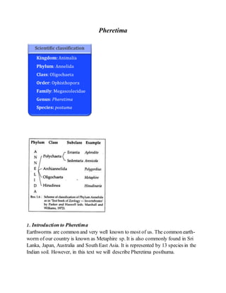
  • 1. Pheretima 1. Introduction to Pheretima Earthworms are common and very well known to most of us. The common earth- worm of our country is known as Metaphire sp. It is also commonly found in Sri Lanka, Japan, Australia and South East Asia. It is represented by 13 species in the Indian soil. However, in this text we will describe Pheretima posthuma.
  • 2. 2. Habitand Habitat of Pheretima: Pheretima is a terrestrial earthworm, living in burrows made in moist soil. It prefers to live in burrow during daytime and at night and rainy seasonthey come out of their shelter. It is thus nocturnal in habit. They are, however, absent in regions where the soil is sandy and deficient in humus. The castings of earthworm are small rounded pellets or balls that lie at the opening of the burrow. These castings are formed when it goes deep into the hard and closely packed soil. The soil upon which it feeds, passes through the bodyand are deposited as castings. While burrowing, the earthworm makes the soil loose and porous. The bodywastes of earthworm increases the fertility of the soil. So, earthworms are often said to be the natural tillers of land. Vermiform culture or vermiculture is of much practice as earthworms are capable of converting kitchen wastes and other human wastes into fertilizers. 3. Structure of Pheretima The body of earthworm is elongated, narrow and cylindrical (Fig. 1.93) measuring about20 cm in length and 3 to 5 mm in width. The anterior end is more pointed than the posterior end. Thedorsal side of the body is brown in colour and can be distinguished from the ventral side which is lighter in colour. The brown colour is due to the pigment porphyrin which is present in the body wall and it protects the bodyfrom bright and strong light. The impression of the dorsalblood vessel can be seen on the dorsal side as a dark median line extending throughout the length of the body. The softand naked bodyis made up of distinct segments or metameres separated from each other by inter-segmental ring-like grooves or annuli. The bodyis made up of a series of 100-120 similar segments. This external segmentation corresponds to internal segmentation and is referred to as metameric segmentation. The segments 14 to 16 from the anterior end are encased in a thick glandular tissue sheet called the clitellum (saddle) or cingulum (belt) (Fig. 1.93). Considering the clitellum as the index, the bodymay be divided into three regions, namely the pre- clitellar, clitellar and post-clitellar regions. Some of the anterior segments bear
  • 3. superficial furrows and may appear to be subdivided, but these are merely external subdivisions. A distinct ‘head’ is absent in Pheretima. The first bodysegment is called peristomium (Greek : peri, around; stoma, mouth) which bears the mouth aperture on the ventral surface. The peristomium is prolonged anteriorly into a small, fleshy lobe, the prostomium (Greek: pro, anterior). The prostomium is thus considered as a projecting part of the first segment and not a segment by itself . The last segment, at its posterior end bears the anus.
  • 4. Digestive System of Earthworm Digestive system of earthworm consists of alimentary canal and glands along with physiology of digestion. Alimentary Canal Alimentary canal is long and straight, extending from mouth to anus. It consists of following parts: 1. Mouth : 1st segment 2. Buccal Cavity : 2nd-3rd segment or middle of 3rd segment 3. Pharynx : 3rd-4th segment 4. Oesophagus : 5th -7th segment 5. Gizzard : 8th or 8th-9th segment 6. Stomach : 9th or 10th-14th segment 7. Intestine : 15th up to last segment except anus 8. Anus : last segment
  • 5. 1. Mouth It is crescentic aperture situated in the 1st segment below the prostomium. Mouth leads into a buccal cavity.Ingestion of food takes place through it. 2. BuccalCavity It is a short, wider, thin-walled tube extending from 2nd up to 3rd or middle of 3rd segment. It consists of two kinds of muscle. They are: protractile muscle and retractile muscle.
  • 6. Buccal cavity protrude out through mouth with the help of special muscle for holding the food particles during feeding.Buccal cavity leads into spacious organ called pharynx. 3. Pharynx It is small, swollen, wider, thick-walled pear-shaped chamber, which extends up to 4th segment.It is wider than buccalcavity.It is distinguished from buccal cavity by means of constriction.It has pharyngeal gland, located in the dorsal salivary chamber. Pharyngeal gland is composed ofmany chromaphil cells, which producesaliva containing proteolytic enzyme; protease and mucin. Mucin makes the food softand protease converts protein into amino acid. 4. Oesophagus It is narrow thin-walled tubular structure extending from 5th to 7th segment. It has no gland.It passes the food particles from pharynx to gizzard. It leads into gizzard. 5. Gizzard It is oval, thick-walled and highly muscular organ lying in the 8th or 8th- 9th segment. It is the hardest part of alimentary canal due to the presence of inner lining of cuticle. It also possess chitinuous teeth like projection. It helps in grinding or crushing food so act as grinder during feeding. 6. Stomach Gizzard leads to short, narrow, thin-walled, highly vascular tubular structure called stomach, which extends from 9th or 10th to 14th segment. It is wider than oesophagus.Ithas calciferous gland which helps in neutralization of food by calcification process.Stomachleads to intestine.The glandular cell of stomach produceproteolytic enzymes for the digestion of protein. 7. Intestine It is long, wide and thin-walled tube which extends from 15th to last segment except anus. Its inner lining is ciliated, vascular, folded and glandular. Its intestinal lining is folded to form villi. One of the villi becomes larger and well developed than other called typhlosole which runs mid-dorsally from 27th to last 25th segment.Typhlosole divides the intestine into 3 regions. They are: i.Pre-typhlosolarregion It extends from 15th segment to 26th segment so it is the first part of the intestine.It consists of villi but no typhlosole.In 26th segment, there is a pair of
  • 7. short and conical lateral outgrowth called intestinal caeca which extends upward up to 23rd segment. Intestinal caeca produceamylase which helps in starch(carbohydrate) digestion. ii.Typhlosolarregion It is the 2nd or middle part of the intestine which extends from 27th segment to last from 25thsegment. It has both villi and typhlosole.The typhlosole is highly vascular and glandular fold that increases the absorptive surface area of the intestine. iii.Post-typhlosolarregion It is the last part of the intestine lying in the last 23rd-25th segment in front of anus. It is also called rectum.It lacks villi and typhlosole.It contains small pellets of mud which are thrown out through the anus to form casting. 8. Anus It is a circular opening in the last segment called anal segment. Undigested food materials release out through anus in the form of worm casting. Digestive glands There are different types of digestive glands associated with alimentary canal of earthworm. 1. Pharyngeal gland 2. Gastric gland 3. Intestinal glands 4. Intestinal caeca Physiology of digestion Digestion is the bio-chemical process inwhich complex organic food is brokendown into simple, soluble and diffusible form in the presence of respective enzymes. Earthworms feed uponall kinds oforganic humus and debris suchas decaying leaves and seeds, protozoan, etc. present in soil. They also feed directly on leaves, grasses and other vegetation. During feeding the buccal cavity is protrude out with the help of protractile and retractile muscle. Then the food is drawn into the mouth. The ingested food enters into pharynx through buccal cavity. The dorsal chamber of pharynx consists of pharyngeal gland which is composed of chromaphil cell which produce saliva
  • 8. containing mucin and protease. Mucin lubricates the food and protease converts protein into amino acids. The foods then pass through oesophagus into gizzard, where grinding or crushing of food material takes place into fine state due to the contraction of circular muscles of gizzard. The grinded food material enters into the stomach where the neutralization of food takes place by calcification process. Also there occurs the complete digestion of protein byproteolytic enzymes. Now the food material enters into intestine. In intestine, intestinal caeca produce amylase which converts starch into glucose. In intestine several enzymes are secreted and acts on the substrate as follows:  Protease: converts protein into amino acid.  Amylase: converts starch into two molecules of glucose i.e. maltose.  Cellulase: converts cellulose into glucose  Chitinase: digest chitin of exoskeleton of insects.  Lipase: converts fats into fatty acids and glycerol. Digestion occurs mostly in the intestine and the digested food is absorbed by villi. Then pass into blood stream through capillaries. Undigested food and the soil are released out in the form of casting through anus. T.S. of Pharynx Pharynx is small, wide, thick-walled, pear-shaped chamber. Two chambers are found in pharynx. They are: dorsal chamber and ventral chamber. The dorsalchamber is also called salivary chamber which contains pharyngeal gland
  • 9. that is composed of many chromaphil cells. Dorsally pharyngeal gland is lined by villi. The chromaphil cells produce saliva containing proteolytic enzymes and mucin. Mucin makes the food soft and protease converts protein into amino acid. The ventral chamber is a conducting chamber which conducts digested orundigested food material from pharynx to oesophagus. Pharynx acts as sucking and pumping organ during feeding.
  • 10. Circulatory System of Earthworm Introduction The blood vascular system of Pheretima is of closed type. It consists of the blood vessels, hearts, loops, capillaries and the blood glands. Blood: The blood of Pheretima is red coloured due to the presence of a respiratory pigment haemoglobin in it. The haemoglobin is not contained in the corpuscles like the vertebrates but it is found dissolved in the plasma. The plasma also contains many corpuscles which are colourless and nucleated. Blood Vessels: The blood vessels are of two types collecting blood vessels and distributing blood vessels which are closed tubes with definite walls and they break up into capillaries to ramify in the different parts of the body. The arrangement of blood vessels in the anterior thirteen segments is somewhat different from that behind the thirteen segment, i.e., in the region of intestine. : Therefore, for convenience the blood vessels canbe described under the following two heads: A. Bloodvessels and their arrangementin the segments behind 13th, i.e., intestinal region. B. Bloodvessels and their arrangement in the anterior thirteen segments. A. BloodVessels and their Arrangement in the Segments behind 13th, i.e., Intestinal Region: The blood vessels ofthis regioninclude: 1. Median longitudinal blood vessels; 2. The intestinal blood plexus;
  • 11. 3. The commissural vessel; 4. The integumentary vessel; and 5. The nephridial vessels. 1. Median Longitudinal Blood Vessels: (i) DorsalVessel: It runs mid-dorsally above the alimentary canal from the posterior to the anterior end of the body. It is the thickest vessel with contractile muscular walls visible from outside as a dark line through the thin and semitransparent bodywall. The direction of flow of blood in this vessel remains from behind to forward (from posterior to anterior). It is contractile and pulsates rhythmically to force the blood from posterior to anterior side. In each segment it has a pair of valves internally which check the backward flow of blood. It is the main collecting vessel behind the 14th segment, but in front it distributes the blood. From the posterior segment up to the 14th segment it receives two pairs of dorso intestinal vessels from the intestine in each segment and a pair of commissural vessels from the sub-neural vessel. The commissural vessels form a loop behind each septum and they receive blood from the bodywall, nephridia and prostate glands. The commissural vessels also give out blood in each segment through a septo intestinal branch to the intestine.
  • 12. (ii) Ventral Vessel: It is also a long vessel and runs ventrally below the alimentary canal and above the ventral nerve cord from second segment up to the last segment of the body. It is thin- walled without muscles and valves. The direction of flow of blood in this vessel remains from anterior to the posterior side or from in front to backwards. It is a distributing vessel. It gives out a pair of ventro in tegumentary vessels, one on each side in front of the septum in all segments. The ventrointegumentary vessels run upwards along the bodywall and supply blood to the bodywall, integumentary nephridia, septal nephridia, gonads, seminal vesicles and spermathecae. The ventral vessel also gives out a ventrointestinal vessel in each segment behind the 13th segment, these take blood to the lower part of the intestine. The branches in intestine form blood plexuses consisting of two networks in the intestinal wall. (iii) Sub-neural Vessel: It is also a long but thin vessel extending from anterior 14th segment up to the last segment situated mid-ventrally below the ventral nerve cord. It is without muscular walls and internal valves. The direction of flow of blood in this vessel remains from anterior to posterior side and it is mainly a collecting vessel.
  • 13. It receives a pair of slender branches in each segment which bring blood from the ventral bodywall and the nerve cord. It gives a pair of commissural vessels in each segment which join the dorsal vessel, as already mentioned while describing the dorsalvessel. Thus, it collects blood from the ventral bodywall and supplies some blood to the intestine. 2. Intestinal Blood Plexus: The intestine of Pheretima is richly supplied with blood capillaries which form a close network. The intestinal blood plexus consists of a close network of capillaries in the wall of intestine. In fact, there are two capillary networks in the intestine: (i) The external and (ii) The internal. The capillary network which is present at the outer surface of intestine is known as external plexus which receives blood from the ventral vessel through ventrointestinal and passes it on to the internal plexus. The capillary network which is present between the circular muscle layer of intestine and its internal epithelial lining is known as internal plexus which serves to absorb the nutrients from the gut and is connected with dorsalblood vessel through the dorsointestinals. 3. Commissural Vessels: These vessels connect the dorsaland sub-neural vessels. These vessels receive blood from nephridia, bodywall and reproductive organs through capillaries and then they send it to dorsalblood vessel. 4. IntegumentaryVessels: These vessels coming from ventral vessels supply the blood to integument for aeration and the aerated blood is collected by numerous capillaries of commissural vessel in each segment. Thus, there is a close parallelism between venous and arterial capillaries throughout the bodywall.
  • 14. 5. Nephridial Vessels: These vessels originate from the ventrotegumentary vessels of ventral vessel and supply the blood to the nephridia. B. Blood Vessels and their Arrangement in the Anterior 13 Segments: The blood vascular system in the first thirteen segments is modified considerably and differs markedly from that of the intestinal region. It consists ofthe following: 1. Median longitudinal vessels; 2. Hearts and anterior loops; 3. Blood vessels of the gut. The function of collecting blood from the anterior region of the gut is taken over by a new vessel supra-oesophageal, while the blood from the peripheral structures is collected by the right and left lateral oesophageal. 1. Median Longitudinal Blood Vessels: (i) Dorsalvessel: This blood vessel becomes the distributing vessel in these segments instead of collecting vessel. Structurally, it retains its original identity as it was in the posterior segments. But is has neither dorsointestinals nor commissural vessels opening into it. It sends out all the collected blood from the posterior region of the body into hearts and the anterior region of the gut where it divides into three branches distributed over the pharyngeal bulb and the roof of the buccal chamber. However, it supplies to stomach, gizzard, oesophagus, pharynx and other related parts. (ii) Ventral vessel: This blood vessel remains distributing in these segments also but extends only up to the second segment. The ventrointestinals are absent, hence, it does not supply to the alimentary canal in this region. However, the ventrotegumentary vessels, a pair in each segment, supply blood to the integument, nephridia, septa and reproductive organs.
  • 15. (iii) Supra-oesophageal vessel: It is the shortestlongitudinal vessel extending from 9th to 13th segment situated above the stomach. It receives blood from the lateral oesophageals by two pairs of anterior loops that encircle the stomach in the 10th and 11th segments. It sends its collected blood by the latero-oesophageal hearts in segments 12th and 13th to the ventral vessel. (iv)Lateral oesophageals: In fact, the sub-neural vessel bifurcates in the 14th segment to form two lateral oesophageals. These vessels are considerably thick and situated along the ventro- lateral margins of alimentary canal in the anterior thirteen segments. These vessels are closely attached to the wall of the stomach from 10th to 13th segments and communicate with the ring vessels. But in the region of gizzard and further forwards, they remain free from the wall of the alimentary canal even though they receive branches from it in each segment. These vessels receive a pair of branches in each segment bringing blood from the bodywall and the septum. They also collect blood from the reproductive organs and nephridia, thus, functioning like the sub-neural and commissural vessels of the posterior region, i.e., these are collecting vessels. 2. Hearts and Anterior Loops: In the posterior segments behind 13th the dorsaland ventral blood vessels have no direct connections but anteriorly both these vessels are connected together by 4 pairs of pulsatile hearts which are neurogenic, i.e., the heart beat originates in the nerve cells of the hearts. The hearts are contractile and encircle the alimentary canal, they are in the segments 7th, 9th, 12th and 13th.
  • 16. The hearts of segments 12th and 13th are joined above to both the dorsal and the oesophageal vessels, these are called latero-oesophageal hearts. These hearts have thick muscular walls and a pair of valves at each junction with the dorsal vessels and supra-oesophagealvessel, and another pair of valves at the ventral end. These valves allow blood to flow downwards only. The other hearts of the segment 7th and 9th are called lateral hearts. These connect the dorsalvessels to the ventral vessel. They have four pairs of valves which allow blood to flow only downwards. Besides four pairs of hearts there are two pairs of loop-like vessels in the 10th and 11th segments which connect the supra-oesophagealwith the lateral oesophageals. These vessels are neither muscular nor pulsatile and are called anterior loops. These are devoid of valves. The blood from lateral oesophageals flows through these loops into supra- oesophageal which sends all its collected blood into ventral vessel through the hearts of 12th and 13th segments. 3. Blood Vessels of the Gut: On either side of stomach are situated ring-like vessels which connectthe supra- oesophageal and lateral-oesophageal vessels. Through these vessels blood flows upwards from the lateral- oesophageals into the supra-oesophageal. Buccal cavity, pharynx and gizzard receive their blood supply from dorsal blood vessel directly.
  • 17. Circulation of Blood: The blood collected by the dorsalvessel through the dorsointestinals, blood plexuses of intestine, and commissurals is given out partly to the anterior alimentary canal, but mainly through the hearts to the ventral vessel. In the ventral vessel the blood flows forwards to the anterior region in front of the hearts, but the main portion of blood flows backwards, this is distributed through ventrotegumentaries to the bodywall and the organs in the coelom, and through the ventrointestinal vessels to the alimentary canal. In other words all parts receive blood from the ventral vessel. From the ventral bodywall blood is collected by the sub-neural which also receives some blood through the lateral-oesophageal from the anterior region. This blood passes from the sub-neural to the dorsalvessel through the commissurals. The lateral-oesophageals also send blood through the anterior loops to the supra- oesophageal vessel which then passes it through the latero- oesophageal hearts to the ventral vessel. The courseof circulation of blood in Pheretima has been shown in Fig. 66.20.
  • 18. Functions: The blood distributes digested food to various bodyregions, and it collects waste substances like nitrogenous waste and C02 which are given up to nephridia, skin and the coeiomic fluid. Respiration in almost all aquatic and terrestrial oligochaetes takes place by diffusion of gases through the integument which in larger forms contains a capillary network in the outer epidermal layer. In terrestrial species the film of moisture necessary for diffusion of gases is supplied by mucous glands, coeiomic fluid, and nephridial excretions. The haemoglobin of plasma extracts O2 from the capillaries of the skin, but there must be a moist skin where O2 can combine with haemoglobin to be transported by the blood. Haemoglobin is an efficient pigment and it can take up O2 either from the surrounding air or from an environment comparatively deficient in oxygen. Hence, earthworms can live in well aerated water and are not drowned. They can also live for several hours without O2, in this condition they probably carry on anaerobic respiration.
  • 19. Blood Glands: In the segments 4th, 5th and 6th above the pharyngeal mass are several groups of small rounded follicles of red colour, which are called blood glands. The follicles have a syncytial wall enclosing a capsule containing a mass of loose cells. The blood glands are connected with pharyngeal nephridia and with salivary glands. These glands manufacture blood corpuscles and haemoglobin. These glands are probably also excretory.
  • 20. Excretory System of Nephridia (Earthworm) These are of three types according to their locationin the body: 1. Septal nephridia; 2. Integumentary nephridia 3. Pharyngeal nephridia. 1. SeptalNephridia: These are found situated on the inter-segmental septum between 15th and 16th segments to the posterior side of the body. Each septum bears nephridia on both the surfaces arranged in semicircles around the intestine, two rows in front of the septum and two behind it. Each septum has about 40 to 50 nephridia in front and the same number behind, so that each segment possesses80 to 100 septal nephridia except the 15th segment which has only 40 to 50 nephridia. These are not found in the segments up to 14th.
  • 21. Structure: The septal nephridia may be considered typical of all the nephridia of Pheretima. Each septal nephridium (Fig. 66.22) consists of nephrostome, neck, bodyof nephridium and the terminal duct. (i) Nephrostome: It is also known as ciliated funnel or nephridiostome. It is the proximal flattened funnel-shaped structure of the nephridium lying in the coelom. It has an elliptical mouth-like opening leading into an intracellular canal of the large central cell, the margins of the opening are surrounded by a large upper lip and a smaller lower lip. The lips are provided with several rows of small ciliated marginal cells and the central canal is also ciliated. (ii)Neck: The nephrostome leads into a short and narrow ciliated canal forming the neck. It joins the nephrostome to the bodyof nephridium.
  • 22. (iii)Body of Nephridium: The bodyof nephridium has two parts a short straight lobe and a long twisted loop. The loop is formed by two limbs— the proximal limb and the distal limb.Both these limbs are twisted spirally around each other, the number of twists varies from nine to thirteen. The neck of nephridium and the terminal duct join together and remain connected with the proximal limb of the twisted loop, while the distal limb becomes the straight lobe. Internally the nephridium is made of a connective tissue matrix having long coiled nephridial duct forming loops. There are four such canals in the straight lobe, three in the lower part and two in the upper part of the limbs of twisted loop. Two canals of the straight lobe out of the four are ciliated like the ciliated canal of the neck. (iv) TerminalDuct: It is short and narrow with a terminal excretory duct. It joins the nephridium with septal excretory canal. Relationof septal nephridia with intestine: The nephridia hang freely in the coelom and are attached only by their terminal ducts. They open by their terminal ducts into two septal excretory canals lying on the posterior surface of the septum, one on each side of the intestine, each begins ventrally but dorsally it opens in the supra-intestinal excretory duct of its own side. The supra-intestinal excretory ducts are two parallel longitudinal canals lying above the gut and below the dorsalvessel (Fig. 66.24). These excretory ducts begin from the 15th segment and run to the last segment, they communicate- with each
  • 23. other for a short space behind each septum, then either the right or the left duct opens by a ductule into the lumen of the intestine near the septum. Thus, each segment has one such opening into the intestine of either the left or the right supra-intestinal excretory duct. The waste collected by the nephridia is discharged through the excretory canals and ducts into the lumen of the intestine. Such nephridia opening into the intestine are called enteronephric nephridia. 2. Integumentary Nephridia: In each segment of the bodyfrom 7th to the last segment, numerous nephridia are found attached inside the lining of the bodywall. These are called integumentary nephridia which are about 200-250 in each segment except the segment of the clitellar region where they number 2,000-2,500 in each segment. These nephridia are small-sized, without nephrostome and without any opening into the coelom. Hence, they are called closed type of nephridia. Each integumentary nephridium is V-shaped with a short straight lobe and a twisted loop, its lumen has two ciliated canals. Each nephridium opens by a nephridiopore on the outer surface of the body wall directly. Since the integumentary nephridia discharge the excretory wastes directly outside, hence, they are called exonephric nephridia.
  • 24. 3. Pharyngeal Nephridia: These nephridia lie in three paired tufts, one on either side of the anterior region of the alimentary canal in the segments 4th, 5th and 6th. The tufts of pharyngeal nephridia also contain blood glands. Each pharyngeal nephridium is about the size of a septal nephridium but it is of the closed type having no funnel or nephrostome. It has a short straight lobe and a spirally twisted loop, its lumen has ciliated canals. Ductules arise from each nephridium and unite to form a single thick- walled ducton each side in each segment. The two ducts of nephridia of segment 6th open into the buccalcavity in segment 2nd and the paired ducts of nephridia of segments 4th and 5th open into the pharynx in segment 4th. These nephridia also discharge their wastes into the alimentary canal and are, therefore, enteronephric but suchenteronephric nephridia which open into the anterior region of the alimentary canal (buccal cavity and pharynx) are called peptonephridia becausethey may have taken the function of digestive glands. Recently it has been reported that the pharyngeal nephridia of P. posthuma produce a variety of enzymes like amylase, chimosin, prolinase, prolidase, dipeptidases, aminopeptidase, lipase, etc., which hydrolyse various foodstuffs. Thus, such nephridia work like the salivary glands. Physiology of Excretion: Like other animals, in earthworms also, the protein catabolism results in the formation of nitrogenous waste substances like certain amino acids, ammonia and urea. Uric acid is not found in the earthworms. However, the amino acids are degraded to form free ammonia and the urea is synthesised in the chloragogen cells which are released into the coelomic fluid and also in the blood for its removal. Free amino acids are not excreted but traces of creatinine occurin the urine. Moreover, the nitrogen excreted in different forms in a well fed worm is about 72% NH3, 5% urea and remaining other compounds,while in a starved worm NH3 8.6%, urea 84.5% and remaining being other compounds. But generally, the
  • 25. excretion is 42% NH3, 50% urea, 0.6% amino acids and remaining being other compounds. So, we can say that in a well fed earthworm, NH3 predominates the nitrogenous excretory wastes, hence, it is ammonotelic, while a starved one is ureotelic. An earthworm excretes the nitrogenous wastes in the form of urine which generally contains urea, water, traces of ammonia and creatinine. Nephridia excrete these substances from the bodyof earthworm. The various excretory wastes from the coelomic fluid are drawn into the nephrostomes of septal nephridia or into the excretory canals of other nephridia along with some other useful substances. These products are either discharged into the intestine (by enteronephric nephridia) or outside by the nephridiopores (by exonephric nephridia). The bodyof nephridia also absorbs somewastes. However, the useful substances are reabsorbed and the passing out waste remains concentrated for various nitrogenous compounds. The excreted waste substances are removed out from the bodywith faeces. The nephridia, in addition to excretory, are also osmoregulatory in function. The nephridia help in conserving water by reabsorption from the excreted products during summers and winters, so they pass hypertonic urine in relation to blood. During rainy season, the urine is dilute due to lesser reabsorption of water. The enteronephric nature of nephridia provides another device for conserving water.
  • 26. Reproductive System of Pheretima (Earthworm) Male Reproductive Organs of Earthworm: The male reproductive organs consist of testes, testis sacs, seminal vesicles, vasa deferentia, prostate glands and accessoryglands. Testes and Testis Sacs: The testes are two pairs, very minute, whitish and lobed structures situated one pair each in segment 10th and 11th found attached with the posterior surface of 9/10 and 10/11 inter-segmental septa. Each testis consists of 4 to 8 finger-like lobules projecting from a compactbase. Each lobule of the testis contains rounded cells in masses called spermatogonia. The testes are well developed only during the young stage of the worm and become degenerated in the adult worm. The testes of each segment are found enclosed in a thin-walled, wide bilobed and fluid-filled sacs called testis sacs. Thus, there are two testis sacs situated in the segments 10th and 11th on the ventrolateral sides of the ventral nerve cord beneath the stomach. Behind each of the four testes, in the testis sacs, is a large spermatic funnel having folded and ciliated margins. The testis sacs remain in communication with the seminal vesicles. The spermatogonia are shed into the testis sacs and pass on into the seminal vesicles where they undergo maturation and form spermatozoa. SeminalVesicles: These are two pairs of large, white, sac-like bodies, one pair each in the segments 11th and 12th into which the testis sacs open by narrow ducts. The anterior seminal vesicles are smaller than the posterior ones. The seminal vesicles of the 11th segment are found enclosed in the posterior larger testis sac, while those of the 12th segment are exposed in the coeiomic cavity. These are also placed ventro-laterally beneath the stomach and since they develop as septal outgrowths, hence, they are also known as septal pouches. Vasa Deferentia: Each spermatic funnel (posteriorly) leads into a thin, narrow, thread-like sperm duct or vas deferens. Thus, the two vasa deferentia of each side run very closely
  • 27. and laterally to the nerve cord beneath the alimentary canal on the ventral body wall, up to 18th segment where they join the prostatic duct. Prostate Glands: These are a pair of large, white, solid and irregularly- shaped glandular masses situated one on either side of the gut in the segments from 16th or 17th to 20th or 21st. Each prostate gland consists of a maximum glandular region and a small part of non-glandular region. From each prostate gland emerges a thick curved prostatic duct in 18th segment. This duct joins the two vasa deferentia of its side and these three ducts are enclosed in a common sheath to form a common prostatic spermatic duct which opens separately through a male genital aperture on the ventral side of the 18th segment. Thus, each genital aperture has three separate apertures—two of the vasa deferentia and one of the prostatic gland. The function of the secretion of prostatic duct is not definitely known.
  • 28. Accessory Glands: These are two pairs of rounded structures situated one pair each in the segments 17th and 18th on the ventral body wall at the lateral sides of the nerve cord. These glands open to the outside by a number of small ductless on the two pairs of genital papillae situated on the either side of the mid-ventral line in 17th and 19th segments externally. The secretion of these glands is believed to keep together the two worms during the process ofcopulation. As mentioned earlier, the spermatogonia from the testes are shed into the testis sacs and pass on to the seminal vesicles where spermatogenesis is completed and tailed spermatozoa are formed. Now, these spermatozoa again enter the testis sacs and through the spermatic funnels go down the vasa deferentia and are discharged through the male genital apertures along with the secretion of prostateglands. Female Reproductive Organs of Earthworm: Female reproductive organs consist of the 1. Ovaries 2 oviducts 3 sperm thecae.
  • 29. Ovaries: There is a pair of white, small, lobulated ovaries, one on either side of the ventral nerve cord in the 13th segment attached with the posterior face of the inter- segmental septum of 12/13 segments. Each ovary has several finger-like processeswith developing ova in a row providing it beaded appearance. The ova remain in various stages of their development in each ovarian lobe being mature in the distal part and immature in the proximal part. Oviducts: Below each ovary in 13th segment, there is a small ovarian funnel with folded and ciliated margins. Each ovarian funnel leads into a short, conical and ciliated oviduct. The oviducts of both the sides converge to meet below the nerve cord and open by a single median female genital aperture ventrally in the 14th segment. The mature ova are released from the ovaries and received by the ovarian funnel, pass through the oviducts and which go out through the female genital aperture.
  • 30. Spermathecae: These are four pairs, one pair each in the segments 6th, 7th, 8th, and 9th situated ventro-laterally. Each spermatheca is flask-shaped, the main bodyis the ampulla which is continued into narrow duct— the neck with a small diverticulum on its inner side. These are also called seminal receptacles as they store the spermatozoa from the another worm during copulation. In Pheretima, unlike the other earthworms, the diverticula store the spermatozoa and not the ampulla. The spermatheca open externally by their small ducts as spermatheca pores situated inter-segmentally between segments 5/6, 6/7, 7/8 and 8/9. Copulation, CocoonFormationand Fertilization of Earthworm:
  • 31. Copulation has been observed in different species of earthworms. It usually occurs in the rainy seasonduring the months of July to Octoberat dawn, i.e., in the morning hours before sunrise. During copulation two earthworms of adjacent burrows half emerge from their burrows and come closer to lie in contactto each other by their ventral surfaces with their anterior ends pointing in oppositedirections. In this position the male genital apertures of each worm lie oppositethe spermathecal pores of the other. The areas of the male genital apertures are raised into papillae and inserted into the spermathecal pores, so that there is a mutual exchange of spermatozoa and prostatic fluid between the two copulating worms. Copulation lasts for about an hour or so, then the worms separate and recede into their burrows. Cocoonorootheca formation starts after copulation when ovaries mature. The epidermis of clitellar segments, i.e., 14th, 15th and 16th segments contain three kinds of glands, they are unicellular mucous glands which producemucus for copulation, cocoon-secreting glands secrete the wall of the cocoonand albumen glands producealbumen in which eggs are deposited in the cocoon. A membranous girdle is secreted by cocoon-secreting glands of the clitellum, this girdle soonhardens, then albumen is deposited between the girdle and the body wall. The worm starts withdrawing itself backwards from the girdle. As the girdle passes over the female genital aperture it receives eggs, and when it passes over the spermathecae sperms are extruded into it through spermathecal pores.
  • 32. Lastly, the girdle is thrown off from the anterior end of the worm and soonthe elasticity of its wall closes up its two ends to form a cocoonorootheca. Several cocoonsare formed after each copulation because the spermatozoa contained in the spermatheca do not pass out all at one time. The cocoons are oval, light yellow in colour and are about 2 to 2.4 mm long and 1.5 to 2 mm broad. Fertilisation takes place inside the cocoonand generally there is only one embryo in a cocoon. Cocoons are laid from August to Octoberin damp situations.