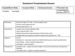
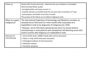
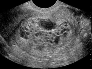
Common
Gynaecological
  Carcinoma
Top killers of the female gender
  All Females, All Ages                    Percent*
  1) Heart disease                        25.1
  2) Cancer                               22.1
  3) Stroke                               6.7
  4) Chronic lower respiratory diseases   5.5
  5) Alzheimer's disease                  4.3
  6) Unintentional injuries               3.6
  7) Diabetes                             2.9
  8) Influenza and pneumonia              2.3
  9) Kidney disease                       2.0
  10) Septicemia                          1.6
: 10 Principle Cause of Deaths in
 Ministry of Health, Malaysia (MOH)
           Hospitals, 2006
1. Septicaemia                                              16.87
2. Heart Diseases & Diseases of Pulmonary Circulation       15.70
3. Malignant Neoplasms                                      10.59
4. Cerebrovascular Diseases                                  8.49
5. Pneumonia                                                 5.81
6. Accidents                                                 5.59
7. Diseases of the Digestive System                          4.47
8. Certain Conditions Originating in The Perinatal Period    4.20
9. Nephritis, Nephrotic Syndrome & Nephrosis                 3.83
10. Ill-defined conditions                                   3.03
All causes                                                  100.0
Prevalence of various types of cancer amongst Malaysian ladies
of all ages
BREAST                     3525            29.9
COLORECTAL                 1247            10.6
CERVIX UTERI               1074             9.1
OVARY                       685             5.8
THYROID GLAND                670             5.7
LUNG                        603              5.1
CORPUS UTERI                372              3.2
STOMACH                     324              2.7
BRAIN, OTHER
NS                           303             2.6
LYMPHOMA                     279            2.4

        Taken from the Malaysian Cancer Statistics 2006
PATHOPHYSIOLOGY

          CERVICAL                        ENDOMETRIAL                       OVARIAN
The squamo-columnar               -Most commonly                   80% from Ovarian
junction and the                  Adenocarcinoma                   Epithelium
Transformation zone               -90%: Endometriod adenoCA        20% others: Germ cell, sex-
                                  -10%:Serious papillary AdenoCA
                                                                   cord stromal, mixed
Common site for HPV               -Rarely: Clear cell              mullerian,
infection
HPV infection persists in                                          Mechanism poorly understood
certain individuals, eventually                                    Two main theories
triggering oncogenic processes
within the TZ
Cell metaplasia occurs:                                            1. Incessant
Immortalization of the basal                                       ovulationrepeated trauma
cells leading to rapid turnover                                    to ovarian epithelium
and subsequent immature
cells
These immature cells picked                                        2. Excess gonadotrophin
up on PAP smear as Cervical                                        secretionhigher level of
Intraepithelial Neoplasia CIN                                      estrogenEpithelial
                                                                   proliferation
AETIOLOGY & RISK FACTORS

          CERVICAL                     ENDOMETRIAL                       OVARIAN
Infection by HPV                 Conditions that lead to       Endometriosis
16,18,31,33                      high levels of estrogen
                                 -Tamoxifen
                                 -Unopposed estrogen therapy
                                 as HRT


Immunosupressed state            Family history (debatable)    Family history
leading to increased risk of     -endometrial cancer           -increased risk from 1.4% in
HPV infection:                   -colorectal/ovarian cancer    general populace to 5%
-RVD                             -HNPCC                        50% if 2 first degree relatives
-Immunosupressive drugs
                                 Nulliparity                   Nulliparity
                                 Late menopause >52 years
                                 Obesity                       Obesity
Smoking                          Diabetes
-theory: immunosupressive
effects of nicotine within the
cx
Mnemonic of risk factors for
         endometrial cancer
• O=Obesity
  L=Late menopause
  D=Diabetes mellitus

 A=cAncer: ovarian, breast, colon
 U=Unopposed estrogen: PCOS, anovulation,
 HRT
 N=Nulliparity
 T=Tamoxifen, chronic use.
RMI score for ovarian CA
Feature                      RMI 1 Score                  RMI 2 Score
Ultrasound features:         0= none                      0= none
• multilocular cyst             1= one abnormality           1= one abnormality
• solid areas                   3= two or more               4= two or more
• bilateral lesions             abnormalities                abnormalities
• scites
• intra-abdominal
    metastases
Premenopausal                1                            1
Postmenopausal               3                            4
CA125                        U/ml                         U/ml
        RMI score = ultrasound score x menopausal score x CA125 level in U/ml.


The RMI scoring system is the method of choice for predicting whether or not an
                    ovarian mass is likely to be malignant.
        Women with an RMI score >200 should be referred to a centre
        with experience in ovarian cancer surgery. Evidence grade 2+
CLINICAL FEATURES

          CERVICAL                    ENDOMETRIAL                              OVARIAN
Abnormal PV bleeding           Abnormal PV bleeding                 Persistent pelvic-
                                                                    abdominal pain
1. Post-coital bleeding        1. Post-Menopausal Bleed Increased abdominal size and
*The cervical cancer is a      ddx: Cervical, endometrial polyps,   bloating
   friable vascular mass on         cervical erosion, vaginal
   the cervix: Mechanical           atrophy
   irritation during coitus:   10% of PMB turns out to be
                                    Endometrial CA
   Post-coital bleed
                               2. Irregular Vaginal
                               bleeding
3. Advanced stage:             3. Advanced stage:                   3. Advanced stage:
-constitutional sx             -constitutional sx                   -constitutional sx
-lungs mets: Recurrent         -lungs mets: Recurrent               -lungs mets: Recurrent
bilateral pleural effusion     bilateral pleural effusion           bilateral pleural effusion
Incontinence                                                        Change in bowel habits
Anemia Sx                                                           Urinary sx
Renal failure                                                       Back ache
INVESTIGATIONS AND SUSPICIONS

           CERVICAL                                ENDOMETRIAL                              OVARIAN
Aim of diagnostics: To provide histopathological evidence of malignant cancer tissue.
To provide sufficient information to properly stage the tumour
To confirm the malignancy                  To confirm the malignancy              To confirm the malignancy
1. Colposcopy : outpatient                 1. Sampling by Pipelle:                and for staging
    examination of the magnified                Outapatient procedure,            1.TAHBSO
    cervix using a light source                 results operator dependant,
    Additional Lugol’s Iodine/ 5% acetic                                          2.CT-scan/MRI
                                                difficult to do if nulliparous,   3.PET-scan
    acid are used to highlight the
    presence of abnormal cells                  retroverted uterus, presence
2. Cervical Biopsy: To confirm                  of fibroids
                                                                                  * In some patients (esp those
    the malignancy and type                2.   Hysteroscopic DD+C: done
                                                if unable to obtain good
                                                                                  unfit for a major surgery such
    of tumour                                                                     as TAHBSO, laparascopic
                                                sample by Pipelle /
                                                Contraindicated for Pipelle       examination and biopsy is
                                                                                  done instead
To assess stage                            To assess stage
1.CT-scan abdomen-pelvis                   1.TAHBSO
2.EUA : Vaginal and rectal                 2.CT-scan/MRI
                                           3.PET-scan
Tumuor markers
1.       Not used for diagnosis for cancer. Diagnosis of cancer is strictly based histopathological
         evidence
2.       Tumour markers can be useful for:
          - Monitoring the response of the malignancy to anti-cancer therapies
         - Monitoring patients on a regular basis to assess for reoccurance


               Tumour marker                    Cancer
     1         Ca-125                           Endometrial, ovarian
     2         CEA                              Pancreas, colon
     3         a-FP                             Liver (HCC)
     4         Ca 19-9                          Pancreas
CERVIX                        ENDOMETRIAL                           OVARIAN

I: Cervical cancer confined to        IA: Endometrium only            I: Ovaries only
cervix                                                                IA: One ovary
Ia1: < 3mm                                                            IB: Two ovaries
                                      IB: <1/2 of myometrium          IC: Ruptured ovary/+ve peritoneal
Ia2: > 3mm
                                                                      wash/ascites
Ib1: < 4cm                            IC: >1/2 of myometrium
1b2:> 4cm
II: Cervical cancer extending         IIA: Endocervical glandular     II: Ovaries + pelvic
beyond the cervix, but not            involvement                     involvement
invading pelvic wall                  IIB: Cervical stroma invasion   IIA: Uterus + Tubes
IIA: No obvious parametric invasion                                   IIB: Pelvic/parametrial tissue
IIB: Obvious parametric invasion +                                    IIC: Rupture/+ve peritoneal
upper 2/3 of vagina                                                   wash/ascites

III: Cervical ca extending to         IIIA: Serosa/Adnexa/+ve         III: Ovaries + peritoneum
pelvic wall                           peritoneal wash                 IIIA: Microscopic seedlings
IIIa: Invasion of lower 1/3 vagina                                    IIIB: < 2cm seedling
                                      IIIB: Invasion of vagina        IIIC: > 2cm seedling
IIIb: Extention to pelvic wall with
hydronephrosis
                                      IIIC: Invasion of pelvic/
                                      Paraaortic LN
IVa: Growth to adjacent               IVA: Invasion to                IV: Distant metastasis
organs                                bladder/rectum                  Pleural effusion
IVb: Distant metastasis               IVB: Distant metastasis
CERVICAL CANCER
FIGO


STAGING
ENDOMETRIAL CANCER FIGO STAGING
Cervical Cancer
STAGE              TREATMENT PROTOCOL
Stage 1a           Surgery: TAH, PLND
                    Modified radical trachelectomy (fertility sparing)
                   : remove 80% of cervix, parametrial tissues, pelvic LN ~ 40% miscarriage
                   Fertility sparing therapy seldom done
                   Radiation therapy: Brachytherapy(internal radiotherapy)
Stage 1b           Wertheim’s hysterectomy
                   TAHBSO+Parametrial tissue excision+PLND+upper 1/3 vagina
                   Radiation therapy has similar success rates, and considered
                   who are unfit for surgery
Stage 2-4          Chemotherapy : Carboplatin + Paclitaxel
                   Radiotherapy: External beam: Teletherapy
                                 Internal radiotherapy: Brachytherapy

                   *Surgery seldom done at this stage:
                   -High rates of surgical complications esp: Bleeding
                   -Unlikely chance of achieving complete clearance of tumour
                   -Requiring post op radiotherapy, and this combined treatment can lead to high
                   complication rates


            Remember: No tumour markers for Cervical Cancer
Endometrial Carcinoma
                                         Surgery
                      *Generally surgery indicated in all
                      patients diagnosed with Endometrial CA
                      C/I: Unfit for surgery, metastasised
                      cancer (Stage IV)
Stage 1A, 1B
TAHBSO (PLND also commonly done             Stage 2C, 3, 4/Papillary serous/Clear
                                            cell
as there is a high chance of spread to
LN at this stage)
                                            TAHBSO + PLND
                                            Adjuvant Chemotherapy
Stage 1C, 2A, 2B
TAHBSO + PLND (50% risk of LN
dissemination at these stages)
                                                           Chemotherapy

                                            -Commence adjuvant chemotherapy
                                            -1st Line: 6 cycles of Carboplatin + Paclitaxel
                                            -2nd Line: 9 cycles (A+B) of Gemcitabine


                            Follow-up: Ca-125, CT-Scan
Ovarian Cancer

                                           Surgery
                          *Studies show that the most important
                          prognostic factor is no residual disease
                          following laparatomy

Non fertility sparing
                                                  Fertility sparing (rarely done)
-Vertical insicion done to gain visual
                                                  -Vertical insicion done to gain visual
access to ALL areas of the abdomen
                                                  access to ALL areas of the abdomen
-Ascites and peritoneal washing samples
                                                  -Unilateral SO, Omentectomy, Peritoneal
are obtained
                                                  biopsies
-TAHBSO + Omentectomy KIV further
                                                  -PLND
debulking if required
                                                  + Endometrial sampling to exclude
- PLND
                                                  concerrent tumour

                           MDT meeting between Oncogynae
                             with pathologist, radiologist
                                         Chemotherapy
                                                  Stage IB, IC , II, III, IV
 Stage 1A                                         -Commence adjuvant chemotherapy
 - Chemotherapy withheld in some cases            -1st Line: 6 cycles of Carboplatin + Paclitaxel
                                                  -2nd Line: 9 cycles (A+B) of Gemcitabine
                                Follow-up: Ca-125, CT-Scan
Drug          Mech of action                          AE
Cisplatin     Platinum based drug                     Nephrotoxic (check RP before giving)
              Acts by binding and cross-              GIT: N+V
              linking DNA leading to cell             Ototoxicity
              apoptosis                               Electrolyte imbalances

Carboplatin                                           Neurotoxicity: peripheral neuropathy
                                                      Myelosupression  Aneamia,neutropenia,TCP
                                                      Less GIT effects compared to Cisplatin
                                                      No nephrotoxic effect


Gemcitabine   Neucloside analogue.                    Flu-like symptoms
              Kills cells in the S-phase of mitosis   GIT: Mild N+V
              : Cell phase specific                   Hair oss
                                                      Myelosupression

5-FU          Pyrimidine analogue                     Myelosupression
              Inhibits Thymidine synthase             Mucositis, dermatitis
              : Cell phase specific (S-phase)         Diarrhoea


Paclitaxel    Stabilizes the microtubules             Myelosupression
              preventing the Interphase thus          Neurotoxicity: Peripheral neuropathy
              interrupting mitosis                    Elevated LFT
              : Cell phase specific                   N+V, hair loss, arthritis
Gestational Throphoblastic Disease

Hydatidiform Mole                Invasive Mole              Choriocarcinoma                 Placental site
                                                                                            throphoblastic
Complete         Incomplete
                                                                                            tumour (PSTT)


  Risk factors         Extremes of age (<15, >40) – risk of complete moles
                       Previous molar pregnancy X 10
                       Ethnicity (East Asians: x 2)
  Presentation         Irregular first-trimester bleeding >90%
                       Uterus large for dates~ 25%
                       Abdominal pain (from hyperstimulation of theca lutien cysts)
                       Exxaggerated early pergnancy symptoms (hyperemesis gravidarum) ~ 10%
  Diagnosis            Clinical
                       Biochemical – excessively raised serum bHCG levels (may be normal in partial moles)
                       Scan findings – classic snowstorm appearance
                       Histological – post evacuation
  Management           Complete mole – surgical evacuation (higher risk of uterine perforation and significant
                       bleeding)
                       Incomplete mole – Surgical or medical evacuation
Follow-up         Review HPE of evacauted mole – determine for sure complete or incomplete
                  Recommended follow-up plan
                  -Fortnightly bHCG until levels normal < 4
                  -4 weekly intervals urine bHCG levels for one year, then 3-monthly in 2nd year
                  -Contraception until bHCG normal for 6 months
                  The purpose of this follow up is to detect malignancy early
When to suspect   The International Federation of Gynecology and Obstetrics considers an
malignancy?       elevated serum hCG level 6 or more months after evacuation of a
                  hydatidiform mole to be diagnostic of malignancy (ie, GTN).
                  *However, serum hCG levels spontaneously return to normal without
                  chemotherapy in most patients with elevated but still declining serum hCG
                  levels 6 months after diagnosis of a hydatidiform mole.
Indication for    1. Serum bHCG levels >20000 4 weeks after uterine evacuation
chemotherapy      2. Static or rising bHCG levels post evacuation
                  3. Histological evidence of choriocarcinoma
                  4. Metastatic disease
                  5. Persistent symptoms
Choriocarcinoma
Incidence        Rare : Amongst Orientals 1:11000 (1:30000 amongst Caucasians)
                 20% of complete hydatidiform moles become ChorioCA
Presentation     Similar to hydatidiform mole
                 -First trimester vaginal bleeding, abdominal pain
                 Symptoms pointing to possibility of chorioCA
                 -Dyspnoea and hemoptysis (lung mets), hematuria, neurological sx
                 -Jaundiced, abdominal tenderness, rebound
Investigations   Biochemical – Persistantly raised / pleateau or increasing trend of bHCG even after surgical
                 evacuation of GTD
                 Scan findings
                 CXR (lung is most frequent site of mets) CT abdomen and chest
Treatment        CHEMOTHERAPY
                 -Regime based on prognostic scoring assestment
                 Low Risk : MTX + Folinic Acid / Actinomycin D
                 Med Risk: MTX + Etoposide
                 High Risk: Intensive weekly EMA (Etoposide,MTX,Dactinomycin) alternating with CE
                 (Cyclophosphamide and Vincristin) – the EMA-CE regiment
Prognosis        Overall survival > 90% - it is one of the most curable forms of malignant cancer
                 Poorer prognosis if
                 -Age >40
                 -Time interval between antecedent pregnancy and start of chemotherapy > 4months
                 -After 12 months of normal hCG levels, less than 1% of patients with GTN have recurrences.
Next             Fertility not impaired by chemo
pregnancy        No increased risk of fetal anomalies

Common gynaecological carcinom final2

  • 1.
  • 2.
    Top killers ofthe female gender All Females, All Ages Percent* 1) Heart disease 25.1 2) Cancer 22.1 3) Stroke 6.7 4) Chronic lower respiratory diseases 5.5 5) Alzheimer's disease 4.3 6) Unintentional injuries 3.6 7) Diabetes 2.9 8) Influenza and pneumonia 2.3 9) Kidney disease 2.0 10) Septicemia 1.6
  • 3.
    : 10 PrincipleCause of Deaths in Ministry of Health, Malaysia (MOH) Hospitals, 2006 1. Septicaemia 16.87 2. Heart Diseases & Diseases of Pulmonary Circulation 15.70 3. Malignant Neoplasms 10.59 4. Cerebrovascular Diseases 8.49 5. Pneumonia 5.81 6. Accidents 5.59 7. Diseases of the Digestive System 4.47 8. Certain Conditions Originating in The Perinatal Period 4.20 9. Nephritis, Nephrotic Syndrome & Nephrosis 3.83 10. Ill-defined conditions 3.03 All causes 100.0
  • 4.
    Prevalence of varioustypes of cancer amongst Malaysian ladies of all ages BREAST 3525 29.9 COLORECTAL 1247 10.6 CERVIX UTERI 1074 9.1 OVARY 685 5.8 THYROID GLAND 670 5.7 LUNG 603 5.1 CORPUS UTERI 372 3.2 STOMACH 324 2.7 BRAIN, OTHER NS 303 2.6 LYMPHOMA 279 2.4 Taken from the Malaysian Cancer Statistics 2006
  • 5.
    PATHOPHYSIOLOGY CERVICAL ENDOMETRIAL OVARIAN The squamo-columnar -Most commonly 80% from Ovarian junction and the Adenocarcinoma Epithelium Transformation zone -90%: Endometriod adenoCA 20% others: Germ cell, sex- -10%:Serious papillary AdenoCA cord stromal, mixed Common site for HPV -Rarely: Clear cell mullerian, infection HPV infection persists in Mechanism poorly understood certain individuals, eventually Two main theories triggering oncogenic processes within the TZ Cell metaplasia occurs: 1. Incessant Immortalization of the basal ovulationrepeated trauma cells leading to rapid turnover to ovarian epithelium and subsequent immature cells These immature cells picked 2. Excess gonadotrophin up on PAP smear as Cervical secretionhigher level of Intraepithelial Neoplasia CIN estrogenEpithelial proliferation
  • 7.
    AETIOLOGY & RISKFACTORS CERVICAL ENDOMETRIAL OVARIAN Infection by HPV Conditions that lead to Endometriosis 16,18,31,33 high levels of estrogen -Tamoxifen -Unopposed estrogen therapy as HRT Immunosupressed state Family history (debatable) Family history leading to increased risk of -endometrial cancer -increased risk from 1.4% in HPV infection: -colorectal/ovarian cancer general populace to 5% -RVD -HNPCC 50% if 2 first degree relatives -Immunosupressive drugs Nulliparity Nulliparity Late menopause >52 years Obesity Obesity Smoking Diabetes -theory: immunosupressive effects of nicotine within the cx
  • 8.
    Mnemonic of riskfactors for endometrial cancer • O=Obesity L=Late menopause D=Diabetes mellitus A=cAncer: ovarian, breast, colon U=Unopposed estrogen: PCOS, anovulation, HRT N=Nulliparity T=Tamoxifen, chronic use.
  • 9.
    RMI score forovarian CA Feature RMI 1 Score RMI 2 Score Ultrasound features: 0= none 0= none • multilocular cyst 1= one abnormality 1= one abnormality • solid areas 3= two or more 4= two or more • bilateral lesions abnormalities abnormalities • scites • intra-abdominal metastases Premenopausal 1 1 Postmenopausal 3 4 CA125 U/ml U/ml RMI score = ultrasound score x menopausal score x CA125 level in U/ml. The RMI scoring system is the method of choice for predicting whether or not an ovarian mass is likely to be malignant. Women with an RMI score >200 should be referred to a centre with experience in ovarian cancer surgery. Evidence grade 2+
  • 10.
    CLINICAL FEATURES CERVICAL ENDOMETRIAL OVARIAN Abnormal PV bleeding Abnormal PV bleeding Persistent pelvic- abdominal pain 1. Post-coital bleeding 1. Post-Menopausal Bleed Increased abdominal size and *The cervical cancer is a ddx: Cervical, endometrial polyps, bloating friable vascular mass on cervical erosion, vaginal the cervix: Mechanical atrophy irritation during coitus: 10% of PMB turns out to be Endometrial CA Post-coital bleed 2. Irregular Vaginal bleeding 3. Advanced stage: 3. Advanced stage: 3. Advanced stage: -constitutional sx -constitutional sx -constitutional sx -lungs mets: Recurrent -lungs mets: Recurrent -lungs mets: Recurrent bilateral pleural effusion bilateral pleural effusion bilateral pleural effusion Incontinence Change in bowel habits Anemia Sx Urinary sx Renal failure Back ache
  • 11.
    INVESTIGATIONS AND SUSPICIONS CERVICAL ENDOMETRIAL OVARIAN Aim of diagnostics: To provide histopathological evidence of malignant cancer tissue. To provide sufficient information to properly stage the tumour To confirm the malignancy To confirm the malignancy To confirm the malignancy 1. Colposcopy : outpatient 1. Sampling by Pipelle: and for staging examination of the magnified Outapatient procedure, 1.TAHBSO cervix using a light source results operator dependant, Additional Lugol’s Iodine/ 5% acetic 2.CT-scan/MRI difficult to do if nulliparous, 3.PET-scan acid are used to highlight the presence of abnormal cells retroverted uterus, presence 2. Cervical Biopsy: To confirm of fibroids * In some patients (esp those the malignancy and type 2. Hysteroscopic DD+C: done if unable to obtain good unfit for a major surgery such of tumour as TAHBSO, laparascopic sample by Pipelle / Contraindicated for Pipelle examination and biopsy is done instead To assess stage To assess stage 1.CT-scan abdomen-pelvis 1.TAHBSO 2.EUA : Vaginal and rectal 2.CT-scan/MRI 3.PET-scan
  • 12.
    Tumuor markers 1. Not used for diagnosis for cancer. Diagnosis of cancer is strictly based histopathological evidence 2. Tumour markers can be useful for: - Monitoring the response of the malignancy to anti-cancer therapies - Monitoring patients on a regular basis to assess for reoccurance Tumour marker Cancer 1 Ca-125 Endometrial, ovarian 2 CEA Pancreas, colon 3 a-FP Liver (HCC) 4 Ca 19-9 Pancreas
  • 13.
    CERVIX ENDOMETRIAL OVARIAN I: Cervical cancer confined to IA: Endometrium only I: Ovaries only cervix IA: One ovary Ia1: < 3mm IB: Two ovaries IB: <1/2 of myometrium IC: Ruptured ovary/+ve peritoneal Ia2: > 3mm wash/ascites Ib1: < 4cm IC: >1/2 of myometrium 1b2:> 4cm II: Cervical cancer extending IIA: Endocervical glandular II: Ovaries + pelvic beyond the cervix, but not involvement involvement invading pelvic wall IIB: Cervical stroma invasion IIA: Uterus + Tubes IIA: No obvious parametric invasion IIB: Pelvic/parametrial tissue IIB: Obvious parametric invasion + IIC: Rupture/+ve peritoneal upper 2/3 of vagina wash/ascites III: Cervical ca extending to IIIA: Serosa/Adnexa/+ve III: Ovaries + peritoneum pelvic wall peritoneal wash IIIA: Microscopic seedlings IIIa: Invasion of lower 1/3 vagina IIIB: < 2cm seedling IIIB: Invasion of vagina IIIC: > 2cm seedling IIIb: Extention to pelvic wall with hydronephrosis IIIC: Invasion of pelvic/ Paraaortic LN IVa: Growth to adjacent IVA: Invasion to IV: Distant metastasis organs bladder/rectum Pleural effusion IVb: Distant metastasis IVB: Distant metastasis
  • 14.
  • 15.
  • 16.
    Cervical Cancer STAGE TREATMENT PROTOCOL Stage 1a Surgery: TAH, PLND Modified radical trachelectomy (fertility sparing) : remove 80% of cervix, parametrial tissues, pelvic LN ~ 40% miscarriage Fertility sparing therapy seldom done Radiation therapy: Brachytherapy(internal radiotherapy) Stage 1b Wertheim’s hysterectomy TAHBSO+Parametrial tissue excision+PLND+upper 1/3 vagina Radiation therapy has similar success rates, and considered who are unfit for surgery Stage 2-4 Chemotherapy : Carboplatin + Paclitaxel Radiotherapy: External beam: Teletherapy Internal radiotherapy: Brachytherapy *Surgery seldom done at this stage: -High rates of surgical complications esp: Bleeding -Unlikely chance of achieving complete clearance of tumour -Requiring post op radiotherapy, and this combined treatment can lead to high complication rates Remember: No tumour markers for Cervical Cancer
  • 17.
    Endometrial Carcinoma Surgery *Generally surgery indicated in all patients diagnosed with Endometrial CA C/I: Unfit for surgery, metastasised cancer (Stage IV) Stage 1A, 1B TAHBSO (PLND also commonly done Stage 2C, 3, 4/Papillary serous/Clear cell as there is a high chance of spread to LN at this stage) TAHBSO + PLND Adjuvant Chemotherapy Stage 1C, 2A, 2B TAHBSO + PLND (50% risk of LN dissemination at these stages) Chemotherapy -Commence adjuvant chemotherapy -1st Line: 6 cycles of Carboplatin + Paclitaxel -2nd Line: 9 cycles (A+B) of Gemcitabine Follow-up: Ca-125, CT-Scan
  • 18.
    Ovarian Cancer Surgery *Studies show that the most important prognostic factor is no residual disease following laparatomy Non fertility sparing Fertility sparing (rarely done) -Vertical insicion done to gain visual -Vertical insicion done to gain visual access to ALL areas of the abdomen access to ALL areas of the abdomen -Ascites and peritoneal washing samples -Unilateral SO, Omentectomy, Peritoneal are obtained biopsies -TAHBSO + Omentectomy KIV further -PLND debulking if required + Endometrial sampling to exclude - PLND concerrent tumour MDT meeting between Oncogynae with pathologist, radiologist Chemotherapy Stage IB, IC , II, III, IV Stage 1A -Commence adjuvant chemotherapy - Chemotherapy withheld in some cases -1st Line: 6 cycles of Carboplatin + Paclitaxel -2nd Line: 9 cycles (A+B) of Gemcitabine Follow-up: Ca-125, CT-Scan
  • 19.
    Drug Mech of action AE Cisplatin Platinum based drug Nephrotoxic (check RP before giving) Acts by binding and cross- GIT: N+V linking DNA leading to cell Ototoxicity apoptosis Electrolyte imbalances Carboplatin Neurotoxicity: peripheral neuropathy Myelosupression  Aneamia,neutropenia,TCP Less GIT effects compared to Cisplatin No nephrotoxic effect Gemcitabine Neucloside analogue. Flu-like symptoms Kills cells in the S-phase of mitosis GIT: Mild N+V : Cell phase specific Hair oss Myelosupression 5-FU Pyrimidine analogue Myelosupression Inhibits Thymidine synthase Mucositis, dermatitis : Cell phase specific (S-phase) Diarrhoea Paclitaxel Stabilizes the microtubules Myelosupression preventing the Interphase thus Neurotoxicity: Peripheral neuropathy interrupting mitosis Elevated LFT : Cell phase specific N+V, hair loss, arthritis
  • 20.
    Gestational Throphoblastic Disease HydatidiformMole Invasive Mole Choriocarcinoma Placental site throphoblastic Complete Incomplete tumour (PSTT) Risk factors Extremes of age (<15, >40) – risk of complete moles Previous molar pregnancy X 10 Ethnicity (East Asians: x 2) Presentation Irregular first-trimester bleeding >90% Uterus large for dates~ 25% Abdominal pain (from hyperstimulation of theca lutien cysts) Exxaggerated early pergnancy symptoms (hyperemesis gravidarum) ~ 10% Diagnosis Clinical Biochemical – excessively raised serum bHCG levels (may be normal in partial moles) Scan findings – classic snowstorm appearance Histological – post evacuation Management Complete mole – surgical evacuation (higher risk of uterine perforation and significant bleeding) Incomplete mole – Surgical or medical evacuation
  • 21.
    Follow-up Review HPE of evacauted mole – determine for sure complete or incomplete Recommended follow-up plan -Fortnightly bHCG until levels normal < 4 -4 weekly intervals urine bHCG levels for one year, then 3-monthly in 2nd year -Contraception until bHCG normal for 6 months The purpose of this follow up is to detect malignancy early When to suspect The International Federation of Gynecology and Obstetrics considers an malignancy? elevated serum hCG level 6 or more months after evacuation of a hydatidiform mole to be diagnostic of malignancy (ie, GTN). *However, serum hCG levels spontaneously return to normal without chemotherapy in most patients with elevated but still declining serum hCG levels 6 months after diagnosis of a hydatidiform mole. Indication for 1. Serum bHCG levels >20000 4 weeks after uterine evacuation chemotherapy 2. Static or rising bHCG levels post evacuation 3. Histological evidence of choriocarcinoma 4. Metastatic disease 5. Persistent symptoms
  • 23.
    Choriocarcinoma Incidence Rare : Amongst Orientals 1:11000 (1:30000 amongst Caucasians) 20% of complete hydatidiform moles become ChorioCA Presentation Similar to hydatidiform mole -First trimester vaginal bleeding, abdominal pain Symptoms pointing to possibility of chorioCA -Dyspnoea and hemoptysis (lung mets), hematuria, neurological sx -Jaundiced, abdominal tenderness, rebound Investigations Biochemical – Persistantly raised / pleateau or increasing trend of bHCG even after surgical evacuation of GTD Scan findings CXR (lung is most frequent site of mets) CT abdomen and chest Treatment CHEMOTHERAPY -Regime based on prognostic scoring assestment Low Risk : MTX + Folinic Acid / Actinomycin D Med Risk: MTX + Etoposide High Risk: Intensive weekly EMA (Etoposide,MTX,Dactinomycin) alternating with CE (Cyclophosphamide and Vincristin) – the EMA-CE regiment Prognosis Overall survival > 90% - it is one of the most curable forms of malignant cancer Poorer prognosis if -Age >40 -Time interval between antecedent pregnancy and start of chemotherapy > 4months -After 12 months of normal hCG levels, less than 1% of patients with GTN have recurrences. Next Fertility not impaired by chemo pregnancy No increased risk of fetal anomalies