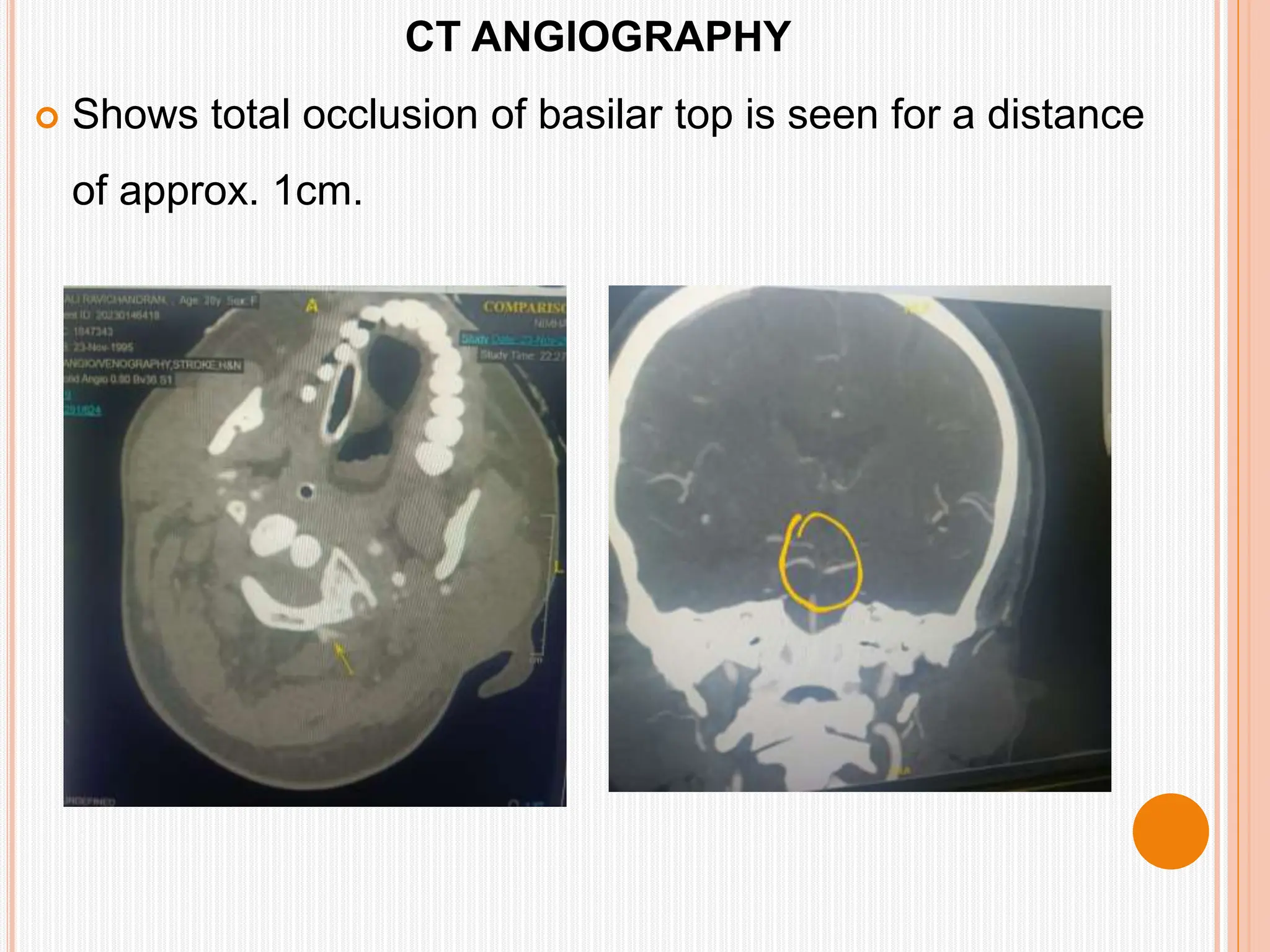
A CASE PRESENTATION ON
POSTERIOR CIRCULATION STROKE
STROKE ICU TEAM
CLINICAL NURSING SERVICES
INTRODUCTION
28 years old female brought to casualty with complaints
of
 Neck pain &Headache (2 days) 17.11.23
 Vomiting – 1episode(18.11.23) followed by
unconsciousness
 Weakness of left side limbs from 19.11.23
DEMOGRAPHIC DATA
 Full Name : Ms. Anjali Ravichandran
 Age : 28 years
 Gender : Female
 Education: Post Graduate
 Profession: Student Advisor / Counsellor
 Ward : Stroke Intensive Care Unit
 Date of admission in ICU : 23/11/2023
 Date of discharge: 17.04.2024
HISTORY OF PRESENT ILLNESS:
 The patient was apparently normal till 3/11/23, when
she developed fever which was moderate grade,
associated with chills and myalgia for which was
admitted in local hospital in Chennai for 3 days and was
detected to be dengue positive and was treated for the
same in IP care for 3 days.
 Patient was asymptomatic till 17/11/23 ,when she
developed neck pain, mild to moderate in severity
reduced on taking analgesics.
• On 18/11/23 patient developed sudden onset of severe
headache following which she had one episode of vomiting
followed by unconsciousness and was taken to a private
hospital, there they told she had hypotension and was
treated for the same. however patient remained in altered
sensorium.
HISTORY OF PRESENT ILLNESS:
• Next day on 19/11/23 patient developed weakness of
left upper and lower limbs, MRI done and was
diagnosed as midbrain and right hemi pontine infarct
• In view of low GCS score of 6/15, she was intubated on
20.11.2023 and kept on oxygen support via T piece
• Patient was then referred to NIMHANS on 23/11/23 for
further management
HISTORY OF PRESENT ILLNESS:
PAST HISTORY :
Past Medical – not a known case of T2DM ,HTN or
Hypothyroidism
Past Surgical – None
FAMILY HISTORY :
No of members : 4
Type : Nuclear
Family history of illness : Nil
Congenital Illness : Nil
HISTORY AND PHYSICAL EXAMINATION
PERSONAL HISTORY :
• Appetite – she consumed mixed diet
• Sleep – no prior sleep issues
• Narcotic addiction - None
• Alcohol addiction - None
HISTORY AND PHYSICAL EXAMINATION
SOCIOECONOMIC HISTORY
Monthly income : Rs. 50000/Month
housing : lives in Pucca house
PSYCHOLOGICAL STATUS
Ethnic background: Patient follows Hindu religion and culture
support system : Patient had good support system of family
and friends
PHYSICAL EXAMINATION
 GENERAL EXAMINATION :
 Blood pressure : 130/90 mmHg
 Temperature : 98.2 F
 Pulse rate : 90 beats/min
- regular rhythm, normal volume, all peripheral pulses
present equally on both sides
 Respiratory rate : 19 breaths/min
PHYSICAL EXAMINATION
SYSTEMIC EXAMINATION :
On Observation
 Built : Endomorphic
 Nutrition : Mixed
 Pallor : Absent
 Icterus : Absent
 Oedema : Absent
 Cardiac : s1 s2 heard normal, no murmurs
 Abdomen : soft, no organomegaly
NERVOUS SYSTEM EXAMINATION:
 Higher Mental Function and Coordination Cannot be assessed
due to weakness and altered sensorium
NERVOUS SYSTEM EXAMINATION:
 GCS: E3VTM5
 Pupils B/L 2 mm equal and reactive to light
 EOM – Right eye abduction and adduction restriction
Left eye adduction restriction
NERVOUS SYSTEM EXAMINATION:
• No facial asymmetry present
• Motor Tone: Grade 2 spasticity
in left upper and lower limb
• Grade 1 spasticity in Right
upper and lower limb

• Plantar responses: Bilateral extensor
• Sensory system: withdraw limb to pain
• Cerebellar signs: Could not be
assessed.
INVESTIGATIONS
- STROKE WORK UP
 CT Brain and CT Angiography and MRI brain
 Cardiac evaluation ( ECG and 2 D echo)
 Chest X- ray and HRCT
 Blood investigations
 ANA profile, ANCA, ESR,CRP,RA factor,
 APLA
 PT/INR/APTT, CBC, LFT,RFT, Serum Electrolytes, Lipid
profile, vitamin B12, homocysteine ,
 Renal Doppler and bilateral lower limb Doppler
BLOOD INVESTIGATIONS
 CBC, LFT, RFT, SERUM ELECTROLYTES and TFT was normal
 Vitamin B12, FOLATE AND HOMOCYSTEINE was normal.
 Vasculitis work up including ANA, ANCA, ESR AND RA factor which was
negative.
 APLA Profile was sent, which showed anti-cardiolipin antibody and beta 2
glycoprotein antibody was positive.
TEST PATIENT VALUE NORMAL VALUE
ANTICARDIOLIPIN
ANTIBODY
78.60 MPLU/ML < 20:NEGATIVE
BETA 2
GLYCOPROTEIN (IgM)
149.60RU/ML <20RU/MLNEGATIVE
BLOOD SUPPLY TO THE BRAIN
 CT BRAIN PLAIN
 Shows infract in the right pons and vermis.
 MRI BRAIN
 Showed acute infract involving
pons (right half predominantly),
medulla and left cerebellar
hemisphere.
 MRI reveals absence of flow
related enhancement with
thrombus induced susceptibility of
basilar top and dissecting
aneurysm was noted at the left V3
VA just proximal to its intradural
course.
CT ANGIOGRAPHY
 Shows total occlusion of basilar top is seen for a distance
of approx. 1cm.
 ULTRASOUND VENOUS
DOPPLER
Did not shows evidence
of DVT.
 HRCT THORAX
Showed bilateral lower
lobe ground glass opacity
suggestive of aspiration
pneumonia and patient was
started on empirical antibiotics.
ECG
2D ECHO
NORMAL STUDY
USG ABDOMEN
NORMAL STUDY
MEDICAL DIAGNOSIS
POSTERIOR CIRCULATION STROKE DISSECTING
ANEURYSM OF LEFT VERTEBRAL ARTERY V3 – V4
JUNCTION
ANTIPHOSPHOLIPID ANTIBODY SYNDROME
SIGNS AND SYMPTOMS
BOOK PICTURE PATIENT PICTURE
Vertigo COULD NOT ASSESS BECAUSE OF ALTERED
SENSORIUM
Ataxia
Motor deficits SPASTICITY GRADE 2 IN LEFT UL & LL
GRADE 1 IN RIGHTT UL & LL
Vomiting PRESENT
Headache PRESENT
CRANIAL NERVE
ABNORMALITIY 3rd CN and
6th CN
EOM- RT EYE ABDUCTION AND ADDUCTION RESTRICTION
LT EYE ADDUCTION RESTRICTION
ALTERED SENSORIUM GCS – E3VTM5
BOOK PICTURE PATIENT PICTURE
Dysphagia COULD NOT ASSESS BECAUSE OF
ALTERED SENSORIUM
Diplopia
Dysarthria
Dizziness
Dyspraxia
ETIOLOGY AND RISK FACTORS
Book picture Patient picture
Arterial atherosclerosis _
cardiac embolization _
Previous stroke or TIA _
Carotid artery disease _
Vascular obstruction or occlusion _
Auto immune and hyper-coagulable
disorders.
Antiphospholipid antibodies syndrome.
immune system mistakenly create
antibodies that attacks tissues in the
body. These antibodies can cause blood
clots to form in arteries and veins. APLA
Profile was sent, which showed anti-
cardiolipin antibody and beta 2
Glycoprotein antibody was positive
Aneurysms Dissecting aneurysm was noted at the
left V3 V4 of VA just proximal to its
intradural course.
PATHOPHYSIOLOGY
DISSECTING ANEURYSM IN THE V3 &V4
JUNCTION IN THE LEFT VERTEBRAL ARTERY
FORMATION OF THROMBUS
THROMBUS DISLOGED INTO THE
BASILAR ARTERY
DISRUPTION OF BLOOD FLOW IN
THE BASILAR AND CEREBELLAR
ARTEY
INFARCT IN THE PONS, VERMIS &
MEDULLA
POSTERIOR CIRCULATION STROKE
COURSE IN HOSPITAL
 The patient was received in stroke ICU on 23/11/23 and was kept on
mechanical ventilator support with spontaneous mode. GCS E3VTM5
 Patient was kept on T piece trial with Oxygen from 27.11.24
 Patient developed continuous fever, high spikes with rise in inflammatory
markers(CRP& Procal) and increased TC count 16,000 ,culture (4/12/23) was
sent which showed growth of streptococcus haemolyticus sensitive to
vancomycin and was started with Inj. Vancomycin 1 gm IV BD on 06.12.23
 Patient developed involuntary clonic movement of left hand and anti-seizure
Rx was initiated . Patient was tried to wean off from the ventilator, however
failed and hence surgical tracheostomy was done on 9/12/2023 and
continued with mechanical ventilation with spontaneous mode
COURSE IN HOSPITAL
 Since patient had continuous fever spikes, started with dual antibiotic inj.
Meropenem 1gm IV BD from 09.12.23
 She was started with weaning from 25/12/23 and tolerated off vent
 Patient also started obeying commands evidenced by eye movements but
motor deficit persists
 On 05/ 12/23 patient was shifted to EICU
 On 08/01/24 patient BP was dropped to 70/45 mmHg and started with
inotropic support (inj. Noradrenaline 4/40 at 5 ml / hr which was gradually
weaned and stopped on 10th and patient also maintained normal vital
signs
o The Tracheal culture report sent on 06/01/24 shows growth of
pseudomonas aeruginosa sensitive to Inj. colistin and was started with
Inj. colistin 4.5MIU IV BD from 09/01/24
o Patient was fully kept off vent from 13.01.24 and also afebrile and was
planned and shifted to step down ward on 15.01.24
o Patient showed gradual improvement in sensorium and was weaned
off ventilator.
o Trachestormy tube was gradually downsized from 7.5mm mm to 6 mm.
Plan was made to decannulate the tracheostomy tube, however patient
did not tolerate complete closure of tracheostomy tube.
COURSE IN HOSPITAL
COURSE IN HOSPITAL
o ENT opinion was sought for the same and advise to defer the
procedure and to give trial of oral feeds. Inputs were taken from
department of DPNR and PSW.
o Patient was continued on rehabilitation measures
o Nature and prognosis was explained to parents. Patient was
discharged in hemodynamically stable condition
COURSE IN HOSPITAL
Date of admission in stroke ICU : 23/11/2023
On ventilator from 23.11.23 to 11.01.24
Weaning from 01.01.24 to 11 .01.24
Fully off vent : 13.01.24
Transfer to EICU: 05/01/24
Transfer to Step down ward:15/01/24
Date of discharge: 17.04.24
Date of intubation:20.11.23(7.5mm)
D.O.T :09/12/23(7.5mm)
Downsizing :25.01.24(6.5 mm)
Downsizing :28.02.24(6mm)
TEAM DYNAMICS IN CARE OF THE PATIENT
MANAGEMENT
ACUTE STOKE CARE
 Airway management and ventilator support
 Blood pressure regulation
 Management of cerebral edema
 Managing and monitoring of elevated ICP
 Pharmacotherapy
PHARMACOLOGICAL MANAGEMENT
1. ANTIPLATELETS
i. Tab Ecospirin 75mg OD
ii.Tab Clopidogrel 75 mg OD
6.LIPID LOWERING AGENTS
i.Tab Atorvastatin 40 mg HS
2. ANTICOAGULANTS
i.Inj Heparin 5000IU s.c BD.
7.ANTIDEPRESSANTS
i.Tab Escitalopram 10 mg HS
2. ANTIEPILEPTIC
i.Tab Levetiracetam 1.5 gm BD
ii.Tab Clobazam 10 mg HS
8. ANTIBIOTICS
1. Inj Ceftriaperazone Salbactum (1.5gm) 1.V BD
2. Inj Meropenem 1 gm I.V BD
3. Inj Colistin 4.5 mIU I.V BD
4. Inj Vancomycin 1gm I.V BD
3. NEUROPROTECTIVE
AGENTS
i. Tab Strocit plus BD
4. ANTACIDS
i.Inj Pantaprazole 40mg I.V OD
5. SUPPLEMETS
i.Tab Homin OD.
NUTRITIONAL MANAGEMENT
 Nutritional assessment of patients and regular review of
nutritional status has been done.
 Nutritional plan has been done and 300ml 0f hospital
feeds were given every 3rd hourly by RT.
 Ruled out any malnutrition or protein deficiency.
 Assessed for diarrhea, constipation and modification of
diet has been done accordingly by dietician.
 Discharge advice including feeding techniques and
consistency of foods were thought.
PHYSIOTHERAPY
 Patient had poor bed mobility and poor transferring
skills, weakness in bilateral upper and lower limbs and
unable to stand.
 Patient was assessed for posture, balance, voluntary
movement, involuntary movement, tone, reflexes,
sensation and functional activities each time.
 Assed for immobility associated complications like DVT
and pulmonary embolism.
 ROM exercises has been given everyday.
 Patient had spasticity grade 2 in left UL & LL has been
advised to put split to leg and brace to shoulder.
PHYSIOTHERAPY
 Patient showed improved
sensory function
flexibility, joint integrity ,
motor control and
reduced spasticity while
discharge.
 Patient has been thought
about exercises and
regular physiotherapy to
be continued after
discharge.
 Currently patient is
walking with support and
has mild weakness in left
upper limb
SPEECH AND LANGUAGE
 Speech, comprehension,
and language deficits are
the most difficult problem
for the patient and family.
 Speech therapists
assessed and formulated
a plan to support
communication.
 Thought speech and
swallowing exercises
PSYCHOSOCIAL SUPPORT
 Initial screening and evaluation of
patients and families had done.
 Helped the patients and family
members to deal with the many
aspects of the patients condition-
social, financial, and emotional
problems.
 Social workers assisted individuals
and families in financial assistance
by referring hospital financial aid
services,
The patient was under APL scheme
And availed financial assistance of Rs.
40000 under ECGC from hospital.
OCCUPATIONAL THERAPY
 Occupational therapy is a
program that helps with
recovery after a stroke,
based on the client's
condition.
 Indepth assessment of the
areas of concern were done.
 Provided Adaptive techniques
and equipment to promote an
independent lifestyle.
 Thought the caregiver to
prevent the development of
any secondary complications
 Provided psychological
support to the client and
caregiver involving the client
in social gatherings and work.
NURSING MANAGEMENT
NURSING DIAGNOSES
- ACTUAL
 Ineffective cerebral tissue perfusion related to basilar artery
occlusion.
 Impaired breathing related to Acute infarcts involving the brain
stem
 Ineffective airway clearance related to acute infection of
bilateral lower lobe of lungs and poor cough reflex
 Impaired physical mobility related to weakness of upper and
lower limb secondary to basilar artery occlusion
 Self care deficit related to motor or sensory deficits
NURSING DIAGNOSES
- ACTUAL
 Hyperthermia related to acute infection of bilateral lower lobe of lungs
 Impaired verbal communication related to inability for articulation
secondary to intubation
 Impaired urinary elimination related to motor or sensory deficits
 Impaired bowel elimination related to motor or sensory deficits
 Disabled family coping related to catastrophic illness, cognitive and
behavioral squeale of stroke and care giving burden
NURSING DIAGNOSES
-POTENTIAL
 Risk for fall related to involuntary clonic movements
of upper limbs
 Risk for infection related to long term mechanical
ventilation and catheterization
 Risk for imbalanced nutrition related to impaired
oral intake
 Risk for impaired skin integrity related to prolonged
immobility
GOALS
 Goals are that the patient will
 Maintain stable or improved level of consciousness
 Maintain patent airway
 Attain maximum physical functioning
 Maximize self-care abilities and skills
 Maintain stable body functions
 Maximize communication abilities.
 Avoid complications of stroke.
 Maintain effective personal and family coping.
PROMOTE RESPIRATORY FUNCTION
MANAGEMENT OF THE
RESPIRATORY SYSTEM IS A
NURSING PRIORITY.
1.Assess the respiratory rate,
rhythm pattern and abnormal
breath sounds.
2. Monitor ABG value and
pulse oximetry
3.Closely monitor ventilator
parameters, alarms and
readiness for weaning.
4. Secure ETT properly.
5. Assess for possible early complications( Rapid
electrolyte changes, Severe alkalosis, Hypotension
secondary to change in Cardiac output).
6. Suction as needed to maintain a patent airway.
7.Turn and reposition every 2 hours along with chest
physiotherapy.
8. Monitor for signs of respiratory distress: Restlessness,
Apprehension, Irritability and increase HR.
9. Administer medications as prescribed ( Mucolytics,
bronchodilators)
PROMOTE RESPIRATORY FUNCTION
NEUROLOGIC SYSTEM
1. Closely assess and monitor neurological status (pupils, GCS and
limb movements) frequently and compare with baseline.
2. Evaluate pupils, noting size, shape, equality, light reactivity.
3. Monitor vital signs: changes in blood pressure, compare BP readings
in both arms.
4. Position with head slightly elevated and in neutral position.
5. Maintain bed rest, provide quiet and relaxing environment, restrict
visitors and activities
6. Assess for nuchal rigidity, twitching, increased restlessness, irritability,
onset of seizure activity
7. Administer medications as indicated:
Antiplatelet agents
Antihypertensives
Neuroprotective agents
 Antiepileptics
NEUROLOGIC SYSTEM
CARDIOVASCULAR SYSTEM
Goals aimed at maintaining homeostasis
1. Monitoring vital signs frequently
2. Monitoring cardiac rhythms
3. Calculating intake and output, noting imbalances
4. Regulating IV infusions
5. Adjusting fluid intake to the individual needs of the patient
6. Monitoring lung sounds for crackles and rhonchi (pulmonary
congestion)
7. Monitoring heart sounds for murmurs
MUSCULOSKELETAL SYSTEM
Goal is to maintain optimal function.
1. Assess extent of impairment initially and on a regular basis
2. Change positions at least every 2 hr (supine, side lying) and
possibly more often if placed on affected side.
3. Evaluate need for positional aids and/or splints during
spastic paralysis ( she was on B/L resting hand splints & B/L
resting AFO)
4. Place pillow under axilla to abduct arm
5. Place finger exercise ball in the palm with fingers and thumb
opposed.
MUSCULOSKELETAL SYSTEM
6. Maintain leg in neutral position
7. Observe affected side for color,
edema, or other signs of
compromised circulation.
 8. Begin active or passive ROM to
all extremities (including splinted) from
admission.
 9. Assist patient with exercise and
perform ROM exercises for both the
affected and unaffected sides.
INTEGUMENTARY SYSTEM
 Susceptible to breakdown related to
 Loss of sensation
 Decreased circulation
 Immobility
 Compounded by patient age, poor nutrition, dehydration,
edema, and incontinence
PREVENTION OF BED SORE
1. Assess pressure points for signs of bed sore development
2. Provide pressure-redistribution surface.
3. Following each incontinent episode, clean area and dry thoroughly.
4. Protect skin with moisture-barrier ointment
5. Establish individualized turning schedule.
6. Change position at least once in two hours and more frequently for
the high risk individuals.
7. Provide adequate nutritional and fluid intake
8. Use comfort devices appropriately.
 GASTROINTESTINAL SYSTEM
 Prevention of stress ulcer/ G.I. bleeding
 Prevention of Constipation.
-Patients may be placed on stool
softeners.
 Physical activity for promoting bowel
function.
 Maintaining adequate nutrition
URINARY SYSTEM
SENSORY SYSTEM
1. Observe behavioural responses: crying, inappropriate affect.
2. Establish and maintain communication with the patient. Set up a
simple method of communicating basic needs. Repeat yourself
quietly and calmly and use gestures when necessary to help in
understanding.
3. Eliminate extraneous noise and stimuli as necessary.
4. Speak in calm, comforting, quiet voice, using short sentences.
Maintain eye contact.
5. Stimulate sense of touch. Give patient objects to touch, and
hold.
COPING
1. Determine outside stressors: family, work, future healthcare needs.
2. Provide psychological support and set realistic short-term goals.
Involve the patient’s caregiver in plan of care when possible and
explain her deficits and strengths.
3. Emphasize small gains either in recovery of function or independence.
4. Support behaviors and efforts such as increased interest/participation
in rehabilitation activities.
5. Monitor for sleep disturbance, increased difficulty concentrating,
statements of inability to cope, lethargy, withdrawal.
6. Refer for neuropsychological evaluation and/or counselling if
indicated.
PREVENTION OF COMPLICATIONS
PREVENTION OF DVT
1. Deep vein thrombosis can be prevented, especially if
patients who are considered high risk are identified and
preventive measures are instituted without delay.
2. Graduated compression stockings. Compression stockings
prevent dislodgement of the thrombus.
3. Pneumatic compression device. Intermittent pneumatic
compression devices increase blood velocity beyond that
produced by the stockings.
4. Leg exercises. Encourage early mobilization and leg
exercises to keep the blood circulating adequately
5. Pharmacological prophylaxis using anticoagulants
PREVENTION OF INFECTION
Prevention of all HAIs by following aseptic techniques and strict
hand hygiene
AMBULATORY AND HOME CARE
 Discharge planning with the patient
and family starts early in the
hospitalization and promotes a
smooth transition from one care
setting to another.
 Evaluation of self-care skills
 Prepare the patient and family for
discharge through
 Education
 Demonstration
 Practice
 Rehabilitation to promote optimal
functioning.
 Physical, mental, and social well-being
Clinical presentation on posterior circulation stroke

Clinical presentation on posterior circulation stroke

  • 1.
    A CASE PRESENTATIONON POSTERIOR CIRCULATION STROKE STROKE ICU TEAM CLINICAL NURSING SERVICES
  • 2.
    INTRODUCTION 28 years oldfemale brought to casualty with complaints of  Neck pain &Headache (2 days) 17.11.23  Vomiting – 1episode(18.11.23) followed by unconsciousness  Weakness of left side limbs from 19.11.23
  • 3.
    DEMOGRAPHIC DATA  FullName : Ms. Anjali Ravichandran  Age : 28 years  Gender : Female  Education: Post Graduate  Profession: Student Advisor / Counsellor  Ward : Stroke Intensive Care Unit  Date of admission in ICU : 23/11/2023  Date of discharge: 17.04.2024
  • 4.
    HISTORY OF PRESENTILLNESS:  The patient was apparently normal till 3/11/23, when she developed fever which was moderate grade, associated with chills and myalgia for which was admitted in local hospital in Chennai for 3 days and was detected to be dengue positive and was treated for the same in IP care for 3 days.  Patient was asymptomatic till 17/11/23 ,when she developed neck pain, mild to moderate in severity reduced on taking analgesics.
  • 5.
    • On 18/11/23patient developed sudden onset of severe headache following which she had one episode of vomiting followed by unconsciousness and was taken to a private hospital, there they told she had hypotension and was treated for the same. however patient remained in altered sensorium. HISTORY OF PRESENT ILLNESS:
  • 6.
    • Next dayon 19/11/23 patient developed weakness of left upper and lower limbs, MRI done and was diagnosed as midbrain and right hemi pontine infarct • In view of low GCS score of 6/15, she was intubated on 20.11.2023 and kept on oxygen support via T piece • Patient was then referred to NIMHANS on 23/11/23 for further management HISTORY OF PRESENT ILLNESS:
  • 7.
    PAST HISTORY : PastMedical – not a known case of T2DM ,HTN or Hypothyroidism Past Surgical – None FAMILY HISTORY : No of members : 4 Type : Nuclear Family history of illness : Nil Congenital Illness : Nil HISTORY AND PHYSICAL EXAMINATION
  • 8.
    HISTORY AND PHYSICALEXAMINATION PERSONAL HISTORY : • Appetite – she consumed mixed diet • Sleep – no prior sleep issues • Narcotic addiction - None • Alcohol addiction - None
  • 9.
    HISTORY AND PHYSICALEXAMINATION SOCIOECONOMIC HISTORY Monthly income : Rs. 50000/Month housing : lives in Pucca house PSYCHOLOGICAL STATUS Ethnic background: Patient follows Hindu religion and culture support system : Patient had good support system of family and friends
  • 10.
    PHYSICAL EXAMINATION  GENERALEXAMINATION :  Blood pressure : 130/90 mmHg  Temperature : 98.2 F  Pulse rate : 90 beats/min - regular rhythm, normal volume, all peripheral pulses present equally on both sides  Respiratory rate : 19 breaths/min
  • 11.
    PHYSICAL EXAMINATION SYSTEMIC EXAMINATION: On Observation  Built : Endomorphic  Nutrition : Mixed  Pallor : Absent  Icterus : Absent  Oedema : Absent  Cardiac : s1 s2 heard normal, no murmurs  Abdomen : soft, no organomegaly
  • 12.
    NERVOUS SYSTEM EXAMINATION: Higher Mental Function and Coordination Cannot be assessed due to weakness and altered sensorium
  • 13.
    NERVOUS SYSTEM EXAMINATION: GCS: E3VTM5  Pupils B/L 2 mm equal and reactive to light  EOM – Right eye abduction and adduction restriction Left eye adduction restriction
  • 14.
    NERVOUS SYSTEM EXAMINATION: •No facial asymmetry present • Motor Tone: Grade 2 spasticity in left upper and lower limb • Grade 1 spasticity in Right upper and lower limb  • Plantar responses: Bilateral extensor • Sensory system: withdraw limb to pain • Cerebellar signs: Could not be assessed.
  • 15.
    INVESTIGATIONS - STROKE WORKUP  CT Brain and CT Angiography and MRI brain  Cardiac evaluation ( ECG and 2 D echo)  Chest X- ray and HRCT  Blood investigations  ANA profile, ANCA, ESR,CRP,RA factor,  APLA  PT/INR/APTT, CBC, LFT,RFT, Serum Electrolytes, Lipid profile, vitamin B12, homocysteine ,  Renal Doppler and bilateral lower limb Doppler
  • 16.
    BLOOD INVESTIGATIONS  CBC,LFT, RFT, SERUM ELECTROLYTES and TFT was normal  Vitamin B12, FOLATE AND HOMOCYSTEINE was normal.  Vasculitis work up including ANA, ANCA, ESR AND RA factor which was negative.  APLA Profile was sent, which showed anti-cardiolipin antibody and beta 2 glycoprotein antibody was positive. TEST PATIENT VALUE NORMAL VALUE ANTICARDIOLIPIN ANTIBODY 78.60 MPLU/ML < 20:NEGATIVE BETA 2 GLYCOPROTEIN (IgM) 149.60RU/ML <20RU/MLNEGATIVE
  • 17.
    BLOOD SUPPLY TOTHE BRAIN
  • 18.
     CT BRAINPLAIN  Shows infract in the right pons and vermis.
  • 19.
     MRI BRAIN Showed acute infract involving pons (right half predominantly), medulla and left cerebellar hemisphere.  MRI reveals absence of flow related enhancement with thrombus induced susceptibility of basilar top and dissecting aneurysm was noted at the left V3 VA just proximal to its intradural course.
  • 20.
    CT ANGIOGRAPHY  Showstotal occlusion of basilar top is seen for a distance of approx. 1cm.
  • 21.
     ULTRASOUND VENOUS DOPPLER Didnot shows evidence of DVT.  HRCT THORAX Showed bilateral lower lobe ground glass opacity suggestive of aspiration pneumonia and patient was started on empirical antibiotics.
  • 22.
  • 23.
  • 24.
  • 25.
    MEDICAL DIAGNOSIS POSTERIOR CIRCULATIONSTROKE DISSECTING ANEURYSM OF LEFT VERTEBRAL ARTERY V3 – V4 JUNCTION ANTIPHOSPHOLIPID ANTIBODY SYNDROME
  • 26.
    SIGNS AND SYMPTOMS BOOKPICTURE PATIENT PICTURE Vertigo COULD NOT ASSESS BECAUSE OF ALTERED SENSORIUM Ataxia Motor deficits SPASTICITY GRADE 2 IN LEFT UL & LL GRADE 1 IN RIGHTT UL & LL Vomiting PRESENT Headache PRESENT CRANIAL NERVE ABNORMALITIY 3rd CN and 6th CN EOM- RT EYE ABDUCTION AND ADDUCTION RESTRICTION LT EYE ADDUCTION RESTRICTION ALTERED SENSORIUM GCS – E3VTM5
  • 27.
    BOOK PICTURE PATIENTPICTURE Dysphagia COULD NOT ASSESS BECAUSE OF ALTERED SENSORIUM Diplopia Dysarthria Dizziness Dyspraxia
  • 28.
    ETIOLOGY AND RISKFACTORS Book picture Patient picture Arterial atherosclerosis _ cardiac embolization _ Previous stroke or TIA _ Carotid artery disease _ Vascular obstruction or occlusion _ Auto immune and hyper-coagulable disorders. Antiphospholipid antibodies syndrome. immune system mistakenly create antibodies that attacks tissues in the body. These antibodies can cause blood clots to form in arteries and veins. APLA Profile was sent, which showed anti- cardiolipin antibody and beta 2 Glycoprotein antibody was positive Aneurysms Dissecting aneurysm was noted at the left V3 V4 of VA just proximal to its intradural course.
  • 29.
    PATHOPHYSIOLOGY DISSECTING ANEURYSM INTHE V3 &V4 JUNCTION IN THE LEFT VERTEBRAL ARTERY FORMATION OF THROMBUS THROMBUS DISLOGED INTO THE BASILAR ARTERY DISRUPTION OF BLOOD FLOW IN THE BASILAR AND CEREBELLAR ARTEY INFARCT IN THE PONS, VERMIS & MEDULLA POSTERIOR CIRCULATION STROKE
  • 30.
    COURSE IN HOSPITAL The patient was received in stroke ICU on 23/11/23 and was kept on mechanical ventilator support with spontaneous mode. GCS E3VTM5  Patient was kept on T piece trial with Oxygen from 27.11.24  Patient developed continuous fever, high spikes with rise in inflammatory markers(CRP& Procal) and increased TC count 16,000 ,culture (4/12/23) was sent which showed growth of streptococcus haemolyticus sensitive to vancomycin and was started with Inj. Vancomycin 1 gm IV BD on 06.12.23  Patient developed involuntary clonic movement of left hand and anti-seizure Rx was initiated . Patient was tried to wean off from the ventilator, however failed and hence surgical tracheostomy was done on 9/12/2023 and continued with mechanical ventilation with spontaneous mode
  • 31.
    COURSE IN HOSPITAL Since patient had continuous fever spikes, started with dual antibiotic inj. Meropenem 1gm IV BD from 09.12.23  She was started with weaning from 25/12/23 and tolerated off vent  Patient also started obeying commands evidenced by eye movements but motor deficit persists  On 05/ 12/23 patient was shifted to EICU  On 08/01/24 patient BP was dropped to 70/45 mmHg and started with inotropic support (inj. Noradrenaline 4/40 at 5 ml / hr which was gradually weaned and stopped on 10th and patient also maintained normal vital signs
  • 32.
    o The Trachealculture report sent on 06/01/24 shows growth of pseudomonas aeruginosa sensitive to Inj. colistin and was started with Inj. colistin 4.5MIU IV BD from 09/01/24 o Patient was fully kept off vent from 13.01.24 and also afebrile and was planned and shifted to step down ward on 15.01.24 o Patient showed gradual improvement in sensorium and was weaned off ventilator. o Trachestormy tube was gradually downsized from 7.5mm mm to 6 mm. Plan was made to decannulate the tracheostomy tube, however patient did not tolerate complete closure of tracheostomy tube. COURSE IN HOSPITAL
  • 33.
    COURSE IN HOSPITAL oENT opinion was sought for the same and advise to defer the procedure and to give trial of oral feeds. Inputs were taken from department of DPNR and PSW. o Patient was continued on rehabilitation measures o Nature and prognosis was explained to parents. Patient was discharged in hemodynamically stable condition
  • 34.
    COURSE IN HOSPITAL Dateof admission in stroke ICU : 23/11/2023 On ventilator from 23.11.23 to 11.01.24 Weaning from 01.01.24 to 11 .01.24 Fully off vent : 13.01.24 Transfer to EICU: 05/01/24 Transfer to Step down ward:15/01/24 Date of discharge: 17.04.24 Date of intubation:20.11.23(7.5mm) D.O.T :09/12/23(7.5mm) Downsizing :25.01.24(6.5 mm) Downsizing :28.02.24(6mm)
  • 35.
    TEAM DYNAMICS INCARE OF THE PATIENT
  • 36.
    MANAGEMENT ACUTE STOKE CARE Airway management and ventilator support  Blood pressure regulation  Management of cerebral edema  Managing and monitoring of elevated ICP  Pharmacotherapy
  • 37.
    PHARMACOLOGICAL MANAGEMENT 1. ANTIPLATELETS i.Tab Ecospirin 75mg OD ii.Tab Clopidogrel 75 mg OD 6.LIPID LOWERING AGENTS i.Tab Atorvastatin 40 mg HS 2. ANTICOAGULANTS i.Inj Heparin 5000IU s.c BD. 7.ANTIDEPRESSANTS i.Tab Escitalopram 10 mg HS 2. ANTIEPILEPTIC i.Tab Levetiracetam 1.5 gm BD ii.Tab Clobazam 10 mg HS 8. ANTIBIOTICS 1. Inj Ceftriaperazone Salbactum (1.5gm) 1.V BD 2. Inj Meropenem 1 gm I.V BD 3. Inj Colistin 4.5 mIU I.V BD 4. Inj Vancomycin 1gm I.V BD 3. NEUROPROTECTIVE AGENTS i. Tab Strocit plus BD 4. ANTACIDS i.Inj Pantaprazole 40mg I.V OD 5. SUPPLEMETS i.Tab Homin OD.
  • 38.
    NUTRITIONAL MANAGEMENT  Nutritionalassessment of patients and regular review of nutritional status has been done.  Nutritional plan has been done and 300ml 0f hospital feeds were given every 3rd hourly by RT.  Ruled out any malnutrition or protein deficiency.  Assessed for diarrhea, constipation and modification of diet has been done accordingly by dietician.  Discharge advice including feeding techniques and consistency of foods were thought.
  • 41.
    PHYSIOTHERAPY  Patient hadpoor bed mobility and poor transferring skills, weakness in bilateral upper and lower limbs and unable to stand.  Patient was assessed for posture, balance, voluntary movement, involuntary movement, tone, reflexes, sensation and functional activities each time.  Assed for immobility associated complications like DVT and pulmonary embolism.  ROM exercises has been given everyday.  Patient had spasticity grade 2 in left UL & LL has been advised to put split to leg and brace to shoulder.
  • 42.
    PHYSIOTHERAPY  Patient showedimproved sensory function flexibility, joint integrity , motor control and reduced spasticity while discharge.  Patient has been thought about exercises and regular physiotherapy to be continued after discharge.  Currently patient is walking with support and has mild weakness in left upper limb
  • 44.
    SPEECH AND LANGUAGE Speech, comprehension, and language deficits are the most difficult problem for the patient and family.  Speech therapists assessed and formulated a plan to support communication.  Thought speech and swallowing exercises
  • 45.
    PSYCHOSOCIAL SUPPORT  Initialscreening and evaluation of patients and families had done.  Helped the patients and family members to deal with the many aspects of the patients condition- social, financial, and emotional problems.  Social workers assisted individuals and families in financial assistance by referring hospital financial aid services, The patient was under APL scheme And availed financial assistance of Rs. 40000 under ECGC from hospital.
  • 46.
    OCCUPATIONAL THERAPY  Occupationaltherapy is a program that helps with recovery after a stroke, based on the client's condition.  Indepth assessment of the areas of concern were done.  Provided Adaptive techniques and equipment to promote an independent lifestyle.  Thought the caregiver to prevent the development of any secondary complications  Provided psychological support to the client and caregiver involving the client in social gatherings and work.
  • 47.
  • 48.
    NURSING DIAGNOSES - ACTUAL Ineffective cerebral tissue perfusion related to basilar artery occlusion.  Impaired breathing related to Acute infarcts involving the brain stem  Ineffective airway clearance related to acute infection of bilateral lower lobe of lungs and poor cough reflex  Impaired physical mobility related to weakness of upper and lower limb secondary to basilar artery occlusion  Self care deficit related to motor or sensory deficits
  • 49.
    NURSING DIAGNOSES - ACTUAL Hyperthermia related to acute infection of bilateral lower lobe of lungs  Impaired verbal communication related to inability for articulation secondary to intubation  Impaired urinary elimination related to motor or sensory deficits  Impaired bowel elimination related to motor or sensory deficits  Disabled family coping related to catastrophic illness, cognitive and behavioral squeale of stroke and care giving burden
  • 50.
    NURSING DIAGNOSES -POTENTIAL  Riskfor fall related to involuntary clonic movements of upper limbs  Risk for infection related to long term mechanical ventilation and catheterization  Risk for imbalanced nutrition related to impaired oral intake  Risk for impaired skin integrity related to prolonged immobility
  • 51.
    GOALS  Goals arethat the patient will  Maintain stable or improved level of consciousness  Maintain patent airway  Attain maximum physical functioning  Maximize self-care abilities and skills  Maintain stable body functions  Maximize communication abilities.  Avoid complications of stroke.  Maintain effective personal and family coping.
  • 52.
    PROMOTE RESPIRATORY FUNCTION MANAGEMENTOF THE RESPIRATORY SYSTEM IS A NURSING PRIORITY. 1.Assess the respiratory rate, rhythm pattern and abnormal breath sounds. 2. Monitor ABG value and pulse oximetry 3.Closely monitor ventilator parameters, alarms and readiness for weaning.
  • 53.
    4. Secure ETTproperly. 5. Assess for possible early complications( Rapid electrolyte changes, Severe alkalosis, Hypotension secondary to change in Cardiac output). 6. Suction as needed to maintain a patent airway. 7.Turn and reposition every 2 hours along with chest physiotherapy. 8. Monitor for signs of respiratory distress: Restlessness, Apprehension, Irritability and increase HR. 9. Administer medications as prescribed ( Mucolytics, bronchodilators) PROMOTE RESPIRATORY FUNCTION
  • 54.
    NEUROLOGIC SYSTEM 1. Closelyassess and monitor neurological status (pupils, GCS and limb movements) frequently and compare with baseline. 2. Evaluate pupils, noting size, shape, equality, light reactivity. 3. Monitor vital signs: changes in blood pressure, compare BP readings in both arms. 4. Position with head slightly elevated and in neutral position. 5. Maintain bed rest, provide quiet and relaxing environment, restrict visitors and activities
  • 55.
    6. Assess fornuchal rigidity, twitching, increased restlessness, irritability, onset of seizure activity 7. Administer medications as indicated: Antiplatelet agents Antihypertensives Neuroprotective agents  Antiepileptics NEUROLOGIC SYSTEM
  • 56.
    CARDIOVASCULAR SYSTEM Goals aimedat maintaining homeostasis 1. Monitoring vital signs frequently 2. Monitoring cardiac rhythms 3. Calculating intake and output, noting imbalances 4. Regulating IV infusions 5. Adjusting fluid intake to the individual needs of the patient 6. Monitoring lung sounds for crackles and rhonchi (pulmonary congestion) 7. Monitoring heart sounds for murmurs
  • 57.
    MUSCULOSKELETAL SYSTEM Goal isto maintain optimal function. 1. Assess extent of impairment initially and on a regular basis 2. Change positions at least every 2 hr (supine, side lying) and possibly more often if placed on affected side. 3. Evaluate need for positional aids and/or splints during spastic paralysis ( she was on B/L resting hand splints & B/L resting AFO) 4. Place pillow under axilla to abduct arm 5. Place finger exercise ball in the palm with fingers and thumb opposed.
  • 58.
    MUSCULOSKELETAL SYSTEM 6. Maintainleg in neutral position 7. Observe affected side for color, edema, or other signs of compromised circulation.  8. Begin active or passive ROM to all extremities (including splinted) from admission.  9. Assist patient with exercise and perform ROM exercises for both the affected and unaffected sides.
  • 59.
    INTEGUMENTARY SYSTEM  Susceptibleto breakdown related to  Loss of sensation  Decreased circulation  Immobility  Compounded by patient age, poor nutrition, dehydration, edema, and incontinence
  • 60.
    PREVENTION OF BEDSORE 1. Assess pressure points for signs of bed sore development 2. Provide pressure-redistribution surface. 3. Following each incontinent episode, clean area and dry thoroughly. 4. Protect skin with moisture-barrier ointment 5. Establish individualized turning schedule. 6. Change position at least once in two hours and more frequently for the high risk individuals. 7. Provide adequate nutritional and fluid intake 8. Use comfort devices appropriately.
  • 62.
     GASTROINTESTINAL SYSTEM Prevention of stress ulcer/ G.I. bleeding  Prevention of Constipation. -Patients may be placed on stool softeners.  Physical activity for promoting bowel function.  Maintaining adequate nutrition
  • 63.
  • 64.
    SENSORY SYSTEM 1. Observebehavioural responses: crying, inappropriate affect. 2. Establish and maintain communication with the patient. Set up a simple method of communicating basic needs. Repeat yourself quietly and calmly and use gestures when necessary to help in understanding. 3. Eliminate extraneous noise and stimuli as necessary. 4. Speak in calm, comforting, quiet voice, using short sentences. Maintain eye contact. 5. Stimulate sense of touch. Give patient objects to touch, and hold.
  • 65.
    COPING 1. Determine outsidestressors: family, work, future healthcare needs. 2. Provide psychological support and set realistic short-term goals. Involve the patient’s caregiver in plan of care when possible and explain her deficits and strengths. 3. Emphasize small gains either in recovery of function or independence. 4. Support behaviors and efforts such as increased interest/participation in rehabilitation activities. 5. Monitor for sleep disturbance, increased difficulty concentrating, statements of inability to cope, lethargy, withdrawal. 6. Refer for neuropsychological evaluation and/or counselling if indicated.
  • 66.
  • 67.
    PREVENTION OF DVT 1.Deep vein thrombosis can be prevented, especially if patients who are considered high risk are identified and preventive measures are instituted without delay. 2. Graduated compression stockings. Compression stockings prevent dislodgement of the thrombus. 3. Pneumatic compression device. Intermittent pneumatic compression devices increase blood velocity beyond that produced by the stockings. 4. Leg exercises. Encourage early mobilization and leg exercises to keep the blood circulating adequately 5. Pharmacological prophylaxis using anticoagulants
  • 69.
    PREVENTION OF INFECTION Preventionof all HAIs by following aseptic techniques and strict hand hygiene
  • 70.
    AMBULATORY AND HOMECARE  Discharge planning with the patient and family starts early in the hospitalization and promotes a smooth transition from one care setting to another.  Evaluation of self-care skills  Prepare the patient and family for discharge through  Education  Demonstration  Practice  Rehabilitation to promote optimal functioning.  Physical, mental, and social well-being