SlideShare a Scribd company logo
1 of 74
Download to read offline
1
Volume 1
Clinical Pathology Atlas
11.01. 2020 The study of microscopic view of clinical Pathology.
[Title]
1
1
Clinical Pathological examination
Technologist : Ajith.A DMLT
Kanthalloor | Marayoor idukki kerala
8637499285 | ajitharun937@gmail.com
[Title]
[Title]
[Title]
ACKNOWLEDGMENTS
I gratefully acknowledge the help of the Principal, Vysnavi Institute of Paramedical science
Udumalpet, Tirupur district in India. Then I want to tank my Professor Mrs. Barathi Mahendran
and Drear respected doctors ,Dr. Vasudevan.MBBS.,
Dr. Nirmala Devi. MD ( Microbiologist ), Dr. R. Renuga. MD( Pathologist )
Senior Technologists Mr. Suresh, Mr. Ayub Ansari, Mr.Raja, Mrs. Meera,
Mrs. Sasi, Mrs. Shamina.
I owe my special thanks to The senior Technologists Deepaa micro lab Mr.Ragavan and Manager
Mr. Krishnamoothi Mr.Srini, Mr. Marimuthu, Ms. Valarmathi, Ms. Sowmiya, Mrs. Ancy, Mr.
Joyi, .Haridas, Mrs. Nandhini, Mrs. Priya, Ms. Reshma,
Ms. Suganthi, Mrs. Valsala Bama, Ms.G.Saranya.
Then I tank to my Father, Mother , Grandma ( Rajamma Baskaran ) and My friends in laboratory,
Mr.srinivasan, Mr. Prasad, Mr.Raju, Mr.Abilash, Mr.Karthi, Mr.Guruprasad,.Buvana,
Mrs.Madhavi, Ms.Maheshwari, Mrs.Shalini, Ms. Karthi Priya, Ms. Logambal, Mrs. Nagajothi,
Ms.Karthipriyanka, Ms.Maha, Ms. Karthika, Ms. Kokila, etc….
[Title]
Contents
Clinical Pathology
1. Physical Examination of Urine,
2. Chemical Examination of Urine,
3. Microscopic Examination of Urine Sediment
4. Stool examination
Seminal Fluid Analysis
1. Physiology
2. Specimen Collection
3. Physical Examination
4. Chemical Examination
5. Microscopic Examination
6. Concentration and sperm count
7. Morphologyic sperm count
8. Reporting format
9. Staining method
Haematology
1. Technique of blood collection
2. Precaution in handling blood and blood products
3. Estimation of haemoglobin
4. Hematocrit
5. Selection and registration of donors
6. Collection of blood from donors
7. ABO blood grouping
8. Erythrocyte sedimentation rate
9. Rhesus blood group system
10. Introduction to peripheral blood smear
11. Staining of peripheral blood smear
12. Interpretation of red cell morphology
[Title]
13. Composition of blood and normal erythropoiesis
14. Maturation and development of leucocytes
15. Formation of platelets and Thrombocytopenia
16. Pretransfusion or compatibility testing
17. Transfusion reactions
18. Introduction of anemia
19. Microcytic hypochromic anemia
20. Microcytic anemia
21. Hemolytic anemia
22. General laboratory features hemolytic anemia
23. Hemolytic anemia due to abnormal haemoglobin
24. Hemolytic anemia due to abnormal erythrocytes
25. Screening of blood transfusion transmitted disease.
26. Hemostasis
27. Autoimmune Hemolytic anemia.
28. Disease affecting erythrocytes
29. Nuclear and cytoplasmic changes in Leucocytes.
30. Acute Myeloid Leukemia.
31. Myeloproliferative Neoplasm.
32. Myelodisplastic Syndrome.
33. Mature Lymphoproliferative Disorders.
34. Morphologic changes after Myeloid Hematopitic growth factor.
35. Microorganisms
36. Miscellaneous cells
37. Normal New Born Peripheral blood morphology.
38. Body fluids
[Title]
[Title]
Clinical Biochemistry
1) General Biochemistry general
2) Carbohydrates
3) Carbohydrat metabolism
4) Lipids
5) Nucleotides
7) Clinical chemistry
8) Enzymes
9) Biological oxidation, Electron Transfer chain and
Oxidative phosphorylation.
10) Vitamins
11) Minerals
12) Hormones
13) Clinical Biochemistry
14) Body water osmolarity and ionic compounds of body fluids.
15) Nutrition
16) Kidney Function Test
17) Liver Function test
18) Spectrophotometry, Light emission and scattering analytical
techniques
19) Basic Principles Of Radioactive Measurements
20) Electro Chemistry
21) Electrophoresis
22) Chromatography And Mass Spectrometer
23) Chemical Enzymology
24) Immunochemical Techniques
25) Automation In Clinical Laboratory
26) Laboratory Quality Measurement
27) Electrolytes and Blood Gases
28) Centrifugation
29) Primary and Secondary Standards.
[Title]
Clinical Pathological Examination
Urine analysis
Physical examination:
Color
Volume
Odor
Appearance
Chemical examination:
Specific gravity
pH
Sugar
Protein (Albumin)
Acetone
Bile salt
Bile pigment
Urobilinogen
Nitrate
Microscopic examination:
Pus cells
RBC's
Epithelial cells
Cast
Crystals
Bacteria
Other parasites & fungi
[Title]
Urine color chart:
Urine :
pH stands for Potential of Hydrogen. It refers to the hydrogen ion concentration in a
solution. It is the measure of the acidity or alkalinity of a solution. The pH value
ranges from 0 to 14 on a pH scale.
[Title]
Urine sugar:
Benedict method for urine sugar procedure :
This is the test for detecting the presence of redusing substance in solutions.
Principe:
With mild alkali, sugar are concerned into an enediol from, which has redusing
property. Enediol reduce Cu2+ to Cu+ forming CuOH. Which on further heating gets
converted into Cu2O. Depending on the amount of reduction of CuSO4 taking place.
The non redusing disaccharides answer this test. After acid hydrolysis.
[Title]
Procedure:
Take a clean test tube. Then the Benedict’s reagent approximately 2ml take the test
tube and gently heat it. After hearing 3 -5 drops urine sample add the reagent. Observe
the color change and not the result.
Substance like creatinine, uric acid, salicylic acid, homogeneous acid, vitamin c etc.
Having redusing properly also give positive test. When Present in appreaciable
amount.
It is semi quantitative test for detection of glucose. Approximate convention of
glucose in urine may be ascertained in clinical purpose. Sodium citrate
Benedict’s quantitative reagent has a slightly different composition. It contains
potassium thiocyanate and potassium ferrocyanide in addition of copper , sodium
citrate and sodium carbonate.
Materials required:
[Title]
sugar level observation chart:
Color of the precipitation
Green. : (+)
Yellow : (++)
Orange : (+++)
Red : (++++)
[Title]
Protein test in urine:
Sulfosalicylic acid test Procedure:
Take a clean test tube. Take approximately 2ml of urine specimen, then sulfosalicylic
acid reagent added to a small amount of urine. The acidification causes precipitation
of protein in the sample (seen as increasing turbidity observe and note the result.
Observation:
[Title]
Acetone test:
Higher levels of ketones in the urine indicate that the body is using fat as the major
source of energy. Ketone bodies that commonly appear in the urine when fats are
burned for energy are acetoacetate and beta-hydroxybutyric acid. Acetone is also
produced and is expired by the lungs.
Procedure:
Take a clean test tube. The 2 ml urine take a test tube. Then Rothera's Mixture Powder
two spatula take and gently mix the urine sample. After the ammonium hydroxide
solution take and add the side wall of the test tube. The junction of urine and ammonia
solution form a pink color ring formation is positive result. No color change the
negative result.
[Title]
Bile salt :
Hay’s Test Procedure:
Hay's test, also known as Hay's sulphur flower test, is a chemical test used for
detecting the presence of bile salts in urine.
Bile pigment in urine:
Procedure:
Bile pigments are breakdown products of the blood pigment hemoglobin that are
excreted in bile. Bilirubin (orange or yellow in color) and its oxidized form biliverdin
[Title]
(green) are two important bile pigments. ... The conjugated bilirubin is excreted
through bile.
Pus cells:
RBC’s:
[Title]
Epithelial cells:
Cast:
Urinary casts, sometimes known as the poor man’s renal biopsy, are objects formed
within renal tubules. Casts are cylindrical and composed mostly of protein and cells.
They may be convoluted (spiral) if formed in distal convoluted tubules, broad if
formed in dilated collecting ducts, and narrow if formed in narrow lumens.
Pus cast:
RBC cast:
[Title]
Red blood cell casts mean there is a microscopic amount of bleeding from the kidney.
They are seen in many kidney diseases.
Epithelial cast
Renal tubular epithelial cell casts reflect damage to tubule cells in the kidney.
These casts are seen in conditions such as renal tubular necrosis, viral disease (such
as CMV nephritis), and kidney transplant rejection. ... White blood cell
(WBC) casts are common with acute kidney infections and interstitial nephritis.
[Title]
Granular cast:
[Title]
Granular casts come in a variety of granular textures. They range from small, fine
granules dispersed throughout the cast matrix to large, coarse granules (Figures 8-48
and 8-49). They are composed primarily of uromodulin protein and cast granulation
is not clinically significant. Easily viewed with bright field microscopy because of
their high refractive index, granu- casts often appear colorless to shades of yellow.
Granular casts can appear in all shapes and sizes, and granular casts are considered
to be an indicator of a poor prognosis.Several mechanisms account for the granular
casts observed in the urine sediment. The granules in finely granular casts have been
identified as by-products of protein metabolism, in part lysosomal, that are excreted
by renal tubular epithelial cells20—this accounts for the appearance of granular casts
in the urine of normal, healthy individuals. A variation of this mechanism is believed
to account for the finding of some casts with large, coarse granulation, particularly
when no accom- panying cellular casts are present. In these cases, as tubular cells
degenerate, their intracellular components are released into the tubular lumen and
become enmeshed in a cast. Other coarsely granular casts result from the
degeneration of cellular casts. These casts often contain identifiable cellular
remnants. In patients with intrinsic renal disease, these coarsely granular casts are
usually accompanied by cellular casts. Further degeneration of granular casts into
waxy casts can occur during urine stasis (see Figure 8-36, 8-42). Urine sediment from
normal healthy individuals may have an occasional finely granular cast. These casts
are not as common as hyaline casts, but their numbers can following exercise.
Patients with various types of renal disease can have varying quantities of coarse and
finely granular casts
[Title]
Hyaline cast:
In healthy individuals, two or fewer hyaline casts per low-power field is considered
normal. Increased numbers of hyaline casts can be found following extreme
physiologic conditions such as strenuous exercise, dehydration, fever, or emotional
stress. They also accompany pathologic casts in renal disease and in cases of
congestive heart failure
[Title]
Crystals in urine:
Uric acid crystals in urinary deposit:
Uric Acid. Uric acid crystals occur in many forms; the most common form
is the rhombic or diamond shape (Figures 8-61 and 8-62). However, the
crystals may be appear as cubes, barrels or bands and may cluster together
to form rosettes (Figures 8-63 and 8-64); they often show layers or
laminations on their surfaces(Figure8-65). Although they present most
often in various forms with four sides, they occasionally have six sides and
may require differentiation from colorless cystine crystals. Uric acid
crystals are yellow to golden brown, and the intensity of their color varies
directly with the thickness of the crystal. As a result, crystals may appear
colorless when they are thin or when the urine is low in uroery-
thrin(aurinepigment).
microscopy, uric acid crystals exhibit strong birefringence and produce a
variety of interference colors.
Uric acid crystals can be present only if the urine pH is less than 5.7. At a
pH greater than 5.7, uric acid is in its ionized form as urate and forms urate
salts (e.g., amorphous urates, sodium urate). Uric acid crystals are
17timeslesssolublethanuratesaltcrystals.Ifurinewith uric acid crystals is
adjusted to an alkaline pH, the crystals readily dissolve. Similarly, if urine
with urate salt crystals is acidified adequately, uric acid crystals form.
Uric acid is a normal urine solute that originates from the catabolism of
purinenucleosides(adenosine and guanosine from RNA and DNA). Hence,
[Title]
uric acid crystals can appear in urine from healthy individuals. Increased
amounts of urinary uric acid can be present following the administration of
cytotoxic drugs (e.g., chemothera- peutic agents) and with gout. With these
conditions, if the urine pH is appropriately acid, large numbers of uric acid
crystals can bepresent.
[Title]
[Title]
[Title]
Different shaped uric acid crystal in urine deposit.
Uric Acid. Uric acid crystals occur in many forms; the most common form is the
rhombic or diamond shape. However, the crystals may be appear as cubes, barrels or
bands and may cluster together to form rosettes they show layers or laminations on
their surfaces.
Although they present most often in various forms with four sides, they occasionally
have six sides and may require differentiation from colorless cystine crystals. Uric
acid crystals are yellow to golden brown, and the intensity of their color varies
directly with the thickness of the crystal. As a result, crystals may appear colorless
when they are thin or when the urine is low in uroerythrin (a urine pigment). With
the use of polarizing.
[Title]
Calcium oxalate crystals in urinary deposit:
[Title]
[Title]
[Title]
Calcium Oxalate. The most common shape of calcium oxalate crystals is their
octahedral or pyramid form (Figures 8-66 and 8-67). This dihydrate form of
calcium oxalate represents two pyramids joined at their bases. When viewed from
one end, they appear as square a scribed with lines that intersect in the center, hence
they are sometimes called envelope crystals. In contrast, calcium oxalate
monohydrate crystals are small and ovoid or dumbbell shaped (Figure 8-68). The less
common monohydrate form can resemble RBCs and may require differentiation by
polarizing microscopy to demonstrate the birefringence of these crystals (see Figure
8-68,B).
Calcium oxalate crystals are colorless and can vary significantly in size. Usually the
crystals are small and require high-power magnification for identification. On
occasion, the crystals may be large enough to be identi- fied under low-power
magnification. Calcium oxalate crystals may cluster together and can stick to mucous
threads. When this occurs, they may be mistaken for crystal casts.
Calcium oxalate crystals are the most frequently observed crystals in human urine, in
part because they can form in urine of any pH. Calcium and oxalate are solutes
normally found in the urine of healthy individu- als. Approximately 50% of the
oxalate typically present in urine is derived from ascorbic acid (vitamin C), an oxalate
precursor (see Figure 7-13) or from oxalic acid. Foodstuffs high in oxalic acid or
ascorbic acid include vegetables (rhubarb, tomatoes, asparagus, spinach) and citrus
fruits. In addition, beverages that are high in oxalic acid include cocoa, tea, coffee,
and chocolate. As urine forms in the renal tubules, oxalate ions associate with calcium
ions to become calcium oxalate. When condi- tions are optimal, calcium oxalate can
precipitate in a crystalline form. Increased numbers of calcium oxalate crystals are
often observed following ingestion of the oxalate precursor ethylene glycol
(antifreeze) and during severe chronic renaldisease.
[Title]
Triple phosphate crystals:
Verity types of Triple Phosphate crystals
[Title]
Triple Phosphate. Triple phosphate (NH4MgPO4, ammonium magnesium
phosphate) crystals arecolorless and appear in several different forms. The most
common and characteristic forms are three- to six-sided prisms with oblique terminal
surfaces, the latter describedas FIGURE 8-71 Calcium phosphate crystals. Prisms are
arranged singly and in rosette forms. Brightfield, 100×.
“coffin lids” (Figure 8-70). Not all crystals are perfectly formed, and their size can
vary greatly. With prolonged storage, these crystals can dissolve, taking on a feathery
form that resembles a fern leaf.
Among the crystals observed in alkaline urine, triple
phosphatecrystalsarecommon.Theycanalsobepresent in neutral urine specimens.
Ammonium magnesium phosphate is a normal urine solute, hence triple phos- phate
crystals can be present in urine from healthy indi- viduals. Triple phosphate crystals
have little clinical significance but have been associated with UTIs charac- terized by
an alkaline pH and have been implicated in theformationofrenalcalculi.
[Title]
[Title]
Calcium Phosphate Crystals:
Calcium Phosphate. Calcium phosphate is present in urine as dibasic calcium
phosphate (i.e., CaHPO4, calcium monohydrogen phosphate) and as monobasic
calcium phosphate (i.e., Ca[H2PO4]2, calcium biphos-
phate).Thesesimilaryetdifferentcompoundsprecipitate out of solution in distinctly
different crystalline shapes. Dibasic calcium phosphate crystals, sometimes called
stellar phosphates, appear as colorless, thin, wedgelike
prismsarrangedinsmallgroupingsorinarosettepattern (Figure 8-71). Each prism has
one tapered orpointed end, with the other end squared off. Another, less com- monly
observed form of dibasic calcium phosphate is seen in those crystals shaped as thin,
long needles arrangedinbundlesorsheaves(Figure8-72).Incontrast, monobasic
calcium phosphate crystals usually appear microscopically as irregular, granular
sheets (Figure8-73) or flat plates that can be large and may be noticed floating on the
top of a urine specimen. These colorless crystalline sheets can resemble large
degenerating squa- mous epithelialcells.
Classified as alkaline crystals because they are usually present in neutral or slightly
alkaline urine specimens, calcium phosphate crystals can also form in slightly acidic
urine. They are weakly birefringent with polariz- ing microscopy. Calcium phosphate
crystals are common and have no clinical significance.
[Title]
Tyrosine crystals
Tyrosine and Leucine. Tyrosine crystals appear as fine, delicate needles that are
colorless or yellow (Figure8-80). They frequently aggregate to form clusters
orsheaves but also may appear singly or in small groups. Leucine crystals are highly
refractile, yellow to brown spheres. They have concentric circles or radial striations
on their surface and can resemble fat globules.
Unlikefat,leucinecrystalsdonotstainwithfatstains. They are birefringent under
polarized microscopy but the light pattern produced is not a true Maltese cross
pattern.
Tyrosine and leucine crystals can form in acidic urineand dissolve in alkaline
solution. They are rarely seentoday because of the rapid turnaround time for a uri-
nalysis, and because they require refrigeration to forcethem out of solution. Of the
two, tyrosine is found moreoften in urine because it is less soluble than
leucine.Sometimes leucine crystals can be forced out of solutionby the addition of
alcohol to tyrosine-containing urines.These amino acid crystals are abnormal and
arepresent in the urine of patients with overflowaminoacidurias—rare inherited
metabolic disorders. Inthese disorders, the concentrations of these amino acidsin the
blood are high (aminoacidemia), resulting inincreased renal excretion. Although rare,
these crystalshave been observed in the urine of patients with severeliver disease.
These abnormal crystals are confirmed,preferably by chromatographic methods,
before they are reported.
[Title]
Acid Urate Crystals
Acid Urates. Acid urate crystals are sodium, potas- sium, and ammonium salts of uric
acid that appear as small, yellow-brown balls or spheres (Figure 8-59).Their color is
distinctive and is similar to that of their alkaline counterpart, ammonium biurate
crystals. Acid urate crystals can be present when the urine pH is neutral toslightly
acidic but frequently are not observed in fresh urine. Because of their small, spherical
shape and color, they may be misidentified as leucine crystals. Similar to other urate
crystals, acid urates dissolve at 60° C and can be converted to uric acid crystals by
the addition of glacial acetic acid. They have no clinical significance and
arereportedas“uratecrystals.”
[Title]
Monosodium Urate:
Monosodium Urate. Monosodium urate crystals, a distinct form of a uric acid salt,
appear as colorless to light-yellow slender, pencil-like prisms (Figure 8-60). They
may be present singly or in small clusters, and their ends are not pointed.
Monosodium urate crystals can be present when the urine pH is acid and dissolve at
60° C. They have no clinical significance and usually are reported as “urate crystals.”
[Title]
Magnesium Phosphate.
Magnesium Phosphate. Magnesium phosphate crystals are large, colorless crystals
that appear as elon- gated rectangular or rhomboid plates. These flattened prisms can
be notched and their edges can be irregular or eroded (Figure 8-74).21They are
weakly birefringent under polarizing microscopy. Althoughmagnesium phosphate
crystals form from normal urine solutes in neutral or alkaline urine, they are rarely
seen.
Sulfonamide
Sulfonamides. Sulfonamides appear in various forms that differ depending on the
particular form of the drug prescribed. When initially manufactured, sul- fonamide
preparations were relatively insoluble and resulted in kidney damage caused by
crystal formation within the renal tubules. Currently, these drugs have
beenmodified,andtheirsolubilityisnolongeraproblem. As a result, sulfonamide
crystals are not found as often in urine sediment, and renal damage caused by them
isuncommon.
Sulfadiazine drug crystals usually appear yellow to brown and as bundles of needles
that resemble sheaves of wheat (Figure 8-85). The constriction of the bundle
maybelocatedcentrallyorextremelyeccentric,resultinin a fan formation.
Sulfamethoxazole (e.g., Bactrim, Septra) is more commonly encountered and appears
as brownrosettesorsphereswithirregularradialstriations (Figure 8-86). All
sulfonamide crystals are highly refrac- tile andbirefringent.
[Title]
Sulfonamide crystals can be present in acid urine and should be confirmed chemically
before they arereported.
[Title]
The diazotization of sulfanilamide followed by an azo- coupling reaction is the
preferred method to confirm their presence. In shape and color, these crystals closely
resemble ammonium biurate crystals but can be differ- entiated from them on the
basis of their pH, their solubil- ity, and the chemical confirmatory test. A list of the
patient’s current and past medications can be of value in confirming the identity of
these urine crystals.
Ammonium Biurate
Ammonium Biurate. Ammonium biurate crystals appear as yellow-brown spheres
with striations on the surface (Figure 8-75). Irregular projections or spicules can also
be present, giving these crystals a “thorny apple” appearance (Figure 8-76). They can
form in alka- line or neutralurine.
[Title]
Ammonium biurate is a normal urine solute. Thesecrystals occur most frequently in
urine specimens that have undergone prolonged storage. However, when they
precipitate out of solution in fresh urine specimens (e.g., following iatrogenically
induced alkalinization), they are clinically significant, because in vivo precipitation
can cause renal tubular damage. Their presence most often indicates inadequate
hydration of the patient. Therefore when ammonium biurate crystals are encountered
in a urine specimen, investigation is required to determine whether (1) the integrity
[Title]
of the urine specimen has been compromised (improper storage), or (2) in vivo forma-
tion is taking place.
Ammonium biurate crystals are strongly birefringent and dissolve in acetic acid or
on heating to approxi- mately 60° C. Similar to other urate salts (amorphous urates,
acid urates), ammonium biurate crystals can be converted to uric acid crystals with
the addition of con- centrated hydrochloric or acetic acid.
An important note is that ammonium biurate crystals can resemble some forms of
sulfonamide crystals. One differentiates between them on the basis of urine pH, a
sulfonamide confirmatory test, and the solubility char- acteristics of the crystals.
Amorphous Crystals:
Amorphous Crystals is two types thy contain Amorphous crystals and Amorphous
urate crystals. The Amorphous Phosphate crystals is alkaline pH and Amorphous
uratas in acid pH. Amorphous Phosphate crystals white color deposit and dissolved
in acetic acid in room temperature. The Amorphous urate crystals yellow or black
color deposit and the crystals disappear in addition of acetic acid and convert to uric
acid crystals in presence of room temperature. The urate crystals is above 60 ℃
temperature dissolved in the presence of acetic acid.
The background shows amorphous. Amorphous urate crystals deposit
[Title]
a single uric acid crystals seen
[Title]
Yeast Cells:
A single hypha seen yeast cell clump in the field.
6 – 8 Squmus epitheliam a single. Candida in cluster seen AFB stain.
Yeast. Yeasts are ovoid, colorless cells that can closely resemble RBCs (Figure 8-
90). More refractile than erythrocytes, yeasts often have characteristic budding
forms and pseudohyphae (Figure 8-91). Yeasts can vary in size, and some species
are very large (10 to 12 µm). Yeasts do not dissolve in acid and usually do not stain
[Title]
with supravital stains; these two characteristics can aid in differentiating them from
erythrocytes.
In women, yeast in the urine sediment most often indicates contamination of the
urine with vaginal secretions. However, because yeasts are ubiquitous—present in
the air and on skin—their presence could indicate contamination from these
sources. Although infrequent, primary UTIs resulting from yeasts are possible,
hence health care providers must correlate the finding of yeast with the patient’s
clinical picture to determine whether esent. Certain situations such as pregnancy,
use of oral contraceptives, and diabetes mellitus promote the development of
vaginal yeast infection.
The most commonly encountered yeast in urine sediment is Candida albicans. The
characteristic budding and the development of pseudohyphaemakeC.albicansreadily
identifiable as yeast. Another species found less frequently is C.glabrata, formerly
called Torulopsis glabrata. This species does not form pseudohyphae, and these
yeast cells may be found phagocytized within white blood cells (Figure 8-92).
Inimmuno suppressed patients, systemic Candida infections are common; for some
unknown reason, yeasts have a predilection for the kidneys. During the microscopic
examination, only the presence of yeast can be determined; identification of the
species present requires fungal culture.
A KOH preparation is often used to detect yeast, hyphae, and other fungal cells in
vaginal secretions.
[Title]
Hypha seen. Yeast cell in cluster seen wet mount plant of pus
cells a single candida seen Yeast cell in cluster
[Title]
Parasites in urine
Trichomonas Vaginalis.Trichomonads, protozoan
,canbeobservedintheurinesediment.Tricho- monads appear as turnip-shaped
flagellates whose uni- cellular bodies average 15 µm in length, although organisms
as small as 5 µm and as large as 30 µm are possible. They have four anterior
flagella, a single pos- terior axostyle, and an undulating membrane that extends
halfway down the body of the organism (Figure8-93). The beating flagella propel
the organism while the undulating membrane rotates it. The result is a charac-
teristic flitting or jerky motility in wet preparations. Because of their similarity in
size to both leukocytes and renal tubular cells, this motility is critical for their iden-
tification (Figure8-94).
Trichomonas vaginalis is the most common cause of parasitic gynecologic infection
in female patients (see Chapter 16, “Analysis of Vaginal Secretions”). Transmit- ted
sexually, trichomonads most frequently represent an
infectionofthevaginaand/orurethra,andtheirpresence in the urine often indicates
contamination with vaginal secretions. In male patients, trichomonad infections of
the urethra are usually asymptomatic. In either case, when observed in urine
[Title]
sediment, trichomonads are not quantitatedbutaresimplyreportedaspresent. means of
positively identifying them, a fresh urine speci- men is needed. Once in urine,
trichomonads proceed todie, first losing their motility; later, their undulating
membrane ceases, and eventually they ball up to resem- ble white blood cells or
renal tubular epithelial cells. With loss of motility or movement of the undulating
membrane, differentiation from other cells in the sedi- ment can be impossible.
Supravital stains do notenhance trichomonad identification, whether they are dead
or alive. Whereas phase-contrast microscopy and interfer- ence contrast microscopy
permit enhanced imaging and visualization of the flagella and undulating
membranes of trichomonads, these techniques depend on movement to identify the
organismsspecifically.
Clue Cells and Gardnerella Vaginalis.
Clue Cells and Gardnerella Vaginalis. Squamous epithelial cells from the vaginal
mucosa with large numbers of bacteria adhering to them are called clue cells; they
can be present in urine specimens contami- nated with vaginal secretions (Figure 8-
95). These char-acteristic cells are indicative of bacterial vaginosis (BV), a
synergistic infection most often involving Gardnerella vaginalis and an anaerobe,
usually Mobiluncus spp. (e.g., Mobiluncus curtisii). A disruption in the normal
[Title]
vaginal flora (lactobacilli) with subsequent proliferation ofthese, usually minor,
endogenous bacterial species results in a foul-smelling vaginal discharge and the
sloughing of clue cells. Clue cells appear soft and finely granular with indistinct cell
borders caused by numerous bacteria adhering to them, hence they are often
described as having shaggy edges. In these bacteria-laden cells, the nucleus may not
be visible. To be considered a clue cell, the bacteria do not need to cover the entire
cell;however, bacterial organisms must extend beyond the cell’s cyto- plasmic
borders. Be aware that with an inexperienced microscopist, normal intracellular
keratohyalin granula- tion can be misidentified as bacteria adhering to squa-
mousepithelialcells. How ever, these granules are variable in size and are usually
larger and more refractile than bacteria. When a health care provider suspects
bacterial vaginosis, a pelvic examination is performed and a
vaginal secretions specimen is collected for evaluation (see Chapter 16).
Parasites. Several parasites, in addition to trichomotrichomonads and yeast, can be
observed in the urine sediment. The eggs or ova of Enterobius vermicularis
(pinworm) can be found in urine from school-agedchildren; however, individuals of
any age can be infected. The adult female pinworm lays eggs in the area around the
rectum; this causes itching. Consequently, the eggs can be present in urine sediment
if the specimen is contaminated during collection. Pinworm eggs are
characteristically American football–shaped, with one side appearing flatter. They
are large, transparent cells (50 to 60 µm long; 20 to 30 µm wide), and the
developing larva can be seen inside (Figure8-96).
Cysts of Giardia lamblia may be observed in urine sediment as the result of fecal
contamination of infected individuals. Giardiasis is most often acquired by drink-
ing contaminated water. It can occur from inadequate sanitation of city water
supplies or from contamination of fresh water lakes and streams. Giardia
organismshave contaminated recreational water sources such as swim-
mingpoolsandwaterparks. Thecystsaresmall, ovoid cells about 8 to 12 µm in length,
with smooth, well- defined cell walls (Figure 8-97). When viewing using bright
field microscopy and high power (400×) magnifi- cation, the cytoplasm appears to
[Title]
be filled with nuclear material, but distinct nuclei (up to four) usually are not
apparent without specific staining.
Schistosoma hae-matobium
Finally, the eggs of the blood fluke Schistosoma hae-matobium can be present in
urine sediment. Schistoso- miasis is endemic in Africa and the Middle East and is
acquired upon exposure to water where infected snails live (e.g., fishermen,
swimmers, workers in irrigation canals). Infections are most often diagnosed when
the eggs are found in urine sediment or in biopsies of the bladder, rectum, or
vaginal wall. Schistosoma eggs are distinctively large (100 to 170 µm long and 40
to 70 µm wide) and shaped like an American football with a spike at one end
(Figure 8-98). Their cell walls are thick but transparent, revealing the larva that fills
its interior. Hematuria is often present as well.
[Title]
Image Gallery Crystals:
[Title]
[Title]
[Title]
Stool Examination:
Color: Yellow, Brown, Green, Redish, Black.
Consistency: solid, Semisolid, Liquid.
Reaction: Acid (6.5), Alkaline (7.5).
Blood: Present, Absent
Mucus: Present, Absent
Ova :
Cyst:
Redusing substance: Age (less than 10 only) Nil,Trace,1+,2+,3+.
Pus cells: Grading.
RBC’s : Grading
Other: Bacteria,Fungus and Paracytes.
[Title]
Ova:
[Title]
[Title]
[Title]
Cysts:
[Title]
[Title]
[Title]
[Title]
Parasitology
General Parasitology
Introduction
Medical parasitology deals with the parasites, which cause human infections and the
diseases they produce.
• It is broadly divided into two parts:
1. Protozoology
2. Helminthology.
• The pioneer Du tch microscopist, Antonie 11an Leeuwenhoek of Holland in 1681,
first introduced single lens microscope and observed Giardia in his own stools.
• Louis Pastuer in 1870, first published scientific study on a protozoa( disease leading
to its control and prevention during investigation of an epidemic silk worm disease
in South Europe.
• A seminal discovery was made in 1878 by Patrick Manson about the role of
mosquitoes in filariasis. This was the first evidence of vector transmission.
• Afterwards, Laveran in Algeria discovered the malarial parasite (1880), and Ronald
Ross in Secunderabad and Calcuna in India, showed its transmission by mosquitoes
(1897). A large nwnber of vector-borne disease have since then been identified.
• By mid 20th century, with dramatic advances in antibiotics and chemotherapy,
insecticides and anti parasitic drugs, and improved lifestyles,all infectious diseases
seemed amenable to control.
[Title]
PARASITES:
Parasites are living organisms, which depend on a living host for their nourishment
and survival. They mulriply or undergo development in the host.
• The term "parasite" is usually applied to Protozoa (unicellular organisms) and
Helminths (multicellular organisms) (Flow chart 1).
• Parasites can also be classified as:
Ectoparasite: Ectoparasites inhabit only the body of the host without penetrating the
tissue. Lice,ticks and mites are examples of ectoparasites. The term infestation is
often employed for parasitization with ectoparasites.
Endoparasite: A parasite, which lives within the body of the host and is said to cause
an infection is called an endoparasite. Most of the protozoan and helminthic parasites
causing human disease are
endoparasites.
Free-living parasite: It refe rs to nonparasitic stages of active existence, which live
independent of the host, e.g. cystic stage of Naegleriafowleri.
Endoparasites can further be classified as:
Obligate parasite: The parasite, wh ich cannot exist without a host, e.g. Toxoplasma
gondii and Plasmodium.
- Facultative para.site: Organism which may either live as parasitic form or as fre
e-living form, e.g. Naegleriafowleri.
- Accidental parasites: Parasites, which infect an unusual host are known as
accidental parasites. Echinococcus granulosus infects man accidentally, giving rise
to hydatid cysts.
[Title]
- Aberrant parasites: Parasites, which infect a host where they cannot develop
further are known as aberrant or wandering parasites, e.g. Toxocara canis (dog
roundworm) infecting lhwnans.
HOST
Host is defined as an organism, which harbors the parasite and provides nourishment
and shelter to latter and is relatively larger than the parasite.
• The host may be of the following types:
- Definitive host: The host, in which the adult parasite lives and undergoes sexual
reproduction is called the definitive host, e.g. mosquito acts as definitive host in
malaria. The definitive host may be a human or any other living being. However, in
majority of human parasitic infections, man is the definitive host (e.g. fil aria,
roundworm, hookworm).
Intermediate host: The host, in which the larval stage of the parasite lives or asexual
multiplication takes place is called the intermediate host. In some parasites, two
different intermediate hosts may be required to compl ete different larval stages.
These are known as first and second intermediate hosts, respectively (Box 1).
- Paratenic host: A host, in which larval stage of the parasite rema ins viable without
furth er development is referred as a paratenic host. Such host transmits the infection
to another host, e.g. fish for plerocercoid larva of D. lalum.
Reservoir host: In an e ndemic area, a parasitic infection is continuously kept up by
the presence of a host, which harbors the parasite and acts as an
important source of infection to other susceptible hosts, e.g. dog is the reservoir host
of hydatid disease.
- Accidental host: The host, in which the parasite is not usually found, e.g. man is
an accidental host for cystic echinococcosis.
ZOONOSIS
[Title]
The word zoonosis was introduced by RudolfVirchow in 1880 to include the diseases
shared in nature by man and animals.
• Later, in 1959, the World Health Organization (WHO) defined wonosis as those
diseases and infeclions, which are naturally transmitted between vertebrate animals
and man.
It is of following types:
 Protozoal zoo noses, e.g. toxoplasmosis, leishmaniasis,
 balanlidiasis and cryptosporidiosis.
 I lelminthic zoonoses, e.g. hydatid disease, taeniasis.
 Anthropozoonoses: Infectio ns tra nsm itted to
 man from lower vertebrate an imals, e.g. cystic
 echinococcosis.
 Zooanthroponoses: Infections transmitted from man
 to lower vertebrate animals, e.g. human n1berculosis
 to cattle.
• HOST-PARASITE RELATIONSHIPS
Host-parasite re lationships a re o f fo ll owing types
(Flow chart 2):
 Symbiosis
 Commensalism
 Parasitism.
[Title]
[Title]
[Title]
[Title]
[Title]
[Title]
[Title]

More Related Content

What's hot

What's hot (20)

Automation in clinical hematology
Automation in clinical hematologyAutomation in clinical hematology
Automation in clinical hematology
 
Red blood cell count
Red blood cell countRed blood cell count
Red blood cell count
 
Erythrocyte Sedimentation Rate (ESR)
Erythrocyte Sedimentation Rate (ESR)Erythrocyte Sedimentation Rate (ESR)
Erythrocyte Sedimentation Rate (ESR)
 
Abnormal morphology of rbc notes
Abnormal morphology of rbc notesAbnormal morphology of rbc notes
Abnormal morphology of rbc notes
 
Pcv
PcvPcv
Pcv
 
Erythrocyte Sedimentation Rate
Erythrocyte Sedimentation RateErythrocyte Sedimentation Rate
Erythrocyte Sedimentation Rate
 
Total leukocyte count
Total leukocyte countTotal leukocyte count
Total leukocyte count
 
Morphological abnormality of red blood cell
Morphological abnormality of red blood cellMorphological abnormality of red blood cell
Morphological abnormality of red blood cell
 
AEC COUNT
AEC COUNTAEC COUNT
AEC COUNT
 
Anticoagulants used in haematology
Anticoagulants used in haematologyAnticoagulants used in haematology
Anticoagulants used in haematology
 
Blood screening, quarantine and release
Blood screening, quarantine and releaseBlood screening, quarantine and release
Blood screening, quarantine and release
 
Diluting fluids
Diluting fluidsDiluting fluids
Diluting fluids
 
Automated blood cell counter by yasin arafat
Automated blood cell counter by yasin arafatAutomated blood cell counter by yasin arafat
Automated blood cell counter by yasin arafat
 
BLOOD BANKING.pptx
BLOOD BANKING.pptxBLOOD BANKING.pptx
BLOOD BANKING.pptx
 
Erythrocyte sedimentation rate
Erythrocyte sedimentation rateErythrocyte sedimentation rate
Erythrocyte sedimentation rate
 
Total leukocyte count - TLC
Total leukocyte count - TLCTotal leukocyte count - TLC
Total leukocyte count - TLC
 
Platelet cell
Platelet cell Platelet cell
Platelet cell
 
Tlc dlc
Tlc dlcTlc dlc
Tlc dlc
 
Erythrocyte Sedimentation Rate (ESR)
Erythrocyte Sedimentation Rate (ESR)Erythrocyte Sedimentation Rate (ESR)
Erythrocyte Sedimentation Rate (ESR)
 
Clinical Hematology Laboratory
Clinical Hematology LaboratoryClinical Hematology Laboratory
Clinical Hematology Laboratory
 

Similar to Clinical pathology Notes

URINE & STOOL EXAMINATION.pptx
URINE & STOOL EXAMINATION.pptxURINE & STOOL EXAMINATION.pptx
URINE & STOOL EXAMINATION.pptxamanjotkaursidhu
 
Urinary Tract Infections and Urine Analysis .pptx
Urinary Tract Infections and Urine Analysis .pptxUrinary Tract Infections and Urine Analysis .pptx
Urinary Tract Infections and Urine Analysis .pptxTagore medical College
 
Evaluation of kidney function
Evaluation of kidney functionEvaluation of kidney function
Evaluation of kidney functionTsegaye Melaku
 
International Journal of Mathematics and Statistics Invention (IJMSI)
International Journal of Mathematics and Statistics Invention (IJMSI) International Journal of Mathematics and Statistics Invention (IJMSI)
International Journal of Mathematics and Statistics Invention (IJMSI) inventionjournals
 
Histopathology,cytology,clinical pathology and haematology
Histopathology,cytology,clinical pathology and haematologyHistopathology,cytology,clinical pathology and haematology
Histopathology,cytology,clinical pathology and haematologysrividhyasowrirajan
 
recent microbial techniques & advancement in identifying, cultivating,& handl...
recent microbial techniques & advancement in identifying, cultivating,& handl...recent microbial techniques & advancement in identifying, cultivating,& handl...
recent microbial techniques & advancement in identifying, cultivating,& handl...Karunanidhan3
 
ISOLATION AND IDENTIFICATION OF NLF BACTERIA IN VARIOUS SAMPLES.
ISOLATION AND IDENTIFICATION OF NLF BACTERIA IN VARIOUS SAMPLES.ISOLATION AND IDENTIFICATION OF NLF BACTERIA IN VARIOUS SAMPLES.
ISOLATION AND IDENTIFICATION OF NLF BACTERIA IN VARIOUS SAMPLES.Daisy Saini
 
RUA(Urineanalysis)3.p and lab experiment
RUA(Urineanalysis)3.p and lab experimentRUA(Urineanalysis)3.p and lab experiment
RUA(Urineanalysis)3.p and lab experimenthassankhadijah000
 
Automation in urine analysis
Automation in urine analysisAutomation in urine analysis
Automation in urine analysisVarun Singh
 
Class-6, Identification of microbes.pptx
Class-6, Identification of microbes.pptxClass-6, Identification of microbes.pptx
Class-6, Identification of microbes.pptxyadavshubham8902
 
Clinical utility of urinalysis
Clinical  utility of urinalysisClinical  utility of urinalysis
Clinical utility of urinalysismohammedlukman
 
Chapter 4 Specimen collection, handling, and processing.ppt
Chapter 4 Specimen collection, handling, and processing.pptChapter 4 Specimen collection, handling, and processing.ppt
Chapter 4 Specimen collection, handling, and processing.pptsrNsy
 
Urine analysis.ppt
Urine analysis.ppt Urine analysis.ppt
Urine analysis.ppt Nam Ly
 

Similar to Clinical pathology Notes (20)

URINE & STOOL EXAMINATION.pptx
URINE & STOOL EXAMINATION.pptxURINE & STOOL EXAMINATION.pptx
URINE & STOOL EXAMINATION.pptx
 
Urinary Tract Infections and Urine Analysis .pptx
Urinary Tract Infections and Urine Analysis .pptxUrinary Tract Infections and Urine Analysis .pptx
Urinary Tract Infections and Urine Analysis .pptx
 
Urine analysis
Urine analysisUrine analysis
Urine analysis
 
Evaluation of kidney function
Evaluation of kidney functionEvaluation of kidney function
Evaluation of kidney function
 
International Journal of Mathematics and Statistics Invention (IJMSI)
International Journal of Mathematics and Statistics Invention (IJMSI) International Journal of Mathematics and Statistics Invention (IJMSI)
International Journal of Mathematics and Statistics Invention (IJMSI)
 
Histopathology,cytology,clinical pathology and haematology
Histopathology,cytology,clinical pathology and haematologyHistopathology,cytology,clinical pathology and haematology
Histopathology,cytology,clinical pathology and haematology
 
recent microbial techniques & advancement in identifying, cultivating,& handl...
recent microbial techniques & advancement in identifying, cultivating,& handl...recent microbial techniques & advancement in identifying, cultivating,& handl...
recent microbial techniques & advancement in identifying, cultivating,& handl...
 
ISOLATION AND IDENTIFICATION OF NLF BACTERIA IN VARIOUS SAMPLES.
ISOLATION AND IDENTIFICATION OF NLF BACTERIA IN VARIOUS SAMPLES.ISOLATION AND IDENTIFICATION OF NLF BACTERIA IN VARIOUS SAMPLES.
ISOLATION AND IDENTIFICATION OF NLF BACTERIA IN VARIOUS SAMPLES.
 
RUA(Urineanalysis)3.p and lab experiment
RUA(Urineanalysis)3.p and lab experimentRUA(Urineanalysis)3.p and lab experiment
RUA(Urineanalysis)3.p and lab experiment
 
Resealed erythrocytes
Resealed erythrocytesResealed erythrocytes
Resealed erythrocytes
 
Automation in urine analysis
Automation in urine analysisAutomation in urine analysis
Automation in urine analysis
 
Ua urinalyisisreview
Ua urinalyisisreviewUa urinalyisisreview
Ua urinalyisisreview
 
Class-6, Identification of microbes.pptx
Class-6, Identification of microbes.pptxClass-6, Identification of microbes.pptx
Class-6, Identification of microbes.pptx
 
Clinical utility of urinalysis
Clinical  utility of urinalysisClinical  utility of urinalysis
Clinical utility of urinalysis
 
Kamal
KamalKamal
Kamal
 
Urine analysis.pdf
Urine analysis.pdfUrine analysis.pdf
Urine analysis.pdf
 
Urine analysis.pdf
Urine analysis.pdfUrine analysis.pdf
Urine analysis.pdf
 
Microscopic examination of Urine sediments.ppt
Microscopic examination of Urine sediments.pptMicroscopic examination of Urine sediments.ppt
Microscopic examination of Urine sediments.ppt
 
Chapter 4 Specimen collection, handling, and processing.ppt
Chapter 4 Specimen collection, handling, and processing.pptChapter 4 Specimen collection, handling, and processing.ppt
Chapter 4 Specimen collection, handling, and processing.ppt
 
Urine analysis.ppt
Urine analysis.ppt Urine analysis.ppt
Urine analysis.ppt
 

Recently uploaded

Call Girls Bommasandra Just Call 👗 7737669865 👗 Top Class Call Girl Service B...
Call Girls Bommasandra Just Call 👗 7737669865 👗 Top Class Call Girl Service B...Call Girls Bommasandra Just Call 👗 7737669865 👗 Top Class Call Girl Service B...
Call Girls Bommasandra Just Call 👗 7737669865 👗 Top Class Call Girl Service B...amitlee9823
 
Digital Advertising Lecture for Advanced Digital & Social Media Strategy at U...
Digital Advertising Lecture for Advanced Digital & Social Media Strategy at U...Digital Advertising Lecture for Advanced Digital & Social Media Strategy at U...
Digital Advertising Lecture for Advanced Digital & Social Media Strategy at U...Valters Lauzums
 
BDSM⚡Call Girls in Mandawali Delhi >༒8448380779 Escort Service
BDSM⚡Call Girls in Mandawali Delhi >༒8448380779 Escort ServiceBDSM⚡Call Girls in Mandawali Delhi >༒8448380779 Escort Service
BDSM⚡Call Girls in Mandawali Delhi >༒8448380779 Escort ServiceDelhi Call girls
 
Cheap Rate Call girls Sarita Vihar Delhi 9205541914 shot 1500 night
Cheap Rate Call girls Sarita Vihar Delhi 9205541914 shot 1500 nightCheap Rate Call girls Sarita Vihar Delhi 9205541914 shot 1500 night
Cheap Rate Call girls Sarita Vihar Delhi 9205541914 shot 1500 nightDelhi Call girls
 
VIP Model Call Girls Hinjewadi ( Pune ) Call ON 8005736733 Starting From 5K t...
VIP Model Call Girls Hinjewadi ( Pune ) Call ON 8005736733 Starting From 5K t...VIP Model Call Girls Hinjewadi ( Pune ) Call ON 8005736733 Starting From 5K t...
VIP Model Call Girls Hinjewadi ( Pune ) Call ON 8005736733 Starting From 5K t...SUHANI PANDEY
 
Capstone Project on IBM Data Analytics Program
Capstone Project on IBM Data Analytics ProgramCapstone Project on IBM Data Analytics Program
Capstone Project on IBM Data Analytics ProgramMoniSankarHazra
 
➥🔝 7737669865 🔝▻ malwa Call-girls in Women Seeking Men 🔝malwa🔝 Escorts Ser...
➥🔝 7737669865 🔝▻ malwa Call-girls in Women Seeking Men  🔝malwa🔝   Escorts Ser...➥🔝 7737669865 🔝▻ malwa Call-girls in Women Seeking Men  🔝malwa🔝   Escorts Ser...
➥🔝 7737669865 🔝▻ malwa Call-girls in Women Seeking Men 🔝malwa🔝 Escorts Ser...amitlee9823
 
➥🔝 7737669865 🔝▻ Thrissur Call-girls in Women Seeking Men 🔝Thrissur🔝 Escor...
➥🔝 7737669865 🔝▻ Thrissur Call-girls in Women Seeking Men  🔝Thrissur🔝   Escor...➥🔝 7737669865 🔝▻ Thrissur Call-girls in Women Seeking Men  🔝Thrissur🔝   Escor...
➥🔝 7737669865 🔝▻ Thrissur Call-girls in Women Seeking Men 🔝Thrissur🔝 Escor...amitlee9823
 
Chintamani Call Girls: 🍓 7737669865 🍓 High Profile Model Escorts | Bangalore ...
Chintamani Call Girls: 🍓 7737669865 🍓 High Profile Model Escorts | Bangalore ...Chintamani Call Girls: 🍓 7737669865 🍓 High Profile Model Escorts | Bangalore ...
Chintamani Call Girls: 🍓 7737669865 🍓 High Profile Model Escorts | Bangalore ...amitlee9823
 
April 2024 - Crypto Market Report's Analysis
April 2024 - Crypto Market Report's AnalysisApril 2024 - Crypto Market Report's Analysis
April 2024 - Crypto Market Report's Analysismanisha194592
 
Probability Grade 10 Third Quarter Lessons
Probability Grade 10 Third Quarter LessonsProbability Grade 10 Third Quarter Lessons
Probability Grade 10 Third Quarter LessonsJoseMangaJr1
 
Call Girls Begur Just Call 👗 7737669865 👗 Top Class Call Girl Service Bangalore
Call Girls Begur Just Call 👗 7737669865 👗 Top Class Call Girl Service BangaloreCall Girls Begur Just Call 👗 7737669865 👗 Top Class Call Girl Service Bangalore
Call Girls Begur Just Call 👗 7737669865 👗 Top Class Call Girl Service Bangaloreamitlee9823
 
Thane Call Girls 7091864438 Call Girls in Thane Escort service book now -
Thane Call Girls 7091864438 Call Girls in Thane Escort service book now -Thane Call Girls 7091864438 Call Girls in Thane Escort service book now -
Thane Call Girls 7091864438 Call Girls in Thane Escort service book now -Pooja Nehwal
 
hybrid Seed Production In Chilli & Capsicum.pptx
hybrid Seed Production In Chilli & Capsicum.pptxhybrid Seed Production In Chilli & Capsicum.pptx
hybrid Seed Production In Chilli & Capsicum.pptx9to5mart
 

Recently uploaded (20)

CHEAP Call Girls in Rabindra Nagar (-DELHI )🔝 9953056974🔝(=)/CALL GIRLS SERVICE
CHEAP Call Girls in Rabindra Nagar  (-DELHI )🔝 9953056974🔝(=)/CALL GIRLS SERVICECHEAP Call Girls in Rabindra Nagar  (-DELHI )🔝 9953056974🔝(=)/CALL GIRLS SERVICE
CHEAP Call Girls in Rabindra Nagar (-DELHI )🔝 9953056974🔝(=)/CALL GIRLS SERVICE
 
Abortion pills in Doha Qatar (+966572737505 ! Get Cytotec
Abortion pills in Doha Qatar (+966572737505 ! Get CytotecAbortion pills in Doha Qatar (+966572737505 ! Get Cytotec
Abortion pills in Doha Qatar (+966572737505 ! Get Cytotec
 
CHEAP Call Girls in Saket (-DELHI )🔝 9953056974🔝(=)/CALL GIRLS SERVICE
CHEAP Call Girls in Saket (-DELHI )🔝 9953056974🔝(=)/CALL GIRLS SERVICECHEAP Call Girls in Saket (-DELHI )🔝 9953056974🔝(=)/CALL GIRLS SERVICE
CHEAP Call Girls in Saket (-DELHI )🔝 9953056974🔝(=)/CALL GIRLS SERVICE
 
Call Girls Bommasandra Just Call 👗 7737669865 👗 Top Class Call Girl Service B...
Call Girls Bommasandra Just Call 👗 7737669865 👗 Top Class Call Girl Service B...Call Girls Bommasandra Just Call 👗 7737669865 👗 Top Class Call Girl Service B...
Call Girls Bommasandra Just Call 👗 7737669865 👗 Top Class Call Girl Service B...
 
Digital Advertising Lecture for Advanced Digital & Social Media Strategy at U...
Digital Advertising Lecture for Advanced Digital & Social Media Strategy at U...Digital Advertising Lecture for Advanced Digital & Social Media Strategy at U...
Digital Advertising Lecture for Advanced Digital & Social Media Strategy at U...
 
BDSM⚡Call Girls in Mandawali Delhi >༒8448380779 Escort Service
BDSM⚡Call Girls in Mandawali Delhi >༒8448380779 Escort ServiceBDSM⚡Call Girls in Mandawali Delhi >༒8448380779 Escort Service
BDSM⚡Call Girls in Mandawali Delhi >༒8448380779 Escort Service
 
Cheap Rate Call girls Sarita Vihar Delhi 9205541914 shot 1500 night
Cheap Rate Call girls Sarita Vihar Delhi 9205541914 shot 1500 nightCheap Rate Call girls Sarita Vihar Delhi 9205541914 shot 1500 night
Cheap Rate Call girls Sarita Vihar Delhi 9205541914 shot 1500 night
 
VIP Model Call Girls Hinjewadi ( Pune ) Call ON 8005736733 Starting From 5K t...
VIP Model Call Girls Hinjewadi ( Pune ) Call ON 8005736733 Starting From 5K t...VIP Model Call Girls Hinjewadi ( Pune ) Call ON 8005736733 Starting From 5K t...
VIP Model Call Girls Hinjewadi ( Pune ) Call ON 8005736733 Starting From 5K t...
 
Capstone Project on IBM Data Analytics Program
Capstone Project on IBM Data Analytics ProgramCapstone Project on IBM Data Analytics Program
Capstone Project on IBM Data Analytics Program
 
➥🔝 7737669865 🔝▻ malwa Call-girls in Women Seeking Men 🔝malwa🔝 Escorts Ser...
➥🔝 7737669865 🔝▻ malwa Call-girls in Women Seeking Men  🔝malwa🔝   Escorts Ser...➥🔝 7737669865 🔝▻ malwa Call-girls in Women Seeking Men  🔝malwa🔝   Escorts Ser...
➥🔝 7737669865 🔝▻ malwa Call-girls in Women Seeking Men 🔝malwa🔝 Escorts Ser...
 
➥🔝 7737669865 🔝▻ Thrissur Call-girls in Women Seeking Men 🔝Thrissur🔝 Escor...
➥🔝 7737669865 🔝▻ Thrissur Call-girls in Women Seeking Men  🔝Thrissur🔝   Escor...➥🔝 7737669865 🔝▻ Thrissur Call-girls in Women Seeking Men  🔝Thrissur🔝   Escor...
➥🔝 7737669865 🔝▻ Thrissur Call-girls in Women Seeking Men 🔝Thrissur🔝 Escor...
 
Chintamani Call Girls: 🍓 7737669865 🍓 High Profile Model Escorts | Bangalore ...
Chintamani Call Girls: 🍓 7737669865 🍓 High Profile Model Escorts | Bangalore ...Chintamani Call Girls: 🍓 7737669865 🍓 High Profile Model Escorts | Bangalore ...
Chintamani Call Girls: 🍓 7737669865 🍓 High Profile Model Escorts | Bangalore ...
 
April 2024 - Crypto Market Report's Analysis
April 2024 - Crypto Market Report's AnalysisApril 2024 - Crypto Market Report's Analysis
April 2024 - Crypto Market Report's Analysis
 
(NEHA) Call Girls Katra Call Now 8617697112 Katra Escorts 24x7
(NEHA) Call Girls Katra Call Now 8617697112 Katra Escorts 24x7(NEHA) Call Girls Katra Call Now 8617697112 Katra Escorts 24x7
(NEHA) Call Girls Katra Call Now 8617697112 Katra Escorts 24x7
 
Probability Grade 10 Third Quarter Lessons
Probability Grade 10 Third Quarter LessonsProbability Grade 10 Third Quarter Lessons
Probability Grade 10 Third Quarter Lessons
 
Call Girls In Shalimar Bagh ( Delhi) 9953330565 Escorts Service
Call Girls In Shalimar Bagh ( Delhi) 9953330565 Escorts ServiceCall Girls In Shalimar Bagh ( Delhi) 9953330565 Escorts Service
Call Girls In Shalimar Bagh ( Delhi) 9953330565 Escorts Service
 
Call Girls Begur Just Call 👗 7737669865 👗 Top Class Call Girl Service Bangalore
Call Girls Begur Just Call 👗 7737669865 👗 Top Class Call Girl Service BangaloreCall Girls Begur Just Call 👗 7737669865 👗 Top Class Call Girl Service Bangalore
Call Girls Begur Just Call 👗 7737669865 👗 Top Class Call Girl Service Bangalore
 
Thane Call Girls 7091864438 Call Girls in Thane Escort service book now -
Thane Call Girls 7091864438 Call Girls in Thane Escort service book now -Thane Call Girls 7091864438 Call Girls in Thane Escort service book now -
Thane Call Girls 7091864438 Call Girls in Thane Escort service book now -
 
Predicting Loan Approval: A Data Science Project
Predicting Loan Approval: A Data Science ProjectPredicting Loan Approval: A Data Science Project
Predicting Loan Approval: A Data Science Project
 
hybrid Seed Production In Chilli & Capsicum.pptx
hybrid Seed Production In Chilli & Capsicum.pptxhybrid Seed Production In Chilli & Capsicum.pptx
hybrid Seed Production In Chilli & Capsicum.pptx
 

Clinical pathology Notes

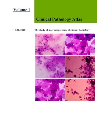
  • 1. 1 Volume 1 Clinical Pathology Atlas 11.01. 2020 The study of microscopic view of clinical Pathology.
  • 2. [Title] 1 1 Clinical Pathological examination Clinical Pathological examination Technologist : Ajith.A DMLT Kanthalloor | Marayoor idukki kerala 8637499285 | ajitharun937@gmail.com
  • 5. [Title] ACKNOWLEDGMENTS I gratefully acknowledge the help of the Principal, Vysnavi Institute of Paramedical science Udumalpet, Tirupur district in India. Then I want to tank my Professor Mrs. Barathi Mahendran and Drear respected doctors ,Dr. Vasudevan.MBBS., Dr. Nirmala Devi. MD ( Microbiologist ), Dr. R. Renuga. MD( Pathologist ) Senior Technologists Mr. Suresh, Mr. Ayub Ansari, Mr.Raja, Mrs. Meera, Mrs. Sasi, Mrs. Shamina. I owe my special thanks to The senior Technologists Deepaa micro lab Mr.Ragavan and Manager Mr. Krishnamoothi Mr.Srini, Mr. Marimuthu, Ms. Valarmathi, Ms. Sowmiya, Mrs. Ancy, Mr. Joyi, .Haridas, Mrs. Nandhini, Mrs. Priya, Ms. Reshma, Ms. Suganthi, Mrs. Valsala Bama, Ms.G.Saranya. Then I tank to my Father, Mother , Grandma ( Rajamma Baskaran ) and My friends in laboratory, Mr.srinivasan, Mr. Prasad, Mr.Raju, Mr.Abilash, Mr.Karthi, Mr.Guruprasad,.Buvana, Mrs.Madhavi, Ms.Maheshwari, Mrs.Shalini, Ms. Karthi Priya, Ms. Logambal, Mrs. Nagajothi, Ms.Karthipriyanka, Ms.Maha, Ms. Karthika, Ms. Kokila, etc….
  • 6. [Title] Contents Clinical Pathology 1. Physical Examination of Urine, 2. Chemical Examination of Urine, 3. Microscopic Examination of Urine Sediment 4. Stool examination Seminal Fluid Analysis 1. Physiology 2. Specimen Collection 3. Physical Examination 4. Chemical Examination 5. Microscopic Examination 6. Concentration and sperm count 7. Morphologyic sperm count 8. Reporting format 9. Staining method Haematology 1. Technique of blood collection 2. Precaution in handling blood and blood products 3. Estimation of haemoglobin 4. Hematocrit 5. Selection and registration of donors 6. Collection of blood from donors 7. ABO blood grouping 8. Erythrocyte sedimentation rate 9. Rhesus blood group system 10. Introduction to peripheral blood smear 11. Staining of peripheral blood smear 12. Interpretation of red cell morphology
  • 7. [Title] 13. Composition of blood and normal erythropoiesis 14. Maturation and development of leucocytes 15. Formation of platelets and Thrombocytopenia 16. Pretransfusion or compatibility testing 17. Transfusion reactions 18. Introduction of anemia 19. Microcytic hypochromic anemia 20. Microcytic anemia 21. Hemolytic anemia 22. General laboratory features hemolytic anemia 23. Hemolytic anemia due to abnormal haemoglobin 24. Hemolytic anemia due to abnormal erythrocytes 25. Screening of blood transfusion transmitted disease. 26. Hemostasis 27. Autoimmune Hemolytic anemia. 28. Disease affecting erythrocytes 29. Nuclear and cytoplasmic changes in Leucocytes. 30. Acute Myeloid Leukemia. 31. Myeloproliferative Neoplasm. 32. Myelodisplastic Syndrome. 33. Mature Lymphoproliferative Disorders. 34. Morphologic changes after Myeloid Hematopitic growth factor. 35. Microorganisms 36. Miscellaneous cells 37. Normal New Born Peripheral blood morphology. 38. Body fluids
  • 9. [Title] Clinical Biochemistry 1) General Biochemistry general 2) Carbohydrates 3) Carbohydrat metabolism 4) Lipids 5) Nucleotides 7) Clinical chemistry 8) Enzymes 9) Biological oxidation, Electron Transfer chain and Oxidative phosphorylation. 10) Vitamins 11) Minerals 12) Hormones 13) Clinical Biochemistry 14) Body water osmolarity and ionic compounds of body fluids. 15) Nutrition 16) Kidney Function Test 17) Liver Function test 18) Spectrophotometry, Light emission and scattering analytical techniques 19) Basic Principles Of Radioactive Measurements 20) Electro Chemistry 21) Electrophoresis 22) Chromatography And Mass Spectrometer 23) Chemical Enzymology 24) Immunochemical Techniques 25) Automation In Clinical Laboratory 26) Laboratory Quality Measurement 27) Electrolytes and Blood Gases 28) Centrifugation 29) Primary and Secondary Standards.
  • 10. [Title] Clinical Pathological Examination Urine analysis Physical examination: Color Volume Odor Appearance Chemical examination: Specific gravity pH Sugar Protein (Albumin) Acetone Bile salt Bile pigment Urobilinogen Nitrate Microscopic examination: Pus cells RBC's Epithelial cells Cast Crystals Bacteria Other parasites & fungi
  • 11. [Title] Urine color chart: Urine : pH stands for Potential of Hydrogen. It refers to the hydrogen ion concentration in a solution. It is the measure of the acidity or alkalinity of a solution. The pH value ranges from 0 to 14 on a pH scale.
  • 12. [Title] Urine sugar: Benedict method for urine sugar procedure : This is the test for detecting the presence of redusing substance in solutions. Principe: With mild alkali, sugar are concerned into an enediol from, which has redusing property. Enediol reduce Cu2+ to Cu+ forming CuOH. Which on further heating gets converted into Cu2O. Depending on the amount of reduction of CuSO4 taking place. The non redusing disaccharides answer this test. After acid hydrolysis.
  • 13. [Title] Procedure: Take a clean test tube. Then the Benedict’s reagent approximately 2ml take the test tube and gently heat it. After hearing 3 -5 drops urine sample add the reagent. Observe the color change and not the result. Substance like creatinine, uric acid, salicylic acid, homogeneous acid, vitamin c etc. Having redusing properly also give positive test. When Present in appreaciable amount. It is semi quantitative test for detection of glucose. Approximate convention of glucose in urine may be ascertained in clinical purpose. Sodium citrate Benedict’s quantitative reagent has a slightly different composition. It contains potassium thiocyanate and potassium ferrocyanide in addition of copper , sodium citrate and sodium carbonate. Materials required:
  • 14. [Title] sugar level observation chart: Color of the precipitation Green. : (+) Yellow : (++) Orange : (+++) Red : (++++)
  • 15. [Title] Protein test in urine: Sulfosalicylic acid test Procedure: Take a clean test tube. Take approximately 2ml of urine specimen, then sulfosalicylic acid reagent added to a small amount of urine. The acidification causes precipitation of protein in the sample (seen as increasing turbidity observe and note the result. Observation:
  • 16. [Title] Acetone test: Higher levels of ketones in the urine indicate that the body is using fat as the major source of energy. Ketone bodies that commonly appear in the urine when fats are burned for energy are acetoacetate and beta-hydroxybutyric acid. Acetone is also produced and is expired by the lungs. Procedure: Take a clean test tube. The 2 ml urine take a test tube. Then Rothera's Mixture Powder two spatula take and gently mix the urine sample. After the ammonium hydroxide solution take and add the side wall of the test tube. The junction of urine and ammonia solution form a pink color ring formation is positive result. No color change the negative result.
  • 17. [Title] Bile salt : Hay’s Test Procedure: Hay's test, also known as Hay's sulphur flower test, is a chemical test used for detecting the presence of bile salts in urine. Bile pigment in urine: Procedure: Bile pigments are breakdown products of the blood pigment hemoglobin that are excreted in bile. Bilirubin (orange or yellow in color) and its oxidized form biliverdin
  • 18. [Title] (green) are two important bile pigments. ... The conjugated bilirubin is excreted through bile. Pus cells: RBC’s:
  • 19. [Title] Epithelial cells: Cast: Urinary casts, sometimes known as the poor man’s renal biopsy, are objects formed within renal tubules. Casts are cylindrical and composed mostly of protein and cells. They may be convoluted (spiral) if formed in distal convoluted tubules, broad if formed in dilated collecting ducts, and narrow if formed in narrow lumens. Pus cast: RBC cast:
  • 20. [Title] Red blood cell casts mean there is a microscopic amount of bleeding from the kidney. They are seen in many kidney diseases. Epithelial cast Renal tubular epithelial cell casts reflect damage to tubule cells in the kidney. These casts are seen in conditions such as renal tubular necrosis, viral disease (such as CMV nephritis), and kidney transplant rejection. ... White blood cell (WBC) casts are common with acute kidney infections and interstitial nephritis.
  • 22. [Title] Granular casts come in a variety of granular textures. They range from small, fine granules dispersed throughout the cast matrix to large, coarse granules (Figures 8-48 and 8-49). They are composed primarily of uromodulin protein and cast granulation is not clinically significant. Easily viewed with bright field microscopy because of their high refractive index, granu- casts often appear colorless to shades of yellow. Granular casts can appear in all shapes and sizes, and granular casts are considered to be an indicator of a poor prognosis.Several mechanisms account for the granular casts observed in the urine sediment. The granules in finely granular casts have been identified as by-products of protein metabolism, in part lysosomal, that are excreted by renal tubular epithelial cells20—this accounts for the appearance of granular casts in the urine of normal, healthy individuals. A variation of this mechanism is believed to account for the finding of some casts with large, coarse granulation, particularly when no accom- panying cellular casts are present. In these cases, as tubular cells degenerate, their intracellular components are released into the tubular lumen and become enmeshed in a cast. Other coarsely granular casts result from the degeneration of cellular casts. These casts often contain identifiable cellular remnants. In patients with intrinsic renal disease, these coarsely granular casts are usually accompanied by cellular casts. Further degeneration of granular casts into waxy casts can occur during urine stasis (see Figure 8-36, 8-42). Urine sediment from normal healthy individuals may have an occasional finely granular cast. These casts are not as common as hyaline casts, but their numbers can following exercise. Patients with various types of renal disease can have varying quantities of coarse and finely granular casts
  • 23. [Title] Hyaline cast: In healthy individuals, two or fewer hyaline casts per low-power field is considered normal. Increased numbers of hyaline casts can be found following extreme physiologic conditions such as strenuous exercise, dehydration, fever, or emotional stress. They also accompany pathologic casts in renal disease and in cases of congestive heart failure
  • 24. [Title] Crystals in urine: Uric acid crystals in urinary deposit: Uric Acid. Uric acid crystals occur in many forms; the most common form is the rhombic or diamond shape (Figures 8-61 and 8-62). However, the crystals may be appear as cubes, barrels or bands and may cluster together to form rosettes (Figures 8-63 and 8-64); they often show layers or laminations on their surfaces(Figure8-65). Although they present most often in various forms with four sides, they occasionally have six sides and may require differentiation from colorless cystine crystals. Uric acid crystals are yellow to golden brown, and the intensity of their color varies directly with the thickness of the crystal. As a result, crystals may appear colorless when they are thin or when the urine is low in uroery- thrin(aurinepigment). microscopy, uric acid crystals exhibit strong birefringence and produce a variety of interference colors. Uric acid crystals can be present only if the urine pH is less than 5.7. At a pH greater than 5.7, uric acid is in its ionized form as urate and forms urate salts (e.g., amorphous urates, sodium urate). Uric acid crystals are 17timeslesssolublethanuratesaltcrystals.Ifurinewith uric acid crystals is adjusted to an alkaline pH, the crystals readily dissolve. Similarly, if urine with urate salt crystals is acidified adequately, uric acid crystals form. Uric acid is a normal urine solute that originates from the catabolism of purinenucleosides(adenosine and guanosine from RNA and DNA). Hence,
  • 25. [Title] uric acid crystals can appear in urine from healthy individuals. Increased amounts of urinary uric acid can be present following the administration of cytotoxic drugs (e.g., chemothera- peutic agents) and with gout. With these conditions, if the urine pH is appropriately acid, large numbers of uric acid crystals can bepresent.
  • 28. [Title] Different shaped uric acid crystal in urine deposit. Uric Acid. Uric acid crystals occur in many forms; the most common form is the rhombic or diamond shape. However, the crystals may be appear as cubes, barrels or bands and may cluster together to form rosettes they show layers or laminations on their surfaces. Although they present most often in various forms with four sides, they occasionally have six sides and may require differentiation from colorless cystine crystals. Uric acid crystals are yellow to golden brown, and the intensity of their color varies directly with the thickness of the crystal. As a result, crystals may appear colorless when they are thin or when the urine is low in uroerythrin (a urine pigment). With the use of polarizing.
  • 29. [Title] Calcium oxalate crystals in urinary deposit:
  • 32. [Title] Calcium Oxalate. The most common shape of calcium oxalate crystals is their octahedral or pyramid form (Figures 8-66 and 8-67). This dihydrate form of calcium oxalate represents two pyramids joined at their bases. When viewed from one end, they appear as square a scribed with lines that intersect in the center, hence they are sometimes called envelope crystals. In contrast, calcium oxalate monohydrate crystals are small and ovoid or dumbbell shaped (Figure 8-68). The less common monohydrate form can resemble RBCs and may require differentiation by polarizing microscopy to demonstrate the birefringence of these crystals (see Figure 8-68,B). Calcium oxalate crystals are colorless and can vary significantly in size. Usually the crystals are small and require high-power magnification for identification. On occasion, the crystals may be large enough to be identi- fied under low-power magnification. Calcium oxalate crystals may cluster together and can stick to mucous threads. When this occurs, they may be mistaken for crystal casts. Calcium oxalate crystals are the most frequently observed crystals in human urine, in part because they can form in urine of any pH. Calcium and oxalate are solutes normally found in the urine of healthy individu- als. Approximately 50% of the oxalate typically present in urine is derived from ascorbic acid (vitamin C), an oxalate precursor (see Figure 7-13) or from oxalic acid. Foodstuffs high in oxalic acid or ascorbic acid include vegetables (rhubarb, tomatoes, asparagus, spinach) and citrus fruits. In addition, beverages that are high in oxalic acid include cocoa, tea, coffee, and chocolate. As urine forms in the renal tubules, oxalate ions associate with calcium ions to become calcium oxalate. When condi- tions are optimal, calcium oxalate can precipitate in a crystalline form. Increased numbers of calcium oxalate crystals are often observed following ingestion of the oxalate precursor ethylene glycol (antifreeze) and during severe chronic renaldisease.
  • 33. [Title] Triple phosphate crystals: Verity types of Triple Phosphate crystals
  • 34. [Title] Triple Phosphate. Triple phosphate (NH4MgPO4, ammonium magnesium phosphate) crystals arecolorless and appear in several different forms. The most common and characteristic forms are three- to six-sided prisms with oblique terminal surfaces, the latter describedas FIGURE 8-71 Calcium phosphate crystals. Prisms are arranged singly and in rosette forms. Brightfield, 100×. “coffin lids” (Figure 8-70). Not all crystals are perfectly formed, and their size can vary greatly. With prolonged storage, these crystals can dissolve, taking on a feathery form that resembles a fern leaf. Among the crystals observed in alkaline urine, triple phosphatecrystalsarecommon.Theycanalsobepresent in neutral urine specimens. Ammonium magnesium phosphate is a normal urine solute, hence triple phos- phate crystals can be present in urine from healthy indi- viduals. Triple phosphate crystals have little clinical significance but have been associated with UTIs charac- terized by an alkaline pH and have been implicated in theformationofrenalcalculi.
  • 36. [Title] Calcium Phosphate Crystals: Calcium Phosphate. Calcium phosphate is present in urine as dibasic calcium phosphate (i.e., CaHPO4, calcium monohydrogen phosphate) and as monobasic calcium phosphate (i.e., Ca[H2PO4]2, calcium biphos- phate).Thesesimilaryetdifferentcompoundsprecipitate out of solution in distinctly different crystalline shapes. Dibasic calcium phosphate crystals, sometimes called stellar phosphates, appear as colorless, thin, wedgelike prismsarrangedinsmallgroupingsorinarosettepattern (Figure 8-71). Each prism has one tapered orpointed end, with the other end squared off. Another, less com- monly observed form of dibasic calcium phosphate is seen in those crystals shaped as thin, long needles arrangedinbundlesorsheaves(Figure8-72).Incontrast, monobasic calcium phosphate crystals usually appear microscopically as irregular, granular sheets (Figure8-73) or flat plates that can be large and may be noticed floating on the top of a urine specimen. These colorless crystalline sheets can resemble large degenerating squa- mous epithelialcells. Classified as alkaline crystals because they are usually present in neutral or slightly alkaline urine specimens, calcium phosphate crystals can also form in slightly acidic urine. They are weakly birefringent with polariz- ing microscopy. Calcium phosphate crystals are common and have no clinical significance.
  • 37. [Title] Tyrosine crystals Tyrosine and Leucine. Tyrosine crystals appear as fine, delicate needles that are colorless or yellow (Figure8-80). They frequently aggregate to form clusters orsheaves but also may appear singly or in small groups. Leucine crystals are highly refractile, yellow to brown spheres. They have concentric circles or radial striations on their surface and can resemble fat globules. Unlikefat,leucinecrystalsdonotstainwithfatstains. They are birefringent under polarized microscopy but the light pattern produced is not a true Maltese cross pattern. Tyrosine and leucine crystals can form in acidic urineand dissolve in alkaline solution. They are rarely seentoday because of the rapid turnaround time for a uri- nalysis, and because they require refrigeration to forcethem out of solution. Of the two, tyrosine is found moreoften in urine because it is less soluble than leucine.Sometimes leucine crystals can be forced out of solutionby the addition of alcohol to tyrosine-containing urines.These amino acid crystals are abnormal and arepresent in the urine of patients with overflowaminoacidurias—rare inherited metabolic disorders. Inthese disorders, the concentrations of these amino acidsin the blood are high (aminoacidemia), resulting inincreased renal excretion. Although rare, these crystalshave been observed in the urine of patients with severeliver disease. These abnormal crystals are confirmed,preferably by chromatographic methods, before they are reported.
  • 38. [Title] Acid Urate Crystals Acid Urates. Acid urate crystals are sodium, potas- sium, and ammonium salts of uric acid that appear as small, yellow-brown balls or spheres (Figure 8-59).Their color is distinctive and is similar to that of their alkaline counterpart, ammonium biurate crystals. Acid urate crystals can be present when the urine pH is neutral toslightly acidic but frequently are not observed in fresh urine. Because of their small, spherical shape and color, they may be misidentified as leucine crystals. Similar to other urate crystals, acid urates dissolve at 60° C and can be converted to uric acid crystals by the addition of glacial acetic acid. They have no clinical significance and arereportedas“uratecrystals.”
  • 39. [Title] Monosodium Urate: Monosodium Urate. Monosodium urate crystals, a distinct form of a uric acid salt, appear as colorless to light-yellow slender, pencil-like prisms (Figure 8-60). They may be present singly or in small clusters, and their ends are not pointed. Monosodium urate crystals can be present when the urine pH is acid and dissolve at 60° C. They have no clinical significance and usually are reported as “urate crystals.”
  • 40. [Title] Magnesium Phosphate. Magnesium Phosphate. Magnesium phosphate crystals are large, colorless crystals that appear as elon- gated rectangular or rhomboid plates. These flattened prisms can be notched and their edges can be irregular or eroded (Figure 8-74).21They are weakly birefringent under polarizing microscopy. Althoughmagnesium phosphate crystals form from normal urine solutes in neutral or alkaline urine, they are rarely seen. Sulfonamide Sulfonamides. Sulfonamides appear in various forms that differ depending on the particular form of the drug prescribed. When initially manufactured, sul- fonamide preparations were relatively insoluble and resulted in kidney damage caused by crystal formation within the renal tubules. Currently, these drugs have beenmodified,andtheirsolubilityisnolongeraproblem. As a result, sulfonamide crystals are not found as often in urine sediment, and renal damage caused by them isuncommon. Sulfadiazine drug crystals usually appear yellow to brown and as bundles of needles that resemble sheaves of wheat (Figure 8-85). The constriction of the bundle maybelocatedcentrallyorextremelyeccentric,resultinin a fan formation. Sulfamethoxazole (e.g., Bactrim, Septra) is more commonly encountered and appears as brownrosettesorsphereswithirregularradialstriations (Figure 8-86). All sulfonamide crystals are highly refrac- tile andbirefringent.
  • 41. [Title] Sulfonamide crystals can be present in acid urine and should be confirmed chemically before they arereported.
  • 42. [Title] The diazotization of sulfanilamide followed by an azo- coupling reaction is the preferred method to confirm their presence. In shape and color, these crystals closely resemble ammonium biurate crystals but can be differ- entiated from them on the basis of their pH, their solubil- ity, and the chemical confirmatory test. A list of the patient’s current and past medications can be of value in confirming the identity of these urine crystals. Ammonium Biurate Ammonium Biurate. Ammonium biurate crystals appear as yellow-brown spheres with striations on the surface (Figure 8-75). Irregular projections or spicules can also be present, giving these crystals a “thorny apple” appearance (Figure 8-76). They can form in alka- line or neutralurine.
  • 43. [Title] Ammonium biurate is a normal urine solute. Thesecrystals occur most frequently in urine specimens that have undergone prolonged storage. However, when they precipitate out of solution in fresh urine specimens (e.g., following iatrogenically induced alkalinization), they are clinically significant, because in vivo precipitation can cause renal tubular damage. Their presence most often indicates inadequate hydration of the patient. Therefore when ammonium biurate crystals are encountered in a urine specimen, investigation is required to determine whether (1) the integrity
  • 44. [Title] of the urine specimen has been compromised (improper storage), or (2) in vivo forma- tion is taking place. Ammonium biurate crystals are strongly birefringent and dissolve in acetic acid or on heating to approxi- mately 60° C. Similar to other urate salts (amorphous urates, acid urates), ammonium biurate crystals can be converted to uric acid crystals with the addition of con- centrated hydrochloric or acetic acid. An important note is that ammonium biurate crystals can resemble some forms of sulfonamide crystals. One differentiates between them on the basis of urine pH, a sulfonamide confirmatory test, and the solubility char- acteristics of the crystals. Amorphous Crystals: Amorphous Crystals is two types thy contain Amorphous crystals and Amorphous urate crystals. The Amorphous Phosphate crystals is alkaline pH and Amorphous uratas in acid pH. Amorphous Phosphate crystals white color deposit and dissolved in acetic acid in room temperature. The Amorphous urate crystals yellow or black color deposit and the crystals disappear in addition of acetic acid and convert to uric acid crystals in presence of room temperature. The urate crystals is above 60 ℃ temperature dissolved in the presence of acetic acid. The background shows amorphous. Amorphous urate crystals deposit
  • 45. [Title] a single uric acid crystals seen
  • 46. [Title] Yeast Cells: A single hypha seen yeast cell clump in the field. 6 – 8 Squmus epitheliam a single. Candida in cluster seen AFB stain. Yeast. Yeasts are ovoid, colorless cells that can closely resemble RBCs (Figure 8- 90). More refractile than erythrocytes, yeasts often have characteristic budding forms and pseudohyphae (Figure 8-91). Yeasts can vary in size, and some species are very large (10 to 12 µm). Yeasts do not dissolve in acid and usually do not stain
  • 47. [Title] with supravital stains; these two characteristics can aid in differentiating them from erythrocytes. In women, yeast in the urine sediment most often indicates contamination of the urine with vaginal secretions. However, because yeasts are ubiquitous—present in the air and on skin—their presence could indicate contamination from these sources. Although infrequent, primary UTIs resulting from yeasts are possible, hence health care providers must correlate the finding of yeast with the patient’s clinical picture to determine whether esent. Certain situations such as pregnancy, use of oral contraceptives, and diabetes mellitus promote the development of vaginal yeast infection. The most commonly encountered yeast in urine sediment is Candida albicans. The characteristic budding and the development of pseudohyphaemakeC.albicansreadily identifiable as yeast. Another species found less frequently is C.glabrata, formerly called Torulopsis glabrata. This species does not form pseudohyphae, and these yeast cells may be found phagocytized within white blood cells (Figure 8-92). Inimmuno suppressed patients, systemic Candida infections are common; for some unknown reason, yeasts have a predilection for the kidneys. During the microscopic examination, only the presence of yeast can be determined; identification of the species present requires fungal culture. A KOH preparation is often used to detect yeast, hyphae, and other fungal cells in vaginal secretions.
  • 48. [Title] Hypha seen. Yeast cell in cluster seen wet mount plant of pus cells a single candida seen Yeast cell in cluster
  • 49. [Title] Parasites in urine Trichomonas Vaginalis.Trichomonads, protozoan ,canbeobservedintheurinesediment.Tricho- monads appear as turnip-shaped flagellates whose uni- cellular bodies average 15 µm in length, although organisms as small as 5 µm and as large as 30 µm are possible. They have four anterior flagella, a single pos- terior axostyle, and an undulating membrane that extends halfway down the body of the organism (Figure8-93). The beating flagella propel the organism while the undulating membrane rotates it. The result is a charac- teristic flitting or jerky motility in wet preparations. Because of their similarity in size to both leukocytes and renal tubular cells, this motility is critical for their iden- tification (Figure8-94). Trichomonas vaginalis is the most common cause of parasitic gynecologic infection in female patients (see Chapter 16, “Analysis of Vaginal Secretions”). Transmit- ted sexually, trichomonads most frequently represent an infectionofthevaginaand/orurethra,andtheirpresence in the urine often indicates contamination with vaginal secretions. In male patients, trichomonad infections of the urethra are usually asymptomatic. In either case, when observed in urine
  • 50. [Title] sediment, trichomonads are not quantitatedbutaresimplyreportedaspresent. means of positively identifying them, a fresh urine speci- men is needed. Once in urine, trichomonads proceed todie, first losing their motility; later, their undulating membrane ceases, and eventually they ball up to resem- ble white blood cells or renal tubular epithelial cells. With loss of motility or movement of the undulating membrane, differentiation from other cells in the sedi- ment can be impossible. Supravital stains do notenhance trichomonad identification, whether they are dead or alive. Whereas phase-contrast microscopy and interfer- ence contrast microscopy permit enhanced imaging and visualization of the flagella and undulating membranes of trichomonads, these techniques depend on movement to identify the organismsspecifically. Clue Cells and Gardnerella Vaginalis. Clue Cells and Gardnerella Vaginalis. Squamous epithelial cells from the vaginal mucosa with large numbers of bacteria adhering to them are called clue cells; they can be present in urine specimens contami- nated with vaginal secretions (Figure 8- 95). These char-acteristic cells are indicative of bacterial vaginosis (BV), a synergistic infection most often involving Gardnerella vaginalis and an anaerobe, usually Mobiluncus spp. (e.g., Mobiluncus curtisii). A disruption in the normal
  • 51. [Title] vaginal flora (lactobacilli) with subsequent proliferation ofthese, usually minor, endogenous bacterial species results in a foul-smelling vaginal discharge and the sloughing of clue cells. Clue cells appear soft and finely granular with indistinct cell borders caused by numerous bacteria adhering to them, hence they are often described as having shaggy edges. In these bacteria-laden cells, the nucleus may not be visible. To be considered a clue cell, the bacteria do not need to cover the entire cell;however, bacterial organisms must extend beyond the cell’s cyto- plasmic borders. Be aware that with an inexperienced microscopist, normal intracellular keratohyalin granula- tion can be misidentified as bacteria adhering to squa- mousepithelialcells. How ever, these granules are variable in size and are usually larger and more refractile than bacteria. When a health care provider suspects bacterial vaginosis, a pelvic examination is performed and a vaginal secretions specimen is collected for evaluation (see Chapter 16). Parasites. Several parasites, in addition to trichomotrichomonads and yeast, can be observed in the urine sediment. The eggs or ova of Enterobius vermicularis (pinworm) can be found in urine from school-agedchildren; however, individuals of any age can be infected. The adult female pinworm lays eggs in the area around the rectum; this causes itching. Consequently, the eggs can be present in urine sediment if the specimen is contaminated during collection. Pinworm eggs are characteristically American football–shaped, with one side appearing flatter. They are large, transparent cells (50 to 60 µm long; 20 to 30 µm wide), and the developing larva can be seen inside (Figure8-96). Cysts of Giardia lamblia may be observed in urine sediment as the result of fecal contamination of infected individuals. Giardiasis is most often acquired by drink- ing contaminated water. It can occur from inadequate sanitation of city water supplies or from contamination of fresh water lakes and streams. Giardia organismshave contaminated recreational water sources such as swim- mingpoolsandwaterparks. Thecystsaresmall, ovoid cells about 8 to 12 µm in length, with smooth, well- defined cell walls (Figure 8-97). When viewing using bright field microscopy and high power (400×) magnifi- cation, the cytoplasm appears to
  • 52. [Title] be filled with nuclear material, but distinct nuclei (up to four) usually are not apparent without specific staining. Schistosoma hae-matobium Finally, the eggs of the blood fluke Schistosoma hae-matobium can be present in urine sediment. Schistoso- miasis is endemic in Africa and the Middle East and is acquired upon exposure to water where infected snails live (e.g., fishermen, swimmers, workers in irrigation canals). Infections are most often diagnosed when the eggs are found in urine sediment or in biopsies of the bladder, rectum, or vaginal wall. Schistosoma eggs are distinctively large (100 to 170 µm long and 40 to 70 µm wide) and shaped like an American football with a spike at one end (Figure 8-98). Their cell walls are thick but transparent, revealing the larva that fills its interior. Hematuria is often present as well.
  • 56. [Title] Stool Examination: Color: Yellow, Brown, Green, Redish, Black. Consistency: solid, Semisolid, Liquid. Reaction: Acid (6.5), Alkaline (7.5). Blood: Present, Absent Mucus: Present, Absent Ova : Cyst: Redusing substance: Age (less than 10 only) Nil,Trace,1+,2+,3+. Pus cells: Grading. RBC’s : Grading Other: Bacteria,Fungus and Paracytes.
  • 64. [Title] Parasitology General Parasitology Introduction Medical parasitology deals with the parasites, which cause human infections and the diseases they produce. • It is broadly divided into two parts: 1. Protozoology 2. Helminthology. • The pioneer Du tch microscopist, Antonie 11an Leeuwenhoek of Holland in 1681, first introduced single lens microscope and observed Giardia in his own stools. • Louis Pastuer in 1870, first published scientific study on a protozoa( disease leading to its control and prevention during investigation of an epidemic silk worm disease in South Europe. • A seminal discovery was made in 1878 by Patrick Manson about the role of mosquitoes in filariasis. This was the first evidence of vector transmission. • Afterwards, Laveran in Algeria discovered the malarial parasite (1880), and Ronald Ross in Secunderabad and Calcuna in India, showed its transmission by mosquitoes (1897). A large nwnber of vector-borne disease have since then been identified. • By mid 20th century, with dramatic advances in antibiotics and chemotherapy, insecticides and anti parasitic drugs, and improved lifestyles,all infectious diseases seemed amenable to control.
  • 65. [Title] PARASITES: Parasites are living organisms, which depend on a living host for their nourishment and survival. They mulriply or undergo development in the host. • The term "parasite" is usually applied to Protozoa (unicellular organisms) and Helminths (multicellular organisms) (Flow chart 1). • Parasites can also be classified as: Ectoparasite: Ectoparasites inhabit only the body of the host without penetrating the tissue. Lice,ticks and mites are examples of ectoparasites. The term infestation is often employed for parasitization with ectoparasites. Endoparasite: A parasite, which lives within the body of the host and is said to cause an infection is called an endoparasite. Most of the protozoan and helminthic parasites causing human disease are endoparasites. Free-living parasite: It refe rs to nonparasitic stages of active existence, which live independent of the host, e.g. cystic stage of Naegleriafowleri. Endoparasites can further be classified as: Obligate parasite: The parasite, wh ich cannot exist without a host, e.g. Toxoplasma gondii and Plasmodium. - Facultative para.site: Organism which may either live as parasitic form or as fre e-living form, e.g. Naegleriafowleri. - Accidental parasites: Parasites, which infect an unusual host are known as accidental parasites. Echinococcus granulosus infects man accidentally, giving rise to hydatid cysts.
  • 66. [Title] - Aberrant parasites: Parasites, which infect a host where they cannot develop further are known as aberrant or wandering parasites, e.g. Toxocara canis (dog roundworm) infecting lhwnans. HOST Host is defined as an organism, which harbors the parasite and provides nourishment and shelter to latter and is relatively larger than the parasite. • The host may be of the following types: - Definitive host: The host, in which the adult parasite lives and undergoes sexual reproduction is called the definitive host, e.g. mosquito acts as definitive host in malaria. The definitive host may be a human or any other living being. However, in majority of human parasitic infections, man is the definitive host (e.g. fil aria, roundworm, hookworm). Intermediate host: The host, in which the larval stage of the parasite lives or asexual multiplication takes place is called the intermediate host. In some parasites, two different intermediate hosts may be required to compl ete different larval stages. These are known as first and second intermediate hosts, respectively (Box 1). - Paratenic host: A host, in which larval stage of the parasite rema ins viable without furth er development is referred as a paratenic host. Such host transmits the infection to another host, e.g. fish for plerocercoid larva of D. lalum. Reservoir host: In an e ndemic area, a parasitic infection is continuously kept up by the presence of a host, which harbors the parasite and acts as an important source of infection to other susceptible hosts, e.g. dog is the reservoir host of hydatid disease. - Accidental host: The host, in which the parasite is not usually found, e.g. man is an accidental host for cystic echinococcosis. ZOONOSIS
  • 67. [Title] The word zoonosis was introduced by RudolfVirchow in 1880 to include the diseases shared in nature by man and animals. • Later, in 1959, the World Health Organization (WHO) defined wonosis as those diseases and infeclions, which are naturally transmitted between vertebrate animals and man. It is of following types:  Protozoal zoo noses, e.g. toxoplasmosis, leishmaniasis,  balanlidiasis and cryptosporidiosis.  I lelminthic zoonoses, e.g. hydatid disease, taeniasis.  Anthropozoonoses: Infectio ns tra nsm itted to  man from lower vertebrate an imals, e.g. cystic  echinococcosis.  Zooanthroponoses: Infections transmitted from man  to lower vertebrate animals, e.g. human n1berculosis  to cattle. • HOST-PARASITE RELATIONSHIPS Host-parasite re lationships a re o f fo ll owing types (Flow chart 2):  Symbiosis  Commensalism  Parasitism.