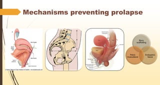
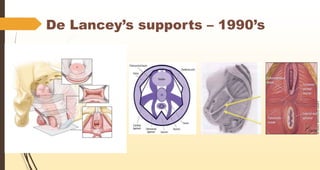
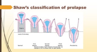
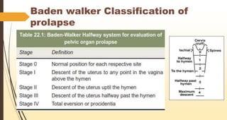
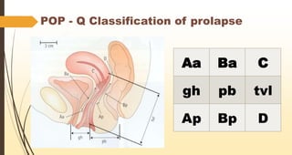
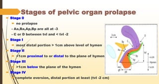
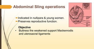
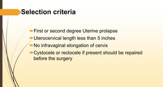
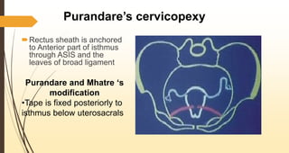
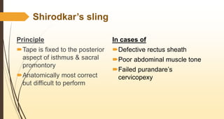
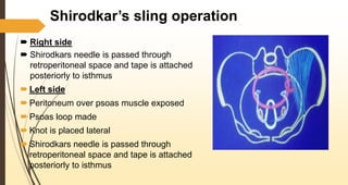
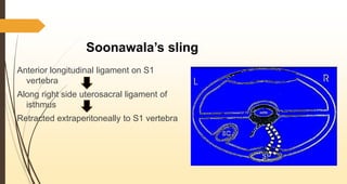
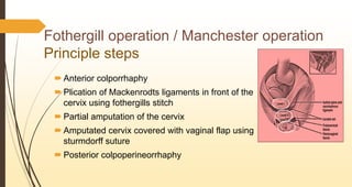
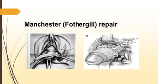
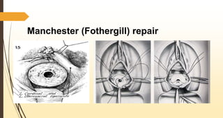
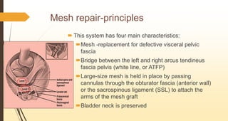
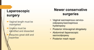
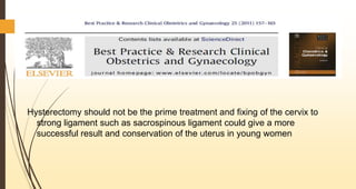
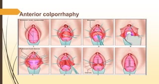
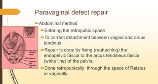
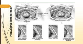
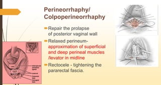
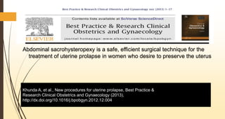
Classification & conservative
surgeries for prolapse
Presenter:- Dr. Indraneel Jadhav
MBBS, DGO, DNB
Consultant Gynecologist and IVF
Indira IVF, Kolhapur
Mechanisms preventing prolapse
Bony
scaffolding
Endopelvic
fascia
Pelvic
musculature
De Lancey’s supports – 1990’s
Shaw’s classification of prolapse
Baden walker Classification of
prolapse
POP - Q Classification of prolapse
Aa Ba C
gh pb tvl
Ap Bp D
Stages of pelvic organ prolapse
 Stage 0
 no prolapse
- Aa,Ba,Ap,Bp are all at -3
- C or D between tvl and < tvl -2
 Stage I
 most distal portion > 1cm above level of hymen
 Stage II
 <1cm proximal to or distal to the plane of hymen
 Stage III
 >1cm below the plane of the hymen
 Stage IV
 complete eversion, distal portion at least (tvl -2 cm)
Abdominal Sling operations
Indicated in nullipara & young women.
Preserves reproductive function.
Objective
• Buttress the weakened support Mackenrodts
and uterosacral ligaments
Selection criteria
First or second degree Uterine prolapse
Uterocervical length less than 5 inches
No infravaginal elongation of cervix
Cystocele or rectocele if present should be repaired
before the surgery
Purandare’s cervicopexy
Rectus sheath is anchored
to Anterior part of isthmus
through ASIS and the
leaves of broad ligament
Purandare and Mhatre ‘s
modification
•Tape is fixed posteriorly to
isthmus below uterosacrals
Shirodkar’s sling
Principle
Tape is fixed to the posterior
aspect of isthmus & sacral
promontory
Anatomically most correct
but difficult to perform
In cases of
Defective rectus sheath
Poor abdominal muscle tone
Failed purandare’s
cervicopexy
Shirodkar’s sling operation
 Right side
 Shirodkars needle is passed through
retroperitoneal space and tape is attached
posteriorly to isthmus
 Left side
 Peritoneum over psoas muscle exposed
 Psoas loop made
 Knot is placed lateral
 Shirodkars needle is passed through
retroperitoneal space and tape is attached
posteriorly to isthmus
Khanna’s sling
 Support is from bony point
 Tape is fixed to the posterior
aspect of the isthmus to the
anterior superior iliac spine
Virkud’s composite sling operation
 End of mersilene tape - sacral
promontory to posterior surface
of isthmus - sutured to rectus
sheath
 Plication of left side uterosacral
ligament to correct
dextrorotation
Virkud’s composite sling operation
Advantages
 Easy to perform
 Double support- bony + dynamic
 Tape is posterior-no risk during
LSCS
 No enterocele
 No injury to sigmoid colon
Joshi’s sling
Anterior surface of the
uterus at the level of the
internal os is suspended to
the pectineal ligament on
both side with merciline
tape
Soonawala’s sling
Anterior longitudinal ligament on S1
vertebra
Along right side uterosacral ligament of
isthmus
Retracted extraperitoneally to S1 vertebra
Fothergill operation / Manchester operation
Principle steps
 Anterior colporrhaphy
 Plication of Mackenrodts ligaments in front of the
cervix using fothergills stitch
 Partial amputation of the cervix
 Amputated cervix covered with vaginal flap using
sturmdorff suture
 Posterior colpoperineorrhaphy
Manchester (Fothergill) repair
Shirodkar’s modification
of Fothergill’s operation
Amputation of cervix is not done
Plication of uterosacral ligaments
Nadkarni’s sleeve
operation
• Modification of Fothergill’s
operation
• Supravaginal portion is
excised
• Fertility is not affected
LeFort’s Operation/ Partial colpocleisis
Old age and unfit for surgery
Uterine pathology to be ruled out
Pap smear to be done
Complete colpocleisis/ colpectomy
Entire vaginal mucosa is
excised
High risk of post
operative stress urinary
incontinence
Grafts
Types
1. Autologous grafts
Fascia lata
Rectus sheath
2. Synthetic
Macroporous
>75microns
Mersilene,
Marlex , Prolene
Gynemesh
Pore size < 75microns
• Complications
• Mesh erosion
• Infection
Prosthetic repair
Tension-free vaginal mesh (TVM) systems
Prolift,
Apogee/Perigee
Avaulta
 all of which vary in
terms of mesh size,
shape and surgical
technique
 This system has four main characteristics:
Mesh -replacement for defective visceral pelvic
fascia
Bridge between the left and right arcus tendineus
fascia pelvis (white line, or ATFP)
Large-size mesh is held in place by passing
cannulas through the obturator fascia (anterior wall)
or the sacrospinous ligament (SSL) to attach the
arms of the mesh graft
Bladder neck is preserved
Mesh repair-principles
Laparoscopic
surgery
 Vaginal length must be
maintained
 Ureters must be
identified and dissected
 Requires great skill and
expertise
Newer conservative
surgeries
• Vaginal sacrospinous cervico-
colpopexy/sacrospinous
hysteropexy
• Posterior intravaginal slingplasty
• Abdominal /laparoscopic
sacrocolpopexy
• Posterior mesh repair
Hysterectomy should not be the prime treatment and fixing of the cervix to
strong ligament such as sacrospinous ligament could give a more
successful result and conservation of the uterus in young women
Anterior colporrhaphy
• To correct cystocele and urethrocele
• The underlying principles are to excise a portion of the
relaxed anterior vaginal wall
• To mobilize the bladder and push it upwards after cutting
the vesicocervical ligament
• The bladder is then permanently supported by plicating
the endopelvic fascia and the pubocervical fascia
under the bladder neck in the midline
Anterior colporrhaphy
Paravaginal defect repair
Abdominal method
Entering the retropubic space
To correct detachment between vagina and arcus
tendinus
Repair is done by fixing (reattaching) the
endopelvic fascia to the arcus tendineus fascia
(white line) of the pelvis.
Done retropubically through the space of Retzius
or vaginally.
Paravaginaldefectrepair
Perineorrhaphy/
Colpoperineorrhaphy
Repair the prolapse
of posterior vaginal wall
Relaxed perineum-
approximation of superficial
and deep perineal muscles
/levator in midline
Rectocele - tightening the
pararectal fascia.
Abdominal sacrohysteropexy is a safe, efficient surgical technique for the
treatment of uterine prolapse in women who desire to preserve the uterus
Khunda A, et al., New procedures for uterine prolapse, Best Practice &
Research Clinical Obstetrics and Gynaecology (2013),
http://dx.doi.org/10.1016/j.bpobgyn.2012.12.004
Laparoscopic uterine suspension techniques seem
promising. Advantages are improved visualisation of pelvic
anatomy, shorter hospitalisation, less postoperative pain, and
a quicker return to normal activities
References
 Rock, John A.; Jones, Howard W.Te Linde's Operative Gynecology, 10th
Edition:Lippincott Williams & Wilkins 2008 section VII chapter 36A
 Schorge et al -Williams gynaecology 1st edition 2008 Section III Chapter 24 ,Page
no- 1023-53
 Khunda A, et al., New procedures for uterine prolapse, Best Practice &
Research Clinical Obstetrics and Gynaecology (2013)
 Jonathan S Berek , Emil Novak :Berek and Novak’s Gynecology 15th edition
Lippincott Williams & Wilkins, 2007:Page no-1211-29
 Practical Obstetrics and gynecology , Virkud, 3rd Edition,Chapter 18 : Page no-
323-47
Thank you

Classification & conservative surgeries for prolapse

  • 1.
    Classification & conservative surgeriesfor prolapse Presenter:- Dr. Indraneel Jadhav MBBS, DGO, DNB Consultant Gynecologist and IVF Indira IVF, Kolhapur
  • 2.
  • 3.
  • 4.
  • 5.
  • 6.
    POP - QClassification of prolapse Aa Ba C gh pb tvl Ap Bp D
  • 7.
    Stages of pelvicorgan prolapse  Stage 0  no prolapse - Aa,Ba,Ap,Bp are all at -3 - C or D between tvl and < tvl -2  Stage I  most distal portion > 1cm above level of hymen  Stage II  <1cm proximal to or distal to the plane of hymen  Stage III  >1cm below the plane of the hymen  Stage IV  complete eversion, distal portion at least (tvl -2 cm)
  • 8.
    Abdominal Sling operations Indicatedin nullipara & young women. Preserves reproductive function. Objective • Buttress the weakened support Mackenrodts and uterosacral ligaments
  • 9.
    Selection criteria First orsecond degree Uterine prolapse Uterocervical length less than 5 inches No infravaginal elongation of cervix Cystocele or rectocele if present should be repaired before the surgery
  • 10.
    Purandare’s cervicopexy Rectus sheathis anchored to Anterior part of isthmus through ASIS and the leaves of broad ligament Purandare and Mhatre ‘s modification •Tape is fixed posteriorly to isthmus below uterosacrals
  • 11.
    Shirodkar’s sling Principle Tape isfixed to the posterior aspect of isthmus & sacral promontory Anatomically most correct but difficult to perform In cases of Defective rectus sheath Poor abdominal muscle tone Failed purandare’s cervicopexy
  • 12.
    Shirodkar’s sling operation Right side  Shirodkars needle is passed through retroperitoneal space and tape is attached posteriorly to isthmus  Left side  Peritoneum over psoas muscle exposed  Psoas loop made  Knot is placed lateral  Shirodkars needle is passed through retroperitoneal space and tape is attached posteriorly to isthmus
  • 13.
    Khanna’s sling  Supportis from bony point  Tape is fixed to the posterior aspect of the isthmus to the anterior superior iliac spine
  • 14.
    Virkud’s composite slingoperation  End of mersilene tape - sacral promontory to posterior surface of isthmus - sutured to rectus sheath  Plication of left side uterosacral ligament to correct dextrorotation
  • 15.
    Virkud’s composite slingoperation Advantages  Easy to perform  Double support- bony + dynamic  Tape is posterior-no risk during LSCS  No enterocele  No injury to sigmoid colon
  • 16.
    Joshi’s sling Anterior surfaceof the uterus at the level of the internal os is suspended to the pectineal ligament on both side with merciline tape
  • 17.
    Soonawala’s sling Anterior longitudinalligament on S1 vertebra Along right side uterosacral ligament of isthmus Retracted extraperitoneally to S1 vertebra
  • 18.
    Fothergill operation /Manchester operation Principle steps  Anterior colporrhaphy  Plication of Mackenrodts ligaments in front of the cervix using fothergills stitch  Partial amputation of the cervix  Amputated cervix covered with vaginal flap using sturmdorff suture  Posterior colpoperineorrhaphy
  • 19.
  • 20.
  • 21.
    Shirodkar’s modification of Fothergill’soperation Amputation of cervix is not done Plication of uterosacral ligaments Nadkarni’s sleeve operation • Modification of Fothergill’s operation • Supravaginal portion is excised • Fertility is not affected
  • 22.
    LeFort’s Operation/ Partialcolpocleisis Old age and unfit for surgery Uterine pathology to be ruled out Pap smear to be done
  • 23.
    Complete colpocleisis/ colpectomy Entirevaginal mucosa is excised High risk of post operative stress urinary incontinence
  • 24.
    Grafts Types 1. Autologous grafts Fascialata Rectus sheath 2. Synthetic Macroporous >75microns Mersilene, Marlex , Prolene Gynemesh Pore size < 75microns • Complications • Mesh erosion • Infection Prosthetic repair
  • 25.
    Tension-free vaginal mesh(TVM) systems Prolift, Apogee/Perigee Avaulta  all of which vary in terms of mesh size, shape and surgical technique
  • 26.
     This systemhas four main characteristics: Mesh -replacement for defective visceral pelvic fascia Bridge between the left and right arcus tendineus fascia pelvis (white line, or ATFP) Large-size mesh is held in place by passing cannulas through the obturator fascia (anterior wall) or the sacrospinous ligament (SSL) to attach the arms of the mesh graft Bladder neck is preserved Mesh repair-principles
  • 27.
    Laparoscopic surgery  Vaginal lengthmust be maintained  Ureters must be identified and dissected  Requires great skill and expertise Newer conservative surgeries • Vaginal sacrospinous cervico- colpopexy/sacrospinous hysteropexy • Posterior intravaginal slingplasty • Abdominal /laparoscopic sacrocolpopexy • Posterior mesh repair
  • 28.
    Hysterectomy should notbe the prime treatment and fixing of the cervix to strong ligament such as sacrospinous ligament could give a more successful result and conservation of the uterus in young women
  • 29.
    Anterior colporrhaphy • Tocorrect cystocele and urethrocele • The underlying principles are to excise a portion of the relaxed anterior vaginal wall • To mobilize the bladder and push it upwards after cutting the vesicocervical ligament • The bladder is then permanently supported by plicating the endopelvic fascia and the pubocervical fascia under the bladder neck in the midline
  • 30.
  • 31.
    Paravaginal defect repair Abdominalmethod Entering the retropubic space To correct detachment between vagina and arcus tendinus Repair is done by fixing (reattaching) the endopelvic fascia to the arcus tendineus fascia (white line) of the pelvis. Done retropubically through the space of Retzius or vaginally.
  • 32.
  • 33.
    Perineorrhaphy/ Colpoperineorrhaphy Repair the prolapse ofposterior vaginal wall Relaxed perineum- approximation of superficial and deep perineal muscles /levator in midline Rectocele - tightening the pararectal fascia.
  • 34.
    Abdominal sacrohysteropexy isa safe, efficient surgical technique for the treatment of uterine prolapse in women who desire to preserve the uterus Khunda A, et al., New procedures for uterine prolapse, Best Practice & Research Clinical Obstetrics and Gynaecology (2013), http://dx.doi.org/10.1016/j.bpobgyn.2012.12.004
  • 35.
    Laparoscopic uterine suspensiontechniques seem promising. Advantages are improved visualisation of pelvic anatomy, shorter hospitalisation, less postoperative pain, and a quicker return to normal activities
  • 36.
    References  Rock, JohnA.; Jones, Howard W.Te Linde's Operative Gynecology, 10th Edition:Lippincott Williams & Wilkins 2008 section VII chapter 36A  Schorge et al -Williams gynaecology 1st edition 2008 Section III Chapter 24 ,Page no- 1023-53  Khunda A, et al., New procedures for uterine prolapse, Best Practice & Research Clinical Obstetrics and Gynaecology (2013)  Jonathan S Berek , Emil Novak :Berek and Novak’s Gynecology 15th edition Lippincott Williams & Wilkins, 2007:Page no-1211-29  Practical Obstetrics and gynecology , Virkud, 3rd Edition,Chapter 18 : Page no- 323-47
  • 37.

Editor's Notes

  • #3 Anteflexion of 170 degrees and anteversion of 90 degrees. Retroversion is the 1st step in POP When the levator muscles contract forcibly, the genital hiatus narrows as it is pulled posteriorly towards sacrum, this mechanism works when the pressure forces on the uterus are transmitted onto the levator muscle
  • #4 Michigan, identifies specific structural goals for each of the 3 levels of support of the vagina, defines a set of goals for the pelvic reconstructive surgeon