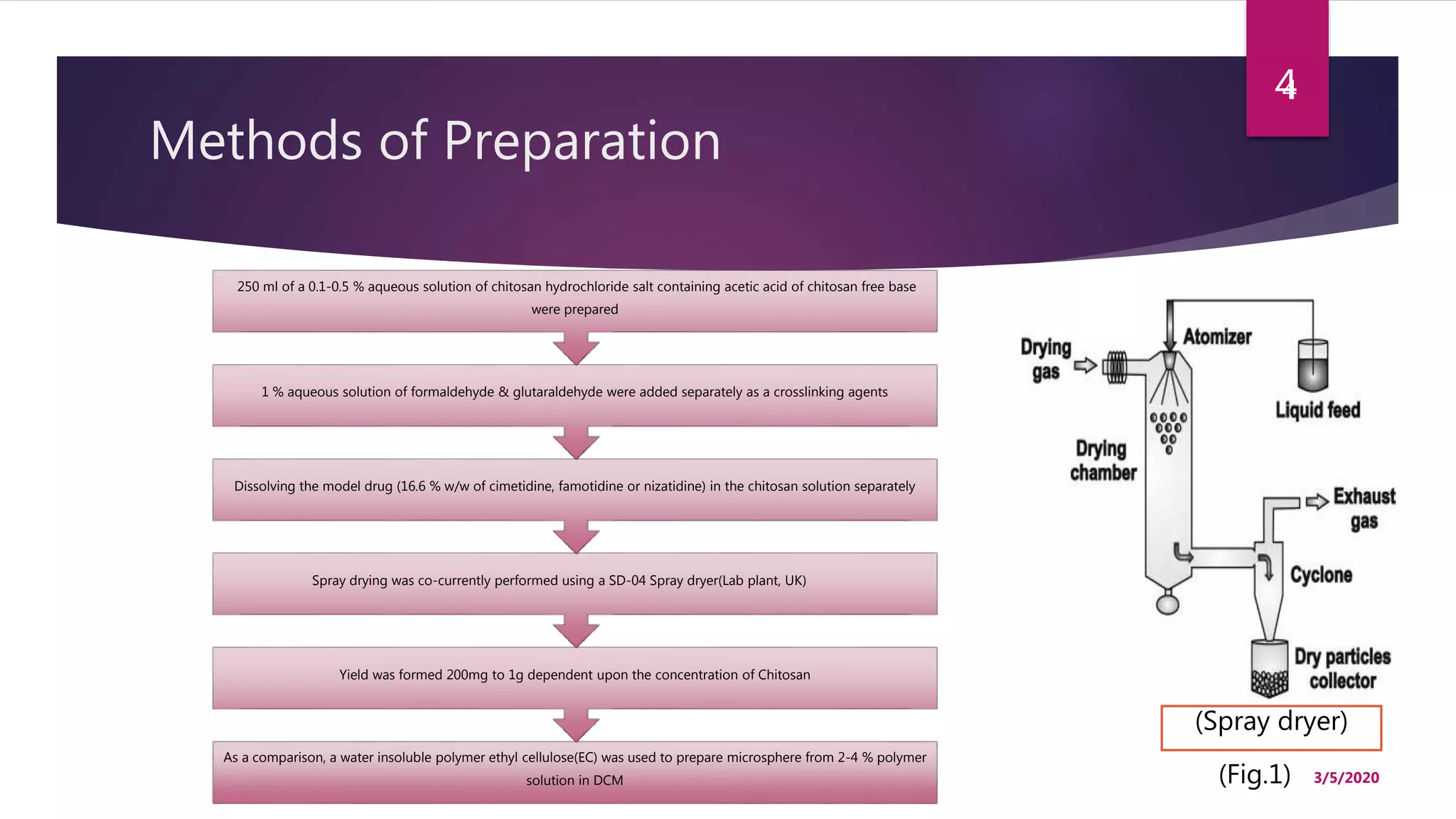
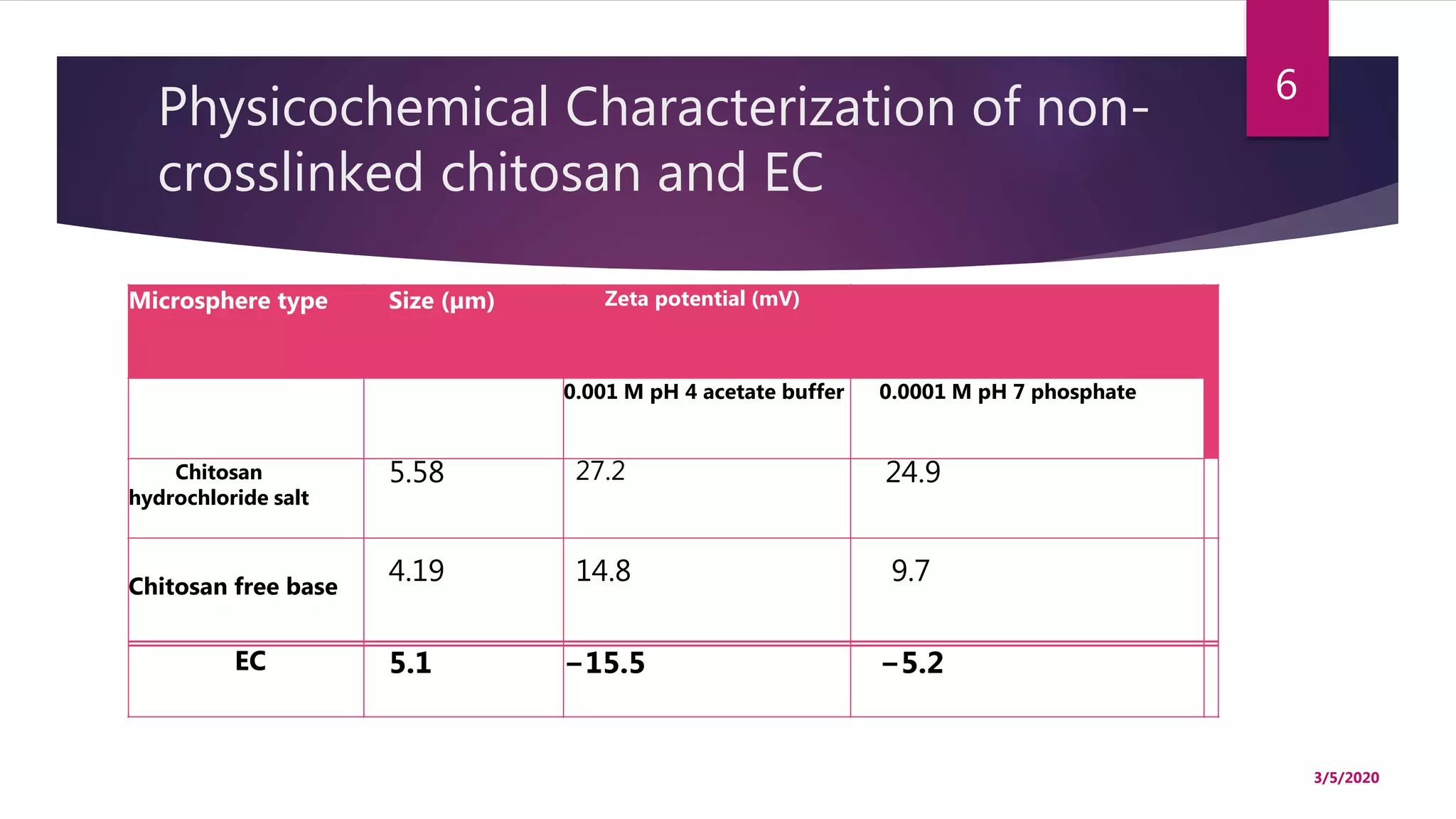
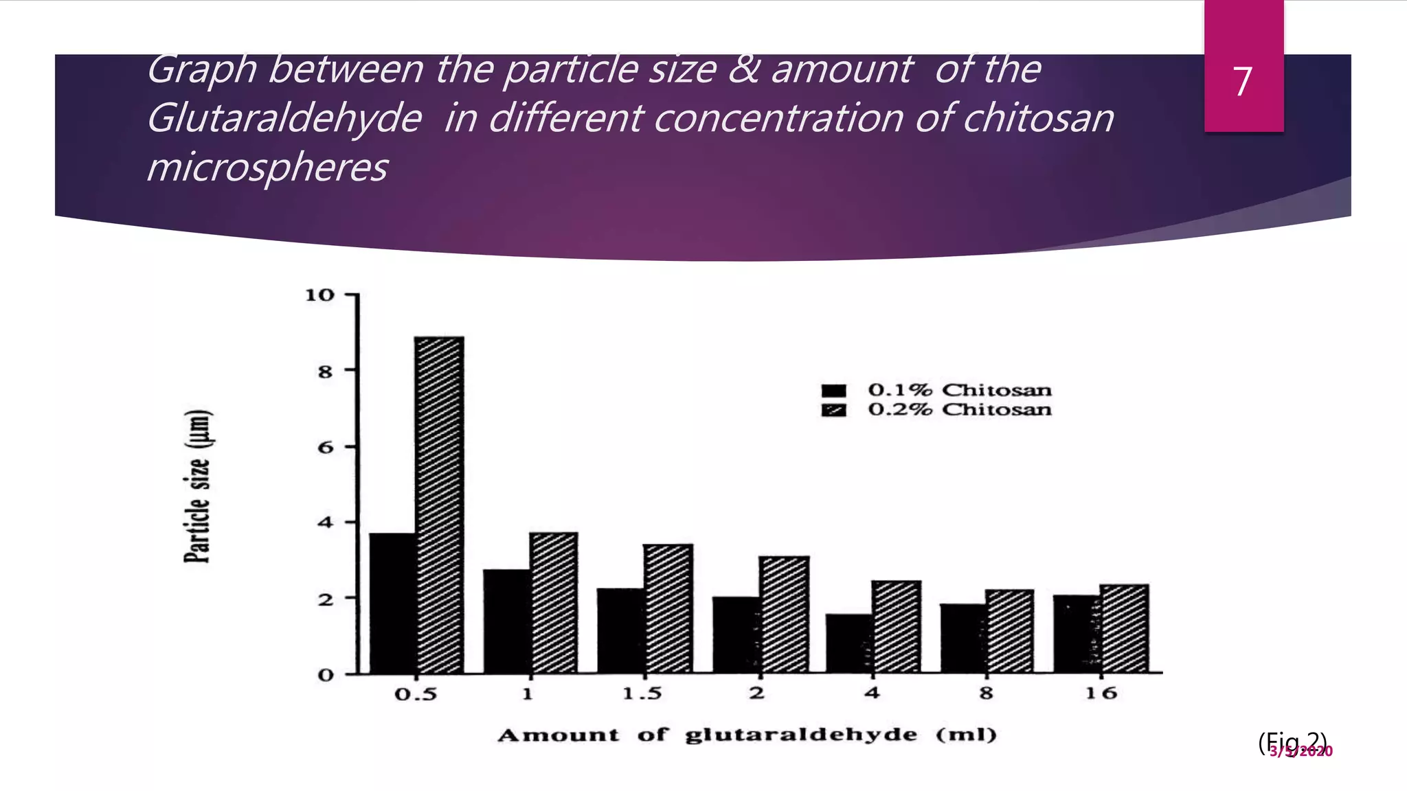
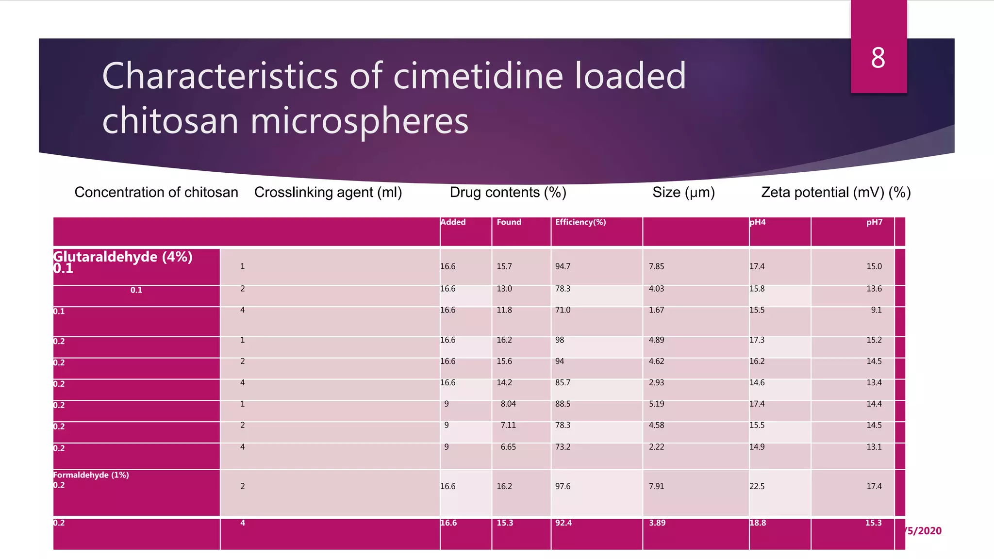
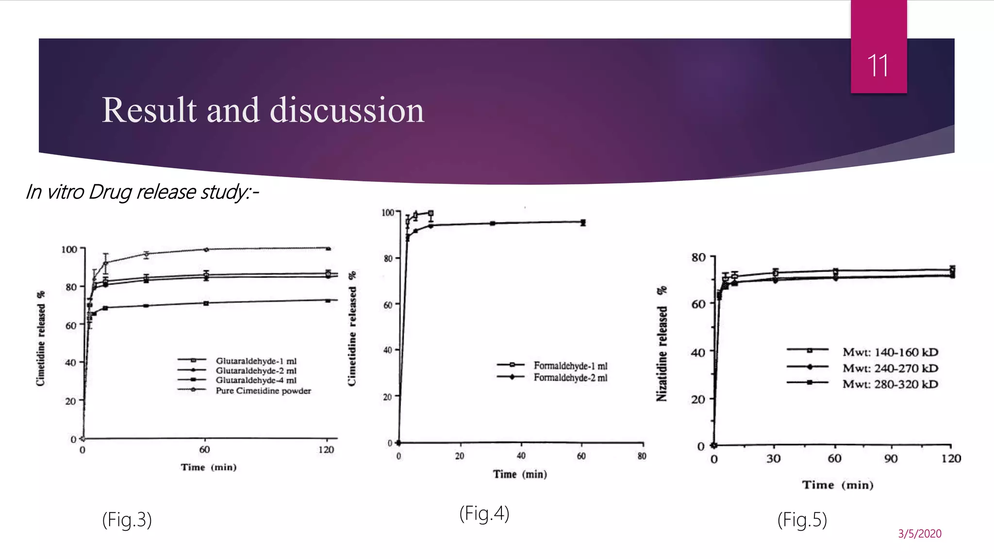
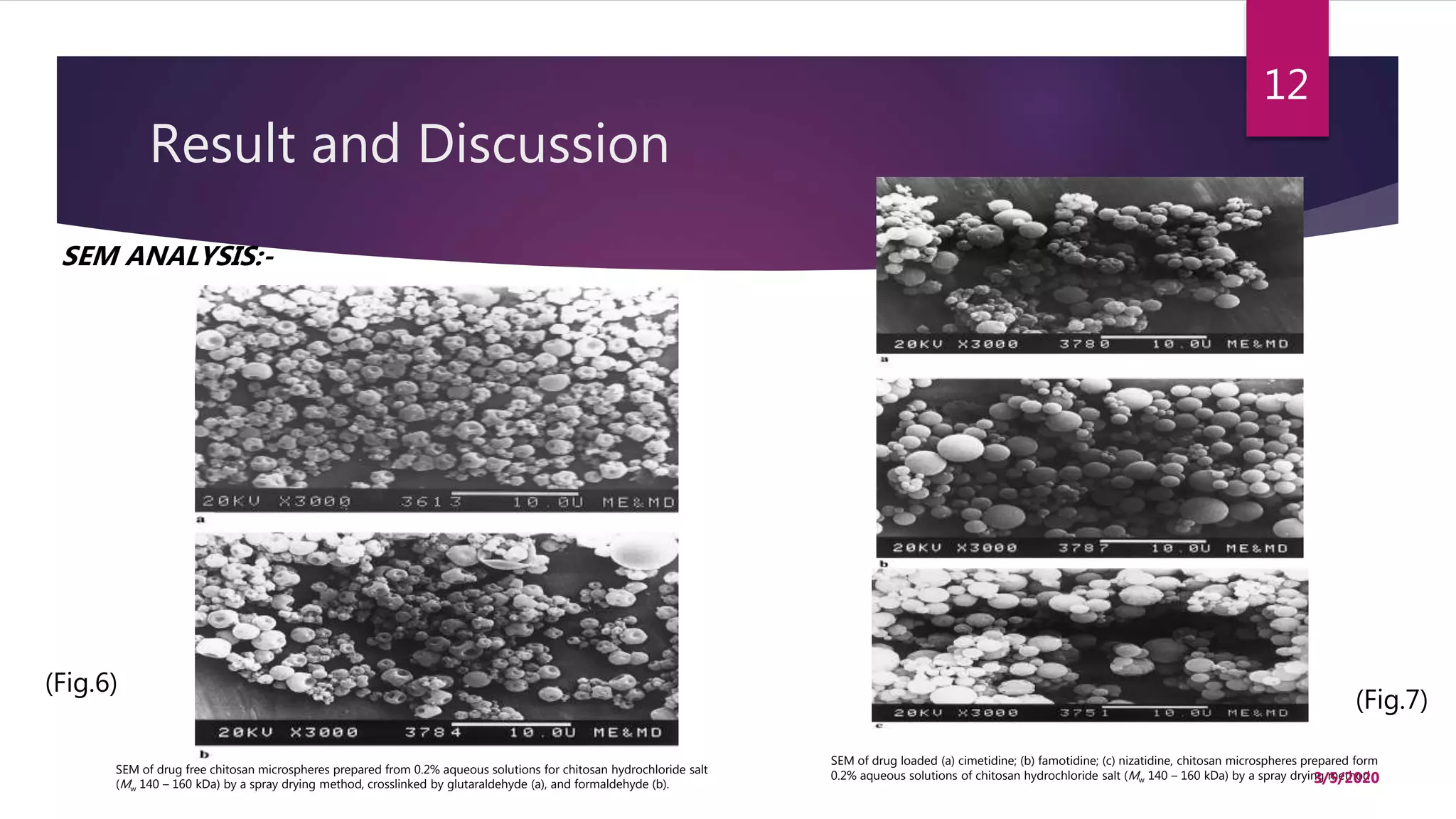
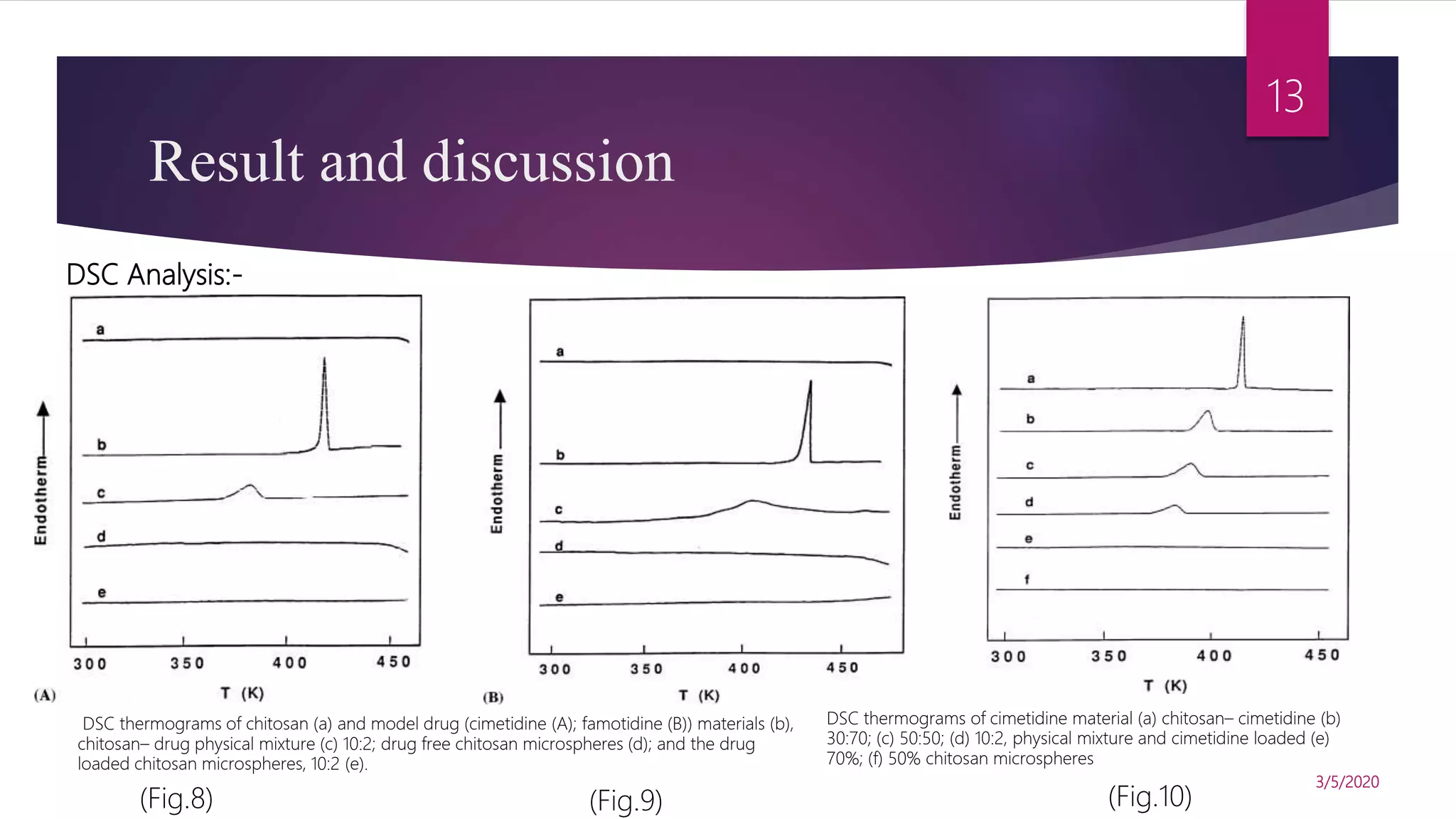
Title : Chitosan microspheres prepared by
spray drying
Department of Pharmaceutics
ISF College of Pharmacy,Moga,Punjab
Presented By:
OMPRAKASH SAHU
M.Pharm 1st year
INTERNATIONAL
JOURNAL OF
PHARMACEUTICS
3/5/2020
1
Flow of presentation
 Introduction
 Methods of preparation
 Characterisation of microsphere
 Result and discussion
 Conclusion
 References
3/5/2020
2
Introduction
 Chitosan is a hydrophilic, biocompatible and biodegradable
polysaccharide of low toxicity, which in recent years has been used
for development of drug delivery systems.
 Non-crosslinked and crosslinked microspheres were prepared by a
spray drying method.
 The microspheres so prepared had a good sphericity and a smooth.
 They were positively charged and particle size range from 2 to 10
µm.
3/5/2020
3
Methods of Preparation
As a comparison, a water insoluble polymer ethyl cellulose(EC) was used to prepare microsphere from 2-4 % polymer
solution in DCM
Yield was formed 200mg to 1g dependent upon the concentration of Chitosan
Spray drying was co-currently performed using a SD-04 Spray dryer(Lab plant, UK)
Dissolving the model drug (16.6 % w/w of cimetidine, famotidine or nizatidine) in the chitosan solution separately
1 % aqueous solution of formaldehyde & glutaraldehyde were added separately as a crosslinking agents
250 ml of a 0.1-0.5 % aqueous solution of chitosan hydrochloride salt containing acetic acid of chitosan free base
were prepared
4
(Fig.1)
(Spray dryer)
3/5/2020
4
Characterisation of Microsphere
Droplet size
• Microsphere were sized
using a Malvern
MasterSizer(MS
1002),determines the
volume diameter(VMD) &
polydispersive index(PDI)
Zeta potential
• Zeta potential of the
microsphere was measured
by laser doppler(Malven
zetasizer 4)employing
0.005,0.0005 & 0.001 M
phosphate buffer at pH 7.0
& 0.001 M acetate buffer
at pH 4.0
DSC
• DSC study was performed
using a perkin elmer DSC-2
• Sample was purged with
atmosphere of
Nitrogen,heat flow rate
was recorded from 280 to
520 K,at a rate of 10 K/min.
3/5/2020
5
Physicochemical Characterization of non-
crosslinked chitosan and EC
Microsphere type Size (µm) Zeta potential (mV)
0.001 M pH 4 acetate buffer 0.0001 M pH 7 phosphate
Chitosan
hydrochloride salt
5.58 27.2 24.9
Chitosan free base
4.19 14.8 9.7
EC 5.1 −15.5 −5.2
3/5/2020
6
Graph between the particle size & amount of the
Glutaraldehyde in different concentration of chitosan
microspheres
(Fig.2)3/5/2020
7
Characteristics of cimetidine loaded
chitosan microspheres
Added Found Efficiency(%) pH4 pH7
Glutaraldehyde (4%)
0.1 1 16.6 15.7 94.7 7.85 17.4 15.0
0.1 2 16.6 13.0 78.3 4.03 15.8 13.6
0.1 4 16.6 11.8 71.0 1.67 15.5 9.1
0.2 1 16.6 16.2 98 4.89 17.3 15.2
0.2 2 16.6 15.6 94 4.62 16.2 14.5
0.2 4 16.6 14.2 85.7 2.93 14.6 13.4
0.2 1 9 8.04 88.5 5.19 17.4 14.4
0.2 2 9 7.11 78.3 4.58 15.5 14.5
0.2 4 9 6.65 73.2 2.22 14.9 13.1
Formaldehyde (1%)
0.2 2 16.6 16.2 97.6 7.91 22.5 17.4
0.2 4 16.6 15.3 92.4 3.89 18.8 15.3
Concentration of chitosan Crosslinking agent (ml) Drug contents (%) Size (µm) Zeta potential (mV) (%)
3/5/2020
8
Determination of drug Content
 For the determination of drug content UV
spectrophotometer(UVIKON 860) were used ,For cimetidine UV
absorption spectra of the sample solution (2-20 µg/mL) were
recorded.
 The absorbance difference at 260 nm were calculated.
 For the determination of famotidine & nizatidine contents, the
absorbance of sample solution (4-20 µg/mL) were recorded at 284
nm & 313 nm respectively.
9
3/5/2020
9
In-vitro drug release
 The rate of release of the model drugs from the
microspheres in phosphate buffer saline(PSB) was
determined in a dissolution apparatus with the dissolution
paddle assembly(USP apparatus 2).
 30-50 mg of microsphere were suspended in 300 ml of
PBS, pH=7.4 at 37℃ & at 50 rpm agitation rate.
3/5/2020
10
Result and discussion
In vitro Drug release study:-
(Fig.3) (Fig.4) (Fig.5)
3/5/2020
11
Result and Discussion
SEM ANALYSIS:-
SEM of drug free chitosan microspheres prepared from 0.2% aqueous solutions for chitosan hydrochloride salt
(Mw 140 – 160 kDa) by a spray drying method, crosslinked by glutaraldehyde (a), and formaldehyde (b).
SEM of drug loaded (a) cimetidine; (b) famotidine; (c) nizatidine, chitosan microspheres prepared form
0.2% aqueous solutions of chitosan hydrochloride salt (Mw 140 – 160 kDa) by a spray drying method
(Fig.6)
(Fig.7)
3/5/2020
12
Result and discussion
DSC Analysis:-
DSC thermograms of chitosan (a) and model drug (cimetidine (A); famotidine (B)) materials (b),
chitosan– drug physical mixture (c) 10:2; drug free chitosan microspheres (d); and the drug
loaded chitosan microspheres, 10:2 (e).
DSC thermograms of cimetidine material (a) chitosan– cimetidine (b)
30:70; (c) 50:50; (d) 10:2, physical mixture and cimetidine loaded (e)
70%; (f) 50% chitosan microspheres
(Fig.8) (Fig.9) (Fig.10)
3/5/2020
13
Conclusion
 Non-crosslinked and crosslinked chitosan microspheres were prepared by a spray
drying method. The microspheres so prepared had a good sphericity and a
smooth but distorted surface morphology. They were positively charged. The
particle size ranged from 2 to 10 µm.
 The release of model drugs (cimetidine, famotidine and nizatidine) from these
microspheres was fast, and accompanied by a burst effect.
3/5/2020
14
References
 He, Ping, Stanley S. Davis, and Lisbeth Illum.
"Chitosan microspheres prepared by spray
drying." International journal of pharmaceutics 187.1
(1999): 53-65.
3/5/2020
15
THANKYOU
3/5/2020
16

Chitosen based Microsphere

  • 1.
    Title : Chitosanmicrospheres prepared by spray drying Department of Pharmaceutics ISF College of Pharmacy,Moga,Punjab Presented By: OMPRAKASH SAHU M.Pharm 1st year INTERNATIONAL JOURNAL OF PHARMACEUTICS 3/5/2020 1
  • 2.
    Flow of presentation Introduction  Methods of preparation  Characterisation of microsphere  Result and discussion  Conclusion  References 3/5/2020 2
  • 3.
    Introduction  Chitosan isa hydrophilic, biocompatible and biodegradable polysaccharide of low toxicity, which in recent years has been used for development of drug delivery systems.  Non-crosslinked and crosslinked microspheres were prepared by a spray drying method.  The microspheres so prepared had a good sphericity and a smooth.  They were positively charged and particle size range from 2 to 10 µm. 3/5/2020 3
  • 4.
    Methods of Preparation Asa comparison, a water insoluble polymer ethyl cellulose(EC) was used to prepare microsphere from 2-4 % polymer solution in DCM Yield was formed 200mg to 1g dependent upon the concentration of Chitosan Spray drying was co-currently performed using a SD-04 Spray dryer(Lab plant, UK) Dissolving the model drug (16.6 % w/w of cimetidine, famotidine or nizatidine) in the chitosan solution separately 1 % aqueous solution of formaldehyde & glutaraldehyde were added separately as a crosslinking agents 250 ml of a 0.1-0.5 % aqueous solution of chitosan hydrochloride salt containing acetic acid of chitosan free base were prepared 4 (Fig.1) (Spray dryer) 3/5/2020 4
  • 5.
    Characterisation of Microsphere Dropletsize • Microsphere were sized using a Malvern MasterSizer(MS 1002),determines the volume diameter(VMD) & polydispersive index(PDI) Zeta potential • Zeta potential of the microsphere was measured by laser doppler(Malven zetasizer 4)employing 0.005,0.0005 & 0.001 M phosphate buffer at pH 7.0 & 0.001 M acetate buffer at pH 4.0 DSC • DSC study was performed using a perkin elmer DSC-2 • Sample was purged with atmosphere of Nitrogen,heat flow rate was recorded from 280 to 520 K,at a rate of 10 K/min. 3/5/2020 5
  • 6.
    Physicochemical Characterization ofnon- crosslinked chitosan and EC Microsphere type Size (µm) Zeta potential (mV) 0.001 M pH 4 acetate buffer 0.0001 M pH 7 phosphate Chitosan hydrochloride salt 5.58 27.2 24.9 Chitosan free base 4.19 14.8 9.7 EC 5.1 −15.5 −5.2 3/5/2020 6
  • 7.
    Graph between theparticle size & amount of the Glutaraldehyde in different concentration of chitosan microspheres (Fig.2)3/5/2020 7
  • 8.
    Characteristics of cimetidineloaded chitosan microspheres Added Found Efficiency(%) pH4 pH7 Glutaraldehyde (4%) 0.1 1 16.6 15.7 94.7 7.85 17.4 15.0 0.1 2 16.6 13.0 78.3 4.03 15.8 13.6 0.1 4 16.6 11.8 71.0 1.67 15.5 9.1 0.2 1 16.6 16.2 98 4.89 17.3 15.2 0.2 2 16.6 15.6 94 4.62 16.2 14.5 0.2 4 16.6 14.2 85.7 2.93 14.6 13.4 0.2 1 9 8.04 88.5 5.19 17.4 14.4 0.2 2 9 7.11 78.3 4.58 15.5 14.5 0.2 4 9 6.65 73.2 2.22 14.9 13.1 Formaldehyde (1%) 0.2 2 16.6 16.2 97.6 7.91 22.5 17.4 0.2 4 16.6 15.3 92.4 3.89 18.8 15.3 Concentration of chitosan Crosslinking agent (ml) Drug contents (%) Size (µm) Zeta potential (mV) (%) 3/5/2020 8
  • 9.
    Determination of drugContent  For the determination of drug content UV spectrophotometer(UVIKON 860) were used ,For cimetidine UV absorption spectra of the sample solution (2-20 µg/mL) were recorded.  The absorbance difference at 260 nm were calculated.  For the determination of famotidine & nizatidine contents, the absorbance of sample solution (4-20 µg/mL) were recorded at 284 nm & 313 nm respectively. 9 3/5/2020 9
  • 10.
    In-vitro drug release The rate of release of the model drugs from the microspheres in phosphate buffer saline(PSB) was determined in a dissolution apparatus with the dissolution paddle assembly(USP apparatus 2).  30-50 mg of microsphere were suspended in 300 ml of PBS, pH=7.4 at 37℃ & at 50 rpm agitation rate. 3/5/2020 10
  • 11.
    Result and discussion Invitro Drug release study:- (Fig.3) (Fig.4) (Fig.5) 3/5/2020 11
  • 12.
    Result and Discussion SEMANALYSIS:- SEM of drug free chitosan microspheres prepared from 0.2% aqueous solutions for chitosan hydrochloride salt (Mw 140 – 160 kDa) by a spray drying method, crosslinked by glutaraldehyde (a), and formaldehyde (b). SEM of drug loaded (a) cimetidine; (b) famotidine; (c) nizatidine, chitosan microspheres prepared form 0.2% aqueous solutions of chitosan hydrochloride salt (Mw 140 – 160 kDa) by a spray drying method (Fig.6) (Fig.7) 3/5/2020 12
  • 13.
    Result and discussion DSCAnalysis:- DSC thermograms of chitosan (a) and model drug (cimetidine (A); famotidine (B)) materials (b), chitosan– drug physical mixture (c) 10:2; drug free chitosan microspheres (d); and the drug loaded chitosan microspheres, 10:2 (e). DSC thermograms of cimetidine material (a) chitosan– cimetidine (b) 30:70; (c) 50:50; (d) 10:2, physical mixture and cimetidine loaded (e) 70%; (f) 50% chitosan microspheres (Fig.8) (Fig.9) (Fig.10) 3/5/2020 13
  • 14.
    Conclusion  Non-crosslinked andcrosslinked chitosan microspheres were prepared by a spray drying method. The microspheres so prepared had a good sphericity and a smooth but distorted surface morphology. They were positively charged. The particle size ranged from 2 to 10 µm.  The release of model drugs (cimetidine, famotidine and nizatidine) from these microspheres was fast, and accompanied by a burst effect. 3/5/2020 14
  • 15.
    References  He, Ping,Stanley S. Davis, and Lisbeth Illum. "Chitosan microspheres prepared by spray drying." International journal of pharmaceutics 187.1 (1999): 53-65. 3/5/2020 15
  • 16.