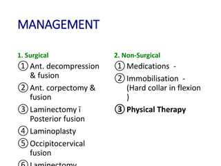
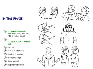
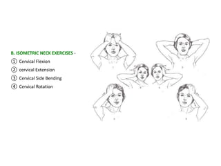
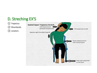
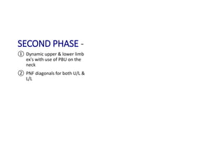
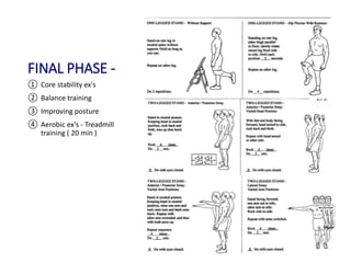
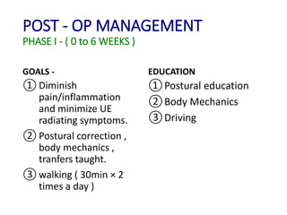
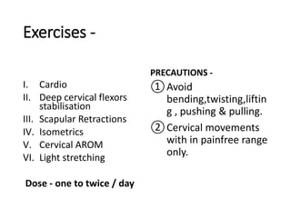
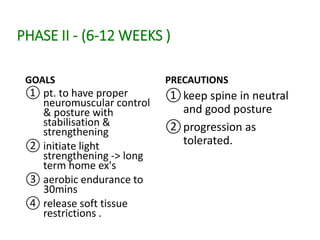
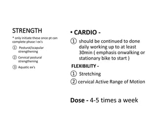
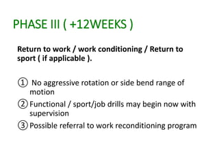
CERVICAL
MYELOPATHY,
PHYSIOTHERAPY
ASSESSMENT AND
MANAGEMENT
PRESENTED BY :
Dr. DWARIKANATH ROUT (PT)
MPT (ORTHOPEDICS)
KIMS HOSPITAL, Odisha
CONTENTS -
• Definition
• Epidemiology
• Etiology
• Clinical presentation
• Clinical sign & Symptoms
• Physiotherapy assessment
• Investigation
• Differential Diagnosis
• Management
• References
DEFINITION -
A form of neurologic impairment caused by
compression of the cervical spinal cord , most
commonly due to degenerative cervical spondylosis .
EPIDEMIOLOGY -
 Most common in men > women
 Earlier in men ( 50 years ) than in women ( 60
years )
It causes hospitalization at a rate of 4.04 per
100,000 person-years.
ETIOLOGY
STATIC FACTORS:
• A narrowing of the spinal canal size commonly results from
degenerative changes in cervical spine such as disc
degeneration, spondylosis, stenosis, osteophyte formation
at the level of facet joints, segmental ossification of
posterior longitudinal ligament and yellow ligament
hypertrophy, classification or ossification.
DYNAMIC FACTORS:
• Due to mechanical abnormalities of the cervical spine or
instability.
VASCULAR AND CELLUALR FACTORS:
• Spinal cord ischemia affects oligodendrocytes which results
in demyelination exhibiting feature of chronic degenerative
disorders.
ETIOLOGY
CONGENITAL
• Myelopathy due to congenital stenosis does not have a
specific lesion but caused by a canal diameter which is
narrower from birth.
SPONDYLOSIS
• Degenerative changes which develop with age, including
ligamentum flavum hypertrophy or buckling, facet joint
hypertrophy and disc protrusion. One or all of these
changes contribute to an overall reduction in canal diameter
which may result in cord compression.
DISC HERNIATION:
• Discogenic disease may cause myelopathy in the acute
setting as a large central soft disc herniation causing cord
compression
POST TRAUMATIC MYELOPATHY:
• Trauma may include myelopathy or precipitate symptoms of
stenosis of spinal cord. Smaller diameter canals have an increased
chance of neurological injury in trauma.
MYELOPATHY DUE TO TUMOR EXPANSION:
• Intraspinal tumors may originate in spinal cord or compress from
outside.
OTHER ETIOLOGIES:
• Cervical rheumatoid arthritis especially upper cervical spine and
to a lesser extend the lower cervical spine may present with the
clinical picture of cervical myelopathy.
• Rare neurologic complications which can cause narrowing of
cervical canal such as ankylosing spondylitis, gouty tophi from
posterior joint and paget’s disease.
CLINICAL PRESENTATION
• Neck stiffness (early complaint)
• Leg weakness, stiffness (proximal-distal)
• Gait abnormalities
• Difficulty with fine motor movements and tasks with hands “clumsy
myelopathic hands”
• Loss of bowel or bladder control
• Heavy feeling in the legs
• Poor exercise tolerance
• Radiculopathy
• Numbness and tingling in the limbs
• Chronic suboccipital headache : suboccipital may radiate to base of the
neck and vertex of the skull
• L’Hermitte’s phenomenon- intermittent electric shock sensations down
the neck, back and limbs, exacerbated by neck flexion
• Myelopathy’s hand: clumsiness, intrinsic wasting
CLINICAL SIGNS AND SYMPTOMS
• Patients may present with pain, paresthesia, weakness
or combination of these symptoms.
• Pain usually is in the cervical region, upper limb,
shoulder and/or intrascapular region, intermittent
shoulder pain.
• >2/3 patients present with unilateral or bilateral
shoulder pain.
• 1/3 patients present with headache
• Radicular signs: often non dermatomal
• Radiculopathy most commonly 6th and 7th roots occurs
from C5-6 or C6-7 spondylosis
• More pain proximally in their limbs, while parasthesias
dominate distally
CLUSTER FOR MYELOPATHY -
According to cook et al, selected combinations of the
following clinical findings are effective in ruling out and
ruling in cervical spine myelopathy. Combinations of 3
or 4 or 5 of these tests enable post-test probability of
the condition to 90-99% :
① Gait deviation
② +ve Hoffman's sign
③ Inverted Supinator sign
④ +ve Babinski sign
⑤ Age > 45 years or older
PHYSIOTHERAPY ASSESSMENT
• DEMOGRAPHIC DATA
• NAME- AGE- SEX- OCCUPATION-
• CHIEF COMPLAINT:
• HISTORY:
• PRESENT HISTORY: Date of onset of symptoms, mechanism of injury,
mode of onset, condition (Improved, stationary, Deteriorated), muscular
weakness
• PAIN HISTORY: Duration, type, aggravating and relieving factors
• PAST HISTORY: Any history of TB, Bronchial asthma, BP, diabetes, cardiac
problems, enquiry made for any accidental injury)
• FAMILY HISTORY: Hereditary?, consanguinity?
• PERSONAL HISTORY: Cigarettes, Alcohol
• SOCIO-ECONOMIC HISTORY:
• MEDICAL HISTORY: Present medication patient is on
• ON OBSERVATION :
• General condition of patient- poor, good, fair built
• Wasting
• Oedema
• Attitude of limbs : supine, sitting, standing
• Types of gait
• Bony contours
• Deformities
ON PALPATION:
• Tenderness- grading (1-patient complains of pain,
2-patient complains of pain and winces, 3- patient
winces and withdraws, 4- patient will not allow
palpation of the joint)
• Tissue tension and texture
• Temperature variation of skin
• Spasm
• Type of skin – Dry or excessive moisture
• Scar – adherent/ Non adherent
Swelling
• Comes on soon after injury- blood
• Comes on after 8-24 hours- synovial
• Boggy, spongy feeling- synovial
• Harder, tense feeling with warmth- blood
• Tough, dry- callus
• Leathery thickening – chronic
• Soft fluctuating- acute
• Hard- bone
• Thick , slow moving- pitting oedema
ON EXAMINATION :
• Patients present with a number of clinical findings which are
predominantly upper motor neuron signs.
• Weakness is more severe in the upper limbs.
• Gait is usually affected with an ataxic broad based gait, usually
spastic and spastic
• Hypertonia- increased resting muscle tone identified by passive
movement
• Hyperreflexia- exaggerated response to normal physiological reflexes
• Exaggerated tendon reflexes (patellar and achilles), presence of
pathological reflexes (e.g. clonus, Babinski and Hoffman’s sign)
• Ankle clonus- forced dorsiflexion at the ankle giving rise to sustained
beats of clonus (more than 3 beats is considered pathological)
• Muscular atrophy: supraspinatus, infraspinatus, deltoid,
triceps and the first dorsal interosseus muscle
• Motor weakness, most commonly in the iliopsoas followed
by quadriceps femoris
• Sensory abnormalities: variable pattern
• Loss of vibratory sense or proprioception in the extremities
can occur
• Spinothalamic sensory loss may be asymmetric
• Most commonly sensory symptoms including:
• Upper extremity numbness & pain, paresthesias initially,
followed by lower extremity sensory changes
• Motor dysfunction may be unilateral or bilateral depending
on the extend and location of cord damage
• Babinski sign – extension of the great toe on scratching
of the sole of the foot
• Hoffman’s reflex- flicking of the terminal phalanx of the
middle or ring finger causing concurrent flexion at the
terminal phalanx of the thumb and index finger
• Finger escape sign – the small finger spontaneously
abducts due to weak intrinsic muscles
• Spurling’s test
• Distraction test
• Hyper reflexic biceps, quadriceps, achilles
• Romberg test
SPURLING TEST & LHERMITTE’S SIGN
NERVE ROOT INVOLVEMENT SYMPTOMS
C3 Nerve Root ① uncommon
② Rx pain may be present as
neck pain or occipital pain
C4 Nerve Root ① C4 Rx ī pain radiating to post
neck , trapezius and ant. chest.
C5 Nerve Root ① presents with Pain and/or
numbness over lat. aspect of
shoulder & deltoid.
② weakness of biceps,
supraspinatus, infraspinatus
③ Abduction relief sign
C6 Nerve Root ① Weakness of biceps , ECR
② Impaired EF & WE
③ sensation loss over thumb &
lat. portion of index finger.
④ dim. biceps & brachioradialis
reflex
⑤ pain radiates from neck-lateral
arm & forearm into the thumb
NERVE ROOT SYMPTOMS
C7 Nerve root ① impaired EE and WF
② dim. Tricep reflex
③ pain & numbnessfrom neck to
arm & digits 2-4
④ Horner's syndrome rarely seen
C8 Nerve root ① Weakness in hands intrinsic
muscles , Wrist extensors , and
flexors
② not sble tofully extend 4th &
5th digits
③ sensory loss- medial forearm &
4th 5th digits
④ pain radiating - neck->medial
forearm-> last 2 digits
⑤ Horner's syndrome-rare
T1 Nerve Root ① weakness- intrinsic muscles
② froment's sign +ve
③ Horner's syndome rare
INVESTIGATION
① Radiographs
• Pavlov index – the antero-
posterior diameter of the
spinal canal measured from
the middle of the posterior
vertebral body to the
nearest point of the
spinous process is equal to
or less than the antero-
posterior diameter of the
vertebral body.
• Should be 1.0 with <0.85
indicating stenosis
2 . MRI
• Compression Ratio -
( < 0.4 indicates poor
prognosis )
CR =
smallest AP diameter of
cord
Largest transverse
diameter of cord
• A canal diameter of 17mm or greater at the mid
vertebral body level is considered normal.
• < 10-13mm - risk of cervical spondylosis
• DIAMETER OF CERVICAL SPINAL CANAL
C1 22.1mm
C2 18.8mm
C3 16.2mm
C4 15.8mm
C5 15.7mm
C6 15.6mm
C7 15.9mm
Differential Diagnosis
MANAGEMENT
1. Surgical
①Ant. decompression
& fusion
②Ant. corpectomy &
fusion
③Laminectomy ī
Posterior fusion
④Laminoplasty
⑤Occipitocervical
fusion
2. Non-Surgical
①Medications -
②Immobilisation -
(Hard collar in flexion
)
③Physical Therapy
PHYSIOTHERAPY MANAGEMENT
GOALS -
①To relieve pain
②To improve function
③To prevent neurological functions
④To reverse or improve neurological deficits
INITIAL PHASE -
① A. Electrotherapeutics
modalities like , TENS, US ,
IFT ( to reduce pain )
②
B. CERVICAL STABLISATION
EX'S
③ Chin Tuck
④ Chin tuck into towel
⑤ Cervical Extension
⑥ Shoulder Shrugs
⑦ Shoulder Rolls
⑧ Scapular Retraction
B. ISOMETRIC NECK EXERCISES -
① Cervical Flexion
② cervical Extension
③ Cervical Side Bending
④ Cervical Rotation
D. Streching EX'S
① Trapezius
② Rhomboids
③ Levators
SECOND PHASE -
① Dynamic upper & lower limb
ex's with use of PBU on the
neck
② PNF diagonals for both U/L &
L/L
FINAL PHASE -
① Core stability ex's
② Balance training
③ Improving posture
④ Aerobic ex's - Treadmill
training ( 20 min )
POST - OP MANAGEMENT
PHASE I - ( 0 to 6 WEEKS )
GOALS -
① Diminish
pain/inflammation
and minimize UE
radiating symptoms.
② Postural correction ,
body mechanics ,
tranfers taught.
③ walking ( 30min × 2
times a day )
EDUCATION
①Postural education
②Body Mechanics
③Driving
Exercises -
I. Cardio
II. Deep cervical flexors
stabilisation
III. Scapular Retractions
IV. Isometrics
V. Cervical AROM
VI. Light stretching
Dose - one to twice / day
PRECAUTIONS -
①Avoid
bending,twisting,liftin
g , pushing & pulling.
②Cervical movements
with in painfree range
only.
PHASE II - (6-12 WEEKS )
GOALS
① pt. to have proper
neuromuscular control
& posture with
stabilisation &
strengthening
② initiate light
strengthening -> long
term home ex's
③ aerobic endurance to
30mins
④ release soft tissue
restrictions .
PRECAUTIONS
①keep spine in neutral
and good posture
②progression as
tolerated.
STRENGTH
* only initiate these once pt can
complete phase I ex's
① Postural/scapular
strengthening
② Cervical postural
strengthening
③ Aquatic ex's
PHASE III ( +12WEEKS )
Return to work / work conditioning / Return to
sport ( if applicable ).
① No aggressive rotation or side bend range of
motion
②Functional / sport/job drills may begin now with
supervision
③Possible referral to work reconditioning program
THANK YOU !

CERVICAL MYELOPATHY

  • 1.
    CERVICAL MYELOPATHY, PHYSIOTHERAPY ASSESSMENT AND MANAGEMENT PRESENTED BY: Dr. DWARIKANATH ROUT (PT) MPT (ORTHOPEDICS) KIMS HOSPITAL, Odisha
  • 2.
    CONTENTS - • Definition •Epidemiology • Etiology • Clinical presentation • Clinical sign & Symptoms • Physiotherapy assessment • Investigation • Differential Diagnosis • Management • References
  • 3.
    DEFINITION - A formof neurologic impairment caused by compression of the cervical spinal cord , most commonly due to degenerative cervical spondylosis . EPIDEMIOLOGY -  Most common in men > women  Earlier in men ( 50 years ) than in women ( 60 years ) It causes hospitalization at a rate of 4.04 per 100,000 person-years.
  • 4.
    ETIOLOGY STATIC FACTORS: • Anarrowing of the spinal canal size commonly results from degenerative changes in cervical spine such as disc degeneration, spondylosis, stenosis, osteophyte formation at the level of facet joints, segmental ossification of posterior longitudinal ligament and yellow ligament hypertrophy, classification or ossification. DYNAMIC FACTORS: • Due to mechanical abnormalities of the cervical spine or instability. VASCULAR AND CELLUALR FACTORS: • Spinal cord ischemia affects oligodendrocytes which results in demyelination exhibiting feature of chronic degenerative disorders.
  • 5.
    ETIOLOGY CONGENITAL • Myelopathy dueto congenital stenosis does not have a specific lesion but caused by a canal diameter which is narrower from birth. SPONDYLOSIS • Degenerative changes which develop with age, including ligamentum flavum hypertrophy or buckling, facet joint hypertrophy and disc protrusion. One or all of these changes contribute to an overall reduction in canal diameter which may result in cord compression. DISC HERNIATION: • Discogenic disease may cause myelopathy in the acute setting as a large central soft disc herniation causing cord compression
  • 6.
    POST TRAUMATIC MYELOPATHY: •Trauma may include myelopathy or precipitate symptoms of stenosis of spinal cord. Smaller diameter canals have an increased chance of neurological injury in trauma. MYELOPATHY DUE TO TUMOR EXPANSION: • Intraspinal tumors may originate in spinal cord or compress from outside. OTHER ETIOLOGIES: • Cervical rheumatoid arthritis especially upper cervical spine and to a lesser extend the lower cervical spine may present with the clinical picture of cervical myelopathy. • Rare neurologic complications which can cause narrowing of cervical canal such as ankylosing spondylitis, gouty tophi from posterior joint and paget’s disease.
  • 7.
    CLINICAL PRESENTATION • Neckstiffness (early complaint) • Leg weakness, stiffness (proximal-distal) • Gait abnormalities • Difficulty with fine motor movements and tasks with hands “clumsy myelopathic hands” • Loss of bowel or bladder control • Heavy feeling in the legs • Poor exercise tolerance • Radiculopathy • Numbness and tingling in the limbs • Chronic suboccipital headache : suboccipital may radiate to base of the neck and vertex of the skull • L’Hermitte’s phenomenon- intermittent electric shock sensations down the neck, back and limbs, exacerbated by neck flexion • Myelopathy’s hand: clumsiness, intrinsic wasting
  • 8.
    CLINICAL SIGNS ANDSYMPTOMS • Patients may present with pain, paresthesia, weakness or combination of these symptoms. • Pain usually is in the cervical region, upper limb, shoulder and/or intrascapular region, intermittent shoulder pain. • >2/3 patients present with unilateral or bilateral shoulder pain. • 1/3 patients present with headache • Radicular signs: often non dermatomal • Radiculopathy most commonly 6th and 7th roots occurs from C5-6 or C6-7 spondylosis • More pain proximally in their limbs, while parasthesias dominate distally
  • 9.
    CLUSTER FOR MYELOPATHY- According to cook et al, selected combinations of the following clinical findings are effective in ruling out and ruling in cervical spine myelopathy. Combinations of 3 or 4 or 5 of these tests enable post-test probability of the condition to 90-99% : ① Gait deviation ② +ve Hoffman's sign ③ Inverted Supinator sign ④ +ve Babinski sign ⑤ Age > 45 years or older
  • 10.
    PHYSIOTHERAPY ASSESSMENT • DEMOGRAPHICDATA • NAME- AGE- SEX- OCCUPATION- • CHIEF COMPLAINT: • HISTORY: • PRESENT HISTORY: Date of onset of symptoms, mechanism of injury, mode of onset, condition (Improved, stationary, Deteriorated), muscular weakness • PAIN HISTORY: Duration, type, aggravating and relieving factors • PAST HISTORY: Any history of TB, Bronchial asthma, BP, diabetes, cardiac problems, enquiry made for any accidental injury) • FAMILY HISTORY: Hereditary?, consanguinity? • PERSONAL HISTORY: Cigarettes, Alcohol • SOCIO-ECONOMIC HISTORY: • MEDICAL HISTORY: Present medication patient is on
  • 11.
    • ON OBSERVATION: • General condition of patient- poor, good, fair built • Wasting • Oedema • Attitude of limbs : supine, sitting, standing • Types of gait • Bony contours • Deformities
  • 12.
    ON PALPATION: • Tenderness-grading (1-patient complains of pain, 2-patient complains of pain and winces, 3- patient winces and withdraws, 4- patient will not allow palpation of the joint) • Tissue tension and texture • Temperature variation of skin • Spasm • Type of skin – Dry or excessive moisture • Scar – adherent/ Non adherent
  • 13.
    Swelling • Comes onsoon after injury- blood • Comes on after 8-24 hours- synovial • Boggy, spongy feeling- synovial • Harder, tense feeling with warmth- blood • Tough, dry- callus • Leathery thickening – chronic • Soft fluctuating- acute • Hard- bone • Thick , slow moving- pitting oedema
  • 14.
    ON EXAMINATION : •Patients present with a number of clinical findings which are predominantly upper motor neuron signs. • Weakness is more severe in the upper limbs. • Gait is usually affected with an ataxic broad based gait, usually spastic and spastic • Hypertonia- increased resting muscle tone identified by passive movement • Hyperreflexia- exaggerated response to normal physiological reflexes • Exaggerated tendon reflexes (patellar and achilles), presence of pathological reflexes (e.g. clonus, Babinski and Hoffman’s sign) • Ankle clonus- forced dorsiflexion at the ankle giving rise to sustained beats of clonus (more than 3 beats is considered pathological)
  • 15.
    • Muscular atrophy:supraspinatus, infraspinatus, deltoid, triceps and the first dorsal interosseus muscle • Motor weakness, most commonly in the iliopsoas followed by quadriceps femoris • Sensory abnormalities: variable pattern • Loss of vibratory sense or proprioception in the extremities can occur • Spinothalamic sensory loss may be asymmetric • Most commonly sensory symptoms including: • Upper extremity numbness & pain, paresthesias initially, followed by lower extremity sensory changes • Motor dysfunction may be unilateral or bilateral depending on the extend and location of cord damage
  • 16.
    • Babinski sign– extension of the great toe on scratching of the sole of the foot • Hoffman’s reflex- flicking of the terminal phalanx of the middle or ring finger causing concurrent flexion at the terminal phalanx of the thumb and index finger • Finger escape sign – the small finger spontaneously abducts due to weak intrinsic muscles • Spurling’s test • Distraction test • Hyper reflexic biceps, quadriceps, achilles • Romberg test
  • 17.
    SPURLING TEST &LHERMITTE’S SIGN
  • 21.
    NERVE ROOT INVOLVEMENTSYMPTOMS C3 Nerve Root ① uncommon ② Rx pain may be present as neck pain or occipital pain C4 Nerve Root ① C4 Rx ī pain radiating to post neck , trapezius and ant. chest. C5 Nerve Root ① presents with Pain and/or numbness over lat. aspect of shoulder & deltoid. ② weakness of biceps, supraspinatus, infraspinatus ③ Abduction relief sign C6 Nerve Root ① Weakness of biceps , ECR ② Impaired EF & WE ③ sensation loss over thumb & lat. portion of index finger. ④ dim. biceps & brachioradialis reflex ⑤ pain radiates from neck-lateral arm & forearm into the thumb
  • 22.
    NERVE ROOT SYMPTOMS C7Nerve root ① impaired EE and WF ② dim. Tricep reflex ③ pain & numbnessfrom neck to arm & digits 2-4 ④ Horner's syndrome rarely seen C8 Nerve root ① Weakness in hands intrinsic muscles , Wrist extensors , and flexors ② not sble tofully extend 4th & 5th digits ③ sensory loss- medial forearm & 4th 5th digits ④ pain radiating - neck->medial forearm-> last 2 digits ⑤ Horner's syndrome-rare T1 Nerve Root ① weakness- intrinsic muscles ② froment's sign +ve ③ Horner's syndome rare
  • 23.
    INVESTIGATION ① Radiographs • Pavlovindex – the antero- posterior diameter of the spinal canal measured from the middle of the posterior vertebral body to the nearest point of the spinous process is equal to or less than the antero- posterior diameter of the vertebral body. • Should be 1.0 with <0.85 indicating stenosis
  • 24.
    2 . MRI •Compression Ratio - ( < 0.4 indicates poor prognosis ) CR = smallest AP diameter of cord Largest transverse diameter of cord
  • 25.
    • A canaldiameter of 17mm or greater at the mid vertebral body level is considered normal. • < 10-13mm - risk of cervical spondylosis • DIAMETER OF CERVICAL SPINAL CANAL C1 22.1mm C2 18.8mm C3 16.2mm C4 15.8mm C5 15.7mm C6 15.6mm C7 15.9mm
  • 29.
  • 30.
    MANAGEMENT 1. Surgical ①Ant. decompression &fusion ②Ant. corpectomy & fusion ③Laminectomy ī Posterior fusion ④Laminoplasty ⑤Occipitocervical fusion 2. Non-Surgical ①Medications - ②Immobilisation - (Hard collar in flexion ) ③Physical Therapy
  • 31.
    PHYSIOTHERAPY MANAGEMENT GOALS - ①Torelieve pain ②To improve function ③To prevent neurological functions ④To reverse or improve neurological deficits
  • 32.
    INITIAL PHASE - ①A. Electrotherapeutics modalities like , TENS, US , IFT ( to reduce pain ) ② B. CERVICAL STABLISATION EX'S ③ Chin Tuck ④ Chin tuck into towel ⑤ Cervical Extension ⑥ Shoulder Shrugs ⑦ Shoulder Rolls ⑧ Scapular Retraction
  • 33.
    B. ISOMETRIC NECKEXERCISES - ① Cervical Flexion ② cervical Extension ③ Cervical Side Bending ④ Cervical Rotation
  • 34.
    D. Streching EX'S ①Trapezius ② Rhomboids ③ Levators
  • 35.
    SECOND PHASE - ①Dynamic upper & lower limb ex's with use of PBU on the neck ② PNF diagonals for both U/L & L/L
  • 36.
    FINAL PHASE - ①Core stability ex's ② Balance training ③ Improving posture ④ Aerobic ex's - Treadmill training ( 20 min )
  • 37.
    POST - OPMANAGEMENT PHASE I - ( 0 to 6 WEEKS ) GOALS - ① Diminish pain/inflammation and minimize UE radiating symptoms. ② Postural correction , body mechanics , tranfers taught. ③ walking ( 30min × 2 times a day ) EDUCATION ①Postural education ②Body Mechanics ③Driving
  • 38.
    Exercises - I. Cardio II.Deep cervical flexors stabilisation III. Scapular Retractions IV. Isometrics V. Cervical AROM VI. Light stretching Dose - one to twice / day PRECAUTIONS - ①Avoid bending,twisting,liftin g , pushing & pulling. ②Cervical movements with in painfree range only.
  • 39.
    PHASE II -(6-12 WEEKS ) GOALS ① pt. to have proper neuromuscular control & posture with stabilisation & strengthening ② initiate light strengthening -> long term home ex's ③ aerobic endurance to 30mins ④ release soft tissue restrictions . PRECAUTIONS ①keep spine in neutral and good posture ②progression as tolerated.
  • 40.
    STRENGTH * only initiatethese once pt can complete phase I ex's ① Postural/scapular strengthening ② Cervical postural strengthening ③ Aquatic ex's
  • 41.
    PHASE III (+12WEEKS ) Return to work / work conditioning / Return to sport ( if applicable ). ① No aggressive rotation or side bend range of motion ②Functional / sport/job drills may begin now with supervision ③Possible referral to work reconditioning program
  • 42.