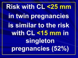
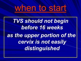
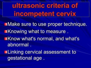
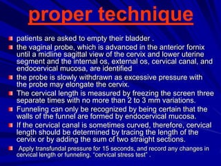
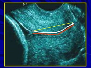
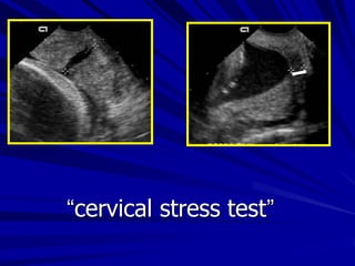
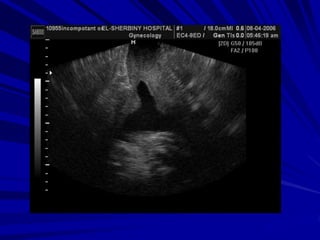
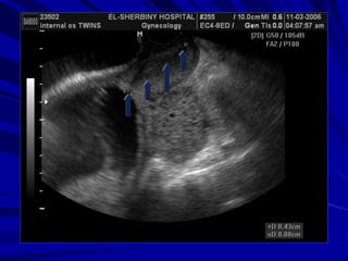
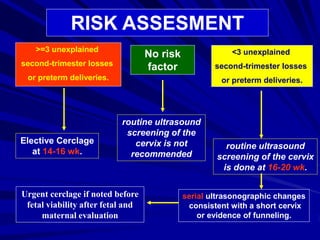
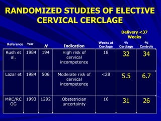
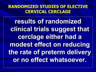
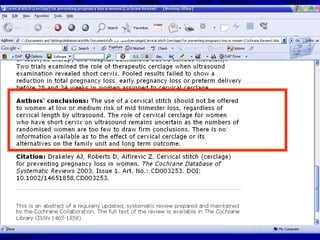
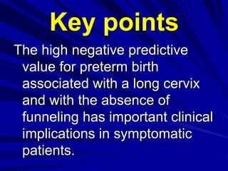
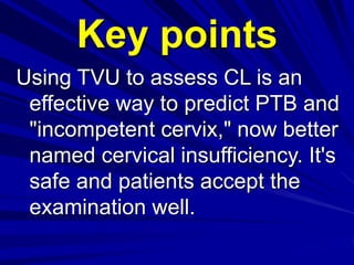
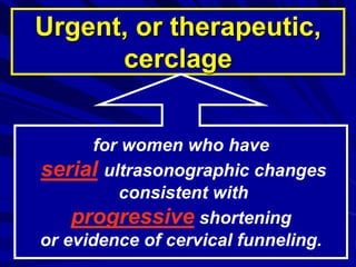
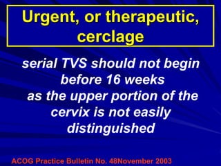
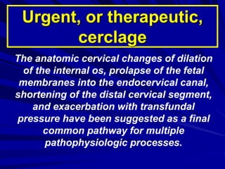
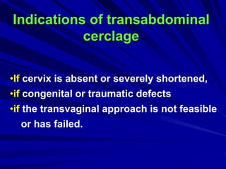
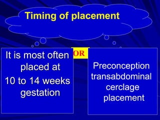
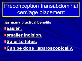
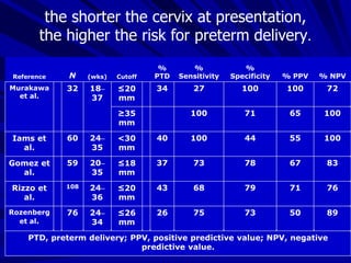
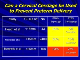
Dr.Mohammed Abdalla
Domiat general hospital
cervical
incompetence
The cervix is said
competent when it
retains pregnancy till
term.
if not it is considered
incompetent .
Mostly incompetence is
idiopathic but it may be
secondary to anatomical,
traumatic, or congenital
connective tissue
disorder.
Although the efficacy of cerclage for
cervical incompetence has never
been fully confirmed in randomized
clinical trials, the role of cerclage
has been expanded to include
women with “risk factors” for
spontaneous preterm birth or
nonreassuring sonographic cervical
findings in the mid trimester.
So before you send
your patient to the
theater for cerclage
your diagnosis
necessities solid
criteria.
But unfortunately there is no
consensus about the
cervical cut off length as
most literatures failed to
state a discriminatory
cervical length, which varied
widely between 15-25mm. in
singleton pregnancy
Cervical cut off
length in singleton
pregnancy
is much more than
in multiple
pregnancy.
Risk with CL <25 mm
in twin pregnancies
is similar to the risk
with CL <15 mm in
singleton
pregnancies (52%(
4%
35%
This wide variation in
discriminatory cervical
length will result in the
categorization of 5% to
10% of pregnant women
as having a short
cervix.
As any controversial
issue we have here
white and black faces
but always within the
grey zone, which lies in
between, we fall in
doubt.
White face here is the
women with
irrelevant obstetric
and gynecological
history, as they need
no screening.
But those who have three or
more midtrimester losses or
preterm births represent the
black face of the problem
and the decision is
a prophylactic cerclage
performed at 13 to 16 weeks
of gestation
.
The grey zone represented
here by those women of
low or moderate risk, and
they need an ultrasound
screening by transvaginal
ultrasonography.
Ultrasound screening
if we are going to screen this group
of patients with mild to moderate
risk :
when to start?
what is the ultrasonic criteria of
incompetent cervix?
and when to intervent?
when to start
TVS should not begin
before 16 weeks
as the upper portion of the
cervix is not easily
distinguished
ultrasonic criteria of
incompetent cervix
Make sure to use proper technique.
Knowing what to measure .
Know what's normal, and what's
abnormal .
Linking cervical assessment to
gestational age .
proper technique
patients are asked to empty their bladder .
the vaginal probe, which is advanced in the anterior fornix
until a midline sagittal view of the cervix and lower uterine
segment and the internal os, external os, cervical canal, and
endocervical mucosa, are identified
the probe is slowly withdrawn as excessive pressure with
the probe may elongate the cervix.
The cervical length is measured by freezing the screen three
separate times with no more than 2 to 3 mm variations.
Funneling can only be recognized by being certain that the
walls of the funnel are formed by endocervical mucosa.
If the cervical canal is sometimes curved, therefore, cervical
length should be determined by tracing the length of the
cervix or by adding the sum of two straight sections.
Apply transfundal pressure for 15 seconds, and record any changes in
cervical length or funneling. “cervical stress test” .
“cervical stress test”
what's normal
In low-risk women, CL during
pregnancy has a mean of 35 to
40 mm from 14 to 30 weeks.
the lower 10th percentile being
25 mm and the upper 10th (90th
percentile) 50 mm.
3812521998Heath et al
421751997Tongsong et al
41411996Cook et al
3529151996Iams et al
371061995Iams et al
421541994Zorzoli et al
371771993Murakawa et al
42771991Andersen et al
48241990Kushnir et al
411251990Andersen et al
48801988Podobnik et al
521501988Ayers et al
Cervical Length (mm)NYearReference
CERVICAL LENGTH (MEAN OR MEDIAN) IN LOW-RISK POPULATIONS IN MIDTRIMESTER
The discriminatory length
of cervical shortening
varies widely between
25mm to 15mm
what's abnormal?
97
4799
8≤1514–24Hassan et
al
99
5299
58≤1523Heath et al
99
39100
6≤2518–22Taipale et al
97
2697
23<2024Iams et al
% NPV% PPV
%
Specificity
%
SensitivityCutoff(wks)Reference
value of cervical sonography
in the screening of preterm
birth
Low %PPV means
that many undue
cerclages were
done.
high %NPV means
that the test is
reassuring when
negative.
So we cannot rely
on cervical length
alone as a predictor
of incompetence
the progressive shortening
detected by serial sonar,
funneling (width and length),
v-shaped lower uterine segment
and dynamic cervical changes
with fundal or suprapubic
pressure.
What are the most
important?
1 2
3 4
bulging of the membranes in the vagina. The fetal
lower limb protruded into the vagina.
But how to avoid
undue cerclage
and
how not to miss a
case?
RISK ASSESMENT
>=3 unexplained
second-trimester losses
or preterm deliveries.
Elective Cerclage
at 14-16 wk.
No risk
factor
routine ultrasound
screening of the
cervix is not
recommended
<3 unexplained
second-trimester losses
or preterm deliveries.
routine ultrasound
screening of the cervix
is done at 16-20 wk.
serial ultrasonographic changes
consistent with a short cervix
or evidence of funneling.
Urgent cerclage if noted before
fetal viability after fetal and
maternal evaluation
Can a Cervical
Cerclage be Used to
Prevent Preterm
Delivery in Patients
with a Short Cervix
or Funneling?
263116Obstetrician
uncertainty
12921993MRC/RC
OG
6.75.5<28Moderate risk of
cervical
incompetence
5061984Lazar et
343218High risk of
cervical
incompetence
1941984Rush et
al.
%
Controls
%
Cerclage
Weeks at
CerclageIndicationNReference Year
Delivery <37
Weeks
RANDOMIZED STUDIES OF ELECTIVE
CERVICAL CERCLAGE
results of randomized
clinical trials suggest that
cerclage either had a
modest effect on reducing
the rate of preterm delivery
or no effect whatsoever.
RANDOMIZED STUDIES OF ELECTIVE
CERVICAL CERCLAGE
Key points
The high negative predictive
value for preterm birth
associated with a long cervix
and with the absence of
funneling has important clinical
implications in symptomatic
patients.
Using TVU to assess CL is an
effective way to predict PTB and
"incompetent cervix," now better
named cervical insufficiency. It's
safe and patients accept the
examination well.
Key points
Screening frequency should
depend on severity of obstetric
history, with serial TVU of the
cervix having a better predictive
accuracy than one, especially in
high-risk populations.
Key points
the shorter the cervix, the
higher the risk of PTB, and
the earlier in gestational age
at which the shortening
occurs, the higher the risk.
Key points
screening high-risk women with TVU
of the cervix and placement of a
cerclage for the short or funneled
cervix should not be considered
standard care until proven by
properly conducted, large
randomized trials
Key points
Thank
you
Relative Risk
(95% Cl)NPVPPV
Specifici
ty
Sensitivi
ty
6.4 (2.8–14.7)86%
(31/36)
89%
(16/18)
94%
(31/33)
76%
(16/21)
Cervical index
≥ 0.52
3.9 (1.8–8.5)83%
(29/35)
67%
(16/24)
78%
(29/37)
73%
(16/22)
Cervical length
≤ 18 mm
2.0 (1.4–6.0)78%
(25/32)
64%
(14/22)
76%
(25/33)
67%
(14/21)
Funnel width
≥ 6 mm
5.0 (2.3–10.7)83%
(30/36)
83%
(15/18)
91%
(30/33)
71%
(15/21)
Funnel length
≥ 9 mm
2.5 (1.1–5.9)80%
(20/25)
50%
(17/34)
54%
(20/37)
77%
(17/22)
Funneling
present
Gomez R, et al., Am J Obstet Gynecol. 1994;171:956–964.
Urgent, or therapeutic,
cerclage
for women who have
serial ultrasonographic changes
consistent with
progressive shortening
or evidence of cervical funneling.
ACOG Practice Bulletin No. 48November 2003
serial TVS should not begin
before 16 weeks
as the upper portion of the
cervix is not easily
distinguished
Urgent, or therapeutic,
cerclage
The anatomic cervical changes of dilation
of the internal os, prolapse of the fetal
membranes into the endocervical canal,
shortening of the distal cervical segment,
and exacerbation with transfundal
pressure have been suggested as a final
common pathway for multiple
pathophysiologic processes.
Urgent, or therapeutic,
cerclage
Transabdominal
cerclage
an alternative
approach to the
incompetent cervix
Indications of transabdominal
cerclage
•If cervix is absent or severely shortened,
•if congenital or traumatic defects
•if the transvaginal approach is not feasible
or has failed.
ORIt is most often
placed at
10 to 14 weeks
gestation
Timing of placement
Preconception
transabdominal
cerclage
placement
has many practical benefits:
easier .
smaller incision.
Safer to fetus.
Can be done laparoscopically.
Preconception transabdominal
cerclage placement
The overall live birth rate for prophylactic
transabdominal cerclage approaches 90%,
in whom transvaginal cerclage has been
unsuccessful.
When cerclage is performed on an
emergent basis-rather than
prophylactically-the success rate drops to
less than 60% due to the increased risk of
rupturing the membranes during the
procedure or trapping the membranes
below the level of the cerclage.
cerclage
placement
Adverse
effects
*Suture displacement,
*rupture of membranes,
*and chorioamnionitis
are the most common complications
associated with vaginal cerclage
placement,
*Transabdominal cerclage can be
complicated by:
rupture of membranes .
chorioamnionitis.
intraoperative hemorrhage.
known risks associated with laparotomy.
Life-threatening complications of
uterine rupture and maternal
septicemia are extremely rare
but have been reported with all
types of cerclage.
Key points
When a cervical length (CL) below 25 mm is found in low-risk
women with an overall incidence of PTB of 4%, the positive
predictive value (PPV) of CL is 18%. When the same 25 mm cut-off
is used in high-risk patients with a history of PTB at less than 32
weeks with an overall incidence of PTB of 26%, PPV jumps to 55%.
The most common gestational age at which a short cervix or
funneling develops is 18 to 22 weeks. So if a screening program is
to only include one CL assessment, perform it during this interval.
While many women would be expected to have a PTB based solely
on their histories, a CL at or above 35 mm between about 18 and 24
weeks was correlated with preterm delivery risk of only 4% in both
high-risk singleton and twin gestations.
Key points
Key points
% NPV% PPV
%
Specificity
%
Sensitivity
%
PTDCutoff(wks)NReference
721001002734≤20
mm
18–
37
32Murakawa
et al.
1006571100≥35
mm
100554410040<30
mm
24–
35
60Iams et
al.
8367787337≤18
mm
20–
35
59Gomez et
al.
7671796843≤20
mm
24–
36
108Rizzo et
al.
8950737526≤26
mm
24–
34
76Rozenberg
et al.
PTD, preterm delivery; PPV, positive predictive value; NPV, negative
predictive value.
the shorter the cervix at presentation,
the higher the risk for preterm delivery.
PTB%
Cerclage gp
PTB%
Expect.gp
NoCL cut offstudy
5%
Shirodkar
52%43<15mmHeath et al
13%
Shirodkar
50%2400<15mmNicolaides et al
27%
McDonald
23%168<25mmBerghella et al
Can a Cervical Cerclage be Used
to Prevent Preterm Delivery
In whom is an urgent
cerclage
indicated?

Cervical incompetence

  • 1.
    Dr.Mohammed Abdalla Domiat generalhospital cervical incompetence
  • 2.
    The cervix issaid competent when it retains pregnancy till term. if not it is considered incompetent .
  • 3.
    Mostly incompetence is idiopathicbut it may be secondary to anatomical, traumatic, or congenital connective tissue disorder.
  • 4.
    Although the efficacyof cerclage for cervical incompetence has never been fully confirmed in randomized clinical trials, the role of cerclage has been expanded to include women with “risk factors” for spontaneous preterm birth or nonreassuring sonographic cervical findings in the mid trimester.
  • 5.
    So before yousend your patient to the theater for cerclage your diagnosis necessities solid criteria.
  • 6.
    But unfortunately thereis no consensus about the cervical cut off length as most literatures failed to state a discriminatory cervical length, which varied widely between 15-25mm. in singleton pregnancy
  • 7.
    Cervical cut off lengthin singleton pregnancy is much more than in multiple pregnancy.
  • 8.
    Risk with CL<25 mm in twin pregnancies is similar to the risk with CL <15 mm in singleton pregnancies (52%(
  • 9.
  • 10.
    This wide variationin discriminatory cervical length will result in the categorization of 5% to 10% of pregnant women as having a short cervix.
  • 11.
    As any controversial issuewe have here white and black faces but always within the grey zone, which lies in between, we fall in doubt.
  • 12.
    White face hereis the women with irrelevant obstetric and gynecological history, as they need no screening.
  • 13.
    But those whohave three or more midtrimester losses or preterm births represent the black face of the problem and the decision is a prophylactic cerclage performed at 13 to 16 weeks of gestation .
  • 14.
    The grey zonerepresented here by those women of low or moderate risk, and they need an ultrasound screening by transvaginal ultrasonography.
  • 15.
    Ultrasound screening if weare going to screen this group of patients with mild to moderate risk : when to start? what is the ultrasonic criteria of incompetent cervix? and when to intervent?
  • 16.
    when to start TVSshould not begin before 16 weeks as the upper portion of the cervix is not easily distinguished
  • 17.
    ultrasonic criteria of incompetentcervix Make sure to use proper technique. Knowing what to measure . Know what's normal, and what's abnormal . Linking cervical assessment to gestational age .
  • 18.
    proper technique patients areasked to empty their bladder . the vaginal probe, which is advanced in the anterior fornix until a midline sagittal view of the cervix and lower uterine segment and the internal os, external os, cervical canal, and endocervical mucosa, are identified the probe is slowly withdrawn as excessive pressure with the probe may elongate the cervix. The cervical length is measured by freezing the screen three separate times with no more than 2 to 3 mm variations. Funneling can only be recognized by being certain that the walls of the funnel are formed by endocervical mucosa. If the cervical canal is sometimes curved, therefore, cervical length should be determined by tracing the length of the cervix or by adding the sum of two straight sections. Apply transfundal pressure for 15 seconds, and record any changes in cervical length or funneling. “cervical stress test” .
  • 20.
  • 23.
    what's normal In low-riskwomen, CL during pregnancy has a mean of 35 to 40 mm from 14 to 30 weeks. the lower 10th percentile being 25 mm and the upper 10th (90th percentile) 50 mm.
  • 24.
    3812521998Heath et al 421751997Tongsonget al 41411996Cook et al 3529151996Iams et al 371061995Iams et al 421541994Zorzoli et al 371771993Murakawa et al 42771991Andersen et al 48241990Kushnir et al 411251990Andersen et al 48801988Podobnik et al 521501988Ayers et al Cervical Length (mm)NYearReference CERVICAL LENGTH (MEAN OR MEDIAN) IN LOW-RISK POPULATIONS IN MIDTRIMESTER
  • 25.
    The discriminatory length ofcervical shortening varies widely between 25mm to 15mm what's abnormal?
  • 26.
    97 4799 8≤1514–24Hassan et al 99 5299 58≤1523Heath etal 99 39100 6≤2518–22Taipale et al 97 2697 23<2024Iams et al % NPV% PPV % Specificity % SensitivityCutoff(wks)Reference value of cervical sonography in the screening of preterm birth
  • 27.
    Low %PPV means thatmany undue cerclages were done.
  • 28.
    high %NPV means thatthe test is reassuring when negative.
  • 29.
    So we cannotrely on cervical length alone as a predictor of incompetence
  • 30.
    the progressive shortening detectedby serial sonar, funneling (width and length), v-shaped lower uterine segment and dynamic cervical changes with fundal or suprapubic pressure. What are the most important?
  • 31.
  • 32.
    bulging of themembranes in the vagina. The fetal lower limb protruded into the vagina.
  • 33.
    But how toavoid undue cerclage and how not to miss a case?
  • 34.
    RISK ASSESMENT >=3 unexplained second-trimesterlosses or preterm deliveries. Elective Cerclage at 14-16 wk. No risk factor routine ultrasound screening of the cervix is not recommended <3 unexplained second-trimester losses or preterm deliveries. routine ultrasound screening of the cervix is done at 16-20 wk. serial ultrasonographic changes consistent with a short cervix or evidence of funneling. Urgent cerclage if noted before fetal viability after fetal and maternal evaluation
  • 35.
    Can a Cervical Cerclagebe Used to Prevent Preterm Delivery in Patients with a Short Cervix or Funneling?
  • 36.
    263116Obstetrician uncertainty 12921993MRC/RC OG 6.75.5<28Moderate risk of cervical incompetence 5061984Lazaret 343218High risk of cervical incompetence 1941984Rush et al. % Controls % Cerclage Weeks at CerclageIndicationNReference Year Delivery <37 Weeks RANDOMIZED STUDIES OF ELECTIVE CERVICAL CERCLAGE
  • 37.
    results of randomized clinicaltrials suggest that cerclage either had a modest effect on reducing the rate of preterm delivery or no effect whatsoever. RANDOMIZED STUDIES OF ELECTIVE CERVICAL CERCLAGE
  • 39.
    Key points The highnegative predictive value for preterm birth associated with a long cervix and with the absence of funneling has important clinical implications in symptomatic patients.
  • 40.
    Using TVU toassess CL is an effective way to predict PTB and "incompetent cervix," now better named cervical insufficiency. It's safe and patients accept the examination well. Key points
  • 41.
    Screening frequency should dependon severity of obstetric history, with serial TVU of the cervix having a better predictive accuracy than one, especially in high-risk populations. Key points
  • 42.
    the shorter thecervix, the higher the risk of PTB, and the earlier in gestational age at which the shortening occurs, the higher the risk. Key points
  • 43.
    screening high-risk womenwith TVU of the cervix and placement of a cerclage for the short or funneled cervix should not be considered standard care until proven by properly conducted, large randomized trials Key points
  • 44.
  • 48.
    Relative Risk (95% Cl)NPVPPV Specifici ty Sensitivi ty 6.4(2.8–14.7)86% (31/36) 89% (16/18) 94% (31/33) 76% (16/21) Cervical index ≥ 0.52 3.9 (1.8–8.5)83% (29/35) 67% (16/24) 78% (29/37) 73% (16/22) Cervical length ≤ 18 mm 2.0 (1.4–6.0)78% (25/32) 64% (14/22) 76% (25/33) 67% (14/21) Funnel width ≥ 6 mm 5.0 (2.3–10.7)83% (30/36) 83% (15/18) 91% (30/33) 71% (15/21) Funnel length ≥ 9 mm 2.5 (1.1–5.9)80% (20/25) 50% (17/34) 54% (20/37) 77% (17/22) Funneling present Gomez R, et al., Am J Obstet Gynecol. 1994;171:956–964.
  • 49.
    Urgent, or therapeutic, cerclage forwomen who have serial ultrasonographic changes consistent with progressive shortening or evidence of cervical funneling.
  • 50.
    ACOG Practice BulletinNo. 48November 2003 serial TVS should not begin before 16 weeks as the upper portion of the cervix is not easily distinguished Urgent, or therapeutic, cerclage
  • 51.
    The anatomic cervicalchanges of dilation of the internal os, prolapse of the fetal membranes into the endocervical canal, shortening of the distal cervical segment, and exacerbation with transfundal pressure have been suggested as a final common pathway for multiple pathophysiologic processes. Urgent, or therapeutic, cerclage
  • 52.
  • 53.
    Indications of transabdominal cerclage •Ifcervix is absent or severely shortened, •if congenital or traumatic defects •if the transvaginal approach is not feasible or has failed.
  • 54.
    ORIt is mostoften placed at 10 to 14 weeks gestation Timing of placement Preconception transabdominal cerclage placement
  • 55.
    has many practicalbenefits: easier . smaller incision. Safer to fetus. Can be done laparoscopically. Preconception transabdominal cerclage placement
  • 56.
    The overall livebirth rate for prophylactic transabdominal cerclage approaches 90%, in whom transvaginal cerclage has been unsuccessful. When cerclage is performed on an emergent basis-rather than prophylactically-the success rate drops to less than 60% due to the increased risk of rupturing the membranes during the procedure or trapping the membranes below the level of the cerclage.
  • 57.
  • 58.
    *Suture displacement, *rupture ofmembranes, *and chorioamnionitis are the most common complications associated with vaginal cerclage placement,
  • 59.
    *Transabdominal cerclage canbe complicated by: rupture of membranes . chorioamnionitis. intraoperative hemorrhage. known risks associated with laparotomy.
  • 60.
    Life-threatening complications of uterinerupture and maternal septicemia are extremely rare but have been reported with all types of cerclage.
  • 61.
    Key points When acervical length (CL) below 25 mm is found in low-risk women with an overall incidence of PTB of 4%, the positive predictive value (PPV) of CL is 18%. When the same 25 mm cut-off is used in high-risk patients with a history of PTB at less than 32 weeks with an overall incidence of PTB of 26%, PPV jumps to 55%. The most common gestational age at which a short cervix or funneling develops is 18 to 22 weeks. So if a screening program is to only include one CL assessment, perform it during this interval. While many women would be expected to have a PTB based solely on their histories, a CL at or above 35 mm between about 18 and 24 weeks was correlated with preterm delivery risk of only 4% in both high-risk singleton and twin gestations.
  • 62.
  • 63.
  • 64.
    % NPV% PPV % Specificity % Sensitivity % PTDCutoff(wks)NReference 721001002734≤20 mm 18– 37 32Murakawa etal. 1006571100≥35 mm 100554410040<30 mm 24– 35 60Iams et al. 8367787337≤18 mm 20– 35 59Gomez et al. 7671796843≤20 mm 24– 36 108Rizzo et al. 8950737526≤26 mm 24– 34 76Rozenberg et al. PTD, preterm delivery; PPV, positive predictive value; NPV, negative predictive value. the shorter the cervix at presentation, the higher the risk for preterm delivery.
  • 65.
    PTB% Cerclage gp PTB% Expect.gp NoCL cutoffstudy 5% Shirodkar 52%43<15mmHeath et al 13% Shirodkar 50%2400<15mmNicolaides et al 27% McDonald 23%168<25mmBerghella et al Can a Cervical Cerclage be Used to Prevent Preterm Delivery
  • 66.
    In whom isan urgent cerclage indicated?