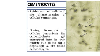
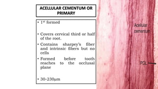
CEMENTUM
By: Dr. SOMRAJ PODDER
1ST YEAR POST GRADUATE
STUDENT
DEPARTMENT OF CONSERVATIVE
DENTISTRY AND ENDODONTICS
CEMENTUM
BY: Dr. SOMRAJ PODDER
1ST YEAR POST GRADUATE STUDENT
DEPARTMENT OF CONSERVATIVE
DENTISTRY AND ENDODONTICS
CONTENTS
PART A
• INTRODUCTION
• DEVELOPMENT OF CEMENTUM
• PHYSICAL PROPERTIES
• CHEMICAL COMPOSITION
• HISTOLOGY
• CLASSIFICATION
• FUNCTION
• CEMENTO-ENAMEL JUNCTION
• CEMENTO-DENTINAL JUNCTION
PART B
• RESORPTION AND REPAIR
• EFFECTS OF AGING
• CEMENTUM IN ORAL
ENVIRONMENT
• ROLE OF CEMENTUM IN
PERIODONTAL DISEASES
• DEVELOPMENTAL ANOMALIES
• CONCLUSION
• REFERENCES
INTRODU
CTION
• Calcified, Avascular
tissue that forms the
outer covering of the
anatomic root.
• Also known as substantia
ossea.
• First demonstrated by
FRANKE AND RACHKOV
(Purkinje)
DEVELOP
MENT
The internal and
external enamel
epithelium
proliferates
downward as a
double layered
sheath of Hertwig
Induce the cells of
dental papillato
differentiateinto
ODONTOBLASTS
Secretes the
organic matrix of
first formed root
pre-dentin
consisting of
ground substance
& collagen fibrils
Break occurs in the
epithelial root
sheath allowing
the newly formed
dentin to come in
direct contact with
the cells of dental
follicle.
Cells derived from
this connective
tissue are called
cementoblasts that
are responsible for
the formation of
cementum.
PHYSICAL PROPERTIES
• Hardnessis less thandentin
• Lightyellowin color.
• Thinnest at the cemento-enamel
junction (20-50µm) & thickest
towards theapex (150-200µm).
• The apical foramen is surrounded by
cementum. Sometimes cementum
extend in the inner wall of the dentin
for a short distance & so a lining of
root canal is formed.
• It formsthroughout the life ofa tooth.
CEMENTUM
INORGANIC
SUBSTANCE
45-50%
Mainly Ca & P in the form of
HYDROXYAPATITE crystal.
It has the highest F content.
ORGANIC
MATERIAL AND
WATER
50-55%
Mainly type 1 collagen & protein
polysaccharides
(proteoglycans).
Collagentype iii, v, vi, xii are also
present.
• The non-collagenous protein plays an important role in
matrix deposition, initiation, control of mineralization &
matrix remodeling.
• BONE SIALOPROTEIN (BSP) & OSTEOPONTIN fills up
the large inter-fibrillar spaces.
• Cementum derived attachment protein (CAP), cementum
derived growth factor are unique to cementum
HISTOLOGY
CEMENTOBLASTS
• Synthesize collagen and
protein polysaccharides
which make up the organic
matrix of cementum.
• At the superficial surface
the collagen fibrils
produced by the
cementoblast form a
fibrous fringe
perpendicular to the PDL
space.
CEMENTOBLAST
• These cells retreat
and intermingle with
the fibroblasts of the
PDL.
• These cells have
mitochondria, golgi
apparatus & large
amount of granular
endoplasmic
reticulam.
CEMENTOCYTES
• Spider shaped cells and
are characteristics of
cellular cementum.
• During formation of
cellular cementum the
cementoblasts get
entrapped into its own
matrix due to its rapid
deposition & are called
cementocytes.
CEMENTOCYTES
• Haphazardly arranged
and widely dispersed.
• Similar to osteocytes and
they lie in a lacunae.
• Canalicular processes
branch and
anastomoses with those
of neighboring cells
CEMENTOCLASTS
• Unilocular/
multilocular cells.
• Present in Howship’s
lacunae.
• Resorption & repair
of cementum
INCREMENTAL LINES OF CEMENTUM
INCREMENTAL LINES OF SALTER
• Represents rhythmic deposition of
cementum.
• Appears as dark lines running
parallel to the root surface.
• Seen in both acellular and cellular
cementum but more prominent in
acellular cementum.
• Highly mineralized area with less
collagen and more ground substance.
CEMENTOID
Unmineralized layer of
cementum on cemental surface
(precementum).
New layer forms as the old
calcifies
3-5µm
Lined by CEMENTOBLASTS.
Connective tissue fibers from
PDL pass between the
cementoblasts.
ARRANGEMENT OF FIBRILS
EXTRINSIC
FIBER SYSTEM
Principal fibers i.e
sharpey’s fibers.
mostly arranged at
right angle to
cementum.
INTRINSIC
FIBRE SYSTEM
dense, irregularly
arranged in the
cemental matrix.
SHARPEY’S FIBERS
• Terminal portions of principal
fibers that inserts into cementum
and bone.
• Produced by cells of dental follicle
during development and later by
PDL fibroblasts.
• Oriented perpendicular to the
root surface.
• 5-7 µm in diameter
• Mineralized partially with
unmineralized core.
INTRINSIC FIBERS
• Produced by cementoblasts.
• Oriented parallel to the root
surface.
• 1-2 µm in diameter
• Uniformly mineralized
• Mainly for repair.
CLASSIFICATION
A. BASED ON LOCATION
CORONAL
CEMENTUM
restricted to
areas of reduced
enamel
epithelium
RADICULAR
CEMENTUM
dental follicle
Covers entire
dentin.
CEJ to apical
foramen
B. Based on Cellularity:
1. Acellular cementum or primary
2. Cellular cementum or secondary
C. Based on presence and absence of collagen
fibrils:
1. Fibrillar cementum
2. Afibrillar cementum.
D. ON THE BASIS OF LOCATION,
STRUCTURE, FUNCTION, RATE OF
FORMATION, BIOCHEMICAL
COMPOSITION AND DEGREE OF
MINERALIZATION:
1. ACELLULAR AFIBRILLAR CEMENTUM
2. ACELLULAR EXTRINSIC FIBER CEMENTUM
3. CELLULAR MIXED STRATIFIED CEMENTUM
4. CELLULAR INTRINSIC FIBER CEMENTUM
5. INTERMEDIATE CEMENTUM
ACELLULAR CEMENTUM OR
PRIMARY
• 1st formed
• Covers cervical third or half
of the root.
• Contains sharpey’s fiber
and intrinsic fibers but no
cells
• Formed before tooth
reaches to the occlusal
plane
• 30-230µm
CELLULAR CEMENTUM OR
SECONDARY
• Formed after tooth reaches the
occlusal plane
• More irregular
• Cementocytes in the lacunae
communicating with each
other through anastomosing
canaliculi.
• Sharpey’s fibre occupy small
space whereas intrinsic fibers
occupy larger space.
ACELLULAR AFIBRILLAR
CEMENTUM
Mineralized ground substance
No intrinsic and extrinsic fibers
Product of cementoblast
As coronal cementum in
dentino-enamel junction.
1-5µm
Deposited as isolated patches
over minor areas of enamel and
dentin
Cemental spurs are found near
the cementoenamel junction
ACELLULAR EXTRINSIC FIBER
CEMENTUM
Cervical margin to apical third.
Product of fibroblast and cementoblast
Sharpey’s fibers are perpendicular to
surface of cementum
Composed almost entirely of densely
packed collagen fibers and lacks cells.
Forms slowly- more uniform and
parallel incremental lines than cellular
cementum.
Main function- anchorage
30-230µm
Cellular mixed stratified
cementum
Extrinsic and intrinsic fibre
and cells forms the bulk of
cellular cementum
Co-product of fibroblast and
cementoblast.
100-1000µm
Adaptation and repair
CELLULAR INTRINSIC FIBER
CEMENTUM
Contains cells, but no
extrinsic collagen fibers.
Formed on root surface
Secreted by
Cementoblasts
Fills the resorption
lacunae
Involved in adaptation
and repair
Less mineralized
INTERMEDIATE
CEMENTUM
Poorly defined zone near
DEJ
Appears hyaline or
structureless also called
hyaline layer
Represents are where HERS
cells become trapped in a
rapidly deposited dentin or
cementum
Apical halves of premolars
and molars
FUNCTION
• medium for attachment of collagen fibers that bind the tooth to the
alveolar bone.
• Connective tissue attachemt to tooth is not possible without cementum
A. ANCHORAGE
• Makes functional adaptation possible
• Deposition is continuous and throughout the life
• Maintain the width of the PDL space
B. ADAPTATION
• Major reparative tissue for root surface.
• Resorption can be repaired
• During repair it resembles cellular cementum as it grows fater.
C. REPAIR:
CEMENTO ENAMEL JUNCTION
CEMENTO ENAMEL JUNCTION-
OVERLAP JUNCTION 60%
Premature degeneration or
retraction of the reduced
enamel epithelium at the
cervical region of enamel
Adjacent mesenchymal cells
invade and intervene between
enamel and its covering
epithelium
The mesenchymal cells
differentiate into cemento
blasts & deposit cementum on
enamel surface
B. CEMENTO ENAMEL JUNCTION- TIGHT
JUNCTION 30%
Cementum meets the cervical end of
cementum in a knife edge or edge to
edge pattern
C. CEMENTO ENAMEL JUNCTION- GAP
JUNCTION 10%
Cementum does not meet enamel
Zone of root dentin appears devoid
of cementum
Causes hypersensitivity
D. SOMETIMES ENAMEL OVERLAPS
CEMENTUM- RARE CASE.
CEMENTO DENTINAL
JUNCTION
• Terminal apical area of the
cementum where it joins the
internal root canal dentin
• Wide zone- 2-3µm wide
• Large quantities of collagen
associated with GAGs
resulting in increased water
content that makes it stiff.
Primary tooth- scalloped
Permanent tooth- smooth
CONTENTS
PART A
• INTRODUCTION
• DEVELOPMENT OF CEMENTUM
• PHYSICAL PROPERTIES
• CHEMICAL COMPOSITION
• HISTOLOGY
• CLASSIFICATION
• FUNCTION
• CEMENTO-ENAMEL JUNCTION
• CEMENTO-DENTINAL JUNCTION
PART B
• RESORPTION AND REPAIR
• EFFECTS OF AGING
• CEMENTUM IN ORAL
ENVIRONMENT
• ROLE OF CEMENTUM IN
PERIODONTAL DISEASES
• DEVELOPMENTAL
ANOMALIES
• CONCLUSION
• REFERENCES
CEMENTUM RESORPTION AND REPAIR
Unlike primaryteeth, permanentteethdo not undergo
physiologic resorption.
Cementumof erupted as wellas un-erupted teethis
subjectto resorptive changes thatmay be of microscopic
level or sufficiently extensive to presenta
radiographicallydetectablealteration in the root
contour.
Cementumresorptionappearsmicroscopicallyas
BAYLIKECONCAVITIES in the root surface.
• Multi nucleated giant cells & mononuclear macrophages are
present in the area adjacent to the resorption site.
• The resorption process may extend into the underlying
dentin and even into the pulp, but is usually painless.
• Cementum resorption is not necessarily continuous and may
alternate with periods of repair and the deposition of new
cementum.
• The newly formed cementum is demarcated from the root by
a deep staining irregular lines known as reversal line, which
delineates the border of the previous resorption.
A. LOCAL CONDITIONS
• Trauma from occlusion.
• Orthodontic movement
• Cysts
• Tumors
• Pressure from mal-aligned erupting teeth
• Teeth with functional antagonist
• Periapical disease
• Periodontal disease
B. SYSTEMIC
CONDITIONS
•Calcium deficiency
•Hypothyroidism
•Hereditary fibrous
osteodystrophy
•Paget’s disease
REPAIR OF CEMENTUM
• Repair of cementum is a process to heal the damage caused by resorption or
cemental fracture.
• Requires the presence of viable connective tissue.
• If epithelium proliferates into the area of resorption, repair will not take place.
• Can occur in both vital & non-vital tooth.
2 TYPES
A. ANATOMIC REPAIR
• Generally occurs when the degree
of destruction is low.
• The root outline is re-established
as it was before cemental
resorption.
• Repair followed by the formation
of acellular and cellular
cementum.
B. FUNCTIONAL REPAIR
In cases of large cemental resorption, repair
does not re-establish the same anatomic
contour as before.
To maintain the width of the PDL space, the
adjacent alveolar bone grows and takes the
shape of defect following the root surface.
Helps to improve the function of the tooth.
EFFECT OF AGEING ON CEMENTUM
• The surface of the cementum becomes more irregular and this is
caused by the calcification of some fiber bundles when they are
attached to cementum.
• Width may increase (apically and lingually) 5-10 times as the
deposition continued throughout the life.
• Continuous deposition of cementum in the apical region may result
in obstruction of apical foramen.
EXPOSURE OF CEMENTUM TO THE
ORAL ENVIRONMENT
Cementum become exposed to the oral environment
in cases of gingival recession or as a consequence of
loss of attachment in pocket formation.
Permeable in nature thus it allows the penetration of
organic substance, inorganic ions & micro-
organisms.
Caries may develop in the cementum.
In periodontal pocket, the pathologically exposed
cementum may interfere in healing.
Hypo-mineralized cementum are formed due to any
defect during the process of formation.
Cementum is avascular & with no nerve supply, so
during scaling no pain occurs. If cementum is removed
from the surface and expose the dentin it will cause
sensitivity.
Pre-cementum is a natural barrier to excessive apical
migration of junctional epithelium.
ROLE OF CEMENTUM IN PERIODONTAL DISEASES
• Cementum provides the surface for attachment of plaque & calculus.
• Forms the inner wall of the periodontal pocket.
• Cemental tissue is relatively static as compared to the other
surrounding dynamic tissues, so any change will have long term
effects.
• To control the progression of periodontal diseases it is very necessary to
return the affected cementum into a healthy state.
• Early & moderate periodontitis- coronal half of the root/ acellular
cementum is affected.
• Advanced & lesions with involvement of furcation- cellular cementum
mostly gets damaged.
CHANGES
STRUCTURAL CHANGES
Pathologic granules in exposed cementum by BASS &
BENSON proves that the vacoule like formations exist.
Partial demineralization by SELVIG.
Loss of cross banding of collagen.
Sub-surface condensation of organic material of
exogenous origin.
CHEMICAL CHANGES
Increase in calcium &
phosphate level by 7-10 times.
Increase in fluoride level with
decrease in sodium level is
appreciated.
DEVELOPMENTAL ANOMELIES OF CEMENTUM
ENAMEL PROJECTIONS
• Deposition of enamel over the cementum in the cervical
region.
• Amelogenesis does not stop before the initiation of root
formation.
ENAMEL PEARL
• Globular or pearl like structure on the root surface in the
cervical region.
• Act as a plaque retentive area.
HYPER-CEMENTOSIS
• Refers to a prominent thickening of the
cementum.
• May be localized to one tooth or affect
the entire dentition (PAGET’S
DISEASE).
• OCCURS As a generalized thickening
of the cementum with nodular
enlargement of the apical third of the
root.
Etiology
1. SPIKE-LIKE TYPE OF
HYPERCEMENTOSIS: results
from excessive tension from orthodontic
appliances or occlusal forces.
2.GENERALIZED TYPE: occurs in
teeth without antagonists.
In teeth subject to low-grade periapical irritation
arising frompulpal disease.
CEMENTOBLASTOMA
• Only neoplasm of cementum.
• Cementum like tissues is deposited in roots of
tooth as irregular or rounded mass.
• Age < 25years.
• Often involves mandibular molars or premolars.
• Affected teeth are mostly vital.
• Attached to the root and may cause resorption.
• Grows slowly & tend to expand the overlying
cortical plates.
• Usually asymptomatic.
CEMENTOMA
• Also known as Benign cementoblastoma /
cemental dysplasia.
• Represents an unusual reaction of bone.
• Caused due to occlusal trauma.
• Present usually at apex of mandibular
incisors.
• Mostly found in BLACKS.
• 20-40 years.
• Asymptomatic expansion of jaw.
CONCRESCENCE
• Fusion of 2 teeth that occurs after root formation
has been completed.
• Arise as a result of traumatic injury or crowding
of teeth with resorption of interdental bone so
that two roots are in approximate contact &
become fused by deposition of cementum
between them.
• May occur before or after teeth have erupted.
CEMENTOPATHIA
• In 1923, GOTTLIEB reported a patient with fatal case of influenza and disease
called diffuse atrophy of bone.
• Characterized by loss of collagen fibers in PDL & their replacement by loose
connective tissue & extensive bone resorption resulting in the widened PDL space.
• Inhibition of continuous cementum formation which is essential for maintenance
of PDL space.
HYPOPHOSPHATASIA
• Hypophosphatasia is an inherited disorder
that affects the development of bones and
teeth.
• This condition disrupts a process called
mineralization, in which minerals like
calcium and phosphorus are deposited in the
developing bone and teeth.
• The mildest form of hypophosphatasia is
odontohypophosphatasia that is caracterized
by total absence of cementum leading to
early loss of teeth.
• Occurs because of the deficiency of enzyme
alkaline phosphatase in serum & tissues.
HYPOPHOSPHATASIA
• Affected infants are born with short limbs, an
abnormally shaped chest & soft skull bone.
• Failure to gain weight due to poor feeding or less
apatite, respiratory problems.
• Hypercalcemia which can lead to recurrent vomiting
& kidney problems.
• Affected children may have short stature with bowed
legs or knock knees.
• Osteomalacia is the characteristic feature of the adult
form.
• Recurrent fractures in foot and thigh bone leads to
chronic pain.
CEMENTICLES
• Calcified bodies sometimes seen in the PDL.
• May be round or ovoid in shape.
• May be single or multiple in number in the cemental
surface.
• Possibly degenerated epithelial cells from the nidus
for their calcification.
• Could be due to trauma.
2 types:
FREE CEMENTICLES
Lamellated cemental bodies
that lie freely in the PDL
ATTACHED CEMENTICLES
Cemental bodies attached to
root surface
ANKYLOSIS
Fusion of cementum & alveolar bone with
obliteration of PDL is termed as ankyloses.
Occurs in teeth with cemental resorption,
may represent a form of abnormal repair.
May develop after chronic periapical
inflammation, tooth replantation, occlusal
trauma.
Results in resorption of root and gradual
replacement by bone tissue.
SIGNIFICANCE OF CEMENTUM IN OPERATIVE AND
ENDODONTIC DENTISTRY
• Root caries occurs on exposed tooth surfaces below the cemento-enamel junction after
gingival recession.
• Cariogenic plaque, rather than periodontitis or gingival inflammation, is the essential
factor responsible for root caries development.
• The different forms of root surface caries, ranging from minor undemineralized and
discolored areas to extensive yellow-brown soft areas, are rarely associated with
cavitation below the Affected cementum.
• The CEJ serves as an important reference for diagnosis of CEPs. These are the extensions
of enamel from the CEJ onto the root surface.
• Masters and Hoskins, classified these unusual anatomical structures based on the degree
of extension of enamel beyond the CEJ and depending on their location in relation to
furcation topography.
SIGNIFICANCE OF CEMENTUM IN OPERATIVE AND
ENDODONTIC DENTISTRY
• Enamel extension apical to the normal CEJ level is a risk factor for periodontal disease because the
periodontal fibers embedded in the cementum to support the tooth, are not in their usual position
and thus, do not act as barrier to the advancement of periodontal disease.
• In effect, the epithelial attachment over the surface of the enamel, which does not have this kind of
attachment, may become detached in the narrow and difficult-to-clean bifurcation area because of
plaque and calculus, increasing the vulnerability to periodontal disease.
• Identification and location of CEJ serves various purposes which can be summarized as follows:
1. CEJ is used as a fixed/static landmark to measure CAL/ periodontal destruction.
2. To assess alveolar bone destruction by measuring CEJ-bone crest distance.
3. To study furcation by finding out furcation-CEJ distance, thus helping in treatment planning for
furcation involvements.
CONCLUSION
Cementum is an important tissue of periodontium, the
attachment apparatus of the tooth & has an important role
in the tooth movements. Cementum deposition is a
continuous process but this tissue is static in comparison to
the surrounding dynamic tissues therefore it has to be
taken in account while performing various dental
procedures.
REFERENCES:
•Shafer’s textbook of oral pathology
•Oral histology and embryology by orbans
•Tencate’s oral histology
•Grossman’s endodontic practice 13th
edition

CEMENTUM .pptx

  • 1.
    CEMENTUM By: Dr. SOMRAJPODDER 1ST YEAR POST GRADUATE STUDENT DEPARTMENT OF CONSERVATIVE DENTISTRY AND ENDODONTICS CEMENTUM BY: Dr. SOMRAJ PODDER 1ST YEAR POST GRADUATE STUDENT DEPARTMENT OF CONSERVATIVE DENTISTRY AND ENDODONTICS
  • 2.
    CONTENTS PART A • INTRODUCTION •DEVELOPMENT OF CEMENTUM • PHYSICAL PROPERTIES • CHEMICAL COMPOSITION • HISTOLOGY • CLASSIFICATION • FUNCTION • CEMENTO-ENAMEL JUNCTION • CEMENTO-DENTINAL JUNCTION PART B • RESORPTION AND REPAIR • EFFECTS OF AGING • CEMENTUM IN ORAL ENVIRONMENT • ROLE OF CEMENTUM IN PERIODONTAL DISEASES • DEVELOPMENTAL ANOMALIES • CONCLUSION • REFERENCES
  • 3.
  • 4.
    • Calcified, Avascular tissuethat forms the outer covering of the anatomic root. • Also known as substantia ossea. • First demonstrated by FRANKE AND RACHKOV (Purkinje)
  • 5.
  • 6.
    The internal and externalenamel epithelium proliferates downward as a double layered sheath of Hertwig Induce the cells of dental papillato differentiateinto ODONTOBLASTS Secretes the organic matrix of first formed root pre-dentin consisting of ground substance & collagen fibrils Break occurs in the epithelial root sheath allowing the newly formed dentin to come in direct contact with the cells of dental follicle. Cells derived from this connective tissue are called cementoblasts that are responsible for the formation of cementum.
  • 8.
  • 9.
    • Hardnessis lessthandentin • Lightyellowin color. • Thinnest at the cemento-enamel junction (20-50µm) & thickest towards theapex (150-200µm). • The apical foramen is surrounded by cementum. Sometimes cementum extend in the inner wall of the dentin for a short distance & so a lining of root canal is formed. • It formsthroughout the life ofa tooth.
  • 11.
    CEMENTUM INORGANIC SUBSTANCE 45-50% Mainly Ca &P in the form of HYDROXYAPATITE crystal. It has the highest F content. ORGANIC MATERIAL AND WATER 50-55% Mainly type 1 collagen & protein polysaccharides (proteoglycans). Collagentype iii, v, vi, xii are also present.
  • 12.
    • The non-collagenousprotein plays an important role in matrix deposition, initiation, control of mineralization & matrix remodeling. • BONE SIALOPROTEIN (BSP) & OSTEOPONTIN fills up the large inter-fibrillar spaces. • Cementum derived attachment protein (CAP), cementum derived growth factor are unique to cementum
  • 13.
  • 14.
    CEMENTOBLASTS • Synthesize collagenand protein polysaccharides which make up the organic matrix of cementum. • At the superficial surface the collagen fibrils produced by the cementoblast form a fibrous fringe perpendicular to the PDL space.
  • 15.
    CEMENTOBLAST • These cellsretreat and intermingle with the fibroblasts of the PDL. • These cells have mitochondria, golgi apparatus & large amount of granular endoplasmic reticulam.
  • 16.
    CEMENTOCYTES • Spider shapedcells and are characteristics of cellular cementum. • During formation of cellular cementum the cementoblasts get entrapped into its own matrix due to its rapid deposition & are called cementocytes.
  • 17.
    CEMENTOCYTES • Haphazardly arranged andwidely dispersed. • Similar to osteocytes and they lie in a lacunae. • Canalicular processes branch and anastomoses with those of neighboring cells
  • 18.
    CEMENTOCLASTS • Unilocular/ multilocular cells. •Present in Howship’s lacunae. • Resorption & repair of cementum
  • 19.
    INCREMENTAL LINES OFCEMENTUM INCREMENTAL LINES OF SALTER • Represents rhythmic deposition of cementum. • Appears as dark lines running parallel to the root surface. • Seen in both acellular and cellular cementum but more prominent in acellular cementum. • Highly mineralized area with less collagen and more ground substance.
  • 20.
    CEMENTOID Unmineralized layer of cementumon cemental surface (precementum). New layer forms as the old calcifies 3-5µm Lined by CEMENTOBLASTS. Connective tissue fibers from PDL pass between the cementoblasts.
  • 21.
    ARRANGEMENT OF FIBRILS EXTRINSIC FIBERSYSTEM Principal fibers i.e sharpey’s fibers. mostly arranged at right angle to cementum. INTRINSIC FIBRE SYSTEM dense, irregularly arranged in the cemental matrix.
  • 22.
    SHARPEY’S FIBERS • Terminalportions of principal fibers that inserts into cementum and bone. • Produced by cells of dental follicle during development and later by PDL fibroblasts. • Oriented perpendicular to the root surface. • 5-7 µm in diameter • Mineralized partially with unmineralized core.
  • 23.
    INTRINSIC FIBERS • Producedby cementoblasts. • Oriented parallel to the root surface. • 1-2 µm in diameter • Uniformly mineralized • Mainly for repair.
  • 24.
  • 25.
    A. BASED ONLOCATION CORONAL CEMENTUM restricted to areas of reduced enamel epithelium RADICULAR CEMENTUM dental follicle Covers entire dentin. CEJ to apical foramen
  • 26.
    B. Based onCellularity: 1. Acellular cementum or primary 2. Cellular cementum or secondary C. Based on presence and absence of collagen fibrils: 1. Fibrillar cementum 2. Afibrillar cementum.
  • 27.
    D. ON THEBASIS OF LOCATION, STRUCTURE, FUNCTION, RATE OF FORMATION, BIOCHEMICAL COMPOSITION AND DEGREE OF MINERALIZATION: 1. ACELLULAR AFIBRILLAR CEMENTUM 2. ACELLULAR EXTRINSIC FIBER CEMENTUM 3. CELLULAR MIXED STRATIFIED CEMENTUM 4. CELLULAR INTRINSIC FIBER CEMENTUM 5. INTERMEDIATE CEMENTUM
  • 28.
    ACELLULAR CEMENTUM OR PRIMARY •1st formed • Covers cervical third or half of the root. • Contains sharpey’s fiber and intrinsic fibers but no cells • Formed before tooth reaches to the occlusal plane • 30-230µm
  • 29.
    CELLULAR CEMENTUM OR SECONDARY •Formed after tooth reaches the occlusal plane • More irregular • Cementocytes in the lacunae communicating with each other through anastomosing canaliculi. • Sharpey’s fibre occupy small space whereas intrinsic fibers occupy larger space.
  • 30.
    ACELLULAR AFIBRILLAR CEMENTUM Mineralized groundsubstance No intrinsic and extrinsic fibers Product of cementoblast As coronal cementum in dentino-enamel junction. 1-5µm Deposited as isolated patches over minor areas of enamel and dentin Cemental spurs are found near the cementoenamel junction
  • 31.
    ACELLULAR EXTRINSIC FIBER CEMENTUM Cervicalmargin to apical third. Product of fibroblast and cementoblast Sharpey’s fibers are perpendicular to surface of cementum Composed almost entirely of densely packed collagen fibers and lacks cells. Forms slowly- more uniform and parallel incremental lines than cellular cementum. Main function- anchorage 30-230µm
  • 32.
    Cellular mixed stratified cementum Extrinsicand intrinsic fibre and cells forms the bulk of cellular cementum Co-product of fibroblast and cementoblast. 100-1000µm Adaptation and repair
  • 33.
    CELLULAR INTRINSIC FIBER CEMENTUM Containscells, but no extrinsic collagen fibers. Formed on root surface Secreted by Cementoblasts Fills the resorption lacunae Involved in adaptation and repair Less mineralized
  • 34.
    INTERMEDIATE CEMENTUM Poorly defined zonenear DEJ Appears hyaline or structureless also called hyaline layer Represents are where HERS cells become trapped in a rapidly deposited dentin or cementum Apical halves of premolars and molars
  • 35.
  • 36.
    • medium forattachment of collagen fibers that bind the tooth to the alveolar bone. • Connective tissue attachemt to tooth is not possible without cementum A. ANCHORAGE • Makes functional adaptation possible • Deposition is continuous and throughout the life • Maintain the width of the PDL space B. ADAPTATION • Major reparative tissue for root surface. • Resorption can be repaired • During repair it resembles cellular cementum as it grows fater. C. REPAIR:
  • 37.
  • 38.
    CEMENTO ENAMEL JUNCTION- OVERLAPJUNCTION 60% Premature degeneration or retraction of the reduced enamel epithelium at the cervical region of enamel Adjacent mesenchymal cells invade and intervene between enamel and its covering epithelium The mesenchymal cells differentiate into cemento blasts & deposit cementum on enamel surface
  • 39.
    B. CEMENTO ENAMELJUNCTION- TIGHT JUNCTION 30% Cementum meets the cervical end of cementum in a knife edge or edge to edge pattern C. CEMENTO ENAMEL JUNCTION- GAP JUNCTION 10% Cementum does not meet enamel Zone of root dentin appears devoid of cementum Causes hypersensitivity D. SOMETIMES ENAMEL OVERLAPS CEMENTUM- RARE CASE.
  • 40.
    CEMENTO DENTINAL JUNCTION • Terminalapical area of the cementum where it joins the internal root canal dentin • Wide zone- 2-3µm wide • Large quantities of collagen associated with GAGs resulting in increased water content that makes it stiff. Primary tooth- scalloped Permanent tooth- smooth
  • 41.
    CONTENTS PART A • INTRODUCTION •DEVELOPMENT OF CEMENTUM • PHYSICAL PROPERTIES • CHEMICAL COMPOSITION • HISTOLOGY • CLASSIFICATION • FUNCTION • CEMENTO-ENAMEL JUNCTION • CEMENTO-DENTINAL JUNCTION PART B • RESORPTION AND REPAIR • EFFECTS OF AGING • CEMENTUM IN ORAL ENVIRONMENT • ROLE OF CEMENTUM IN PERIODONTAL DISEASES • DEVELOPMENTAL ANOMALIES • CONCLUSION • REFERENCES
  • 42.
    CEMENTUM RESORPTION ANDREPAIR Unlike primaryteeth, permanentteethdo not undergo physiologic resorption. Cementumof erupted as wellas un-erupted teethis subjectto resorptive changes thatmay be of microscopic level or sufficiently extensive to presenta radiographicallydetectablealteration in the root contour. Cementumresorptionappearsmicroscopicallyas BAYLIKECONCAVITIES in the root surface.
  • 43.
    • Multi nucleatedgiant cells & mononuclear macrophages are present in the area adjacent to the resorption site. • The resorption process may extend into the underlying dentin and even into the pulp, but is usually painless. • Cementum resorption is not necessarily continuous and may alternate with periods of repair and the deposition of new cementum. • The newly formed cementum is demarcated from the root by a deep staining irregular lines known as reversal line, which delineates the border of the previous resorption.
  • 44.
    A. LOCAL CONDITIONS •Trauma from occlusion. • Orthodontic movement • Cysts • Tumors • Pressure from mal-aligned erupting teeth • Teeth with functional antagonist • Periapical disease • Periodontal disease
  • 45.
  • 46.
    REPAIR OF CEMENTUM •Repair of cementum is a process to heal the damage caused by resorption or cemental fracture. • Requires the presence of viable connective tissue. • If epithelium proliferates into the area of resorption, repair will not take place. • Can occur in both vital & non-vital tooth.
  • 47.
    2 TYPES A. ANATOMICREPAIR • Generally occurs when the degree of destruction is low. • The root outline is re-established as it was before cemental resorption. • Repair followed by the formation of acellular and cellular cementum.
  • 48.
    B. FUNCTIONAL REPAIR Incases of large cemental resorption, repair does not re-establish the same anatomic contour as before. To maintain the width of the PDL space, the adjacent alveolar bone grows and takes the shape of defect following the root surface. Helps to improve the function of the tooth.
  • 49.
    EFFECT OF AGEINGON CEMENTUM • The surface of the cementum becomes more irregular and this is caused by the calcification of some fiber bundles when they are attached to cementum. • Width may increase (apically and lingually) 5-10 times as the deposition continued throughout the life. • Continuous deposition of cementum in the apical region may result in obstruction of apical foramen.
  • 50.
    EXPOSURE OF CEMENTUMTO THE ORAL ENVIRONMENT Cementum become exposed to the oral environment in cases of gingival recession or as a consequence of loss of attachment in pocket formation. Permeable in nature thus it allows the penetration of organic substance, inorganic ions & micro- organisms. Caries may develop in the cementum.
  • 51.
    In periodontal pocket,the pathologically exposed cementum may interfere in healing. Hypo-mineralized cementum are formed due to any defect during the process of formation. Cementum is avascular & with no nerve supply, so during scaling no pain occurs. If cementum is removed from the surface and expose the dentin it will cause sensitivity. Pre-cementum is a natural barrier to excessive apical migration of junctional epithelium.
  • 52.
    ROLE OF CEMENTUMIN PERIODONTAL DISEASES • Cementum provides the surface for attachment of plaque & calculus. • Forms the inner wall of the periodontal pocket. • Cemental tissue is relatively static as compared to the other surrounding dynamic tissues, so any change will have long term effects.
  • 53.
    • To controlthe progression of periodontal diseases it is very necessary to return the affected cementum into a healthy state. • Early & moderate periodontitis- coronal half of the root/ acellular cementum is affected. • Advanced & lesions with involvement of furcation- cellular cementum mostly gets damaged.
  • 54.
    CHANGES STRUCTURAL CHANGES Pathologic granulesin exposed cementum by BASS & BENSON proves that the vacoule like formations exist. Partial demineralization by SELVIG. Loss of cross banding of collagen. Sub-surface condensation of organic material of exogenous origin. CHEMICAL CHANGES Increase in calcium & phosphate level by 7-10 times. Increase in fluoride level with decrease in sodium level is appreciated.
  • 55.
    DEVELOPMENTAL ANOMELIES OFCEMENTUM ENAMEL PROJECTIONS • Deposition of enamel over the cementum in the cervical region. • Amelogenesis does not stop before the initiation of root formation. ENAMEL PEARL • Globular or pearl like structure on the root surface in the cervical region. • Act as a plaque retentive area.
  • 56.
    HYPER-CEMENTOSIS • Refers toa prominent thickening of the cementum. • May be localized to one tooth or affect the entire dentition (PAGET’S DISEASE). • OCCURS As a generalized thickening of the cementum with nodular enlargement of the apical third of the root.
  • 57.
    Etiology 1. SPIKE-LIKE TYPEOF HYPERCEMENTOSIS: results from excessive tension from orthodontic appliances or occlusal forces. 2.GENERALIZED TYPE: occurs in teeth without antagonists. In teeth subject to low-grade periapical irritation arising frompulpal disease.
  • 58.
    CEMENTOBLASTOMA • Only neoplasmof cementum. • Cementum like tissues is deposited in roots of tooth as irregular or rounded mass. • Age < 25years. • Often involves mandibular molars or premolars. • Affected teeth are mostly vital. • Attached to the root and may cause resorption. • Grows slowly & tend to expand the overlying cortical plates. • Usually asymptomatic.
  • 59.
    CEMENTOMA • Also knownas Benign cementoblastoma / cemental dysplasia. • Represents an unusual reaction of bone. • Caused due to occlusal trauma. • Present usually at apex of mandibular incisors. • Mostly found in BLACKS. • 20-40 years. • Asymptomatic expansion of jaw.
  • 60.
    CONCRESCENCE • Fusion of2 teeth that occurs after root formation has been completed. • Arise as a result of traumatic injury or crowding of teeth with resorption of interdental bone so that two roots are in approximate contact & become fused by deposition of cementum between them. • May occur before or after teeth have erupted.
  • 61.
    CEMENTOPATHIA • In 1923,GOTTLIEB reported a patient with fatal case of influenza and disease called diffuse atrophy of bone. • Characterized by loss of collagen fibers in PDL & their replacement by loose connective tissue & extensive bone resorption resulting in the widened PDL space. • Inhibition of continuous cementum formation which is essential for maintenance of PDL space.
  • 62.
    HYPOPHOSPHATASIA • Hypophosphatasia isan inherited disorder that affects the development of bones and teeth. • This condition disrupts a process called mineralization, in which minerals like calcium and phosphorus are deposited in the developing bone and teeth. • The mildest form of hypophosphatasia is odontohypophosphatasia that is caracterized by total absence of cementum leading to early loss of teeth. • Occurs because of the deficiency of enzyme alkaline phosphatase in serum & tissues.
  • 63.
    HYPOPHOSPHATASIA • Affected infantsare born with short limbs, an abnormally shaped chest & soft skull bone. • Failure to gain weight due to poor feeding or less apatite, respiratory problems. • Hypercalcemia which can lead to recurrent vomiting & kidney problems. • Affected children may have short stature with bowed legs or knock knees. • Osteomalacia is the characteristic feature of the adult form. • Recurrent fractures in foot and thigh bone leads to chronic pain.
  • 64.
    CEMENTICLES • Calcified bodiessometimes seen in the PDL. • May be round or ovoid in shape. • May be single or multiple in number in the cemental surface. • Possibly degenerated epithelial cells from the nidus for their calcification. • Could be due to trauma.
  • 65.
    2 types: FREE CEMENTICLES Lamellatedcemental bodies that lie freely in the PDL ATTACHED CEMENTICLES Cemental bodies attached to root surface
  • 66.
    ANKYLOSIS Fusion of cementum& alveolar bone with obliteration of PDL is termed as ankyloses. Occurs in teeth with cemental resorption, may represent a form of abnormal repair. May develop after chronic periapical inflammation, tooth replantation, occlusal trauma. Results in resorption of root and gradual replacement by bone tissue.
  • 67.
    SIGNIFICANCE OF CEMENTUMIN OPERATIVE AND ENDODONTIC DENTISTRY • Root caries occurs on exposed tooth surfaces below the cemento-enamel junction after gingival recession. • Cariogenic plaque, rather than periodontitis or gingival inflammation, is the essential factor responsible for root caries development. • The different forms of root surface caries, ranging from minor undemineralized and discolored areas to extensive yellow-brown soft areas, are rarely associated with cavitation below the Affected cementum. • The CEJ serves as an important reference for diagnosis of CEPs. These are the extensions of enamel from the CEJ onto the root surface. • Masters and Hoskins, classified these unusual anatomical structures based on the degree of extension of enamel beyond the CEJ and depending on their location in relation to furcation topography.
  • 68.
    SIGNIFICANCE OF CEMENTUMIN OPERATIVE AND ENDODONTIC DENTISTRY • Enamel extension apical to the normal CEJ level is a risk factor for periodontal disease because the periodontal fibers embedded in the cementum to support the tooth, are not in their usual position and thus, do not act as barrier to the advancement of periodontal disease. • In effect, the epithelial attachment over the surface of the enamel, which does not have this kind of attachment, may become detached in the narrow and difficult-to-clean bifurcation area because of plaque and calculus, increasing the vulnerability to periodontal disease. • Identification and location of CEJ serves various purposes which can be summarized as follows: 1. CEJ is used as a fixed/static landmark to measure CAL/ periodontal destruction. 2. To assess alveolar bone destruction by measuring CEJ-bone crest distance. 3. To study furcation by finding out furcation-CEJ distance, thus helping in treatment planning for furcation involvements.
  • 69.
    CONCLUSION Cementum is animportant tissue of periodontium, the attachment apparatus of the tooth & has an important role in the tooth movements. Cementum deposition is a continuous process but this tissue is static in comparison to the surrounding dynamic tissues therefore it has to be taken in account while performing various dental procedures.
  • 70.
    REFERENCES: •Shafer’s textbook oforal pathology •Oral histology and embryology by orbans •Tencate’s oral histology •Grossman’s endodontic practice 13th edition