Download to read offline

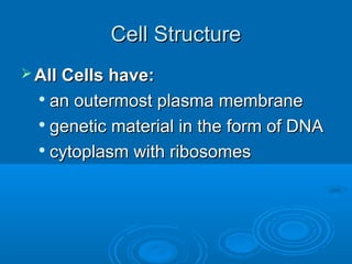
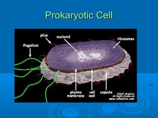
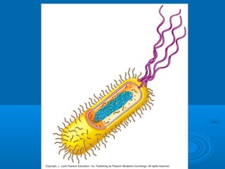
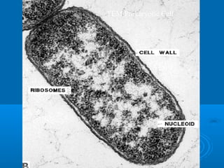
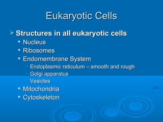
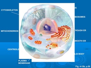

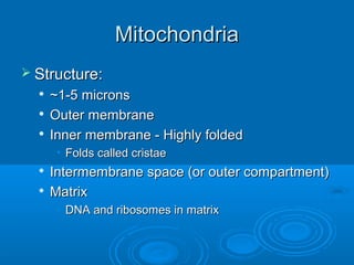
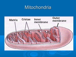
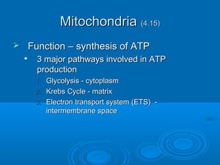
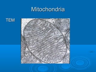
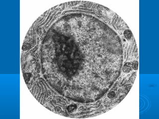
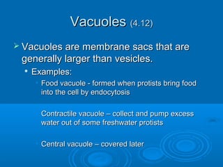
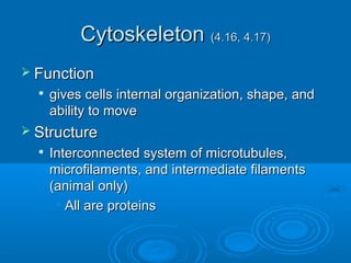
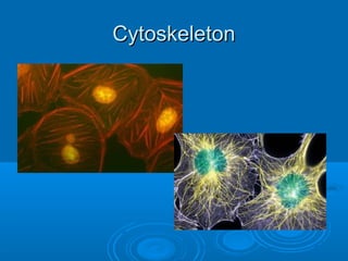

This document provides an overview of cell structure and function. It begins with a brief history of cell theory and outlines the key points of cell theory. It then describes the basic structures found in both prokaryotic and eukaryotic cells, including the plasma membrane, genetic material, cytoplasm, and organelles. Specific organelles like the nucleus, endoplasmic reticulum, Golgi apparatus, vesicles, lysosomes and mitochondria are explained in more detail. The roles of these structures and organelles in protein modification and transport are summarized. The document also addresses why cells are typically small in size.


![THEORY cell_biology__notes_print_1[1].pptx](https://cdn.slidesharecdn.com/ss_thumbnails/cellbiologynotesprint11-251210090642-34c62fe5-thumbnail.jpg?width=640&height=640&fit=bounds)


