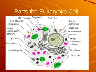
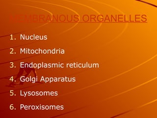
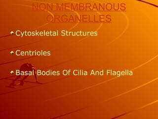
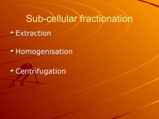
CELL AND SUB
CELLULAR
ORGANELLES
Structure and Function
Dr. Roopa.R
OVERVIEW-
i) History
ii) Properties of cell
SUBCELLULAR
COMPONENTS
CELL TRANSPORT
CELL
Cell from latin word “cella” meaning a
small room.
Cell is the structural & functional unit of
life.
Cell is the smallest build structure of
living organisms that functionalize the
vital process.
Main characteristics of CELL
The cell structure have:
An outer membrane
Genetic material (DNA/RNA)
Cytoplasm
Discovery of Cells
The invention of the lens
Robert Hooke (1665):
observed a thin slice of
cork (dead plant cells)
with a microscope. He
described what he
observed as “little boxes”
(cells).
Discovery of Cells
Anton van Leeuwenhoek
(1675): was the first
person to observe living
cells.
The Cell Theory
Who developed the cell theory?
– Matthias Schleiden (1838):
concluded that all plants are
composed of cells
– Theodor Schwann (1839):
concluded that all animals
are composed of cells
– Rudolph Virchow (1855):
determined that cells come
only from other existing cells
The Cell Theory
What is the cell theory?
1. All living things are composed of
one or more cells.
2. Cells are organisms’ basic units
of structure and function.
3. Cells come only from existing
cells.
PROPERTIES OF THE CELLS
-- Self contained & Self maintained.
-- Sharing Abilities
* reproduction by cell division.
* metabolism
* responds to ext & int stimuli
i.e., temp,pH or nutrients
-- Cell Diversity
* shape, size & function
* surface area
Cell Diversity
Size
Shape
Internal Organization
Cell Diversity- Size
6 inches long, 5 inches wide, 3 pounds
Smallest Cells:
Longest Cells:
Ostrich Egg
Biggest Cells:
In human body:
How many cells do human body have?
About 50-100 trillion cells!
Do all humans have the same number
of cells?
No, It depends on the individual size
A typical human cell size is 10μm and a
typical cell mass is 1nanogram
Which is the largest cell of the human
body?
Which is the smallest cell of the human
body?
5-6
microns
120- 150
microns
Cell Diversity- Shape
Cells differ widely in
shape.
Most cells are roughly
cuboidal or spherical.
Cell Diversity- Internal
Organization
Nucleus: contains DNA which directs
the activity of the cell
Organelle: is a discrete structure of
cell having specialized functions.
Based on internal organization
Eukaryotes: cells that contain a
nucleus and membrane-bound
organelles
Prokaryotes: cells that lack nuclei
and membrane-bound organelles
Eukaryotes vs. Prokaryotes
Eukaryotes (animals,
plants, fungi, protists)
and
prokaryotes (bacteria)
differ greatly in
structure.
Parts the Eukaryotic Cell
MEMBRANOUS ORGANELLES
1. Nucleus
2. Mitochondria
3. Endoplasmic reticulum
4. Golgi Apparatus
5. Lysosomes
6. Peroxisomes
NON MEMBRANOUS
ORGANELLES
Cytoskeletal Structures
Centrioles
Basal Bodies Of Cilia And Flagella
Sub-cellular fractionation
Extraction
Homogenisation
Centrifugation
Extraction:
Extraction of cells from tissues by
suspension in 0.25M/L sucrose at pH
7.4
Homogenisation:
Plasma membrane disrupted using a
lipid solvent Tween 20 or by stirring
the suspension in a blender or by
sonication. Homogenate formed.
Homogenate is subjected to a series of
centrifugation
Homogenate is subjected
to a series of
centrifugation steps at
successively greater
speeds.
At each step a pellet and
supernatant is obtained.
The pellet is removed at
the end of each step.
Supernatant is further
centrifuged
The pellet obtained at each step is
analyzed for “marker enzymes” in order
to identify the organelle obtained
A marker enzyme is one that is almost
exclusively confined to one particular
organelle
The marker enzyme helps to indicate the
presence or absence of the organelle in
which it is located.
Plasma membrane/ Cell
membrane
Membrane that envelopes a cell or a
sub-cellular organelle
Separates the cells from the external
environment and divides the interior
of the cell into compartments
Composition of plasma membrane
Thin, tough but flexible membrane
allowing changes in size and shape of
cell
Made up of
 Lipids (phospholipids, glycolipids and
cholesterol)
 Proteins (peripheral and Integral) and
 Small amounts of carbohydrates
(glycoproteins and glycolipids)
All these components held together by
non-covalent hydrophobic interactions
Fluid Mosaic Model
Proposed by Singer and
Nicolson-1972
Membranes are asymmetric
Composed of PLP, Integral
proteins, Peripheral proteins,
Glyolipids, Glycoprotein's and
Cholesterol
Proteins are similar to shifting
tiles, Producing mosaic effect.
The spaces between the tiles are
filled with fluid-like
phospholipids.
Membrane lipids
Phospholipids, cholesterol and glycolipids
Arranged as two layers
Phospholipids are oriented in such a way
that hydrophobic tails are away from the
cytoplasm and extracellular side
Cholesterol is present in the Inner
hydrophobic core, which makes the bilayer
stronger, more flexible and more
permeable.
Inner hydrophobic core is fluid and
permits free lateral movement of
molecules.
Fluidity depends on cholesterol and MUFA
and allows endocytosis and exocytosis
10A°
15A°
Membrane proteins
Peripheral proteins exist (ionic and polar
bonds)on the surface of the bilayer and
can be removed without disrupting the
membrane.
Integral Membrane proteins are deeply
embedded (hydrophobic bonds) in the bi
layer.
Transmembrane proteins are integral
proteins that span the whole bilayer-
receptors, antigens, ion channels,
membrane based enzymes, Ig…
Functions of Membrane proteins
Transmembrane proteins form
channels and pores, carriers,
enzymes, receptors.
Peripheral proteins form
Enzymes: Ex- 5’ Nucleotidase, ALP
Receptors: Hormone receptors
Membrane carbohydrates
Minor components, always towards the
extracellular side
Present in glycolipids and glycoproteins as
oligosaccharide chains
Play an important role in cell-cell recognition,
adhesion and receptor action
Composition of certain membranes
Functions of plasma membrane
Forms a boundary between a cell and
its external environment and
between organelles
Plays an important role in cell-cell
recognition
Maintains shape of cell
Cellular movement
Controls movement of molecules
between inside and outside of the
cell
Cytoplasm
Cytoplasm is a jelly-like material that fills cells.
The cytosol is made up of water, salts, organic
molecules and many enzymes that catalyze reactions.
The cytoplasm plays an important role in a cell,
serving as a "molecular soup" in which the organelles
are suspended and held together by a fatty membrane
Cytoskeleton
The cytoskeleton is a
cellular "scaffolding" or
"skeleton“ made up of
filamentous proteins
It is a dynamic structure
that maintains cell
shape, enables cell
motion
Functions of Cytoskeleton
 Maintains characteristic
shape and form of cell.
 Provides attachment points
for Organelles
 Separation of chromatin
during cell division
Proteins of Cytoskeleton
1. Microfilaments: 5nm
Made up of Actin
G actin polymerizes to form double
helical F actin
Functions:Muscle contraction
Maintains shape of cell
Helps in cellular movement
Proteins of Cytoskeleton
2.Microtubules: 20 -25 nm
Made up of tubulin
α -tubulin and β –tubulin: α β dimers polymerizes
to protofilament
A microtubule is made up of 13 protofilaments
Functions: Formation of mitotic spindle
Stability of cell
Intracellular movement of vesicles
Cilia and flagella
Components of axons and dendrites
Proteins of Cytoskeleton
3. Intermediate Filaments: 10 nm
Seven classes: Keratin, Lamins, Desmin,
Vimentin, GFAP, Peripherin, Neurofilament,
Functions: Cell strength and maintain shape.
Cell to cell attachment.
Strength and rigidity to axons.
Structural role in skin & hair cells.
Nucleus
Nucleus
It houses the cell's chromosomes.
The nucleus is spherical in shape
and separated from the cytoplasm
by a double membrane called the
nuclear envelope.
The nuclear envelope isolates and
protects a cell's DNA from various
molecules that could accidentally
damage its structure or interfere
with its processing.
It consists of an outer and inner
membrane
Nuclear pores : 90 nm(inner
membrane)
Functions of Nucleus
Nucleus contains DNA, the chemical
basis of genes which governs all the
functions of the cell.
DNA molecules are complexed with
proteins to form chromatin and are
further organized into chromosomes.
DNA replication and RNA synthesis
(transcription) are taking place inside
the nucleus.
Nucleolus
The nucleolus (plural nucleoli) is a
"sub organelle" of the cell nucleus.
A main function of the nucleolus is
the production and assembly of
ribosome components.
The nucleolus is roughly spherical,
and is surrounded by a layer of
condensed chromatin.
No membrane separates the
nucleolus from the nucleoplasm.
Ribosomes
A ribosome is an organelle in
cells that assembles
proteins.
Ribosomes are composed of
ribosomal RNA and
ribosomal proteins.
It translates Messenger RNA
(mRNA) into a polypeptide
chain
Endoplasmic Reticulum
Structure: a system of membranous tubules
and sacs. It’s a network of interconnecting
membranes enclosing channels and cisternae
held together by cytoskeleton
Function: intercellular highway (a path along
which molecules move from one part of the cell
to another)
Two types:
– Rough Endoplasmic Reticulum
– Smooth Endoplasmic Reticulum
Rough Endoplasmic Reticulum
Rough Endoplasmic
Reticulum (rER):
prominent in cells that
make large amounts of
proteins to be exported
from the cell or inserted
into the cell membrane
– Covered with
ribosomes
Smooth Endoplasmic Reticulum
Smooth Endoplasmic
Reticulum (sER):
involved in the synthesis
of lipids and breakdown
of toxic substances
– Not covered with
ribosomes
Golgi Apparatus
Structure:Network of
flattened, smooth membranes
and vesicles
Has cis side facing ER, medial
(intermediate cisterna) and
trans side facing the plasma
membrane.
Function: receives proteins
from the rER and distributes
them to other organelles or
out of the cell
Protein sorting, packaging
and secretion
GOLGI APPARATUS
The finished products
may have one of the
following fate:
1) Pass through plasma
membrane & secreted.
2) They form integral
part of plasma
membrane.
3) Stored in secretory
vesicles and secreted
under proper stimuli.
Mitochondria
Power house of the cell
centre for cellular respiration
Evolved from aerobic bacteria
Vary in number, shape and location
Spherical , oval, rod like or sausage
shape
It is a self-replicating organelles.
Maternal Inheritance
Mitochondria
Structure: folded membrane
within an outer membrane
– The folds of the inner
membrane are called
cristae, Contains enzymes
of ETC
– Mitochondrial matrix
 Contains enzymes of TCA
cycle, B- oxidation, Urea
cycle and heme synthesis
Mitochondria
Function: converts energy stored in
food into usable energy for work.
(cellular respiration)
Mitochondrial DNA
Circular DNA
High rate of mutation
Codes for 13 proteins, 2rRNA and
22tRNA
Inherited Mitochondrial disorders
Nucleotide substitutions-
--Leber’s Hereditary Optic Neuropathy (LHON) and
--Myoclonic Epilepsy, and Ragged-Red Fiber disease
(MERRF)
MtDNA deletions-
Kearns-Sayre Syndrome (KSS)
tRNA mutations-
Mitochondrial Encephalomyopathy Lactic Acidosis
(MELAS)
Lysosomes
Structure: spherical
organelles that contain
hydrolytic enzymes
within single membranes
Function: breaks down
food particles, invading
objects, or worn out cell
parts
The degraded products are reutilised
by the cell, transported out of the
cell or accumulate in the cell
Accumulation of residual products-
Lipofuscin (rich in lipids and proteins)
is a sign of ageing or ‘wear and tear’
of the cell
Lysosomes applied aspect
The lysosome has the thickest membrane
amongst the sub-cellular organelles.
This is a protective mechanism to confine all the
hydrolytic enzymes within the organelle.
Escape of the hydrolytic enzymes into the cytosol
would destroy all the cellular macromolecules.
This mechanism is implicated in the occurrence of
inflammatory reactions in several diseases like
gout, silicosis, arthritis and allergic disorders.
Lysosomes-applied aspect
Lysosomal storage disorders are characterized
by specific enzyme deficiencies. These diseases
show characteristic patterns of the accumulation
of polysaccharides or lipids in the lysosomes.
Some of these diseases are Hurler's syndrome,
Tay-sachs, Pompe's disease
Inclusion cell disease is a condition wherein
lysosomes lack in enzymes. Enzymes are present
in blood but are are not packaged into
lysosomes
Peroxisomes
Oval shaped bodies with a
crystalline core enclosed in a
membrane
Contains catalase and peroxidase
enzymes- involved in the
destruction of peroxides and other
free radicals (antioxidant effect)
Oxidation of long chain fatty acids
Synthesis of plasmalogens and
glycolipids
Peroxisomal deficiency
diseases
Adrenoleukodystrophy: due to deficiency
of peroxisomal matrix proteins,
characterized by progressive destruction
of liver, kidney & brain.
Zellweger syndrome: proteins are not
transported into peroxisomes, leading
empty/ghost peroxisomes inside the cell.
Cilia and Flagella
Structure: hair-like organelles that extend from
the surface of cells
– Short and in large numbers  cilia
– Less numerous and longer  flagella
– Both organelles are composed of nine pairs
of microtubules arranged around a
central pair.
Function: cell motility
 Examples: Cilia: respiratory tract, fallopian
tube, middle ear
Flagella: sperm cell
Cillia and Flagella
Basal Bodies
The microtubule assembly of a cilium or
flagellum is anchored in the cell by a basal
body.
Structurally identical to a centriole
Centrioles
Structure: composed of
nine sets of triplet
microtubules arranged in
a ring
– Exist in pairs
Function: centrioles play
a major role in cell
division (mitosis)
Vacuoles
Structure: a sac of fluid
surrounded by a
membrane
– Very large in plants
Function: used for
temporary storage of
wastes, nutrients, and
water
The following is a list of tissues that have specialized functions
and demonstrate corresponding specialization of subcellular
structure. Match the tissue with the letter of the cell structures
and organelles listed to the right that would be abundant in
these cells.
Tissues Cell Structures
and Organelles
1. Enzyme (protein)-secreting A. plasma membrane
cells of the pancreas B. mitochondria
2. Insect flight muscles C. Golgi
apparatus D. chloroplast
3. Cells lining the respiratory E. ER
passages F. cillia & flagella
4. White blood cells that engulf G. vacuole
and destroy invading bacteria H. ribosome
5. Leaf cells in cacti I. lysosome
J. peroxisomes









Cell and sub cellular oraganelles and their function

  • 1.
  • 2.
    OVERVIEW- i) History ii) Propertiesof cell SUBCELLULAR COMPONENTS CELL TRANSPORT
  • 3.
    CELL Cell from latinword “cella” meaning a small room. Cell is the structural & functional unit of life. Cell is the smallest build structure of living organisms that functionalize the vital process.
  • 4.
    Main characteristics ofCELL The cell structure have: An outer membrane Genetic material (DNA/RNA) Cytoplasm
  • 5.
    Discovery of Cells Theinvention of the lens Robert Hooke (1665): observed a thin slice of cork (dead plant cells) with a microscope. He described what he observed as “little boxes” (cells).
  • 6.
    Discovery of Cells Antonvan Leeuwenhoek (1675): was the first person to observe living cells.
  • 7.
    The Cell Theory Whodeveloped the cell theory? – Matthias Schleiden (1838): concluded that all plants are composed of cells – Theodor Schwann (1839): concluded that all animals are composed of cells – Rudolph Virchow (1855): determined that cells come only from other existing cells
  • 8.
    The Cell Theory Whatis the cell theory? 1. All living things are composed of one or more cells. 2. Cells are organisms’ basic units of structure and function. 3. Cells come only from existing cells.
  • 9.
    PROPERTIES OF THECELLS -- Self contained & Self maintained. -- Sharing Abilities * reproduction by cell division. * metabolism * responds to ext & int stimuli i.e., temp,pH or nutrients -- Cell Diversity * shape, size & function * surface area
  • 10.
  • 11.
    Cell Diversity- Size 6inches long, 5 inches wide, 3 pounds Smallest Cells: Longest Cells: Ostrich Egg Biggest Cells:
  • 12.
    In human body: Howmany cells do human body have? About 50-100 trillion cells! Do all humans have the same number of cells? No, It depends on the individual size A typical human cell size is 10μm and a typical cell mass is 1nanogram
  • 13.
    Which is thelargest cell of the human body? Which is the smallest cell of the human body?
  • 14.
  • 15.
    Cell Diversity- Shape Cellsdiffer widely in shape. Most cells are roughly cuboidal or spherical.
  • 16.
    Cell Diversity- Internal Organization Nucleus:contains DNA which directs the activity of the cell Organelle: is a discrete structure of cell having specialized functions.
  • 17.
    Based on internalorganization Eukaryotes: cells that contain a nucleus and membrane-bound organelles Prokaryotes: cells that lack nuclei and membrane-bound organelles
  • 18.
    Eukaryotes vs. Prokaryotes Eukaryotes(animals, plants, fungi, protists) and prokaryotes (bacteria) differ greatly in structure.
  • 21.
  • 22.
    MEMBRANOUS ORGANELLES 1. Nucleus 2.Mitochondria 3. Endoplasmic reticulum 4. Golgi Apparatus 5. Lysosomes 6. Peroxisomes
  • 23.
  • 24.
  • 25.
    Extraction: Extraction of cellsfrom tissues by suspension in 0.25M/L sucrose at pH 7.4 Homogenisation: Plasma membrane disrupted using a lipid solvent Tween 20 or by stirring the suspension in a blender or by sonication. Homogenate formed.
  • 26.
    Homogenate is subjectedto a series of centrifugation
  • 27.
    Homogenate is subjected toa series of centrifugation steps at successively greater speeds. At each step a pellet and supernatant is obtained. The pellet is removed at the end of each step. Supernatant is further centrifuged
  • 28.
    The pellet obtainedat each step is analyzed for “marker enzymes” in order to identify the organelle obtained A marker enzyme is one that is almost exclusively confined to one particular organelle The marker enzyme helps to indicate the presence or absence of the organelle in which it is located.
  • 32.
    Plasma membrane/ Cell membrane Membranethat envelopes a cell or a sub-cellular organelle Separates the cells from the external environment and divides the interior of the cell into compartments
  • 33.
    Composition of plasmamembrane Thin, tough but flexible membrane allowing changes in size and shape of cell Made up of  Lipids (phospholipids, glycolipids and cholesterol)  Proteins (peripheral and Integral) and  Small amounts of carbohydrates (glycoproteins and glycolipids) All these components held together by non-covalent hydrophobic interactions
  • 34.
    Fluid Mosaic Model Proposedby Singer and Nicolson-1972 Membranes are asymmetric Composed of PLP, Integral proteins, Peripheral proteins, Glyolipids, Glycoprotein's and Cholesterol Proteins are similar to shifting tiles, Producing mosaic effect. The spaces between the tiles are filled with fluid-like phospholipids.
  • 35.
    Membrane lipids Phospholipids, cholesteroland glycolipids Arranged as two layers Phospholipids are oriented in such a way that hydrophobic tails are away from the cytoplasm and extracellular side Cholesterol is present in the Inner hydrophobic core, which makes the bilayer stronger, more flexible and more permeable. Inner hydrophobic core is fluid and permits free lateral movement of molecules. Fluidity depends on cholesterol and MUFA and allows endocytosis and exocytosis 10A° 15A°
  • 36.
    Membrane proteins Peripheral proteinsexist (ionic and polar bonds)on the surface of the bilayer and can be removed without disrupting the membrane. Integral Membrane proteins are deeply embedded (hydrophobic bonds) in the bi layer. Transmembrane proteins are integral proteins that span the whole bilayer- receptors, antigens, ion channels, membrane based enzymes, Ig…
  • 38.
    Functions of Membraneproteins Transmembrane proteins form channels and pores, carriers, enzymes, receptors. Peripheral proteins form Enzymes: Ex- 5’ Nucleotidase, ALP Receptors: Hormone receptors
  • 39.
    Membrane carbohydrates Minor components,always towards the extracellular side Present in glycolipids and glycoproteins as oligosaccharide chains Play an important role in cell-cell recognition, adhesion and receptor action
  • 40.
  • 41.
    Functions of plasmamembrane Forms a boundary between a cell and its external environment and between organelles Plays an important role in cell-cell recognition Maintains shape of cell Cellular movement Controls movement of molecules between inside and outside of the cell
  • 42.
    Cytoplasm Cytoplasm is ajelly-like material that fills cells. The cytosol is made up of water, salts, organic molecules and many enzymes that catalyze reactions. The cytoplasm plays an important role in a cell, serving as a "molecular soup" in which the organelles are suspended and held together by a fatty membrane
  • 43.
    Cytoskeleton The cytoskeleton isa cellular "scaffolding" or "skeleton“ made up of filamentous proteins It is a dynamic structure that maintains cell shape, enables cell motion
  • 44.
    Functions of Cytoskeleton Maintains characteristic shape and form of cell.  Provides attachment points for Organelles  Separation of chromatin during cell division
  • 45.
    Proteins of Cytoskeleton 1.Microfilaments: 5nm Made up of Actin G actin polymerizes to form double helical F actin Functions:Muscle contraction Maintains shape of cell Helps in cellular movement
  • 46.
    Proteins of Cytoskeleton 2.Microtubules:20 -25 nm Made up of tubulin α -tubulin and β –tubulin: α β dimers polymerizes to protofilament A microtubule is made up of 13 protofilaments Functions: Formation of mitotic spindle Stability of cell Intracellular movement of vesicles Cilia and flagella Components of axons and dendrites
  • 47.
    Proteins of Cytoskeleton 3.Intermediate Filaments: 10 nm Seven classes: Keratin, Lamins, Desmin, Vimentin, GFAP, Peripherin, Neurofilament, Functions: Cell strength and maintain shape. Cell to cell attachment. Strength and rigidity to axons. Structural role in skin & hair cells.
  • 48.
  • 49.
    Nucleus It houses thecell's chromosomes. The nucleus is spherical in shape and separated from the cytoplasm by a double membrane called the nuclear envelope. The nuclear envelope isolates and protects a cell's DNA from various molecules that could accidentally damage its structure or interfere with its processing. It consists of an outer and inner membrane Nuclear pores : 90 nm(inner membrane)
  • 50.
    Functions of Nucleus Nucleuscontains DNA, the chemical basis of genes which governs all the functions of the cell. DNA molecules are complexed with proteins to form chromatin and are further organized into chromosomes. DNA replication and RNA synthesis (transcription) are taking place inside the nucleus.
  • 51.
    Nucleolus The nucleolus (pluralnucleoli) is a "sub organelle" of the cell nucleus. A main function of the nucleolus is the production and assembly of ribosome components. The nucleolus is roughly spherical, and is surrounded by a layer of condensed chromatin. No membrane separates the nucleolus from the nucleoplasm.
  • 52.
    Ribosomes A ribosome isan organelle in cells that assembles proteins. Ribosomes are composed of ribosomal RNA and ribosomal proteins. It translates Messenger RNA (mRNA) into a polypeptide chain
  • 53.
    Endoplasmic Reticulum Structure: asystem of membranous tubules and sacs. It’s a network of interconnecting membranes enclosing channels and cisternae held together by cytoskeleton Function: intercellular highway (a path along which molecules move from one part of the cell to another) Two types: – Rough Endoplasmic Reticulum – Smooth Endoplasmic Reticulum
  • 54.
    Rough Endoplasmic Reticulum RoughEndoplasmic Reticulum (rER): prominent in cells that make large amounts of proteins to be exported from the cell or inserted into the cell membrane – Covered with ribosomes
  • 55.
    Smooth Endoplasmic Reticulum SmoothEndoplasmic Reticulum (sER): involved in the synthesis of lipids and breakdown of toxic substances – Not covered with ribosomes
  • 56.
    Golgi Apparatus Structure:Network of flattened,smooth membranes and vesicles Has cis side facing ER, medial (intermediate cisterna) and trans side facing the plasma membrane. Function: receives proteins from the rER and distributes them to other organelles or out of the cell Protein sorting, packaging and secretion
  • 57.
  • 58.
    The finished products mayhave one of the following fate: 1) Pass through plasma membrane & secreted. 2) They form integral part of plasma membrane. 3) Stored in secretory vesicles and secreted under proper stimuli.
  • 59.
    Mitochondria Power house ofthe cell centre for cellular respiration Evolved from aerobic bacteria Vary in number, shape and location Spherical , oval, rod like or sausage shape It is a self-replicating organelles. Maternal Inheritance
  • 60.
    Mitochondria Structure: folded membrane withinan outer membrane – The folds of the inner membrane are called cristae, Contains enzymes of ETC – Mitochondrial matrix  Contains enzymes of TCA cycle, B- oxidation, Urea cycle and heme synthesis
  • 61.
    Mitochondria Function: converts energystored in food into usable energy for work. (cellular respiration)
  • 62.
    Mitochondrial DNA Circular DNA Highrate of mutation Codes for 13 proteins, 2rRNA and 22tRNA
  • 63.
    Inherited Mitochondrial disorders Nucleotidesubstitutions- --Leber’s Hereditary Optic Neuropathy (LHON) and --Myoclonic Epilepsy, and Ragged-Red Fiber disease (MERRF) MtDNA deletions- Kearns-Sayre Syndrome (KSS) tRNA mutations- Mitochondrial Encephalomyopathy Lactic Acidosis (MELAS)
  • 64.
    Lysosomes Structure: spherical organelles thatcontain hydrolytic enzymes within single membranes Function: breaks down food particles, invading objects, or worn out cell parts
  • 66.
    The degraded productsare reutilised by the cell, transported out of the cell or accumulate in the cell Accumulation of residual products- Lipofuscin (rich in lipids and proteins) is a sign of ageing or ‘wear and tear’ of the cell
  • 67.
    Lysosomes applied aspect Thelysosome has the thickest membrane amongst the sub-cellular organelles. This is a protective mechanism to confine all the hydrolytic enzymes within the organelle. Escape of the hydrolytic enzymes into the cytosol would destroy all the cellular macromolecules. This mechanism is implicated in the occurrence of inflammatory reactions in several diseases like gout, silicosis, arthritis and allergic disorders.
  • 68.
    Lysosomes-applied aspect Lysosomal storagedisorders are characterized by specific enzyme deficiencies. These diseases show characteristic patterns of the accumulation of polysaccharides or lipids in the lysosomes. Some of these diseases are Hurler's syndrome, Tay-sachs, Pompe's disease Inclusion cell disease is a condition wherein lysosomes lack in enzymes. Enzymes are present in blood but are are not packaged into lysosomes
  • 69.
    Peroxisomes Oval shaped bodieswith a crystalline core enclosed in a membrane Contains catalase and peroxidase enzymes- involved in the destruction of peroxides and other free radicals (antioxidant effect) Oxidation of long chain fatty acids Synthesis of plasmalogens and glycolipids
  • 70.
    Peroxisomal deficiency diseases Adrenoleukodystrophy: dueto deficiency of peroxisomal matrix proteins, characterized by progressive destruction of liver, kidney & brain. Zellweger syndrome: proteins are not transported into peroxisomes, leading empty/ghost peroxisomes inside the cell.
  • 71.
    Cilia and Flagella Structure:hair-like organelles that extend from the surface of cells – Short and in large numbers  cilia – Less numerous and longer  flagella – Both organelles are composed of nine pairs of microtubules arranged around a central pair. Function: cell motility  Examples: Cilia: respiratory tract, fallopian tube, middle ear Flagella: sperm cell
  • 72.
  • 73.
    Basal Bodies The microtubuleassembly of a cilium or flagellum is anchored in the cell by a basal body. Structurally identical to a centriole
  • 74.
    Centrioles Structure: composed of ninesets of triplet microtubules arranged in a ring – Exist in pairs Function: centrioles play a major role in cell division (mitosis)
  • 75.
    Vacuoles Structure: a sacof fluid surrounded by a membrane – Very large in plants Function: used for temporary storage of wastes, nutrients, and water
  • 76.
    The following isa list of tissues that have specialized functions and demonstrate corresponding specialization of subcellular structure. Match the tissue with the letter of the cell structures and organelles listed to the right that would be abundant in these cells. Tissues Cell Structures and Organelles 1. Enzyme (protein)-secreting A. plasma membrane cells of the pancreas B. mitochondria 2. Insect flight muscles C. Golgi apparatus D. chloroplast 3. Cells lining the respiratory E. ER passages F. cillia & flagella 4. White blood cells that engulf G. vacuole and destroy invading bacteria H. ribosome 5. Leaf cells in cacti I. lysosome J. peroxisomes        