The cell membrane is a semi-permeable membrane that surrounds the cytoplasm of all cell types. It is composed mainly of phospholipids organized into a fluid mosaic structure along with embedded proteins and carbohydrates. The phospholipids are arranged as a bilayer with hydrophilic heads facing outwards and hydrophobic tails in the middle. Membrane proteins can be integral, embedded in the bilayer, or peripheral, attached to the surface. They perform functions like transport, signaling, and structural support.
INTRODUCTION
plasma membrane is also known as cell membrane or cytoplasm membrane.
It is the biological membrane, separates interior of the cell from the outside environment.
Selective permeable to Ions and organic molecules.
Its basic function is to protect the cell from its surroundings.
It consists of the phospholipids bilayer with embedded proteins.
Cell membranes are involved in: cell adhesion, ion conductivity and cell signaling and serve as the attachment surface for several extracellular structures.
Structure and function of plasma membrane 2ICHHA PURAK
The presentation consists of 72 slides,describes following heads
DEFINITION : STRUCTURE OF PLASMA MEMBRANE
COMPONENTS OF PLASMA MEMBRANE ( (BIOCHEMICAL PROPERTIES)
LIPID BILAYER
PROTEINS
CARBOHYDRATES
CHOLESTEROL
MODELS EXPLAINING STRUCTURE OF BIO MEMBRANE
FLUID MOSAIC MODEL
MOBILITY OF MEMBRANE
GLYCOCALYX : GLYCOPROTEINS AND GLYCOLIPIDS
TRANSPORT OF IONS AND MOLECULES ACROSS PLASMA MEMBRANE
FUNCTIONS OF PLASMA MEMBRANE
DIVERSITY OF CELL MEMBRANES
SITE OF ATPASE ION CARRIER CHANNELS AND PUMPS-RECEPTORS
The slides below are about a cell membrane. They define a cell membrane and also discuss how molecules moves in and out of the cell membrane using different channels.
Cancer cell metabolism: special Reference to Lactate PathwayAADYARAJPANDEY1
Normal Cell Metabolism:
Cellular respiration describes the series of steps that cells use to break down sugar and other chemicals to get the energy we need to function.
Energy is stored in the bonds of glucose and when glucose is broken down, much of that energy is released.
Cell utilize energy in the form of ATP.
The first step of respiration is called glycolysis. In a series of steps, glycolysis breaks glucose into two smaller molecules - a chemical called pyruvate. A small amount of ATP is formed during this process.
Most healthy cells continue the breakdown in a second process, called the Kreb's cycle. The Kreb's cycle allows cells to “burn” the pyruvates made in glycolysis to get more ATP.
The last step in the breakdown of glucose is called oxidative phosphorylation (Ox-Phos).
It takes place in specialized cell structures called mitochondria. This process produces a large amount of ATP. Importantly, cells need oxygen to complete oxidative phosphorylation.
If a cell completes only glycolysis, only 2 molecules of ATP are made per glucose. However, if the cell completes the entire respiration process (glycolysis - Kreb's - oxidative phosphorylation), about 36 molecules of ATP are created, giving it much more energy to use.
IN CANCER CELL:
Unlike healthy cells that "burn" the entire molecule of sugar to capture a large amount of energy as ATP, cancer cells are wasteful.
Cancer cells only partially break down sugar molecules. They overuse the first step of respiration, glycolysis. They frequently do not complete the second step, oxidative phosphorylation.
This results in only 2 molecules of ATP per each glucose molecule instead of the 36 or so ATPs healthy cells gain. As a result, cancer cells need to use a lot more sugar molecules to get enough energy to survive.
introduction to WARBERG PHENOMENA:
WARBURG EFFECT Usually, cancer cells are highly glycolytic (glucose addiction) and take up more glucose than do normal cells from outside.
Otto Heinrich Warburg (; 8 October 1883 – 1 August 1970) In 1931 was awarded the Nobel Prize in Physiology for his "discovery of the nature and mode of action of the respiratory enzyme.
WARNBURG EFFECT : cancer cells under aerobic (well-oxygenated) conditions to metabolize glucose to lactate (aerobic glycolysis) is known as the Warburg effect. Warburg made the observation that tumor slices consume glucose and secrete lactate at a higher rate than normal tissues.
A brief information about the SCOP protein database used in bioinformatics.
The Structural Classification of Proteins (SCOP) database is a comprehensive and authoritative resource for the structural and evolutionary relationships of proteins. It provides a detailed and curated classification of protein structures, grouping them into families, superfamilies, and folds based on their structural and sequence similarities.
Earliest Galaxies in the JADES Origins Field: Luminosity Function and Cosmic ...Sérgio Sacani
We characterize the earliest galaxy population in the JADES Origins Field (JOF), the deepest
imaging field observed with JWST. We make use of the ancillary Hubble optical images (5 filters
spanning 0.4−0.9µm) and novel JWST images with 14 filters spanning 0.8−5µm, including 7 mediumband filters, and reaching total exposure times of up to 46 hours per filter. We combine all our data
at > 2.3µm to construct an ultradeep image, reaching as deep as ≈ 31.4 AB mag in the stack and
30.3-31.0 AB mag (5σ, r = 0.1” circular aperture) in individual filters. We measure photometric
redshifts and use robust selection criteria to identify a sample of eight galaxy candidates at redshifts
z = 11.5 − 15. These objects show compact half-light radii of R1/2 ∼ 50 − 200pc, stellar masses of
M⋆ ∼ 107−108M⊙, and star-formation rates of SFR ∼ 0.1−1 M⊙ yr−1
. Our search finds no candidates
at 15 < z < 20, placing upper limits at these redshifts. We develop a forward modeling approach to
infer the properties of the evolving luminosity function without binning in redshift or luminosity that
marginalizes over the photometric redshift uncertainty of our candidate galaxies and incorporates the
impact of non-detections. We find a z = 12 luminosity function in good agreement with prior results,
and that the luminosity function normalization and UV luminosity density decline by a factor of ∼ 2.5
from z = 12 to z = 14. We discuss the possible implications of our results in the context of theoretical
models for evolution of the dark matter halo mass function.
Professional air quality monitoring systems provide immediate, on-site data for analysis, compliance, and decision-making.
Monitor common gases, weather parameters, particulates.
This pdf is about the Schizophrenia.
For more details visit on YouTube; @SELF-EXPLANATORY;
https://www.youtube.com/channel/UCAiarMZDNhe1A3Rnpr_WkzA/videos
Thanks...!
Slide 1: Title Slide
Extrachromosomal Inheritance
Slide 2: Introduction to Extrachromosomal Inheritance
Definition: Extrachromosomal inheritance refers to the transmission of genetic material that is not found within the nucleus.
Key Components: Involves genes located in mitochondria, chloroplasts, and plasmids.
Slide 3: Mitochondrial Inheritance
Mitochondria: Organelles responsible for energy production.
Mitochondrial DNA (mtDNA): Circular DNA molecule found in mitochondria.
Inheritance Pattern: Maternally inherited, meaning it is passed from mothers to all their offspring.
Diseases: Examples include Leber’s hereditary optic neuropathy (LHON) and mitochondrial myopathy.
Slide 4: Chloroplast Inheritance
Chloroplasts: Organelles responsible for photosynthesis in plants.
Chloroplast DNA (cpDNA): Circular DNA molecule found in chloroplasts.
Inheritance Pattern: Often maternally inherited in most plants, but can vary in some species.
Examples: Variegation in plants, where leaf color patterns are determined by chloroplast DNA.
Slide 5: Plasmid Inheritance
Plasmids: Small, circular DNA molecules found in bacteria and some eukaryotes.
Features: Can carry antibiotic resistance genes and can be transferred between cells through processes like conjugation.
Significance: Important in biotechnology for gene cloning and genetic engineering.
Slide 6: Mechanisms of Extrachromosomal Inheritance
Non-Mendelian Patterns: Do not follow Mendel’s laws of inheritance.
Cytoplasmic Segregation: During cell division, organelles like mitochondria and chloroplasts are randomly distributed to daughter cells.
Heteroplasmy: Presence of more than one type of organellar genome within a cell, leading to variation in expression.
Slide 7: Examples of Extrachromosomal Inheritance
Four O’clock Plant (Mirabilis jalapa): Shows variegated leaves due to different cpDNA in leaf cells.
Petite Mutants in Yeast: Result from mutations in mitochondrial DNA affecting respiration.
Slide 8: Importance of Extrachromosomal Inheritance
Evolution: Provides insight into the evolution of eukaryotic cells.
Medicine: Understanding mitochondrial inheritance helps in diagnosing and treating mitochondrial diseases.
Agriculture: Chloroplast inheritance can be used in plant breeding and genetic modification.
Slide 9: Recent Research and Advances
Gene Editing: Techniques like CRISPR-Cas9 are being used to edit mitochondrial and chloroplast DNA.
Therapies: Development of mitochondrial replacement therapy (MRT) for preventing mitochondrial diseases.
Slide 10: Conclusion
Summary: Extrachromosomal inheritance involves the transmission of genetic material outside the nucleus and plays a crucial role in genetics, medicine, and biotechnology.
Future Directions: Continued research and technological advancements hold promise for new treatments and applications.
Slide 11: Questions and Discussion
Invite Audience: Open the floor for any questions or further discussion on the topic.
What is greenhouse gasses and how many gasses are there to affect the Earth.moosaasad1975
What are greenhouse gasses how they affect the earth and its environment what is the future of the environment and earth how the weather and the climate effects.
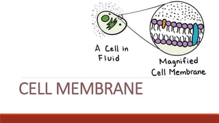
2. Cell Membrane (Plasma Membrane)
Semi-permeable membrane that isolates cytoplasm from the surrounding
environment
Found in all cell types
It is alive
Selectively-permeable to materials
3. Main component of cell membrane is a type of lipid, called
phospholipid
It includes speical proteins and carbohydrates embedded on it .
4. Fluid mosaic cell structure is well-recognized cell membrane model,
discovered by Singer and Nicholson in 1972.
The term ‘fluid’ refers fluidity of the membrane and ‘mosaic’ refers to
different organic compounds (lipid, protein and carbohydrates) which
cell membrane has
5. Cell membrane is made up of phospholipids.
A phospholipid molecule has two parts: hydrophilic head and
hydrophobic tail
Hydrophilic head is composed of glycerol and phosphate and
attracts water
Hydrophobic tail is composed of fatty acid chains and repels water.
This movements make cell membrane alive
6. There are two types of proteins on the cell membrane: integral and
peripheral
If the proteins are embedded into the phospholipid bilayer, they
are integral proteins
They are usually involved in transporting substances across the cell
membrane
If they are only found on the surface of the cell mebrane, they are
peripheral proteins.
They are involved in maintaining the cell’ shape or motility.
7.
8.
9. Membrane proteins may have different roles
Channel proteins allow movements of specific molecules across cell
membrane
Receptor proteins detect hormones arriving cells
Electron carriers allow electrons to pass across the membrane
10. Glycoproteins: function in the cell to cell recognition, selectively permeability of
cell membrane, receptors for chemical signals and recognition of hormones.
Glycolipids: They are called cell markers
or antigens and can be recognized by the
cells of the immune systems as self (of
the organism) or non-self (of cells
belonging to other organisms).
Cholesterol: maintains the integrity and
the fluidity of the cell membrane, also
gives the extra support.