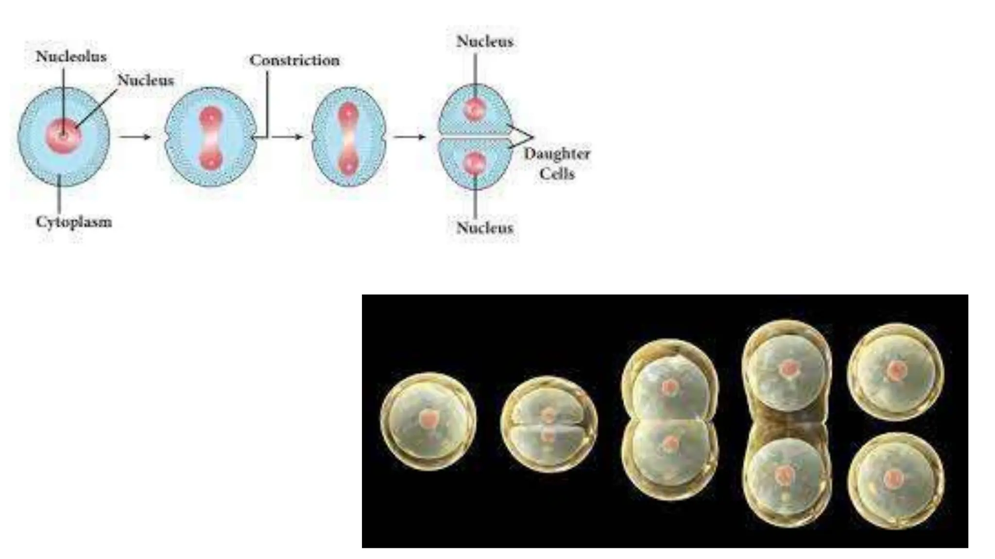
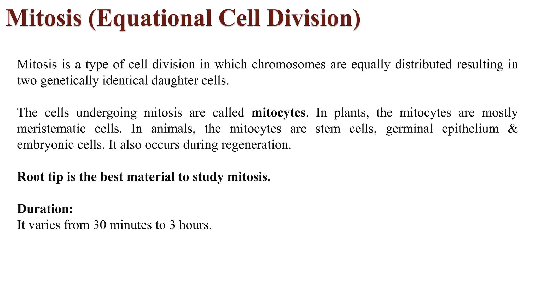
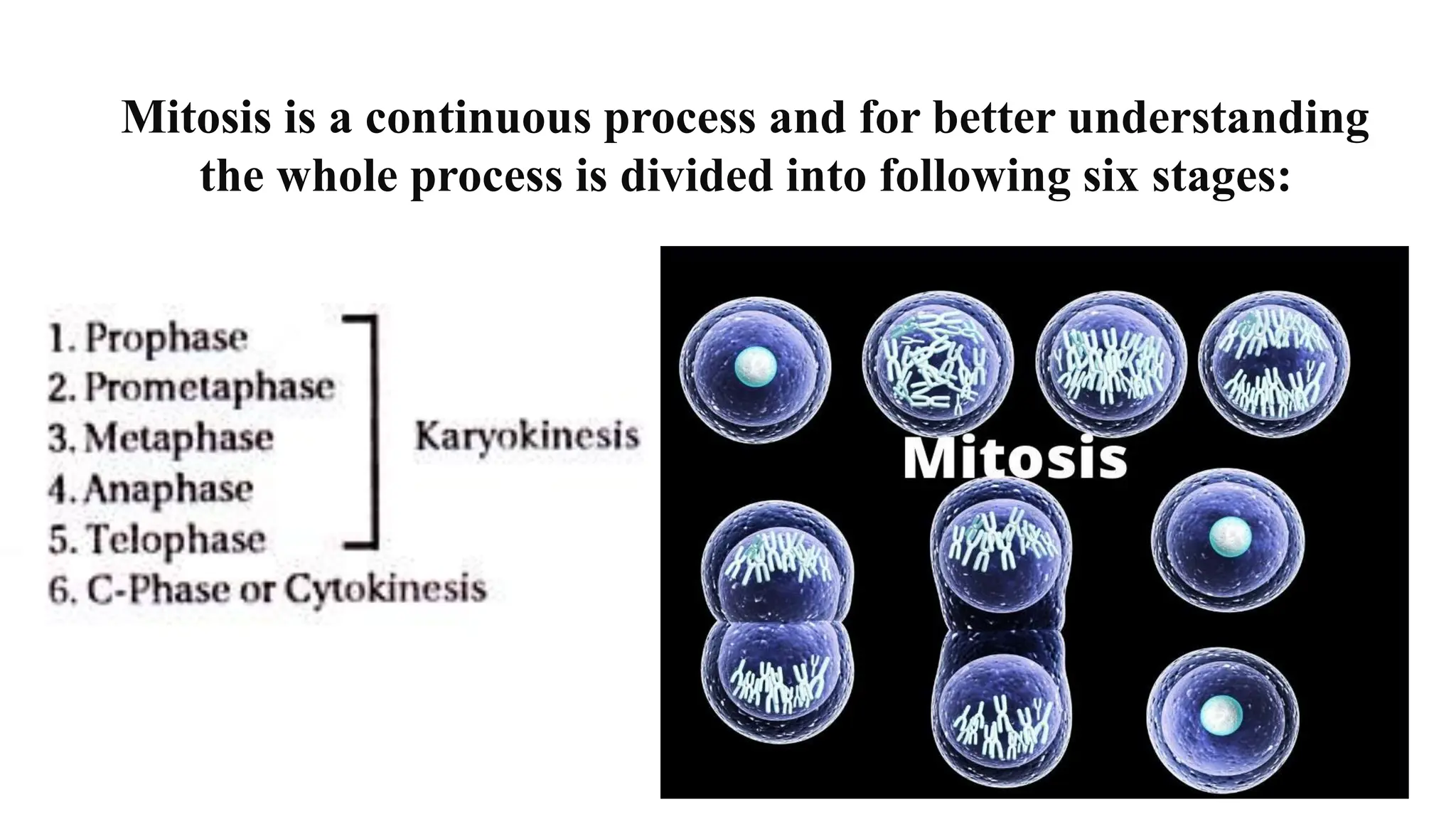
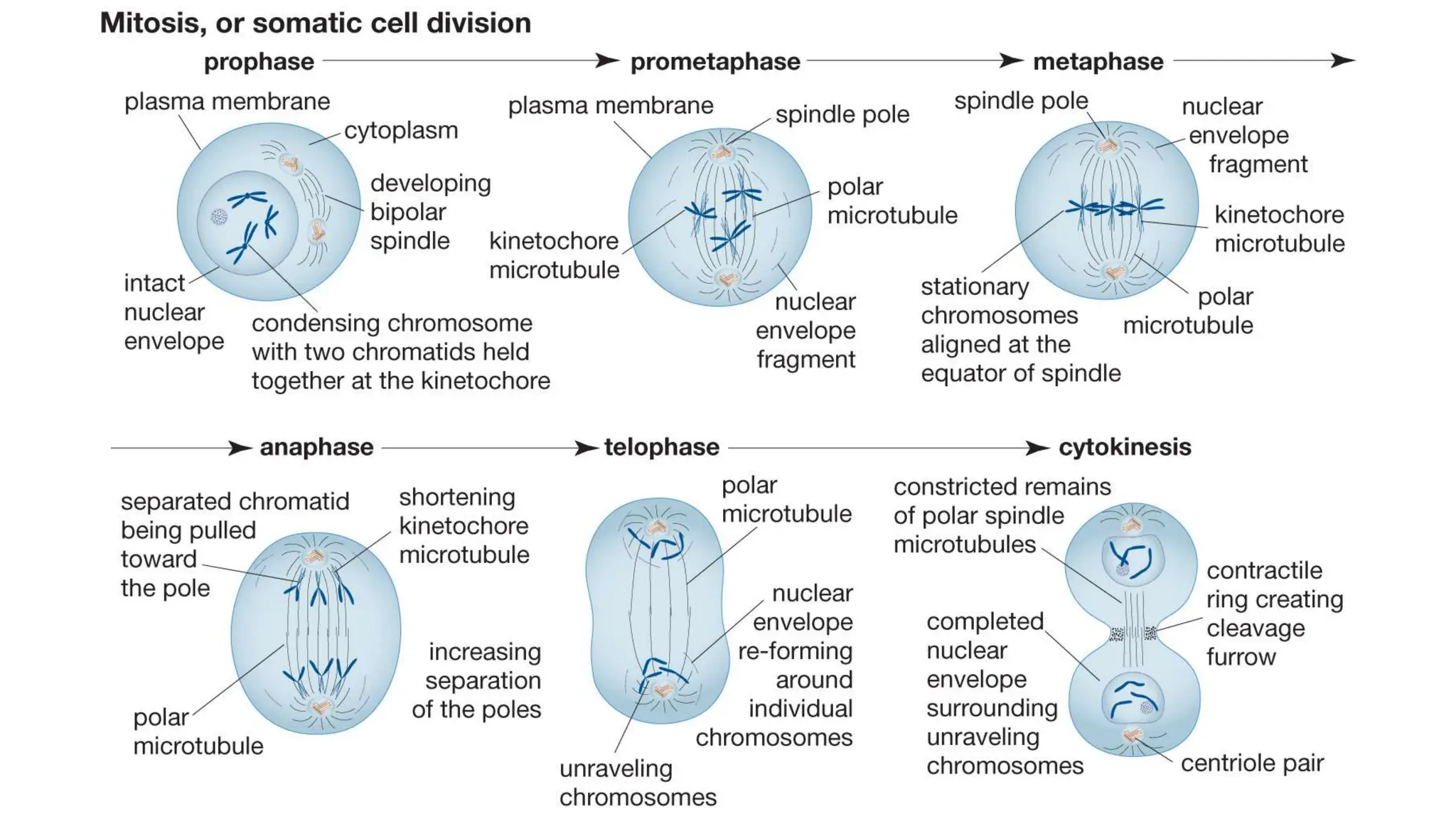
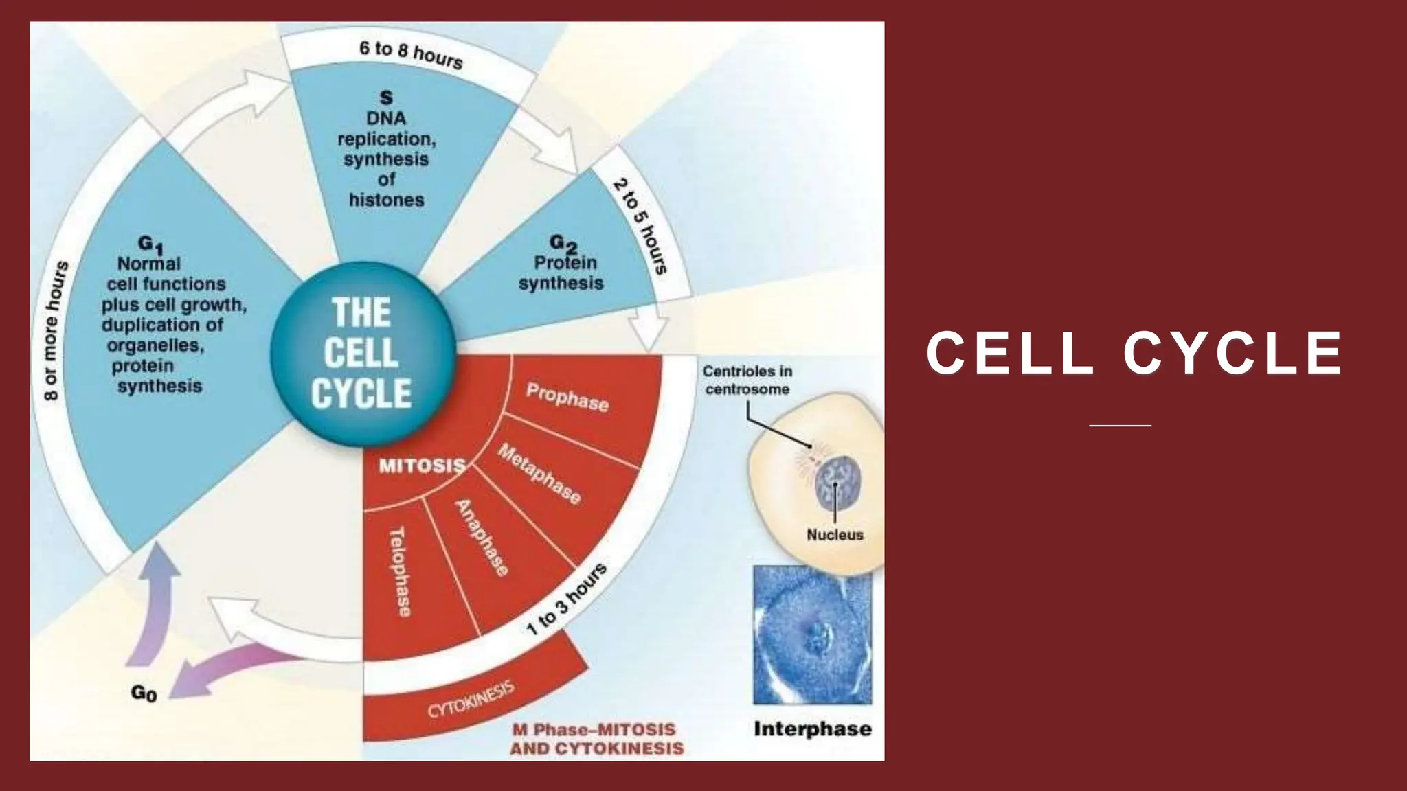
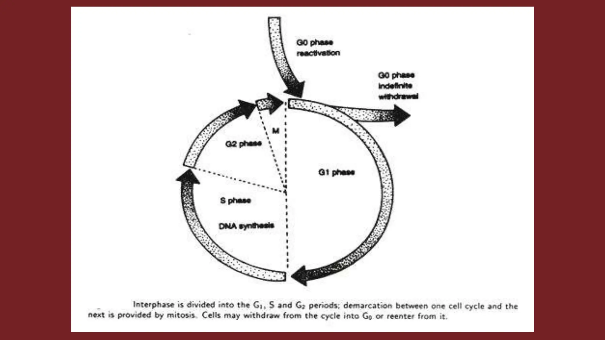
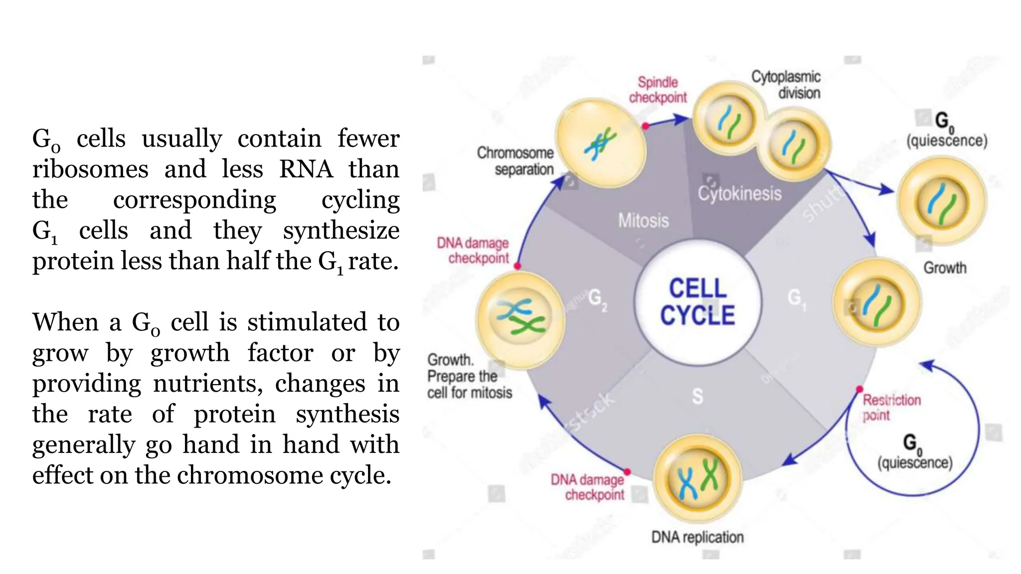
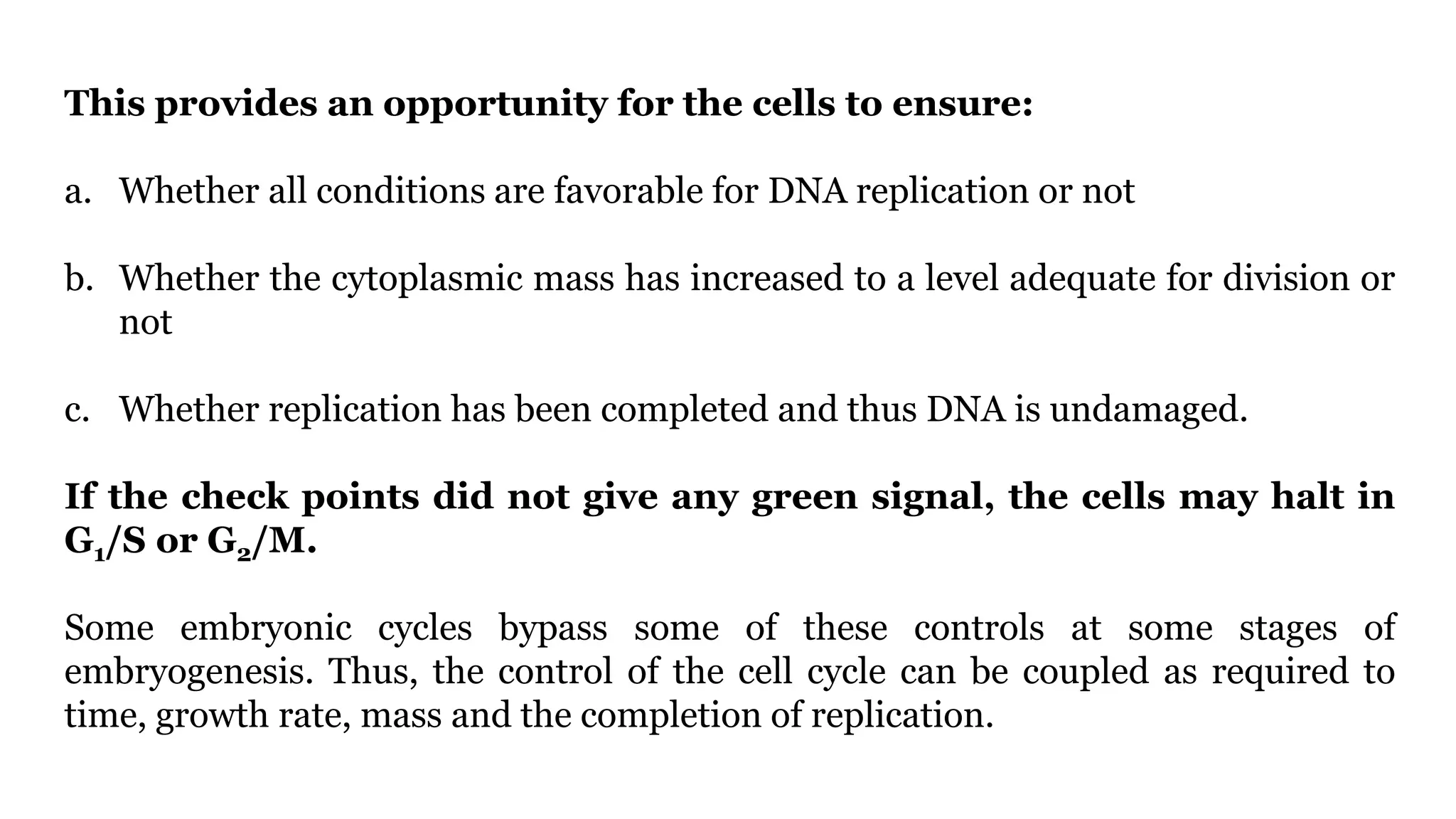
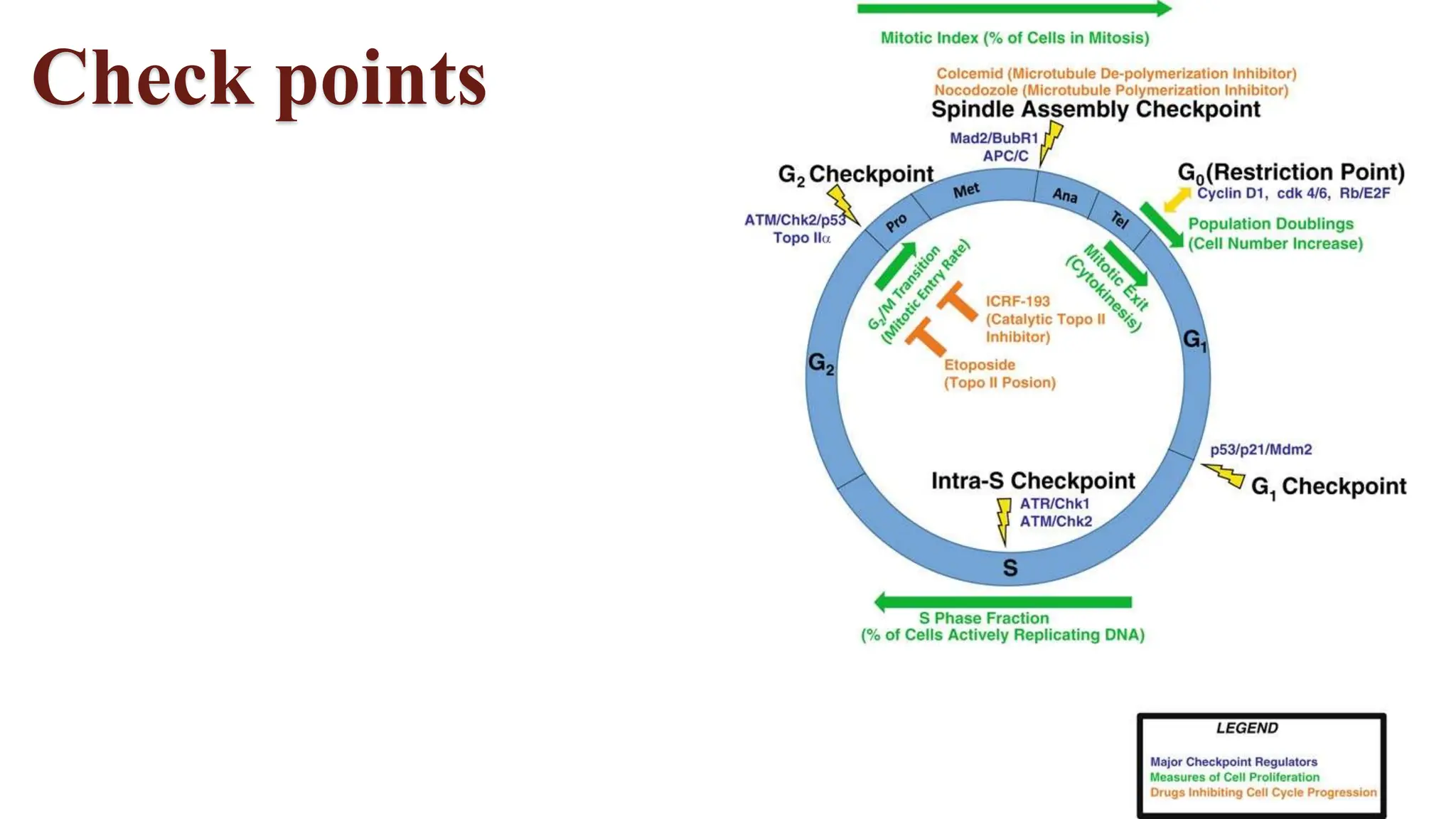
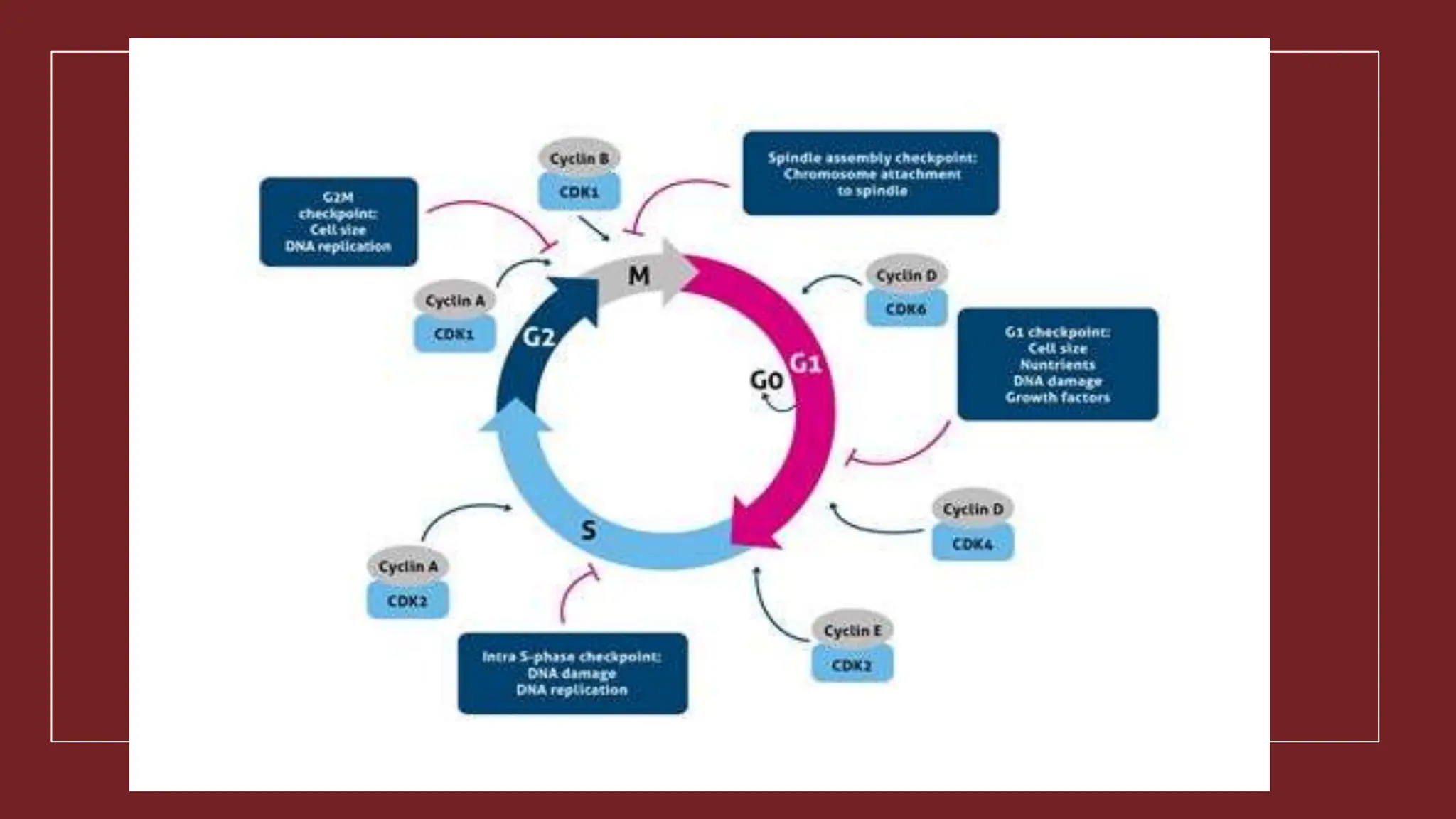
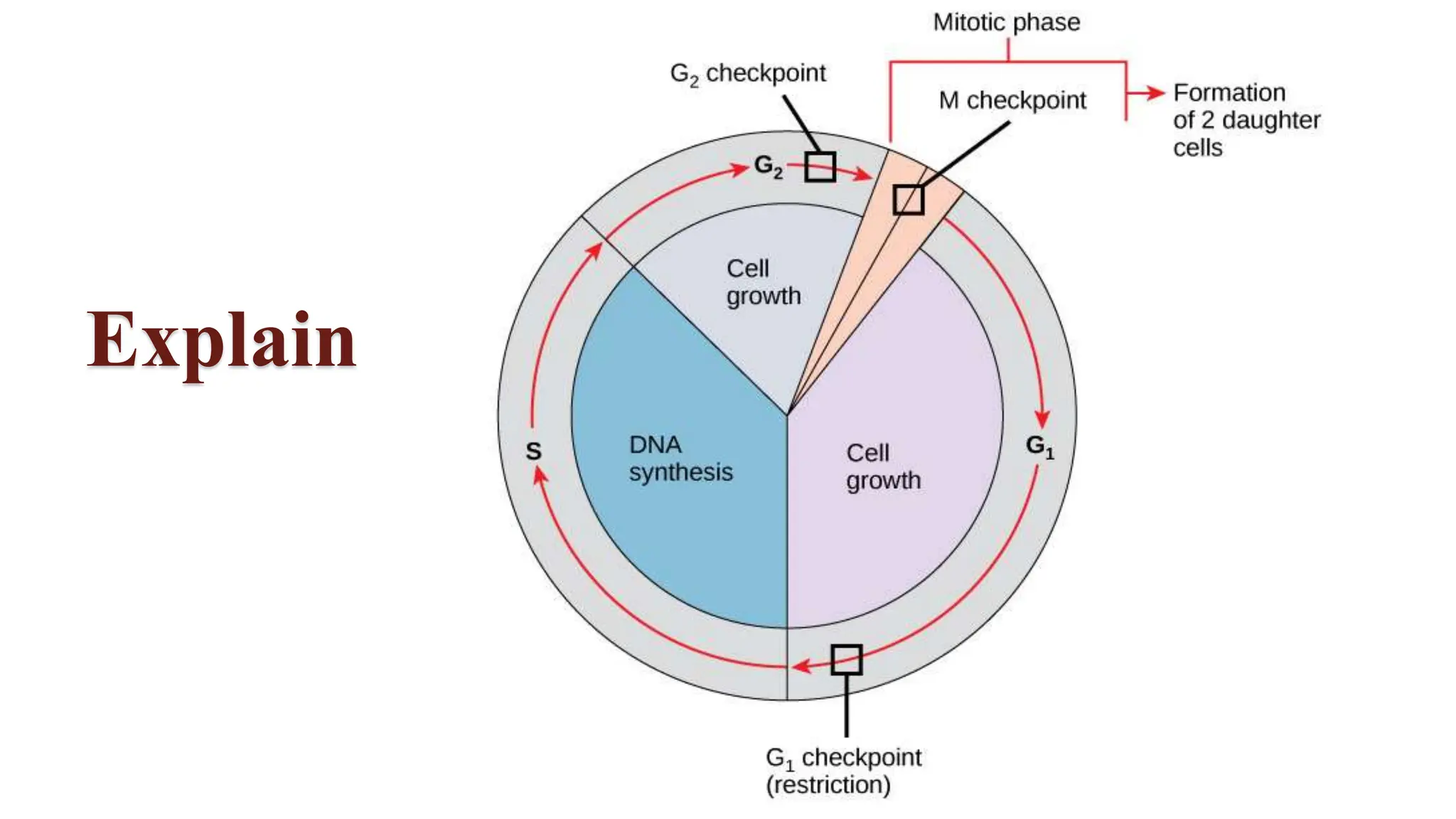
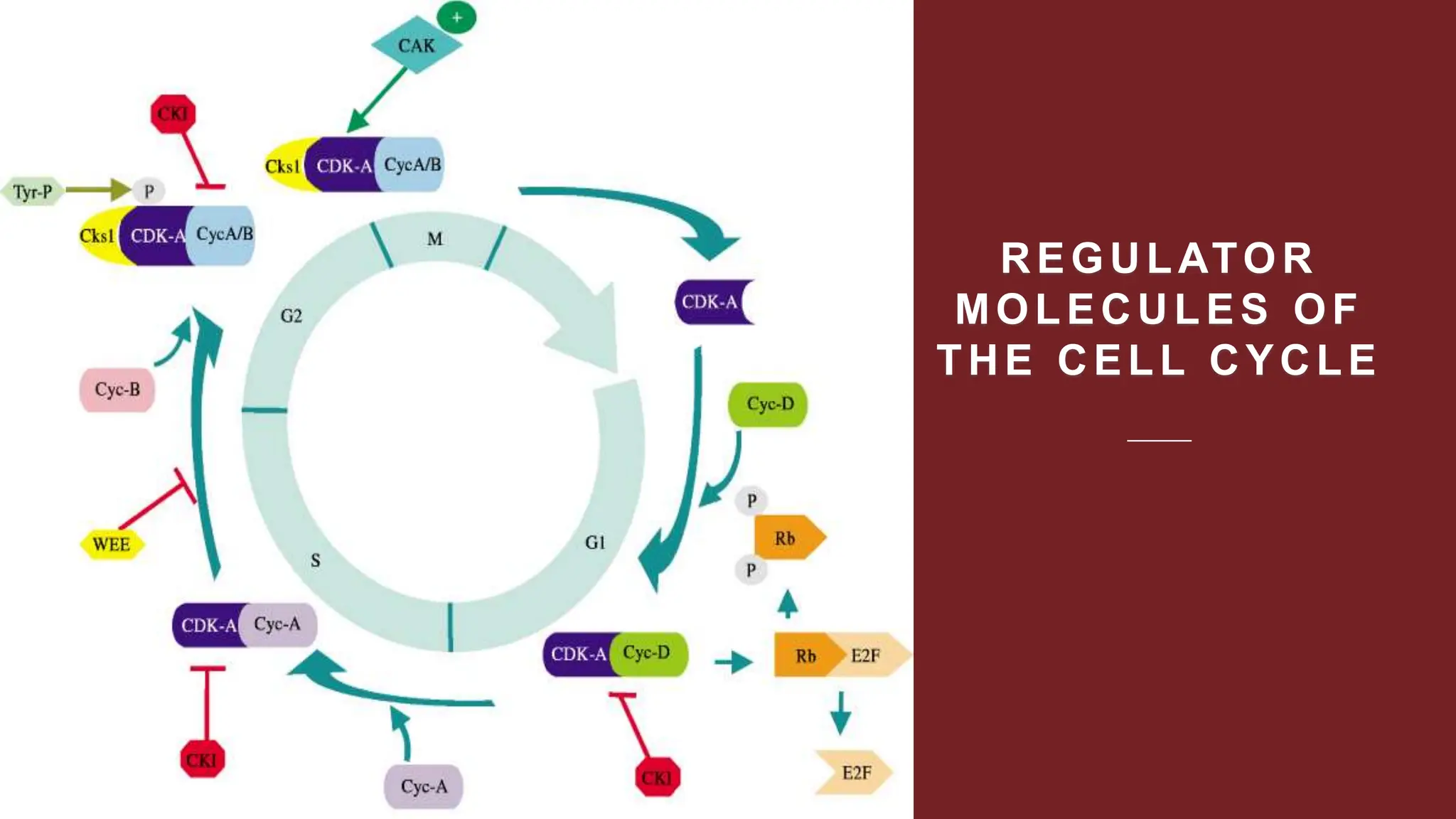
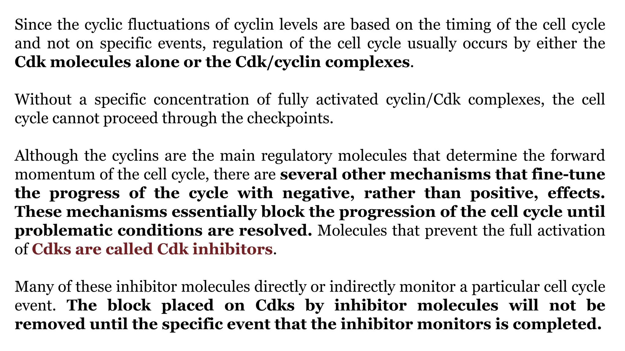
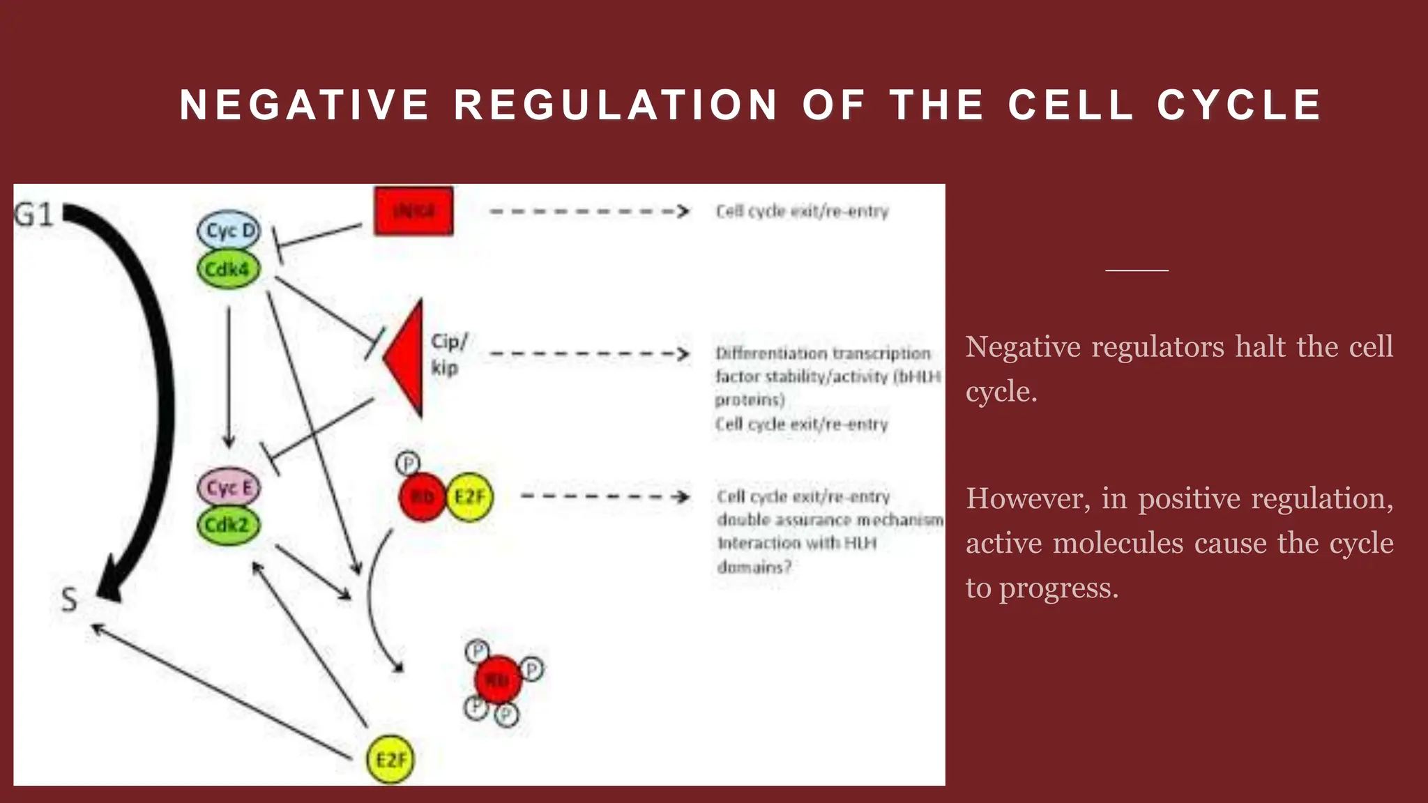
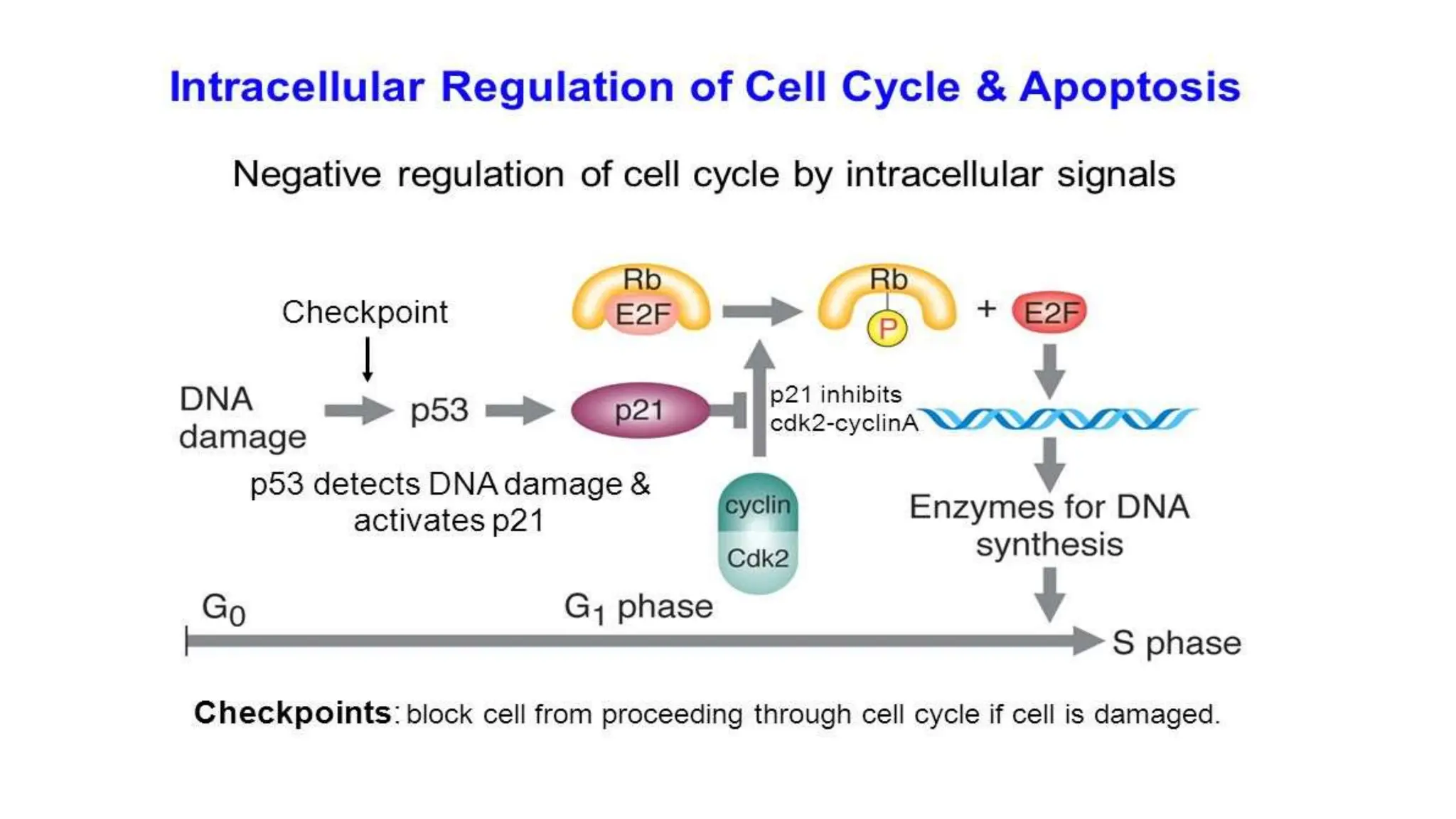
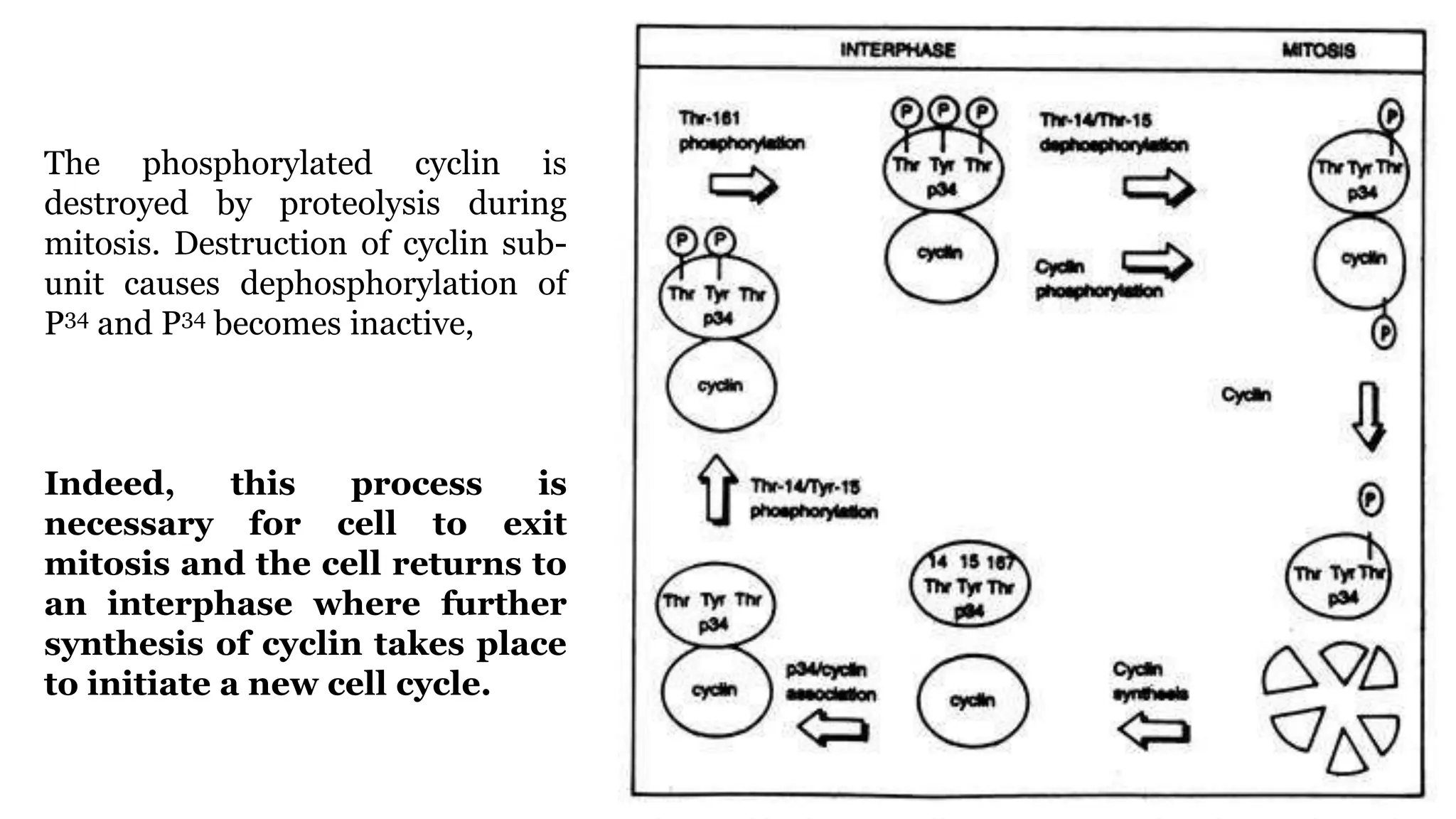
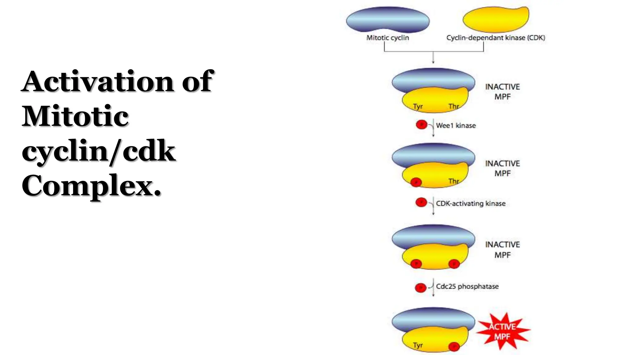
CELL DIVISION
1. Cell division is a pre-requisite for the continuity of life and forms the basis of evolution
to various life forms.
2. In unicellular organisms, cell division is the means of asexual reproduction, which
produces two or more new individuals from the mother cell. The group of such identical
individuals is known as clone.
3. In multi-cellular organisms, life starts from a single cell called zygote (fertilized egg).
The zygote transforms into an adult that is composed of millions of cells formed by
successive divisions.
4. Cell division is the basis of repair and regeneration of old tissues.
Cell Division
Cell Division
Cell division, cell reproduction or cell multiplication is the process of formation of new or
daughter cells from the pre-existing or parent cells.
It occurs in three ways:
1. Amitosis or Direct cell division.
2. Mitosis or Indirect cell division.
3. Meiosis or Reductional cell division
Amitosis (Direct Cell Division)
Amitosis (GK. a = no, mitosis = thread, osis = state)
Amitosis is a mode of division in which nucleus elongates, constricts in the middle and divides
directly into two daughter nuclei. This is followed by centripetal constriction of cytoplasm to
form two daughter cells.
Amitosis is characterized by:
1. Intact nuclear envelope is found through nut the division.
2. Chromatin does not condense into definite chromosomes.
3. A spindle is not formed.
4. Chromatin distribution occurs unequally which causes abnormalities in metabolism and
reproduction.
5. Cytokinesis may or may not follow karyokinesis.
Amitosis occurs in mega-nucleus of paramecium, endosperm cells of seeds, cartilage cells and
diseased cells.
Mitosis (Equational Cell Division)
Mitosis is a type of cell division in which chromosomes are equally distributed resulting in
two genetically identical daughter cells.
The cells undergoing mitosis are called mitocytes. In plants, the mitocytes are mostly
meristematic cells. In animals, the mitocytes are stem cells, germinal epithelium &
embryonic cells. It also occurs during regeneration.
Root tip is the best material to study mitosis.
Duration:
It varies from 30 minutes to 3 hours.
Mitosis is a continuous process and for better understanding
the whole process is divided into following six stages:
CELL CYCLE
All living organisms of the biological world start life as one cell, i.e., unicellular zygote,
the product of the union of gametes. Of course, unicellular organisms live their entire
lives as one cell. But in a multicellular organism, the unicellular zygote undergoes
countless divisions and produces many cells.
These cells ultimately build the organism to a level of cellular complexity and
organization. The process by which any cell produces its own replica is
known as cell division. Thus, simply by cell division a zygote enables an organism
to grow. During this period of growth, many cells undergo a course of specialization
that commits them to perform specific functions.
Some cells function in cell division—either they divide to produce gametes for
sexual reproduction, or they divide to make new cells for growth or to
replace old and damaged cells. Thus, cell division is at the core of life itself. It
helps organisms to grow, reproduce and repair damaged and worn tissue—three
fundamental activities of life.
For hereditary information to be transmitted from generation to
generation, DNA must be replicated before the cells divide so that
each new daughter cell receives a complete copy of hereditary
instruction.
Since DNA is a part of eukaryotic cell’s chromosome, the chromosomes duplicate
as well.
After chromosomal duplication, the rest of the division activities proceed in a way
that ensures each daughter cell receives the same share of genetic information as
well as almost equal proportion of the cell’s cytoplasm and organelles.
Therefore, in order to divide, a cell must double its mass and increase in shape
and size. Cells generally divide when they attain the maximum size.
Phases of Cell Cycle
Most cells divide one or more times during their lifetime. When they do, they pass
through an ordered sequence of events that collectively forms the cell cycle. The
duration of the cell cycle varies greatly from one cell to another.
The shortest cell cycle occurs in early embryo and can last as little as 8 minutes. The
cell cycle of growing eukaryotic cell lasts from 90 minutes to more than 24 hours, its
duration varying considerably within a population of cells.
The cell cycle of the eukaryotic cell is divided into two fundamental
parts:
i. Interphase, and
ii. Mitosis (including Cytokinesis)
Interphase is the period of non-apparent division whereas mitosis is the period of
division. Actually, for many years cell biologists were concerned with the period of
division in which changes visible under the compound microscope could be
observed—whereas during interphase no visible changes under compound
microscope were seen.
Even chromosomes were not visible in the interphase because the refrac-
tive index of the nuclear sap and that of the chromosome present in re-
condensed, hydrated and dispraised state become identical. The whole
nucleus appears as idle. So, interphase was mistakenly considered as resting stage.
During interphase of nucleus several changes take place at the molecular level that are
not visible microscopically. Interphase is a period of intense biosynthetic
activity in which the cell doubles in size and duplicates precisely its
chromosome complement. So, this phase is also known as metabolic
phase and the nucleus is known as the metabolic nucleus.
Interphase
The time from the end of one mitosis to the start of the next mitosis is called
interphase.
This is the longest period in cell division. Feulgen staining of metabolic nucleus
followed by a cytophotometric quantitative assay first suggested that doubling of
DNA takes place during interphase.
Auto-radiographic studies with labelled thymine demonstrated that doubling of
DNA—i.e., replication or synthesis of DNA— did not take place throughout the entire
interphase. It occurs only in a restricted portion of the interphase—the so-called S
phase, i.e., synthetic period.
This period is preceded and followed by two gap periods of interphase (G1 and G2)
in which there is no DNA synthesis. Thus, the interphase can be subdivided into
three successive sub-phases G1, S and G2 and it normally comprises 90% or more of
the total cell cycle.
G1 Phase
The period between the end of
telophase and just before the entry S
phase is called G1 phase.
The period of G1 is usually greater and
is subjected to greater variation.
The S and G2 and mitotic periods are
relatively constant in the cells of the
same organism. But the G1 period is the
most variable in length.
It may constitute 25-50% of the total interphase duration. In some cells G1 may be very
short or absent. Depending on the physiological condition of the cell it may retain in
G1 phase for days, months or years
In cells preparing for cell division there is a marked synthesis of mRNA, tRNA and
proteins during G1 but there is no DNA synthesis. The enzymes and
substrates necessary for DNA synthesis during S phase are also
synthesized during this phase.
Nucleolus produces rRNA and ribosomes are synthesized. This is necessary for the
entry of cells into mitosis as inhibition of their production delays the entry of the cell
into mitosis.
As a whole, cellular metabolic rate is very high in G1. As a result, cell
growth occurs.
Commitment to chromosome or DNA
replication in S phase occurs in
G1 phase.
If conditions to pass the commitment
point are satisfied, after a lag a cell will
enter S phase. The conditions mean the
nutritional state of the medium, the
mass of the cell etc. The commitment
point is clearly observed in yeast cell
where it is called start. The comparable
feature of the animal cell is called the
restriction point.
G0 Phase
Some cells do not divide at all. These cells are often considered to have indefinitely
withdrawn from the cell cycle into another state, resembling G1 but distinct from it
because they are not able to go to S phase, i.e., cells are arrested to non-
cycling state. This non-cycling state is called Go state or resting state
and the cells are called resting cells.
Some cells such as neurons have left the cell cycle irreversibly and can never divide
again. But certain types of cell can be stimulated to leave Go and reenter a cell
cycle. For example, liver cells normally neither grow nor divide, but liver damage
rapidly induces them to divide.
Indefinite withdrawal from or reactivation into the cell cycle takes place effectively
at an early part G1 phase. The absence of nutrients or growth factors cause cells to
enter a resting state. Yeast cells starved of nutrients or mammalian cells deprived
of growth factors arrest early in G1 in the stage G0.
G0 cells usually contain fewer
ribosomes and less RNA than
the corresponding cycling
G1 cells and they synthesize
protein less than half the G1 rate.
When a G0 cell is stimulated to
grow by growth factor or by
providing nutrients, changes in
the rate of protein synthesis
generally go hand in hand with
effect on the chromosome cycle.
S Phase
S phase is the intermediate phase between G1 and G2 phases. When G1 phase ends,
S phase starts. It is a highly specialized phase of interphase, and the word S
stands for synthesis. Actually, DNA synthesis takes place in this phase.
Before a cell can divide, it must produce a new copy of its chromosomes.
For making a new copy of chromosome it needs both the replication of the long
DNA molecule in each chromosome and the assembly of a new set of chromosomal
proteins onto the DNA to form chromatin or chromatid.
By its end each chromosome has been copied to two complete chromatids which
remain joined together at their centromeres until the M phase that soon follows.
G2 Phase
The period from the end of S phase until mitosis is called G2 phase. G2 phase is
usually the shortest part of interphase. In this phase intensive cellular
synthesis occurs. Mitochondria and chloroplasts divide. Energy stores
increase.
Mitotic spindle begins to form. In the interphase there are two control points such
as G1/S and G2/M at which the cell takes a decision on whether to proceed or not
to the next step. Two control points are also called check points.
This provides an opportunity for the cells to ensure:
a. Whether all conditions are favorable for DNA replication or not
b. Whether the cytoplasmic mass has increased to a level adequate for division or
not
c. Whether replication has been completed and thus DNA is undamaged.
If the check points did not give any green signal, the cells may halt in
G1/S or G2/M.
Some embryonic cycles bypass some of these controls at some stages of
embryogenesis. Thus, the control of the cell cycle can be coupled as required to
time, growth rate, mass and the completion of replication.
Check points
It is essential that the daughter cells produced be exact duplicates of the parent
cell. Mistakes in the duplication or distribution of the chromosomes lead to
mutations that may be passed forward to every new cell produced from an
abnormal cell.
To prevent a compromised cell from continuing to divide, there are
internal control mechanisms that operate at three main cell cycle
checkpoints.
A checkpoint is one of several points in the eukaryotic cell cycle at
which the progression of a cell to the next stage in the cycle can be
halted until conditions are favorable.
These checkpoints occur near the end of G1, at the G2/M transition, and the
spindle checkpoint, at the transition from metaphase to anaphase.
The G1 Checkpoint
The G1 checkpoint determines whether all conditions are favorable for cell division
to proceed. The G1 checkpoint, also called the restriction point (in yeast), is a
point at which the cell irreversibly commits to the cell division process.
External influences, such as growth factors, play a large role in carrying the cell
past the G1 checkpoint.
In addition to adequate reserves and cell size, there is a check for genomic DNA
damage at the G1 checkpoint.
A cell that does not meet all the requirements will not be allowed to
progress into the S phase. The cell can halt the cycle and attempt to remedy
the problematic condition, or the cell can advance into G0 and await further
signals when conditions improve.
The G2 Checkpoint
The G2 checkpoint bars entry into the mitotic phase if certain conditions are not
met. As at the G1 checkpoint, cell size and protein reserves are assessed.
The most important role of the G2 checkpoint is to ensure that all of the
chromosomes have been replicated and that the replicated DNA is not
damaged.
If the checkpoint mechanisms detect problems with the DNA, the cell cycle is
halted, and the cell attempts to either complete DNA replication or repair the
damaged DNA.
The M Checkpoint
The M checkpoint occurs near the end of the metaphase stage of
karyokinesis.
The M checkpoint is also known as the spindle checkpoint, because it
determines whether all the sister chromatids are correctly attached
to the spindle microtubules.
Because the separation of the sister chromatids during anaphase is an
irreversible step, the cycle will not proceed until the kinetochores of
each pair of sister chromatids are firmly anchored to at least two
spindle fibers arising from opposite poles of the cell.
Explain
REGULATOR
MOLECULES OF
THE CELL CYCLE
In addition to the internally controlled checkpoints, there are two groups of
intracellular molecules that regulate the cell cycle. These regulatory
molecules either promote progress of the cell to the next phase
(positive regulation) or halt the cycle (negative regulation).
Regulator molecules may act individually, or they can influence the activity or
production of other regulatory proteins. Therefore, the failure of a single regulator
may have almost no effect on the cell cycle, especially if more than one mechanism
controls the same event.
Conversely, the effect of a deficient or non-functioning regulator can be
wide-ranging and possibly fatal to the cell if multiple processes are
affected.
Positive Regulation of the Cell Cycle
Two groups of proteins, called cyclins and cyclin-dependent kinases (Cdks),
are responsible for the progress of the cell through the various checkpoints.
The levels of the four cyclin proteins fluctuate throughout the cell cycle in a
predictable pattern.
Increases in the concentration of cyclin proteins are triggered by both external
and internal signals.
After the cell moves to the next stage of the cell cycle, the cyclins that were active in
the previous stage are degraded.
The concentrations of cyclin proteins change throughout the cell cycle. There is a direct
correlation between cyclin accumulation and the three major cell cycle checkpoints. Also
note the sharp decline of cyclin levels following each checkpoint (the transition
between phases of the cell cycle), as cyclin is degraded by cytoplasmic enzymes.
Cyclins regulate the cell cycle only when they are tightly bound to Cdks.
To be fully active, the Cdk/cyclin complex must also be phosphorylated
in specific locations.
Like all kinases, Cdks are enzymes (kinases) that phosphorylate other proteins.
Phosphorylation activates the protein by changing its shape.
The proteins phosphorylated by Cdks are involved in advancing the cell to the next
phase.
The levels of Cdk proteins are relatively stable throughout the cell cycle; however,
the concentrations of cyclin fluctuate and determine when Cdk/cyclin complexes
form.
The different cyclins and Cdks bind at specific points in the cell cycle
and thus regulate different checkpoints.
Since the cyclic fluctuations of cyclin levels are based on the timing of the cell cycle
and not on specific events, regulation of the cell cycle usually occurs by either the
Cdk molecules alone or the Cdk/cyclin complexes.
Without a specific concentration of fully activated cyclin/Cdk complexes, the cell
cycle cannot proceed through the checkpoints.
Although the cyclins are the main regulatory molecules that determine the forward
momentum of the cell cycle, there are several other mechanisms that fine-tune
the progress of the cycle with negative, rather than positive, effects.
These mechanisms essentially block the progression of the cell cycle until
problematic conditions are resolved. Molecules that prevent the full activation
of Cdks are called Cdk inhibitors.
Many of these inhibitor molecules directly or indirectly monitor a particular cell cycle
event. The block placed on Cdks by inhibitor molecules will not be
removed until the specific event that the inhibitor monitors is completed.
NEGATIVE REGULATION OF THE CELL CYCLE
The best understood negative regulatory molecules are retinoblastoma protein
(Rb), p53, and p21.
Retinoblastoma proteins are a group of tumor-suppressor proteins common in
many cells. The 53 and 21 designations refer to the functional molecular masses of
the proteins (p) in kilodaltons.
All three of these regulatory proteins were discovered to be damaged or
non-functional in cells that had begun to replicate uncontrollably
(became cancerous).
In each case, the main cause of the unchecked progress through the cell
cycle was a faulty copy of the regulatory protein.
Rb, p53, and p21 act primarily at the G1 checkpoint.
p53 is a multi-functional protein that has a major impact on the commitment of a cell to
division because it acts when there is damaged DNA in cells that are undergoing the
preparatory processes during G1.
If damaged DNA is detected, p53 halts the cell cycle and recruits enzymes to repair the
DNA.
If the DNA cannot be repaired, p53 can trigger apoptosis, or cell suicide, to
prevent the duplication of damaged chromosomes.
As p53 levels rise, the production of p21 is triggered. p21 enforces the halt in the
cycle dictated by p53 by binding to and inhibiting the activity of the Cdk/cyclin
complexes.
As a cell is exposed to more stress, higher levels of p53 and p21 accumulate,
making it less likely that the cell will move into the S phase.
F L U C T U AT I O N S I N P 5 3
S I G N A L I N G A L L O W
E S C A P E F R O M C E L L -
C Y C L E A R R E S T
Enzymes that controls cell cycle
It has been observed from the experiments on frog eggs that when an arrested immature
oocyte (equivalent to G2 somatic cell) is injected with cytoplasm extracted from arrested
eggs (equivalent to M phase somatic cells), the oocyte starts to divide. This experiment
suggests that the extract contains an active component that induces the immature
oocyte to enter M phase.
The active component of the extract is, therefore, called maturation promoting
factor (MPF) because MPF causes the cells to enter M phase. MPF is now
understood to stand for M phase promoting factor.
In fact, the MPF is seen to have an enzymatic activity and it has the ability to
phosphorylate target protein and so it is also known as M phase kinase.
M phase kinase consists of two protein sub-units, P34 and P45 (the numbers indicating
molecular weight; P34 = 34,000 Dalton’s).
The two sub-units have different function:
i. The sequence of P34 is a catalytic sub-unit which phosphorylates serine and
threonine residues of target protein.
ii. The other sub-unit, i.e., P54 is a regulatory sub-unit which has kinase activity
with appropriate substrate. This sub-unit is also named as cyclin.
Cyclins can be classified into two general types, viz., A and B. About 30% overall identity is
found between A and B. In mammals and frogs, the B cyclins can be divided into the sub-
types B1 and B2.
The sub-unit P34 is activated by modification at the start of M phase. The other sub-unit,
i.e., P45, or cyclin, is destroyed gradually during mitosis. Its destruction is responsible for
inactivating M phase kinase (P34) and releasing the daughter cells to leave mitosis.
The cell in G2 phase does not enter mitosis (M phase) until and unless M phase
kinase is activated.
During G2 phase two sub-units, i.e., P34 and cyclin, bind with each other to form an inactive
P34-cyclin dimer.
Thereafter P34 undergoes phosphorylation at three sites by two steps.
In the first step threonine at 14 position (Thr 14) and Tyrosine at 15 position (Tyr 15) of the
amino acid chain of P34 are phosphorylated.
In the second step, another phosphorylation occurs on Threonine 167 (Thr 161) of P34. After
phosphorylation, Thr 14 and Tyr 15 are dephosphorylated, and at the same time,
cyclin is phosphorylated. The phosphorylation and dephosphorylation of
P34 and cyclin respectively, are major activities that induce the cell to enter the
mitosis (M phase).
The phosphate at Thr 167 of P34 is required for its activity during M phase.
The phosphorylated cyclin is
destroyed by proteolysis during
mitosis. Destruction of cyclin sub-
unit causes dephosphorylation of
P34 and P34 becomes inactive,
Indeed, this process is
necessary for cell to exit
mitosis and the cell returns to
an interphase where further
synthesis of cyclin takes place
to initiate a new cell cycle.
Mode of Action of M Phase Kinase:
1. M phase kinase phosphorylates target proteins which, in turn, act to regulate other
necessary functions. So, this is an indirect action.
2. M phase kinase directly phosphorylates the crucial substrates that are needed to
regulate M phase.
The action of M phase kinase is always reversible. The M phase kinase directly or indirectly
triggers several activities that causes the onset of M phase.
These activities are:
1. Condensation of chromatin;
2. Dissolution of the nuclear lamina and breakdown of nuclear envelope (except yeast
where the nuclear envelope does not breakdown).
3. Reconstruction of microtubules into a spindle.
4. Reconstruction of actin filaments for cytokinesis.
Molecular Mechanism of Cell Cycle
While many of the checkpoint sensing mechanisms are still unclear, they seem to converge on
two sets of proteins that act together to trigger cell cycle advancement. These proteins are
known as the cyclins and the cyclin-dependent kinases (cdk).
As the names suggest, the cyclins are proteins that regulate progression through the
cell cycle and must be present in sufficient concentration to help activate the
appropriate cdk.
The cyclin-dependent kinase is the active, enzymatic, half of the partnership, and activates
other enzymes by phosphorylation.
Both the cyclins and the cdk’s are families of related proteins, and they can combine in different
ways to govern particular points in the cell cycle.
The intracellular level of cdks is fairly constant. The level of cyclins, on the other hand,
fluctuates dramatically depending on the state of the cell with respect to the cell cycle.
Activation of
Mitotic
cyclin/cdk
Complex.
As more mutant yeast were being screened for changes to their cell cycle, two other genes
were found in which mutations gave rise to similar phenotypes.
Nonfunctional cdc25 or overactive wee1 mutants generated the overly large cells
with a single nucleus.
Both cdc25 and wee1 gene products interact with cdk, and in fact, they are positive
and negative regulators of cdk, respectively.
Acting together with one more enzyme, CAK (cdk-activating kinase), they
activate the cdk.
Using the mitotic cyclin/cdk complex, the cyclin (cdc13) and cdk (cdc2) come together to
form an inactive complex.
The cdk is then phosphorylated by wee1, a kinase. The phosphate it puts on tyrosine-15 is
needed for the rest of the activation sequence, but it is inhibitory: it
actually prevents final activation.
But once Tyr-15 is phosphorylated, CAK can phosphorylate a neighboring threonine (Thr-
161), which is required for activation.
Finally, cdc25, a protein phosphatase, removes the phosphate on Tyr-15, allowing
activation of the cdk by the phosphorylated Thr-161, and the MPF is finally on its way.
There is self-amplification of the activation as well, because one of the targets of MPF is
cdc25, so there is a positive feedback loop in which the activity of cdc25 is upregulated by
phosphorylation.
MPF ensures its own destruction: one of
its phosphorylation targets is cdc20.
Upon phosphorylation, cdc20 is activated
and then activates anaphase promoting
complex (APC). APC is a ubiquitin ligase
(type E3) that polyubiquitinates the cyclin
of the MPF complex, making it a target for
proteolytic degradation by a proteosome.
Note that only the cyclin is destroyed,
while the kinase is left alone.
Without the cyclin, the kinase is inactive
and must wait for cyclin levels to rise
again before it can be re-activated by a
fresh round of phosphorylation and
dephosphorylation.
Cancer & Cell Cycle
Cancer comprises many different diseases caused by a common mechanism: uncontrolled cell
growth. Despite the redundancy and overlapping levels of cell cycle control, errors do occur.
One of the critical processes monitored by the cell cycle checkpoint surveillance mechanism is
the proper replication of DNA during the S phase.
Even when all of the cell cycle controls are fully functional, a small percentage of replication
errors (mutations) will be passed on to the daughter cells. If changes to the DNA
nucleotide sequence occur within a coding portion of a gene and are not
corrected, a gene mutation results. All cancers start when a gene mutation gives
rise to a faulty protein that plays a key role in cell reproduction.
The change in the cell that results from the malformed protein may be minor: perhaps a slight
delay in the binding of Cdk to cyclin. Even minor mistakes, however, may allow subsequent
mistakes to occur more readily.
Over and over, small uncorrected errors are passed from the parent cell to the
daughter cells and amplified as each generation produces more non-functional
proteins from uncorrected DNA damage.
Eventually, the pace of the cell cycle speeds up as the effectiveness of the control
and repair mechanisms decreases.
Uncontrolled growth of the mutated cells outpaces the growth of normal cells in
the area, and a tumor can result.
Proto-oncogenes
The genes that code for the positive cell cycle regulators are called proto-oncogenes.
Proto-oncogenes are normal genes that, when mutated in certain ways,
become oncogenes, genes that cause a cell to become cancerous.
In most instances, the alteration of the DNA sequence will result in a less functional (or non-
functional) protein. The result is detrimental to the cell and will likely prevent the cell from
completing the cell cycle; however, the organism is not harmed because the mutation will
not be carried forward. If a cell cannot reproduce, the mutation is not propagated, and the
damage is minimal.
Occasionally, however, a gene mutation causes a change that increases the activity of a
positive regulator. For example, a mutation that allows Cdk to be activated without being
partnered with cyclin could push the cell cycle past a checkpoint before all of the required
conditions are met. If the resulting daughter cells are too damaged to undergo further cell
divisions, the mutation would not be propagated, and no harm would come to the organism.
However, if the atypical daughter cells are able to undergo further cell divisions,
subsequent generations of cells will probably accumulate even more mutations, some
possibly in additional genes that regulate the cell cycle.
The Cdk gene in the above example is only one of many genes that are considered proto-
oncogenes.
In addition to the cell cycle regulatory proteins, any protein that influences the cycle can
be altered in such a way as to override cell cycle checkpoints. An oncogene is any gene
that, when altered, leads to an increase in the rate of cell cycle progression.
THANK YOU

Cell division: Basic concept of Cytology

  • 1.
  • 2.
    1. Cell divisionis a pre-requisite for the continuity of life and forms the basis of evolution to various life forms. 2. In unicellular organisms, cell division is the means of asexual reproduction, which produces two or more new individuals from the mother cell. The group of such identical individuals is known as clone. 3. In multi-cellular organisms, life starts from a single cell called zygote (fertilized egg). The zygote transforms into an adult that is composed of millions of cells formed by successive divisions. 4. Cell division is the basis of repair and regeneration of old tissues. Cell Division
  • 3.
    Cell Division Cell division,cell reproduction or cell multiplication is the process of formation of new or daughter cells from the pre-existing or parent cells. It occurs in three ways: 1. Amitosis or Direct cell division. 2. Mitosis or Indirect cell division. 3. Meiosis or Reductional cell division
  • 4.
    Amitosis (Direct CellDivision) Amitosis (GK. a = no, mitosis = thread, osis = state) Amitosis is a mode of division in which nucleus elongates, constricts in the middle and divides directly into two daughter nuclei. This is followed by centripetal constriction of cytoplasm to form two daughter cells. Amitosis is characterized by: 1. Intact nuclear envelope is found through nut the division. 2. Chromatin does not condense into definite chromosomes. 3. A spindle is not formed. 4. Chromatin distribution occurs unequally which causes abnormalities in metabolism and reproduction. 5. Cytokinesis may or may not follow karyokinesis. Amitosis occurs in mega-nucleus of paramecium, endosperm cells of seeds, cartilage cells and diseased cells.
  • 6.
    Mitosis (Equational CellDivision) Mitosis is a type of cell division in which chromosomes are equally distributed resulting in two genetically identical daughter cells. The cells undergoing mitosis are called mitocytes. In plants, the mitocytes are mostly meristematic cells. In animals, the mitocytes are stem cells, germinal epithelium & embryonic cells. It also occurs during regeneration. Root tip is the best material to study mitosis. Duration: It varies from 30 minutes to 3 hours.
  • 7.
    Mitosis is acontinuous process and for better understanding the whole process is divided into following six stages:
  • 11.
  • 12.
    All living organismsof the biological world start life as one cell, i.e., unicellular zygote, the product of the union of gametes. Of course, unicellular organisms live their entire lives as one cell. But in a multicellular organism, the unicellular zygote undergoes countless divisions and produces many cells. These cells ultimately build the organism to a level of cellular complexity and organization. The process by which any cell produces its own replica is known as cell division. Thus, simply by cell division a zygote enables an organism to grow. During this period of growth, many cells undergo a course of specialization that commits them to perform specific functions. Some cells function in cell division—either they divide to produce gametes for sexual reproduction, or they divide to make new cells for growth or to replace old and damaged cells. Thus, cell division is at the core of life itself. It helps organisms to grow, reproduce and repair damaged and worn tissue—three fundamental activities of life.
  • 13.
    For hereditary informationto be transmitted from generation to generation, DNA must be replicated before the cells divide so that each new daughter cell receives a complete copy of hereditary instruction. Since DNA is a part of eukaryotic cell’s chromosome, the chromosomes duplicate as well. After chromosomal duplication, the rest of the division activities proceed in a way that ensures each daughter cell receives the same share of genetic information as well as almost equal proportion of the cell’s cytoplasm and organelles. Therefore, in order to divide, a cell must double its mass and increase in shape and size. Cells generally divide when they attain the maximum size.
  • 14.
    Phases of CellCycle Most cells divide one or more times during their lifetime. When they do, they pass through an ordered sequence of events that collectively forms the cell cycle. The duration of the cell cycle varies greatly from one cell to another. The shortest cell cycle occurs in early embryo and can last as little as 8 minutes. The cell cycle of growing eukaryotic cell lasts from 90 minutes to more than 24 hours, its duration varying considerably within a population of cells. The cell cycle of the eukaryotic cell is divided into two fundamental parts: i. Interphase, and ii. Mitosis (including Cytokinesis)
  • 15.
    Interphase is theperiod of non-apparent division whereas mitosis is the period of division. Actually, for many years cell biologists were concerned with the period of division in which changes visible under the compound microscope could be observed—whereas during interphase no visible changes under compound microscope were seen. Even chromosomes were not visible in the interphase because the refrac- tive index of the nuclear sap and that of the chromosome present in re- condensed, hydrated and dispraised state become identical. The whole nucleus appears as idle. So, interphase was mistakenly considered as resting stage. During interphase of nucleus several changes take place at the molecular level that are not visible microscopically. Interphase is a period of intense biosynthetic activity in which the cell doubles in size and duplicates precisely its chromosome complement. So, this phase is also known as metabolic phase and the nucleus is known as the metabolic nucleus.
  • 16.
    Interphase The time fromthe end of one mitosis to the start of the next mitosis is called interphase. This is the longest period in cell division. Feulgen staining of metabolic nucleus followed by a cytophotometric quantitative assay first suggested that doubling of DNA takes place during interphase. Auto-radiographic studies with labelled thymine demonstrated that doubling of DNA—i.e., replication or synthesis of DNA— did not take place throughout the entire interphase. It occurs only in a restricted portion of the interphase—the so-called S phase, i.e., synthetic period. This period is preceded and followed by two gap periods of interphase (G1 and G2) in which there is no DNA synthesis. Thus, the interphase can be subdivided into three successive sub-phases G1, S and G2 and it normally comprises 90% or more of the total cell cycle.
  • 18.
    G1 Phase The periodbetween the end of telophase and just before the entry S phase is called G1 phase. The period of G1 is usually greater and is subjected to greater variation. The S and G2 and mitotic periods are relatively constant in the cells of the same organism. But the G1 period is the most variable in length.
  • 19.
    It may constitute25-50% of the total interphase duration. In some cells G1 may be very short or absent. Depending on the physiological condition of the cell it may retain in G1 phase for days, months or years In cells preparing for cell division there is a marked synthesis of mRNA, tRNA and proteins during G1 but there is no DNA synthesis. The enzymes and substrates necessary for DNA synthesis during S phase are also synthesized during this phase. Nucleolus produces rRNA and ribosomes are synthesized. This is necessary for the entry of cells into mitosis as inhibition of their production delays the entry of the cell into mitosis. As a whole, cellular metabolic rate is very high in G1. As a result, cell growth occurs.
  • 20.
    Commitment to chromosomeor DNA replication in S phase occurs in G1 phase. If conditions to pass the commitment point are satisfied, after a lag a cell will enter S phase. The conditions mean the nutritional state of the medium, the mass of the cell etc. The commitment point is clearly observed in yeast cell where it is called start. The comparable feature of the animal cell is called the restriction point.
  • 21.
    G0 Phase Some cellsdo not divide at all. These cells are often considered to have indefinitely withdrawn from the cell cycle into another state, resembling G1 but distinct from it because they are not able to go to S phase, i.e., cells are arrested to non- cycling state. This non-cycling state is called Go state or resting state and the cells are called resting cells. Some cells such as neurons have left the cell cycle irreversibly and can never divide again. But certain types of cell can be stimulated to leave Go and reenter a cell cycle. For example, liver cells normally neither grow nor divide, but liver damage rapidly induces them to divide. Indefinite withdrawal from or reactivation into the cell cycle takes place effectively at an early part G1 phase. The absence of nutrients or growth factors cause cells to enter a resting state. Yeast cells starved of nutrients or mammalian cells deprived of growth factors arrest early in G1 in the stage G0.
  • 22.
    G0 cells usuallycontain fewer ribosomes and less RNA than the corresponding cycling G1 cells and they synthesize protein less than half the G1 rate. When a G0 cell is stimulated to grow by growth factor or by providing nutrients, changes in the rate of protein synthesis generally go hand in hand with effect on the chromosome cycle.
  • 23.
    S Phase S phaseis the intermediate phase between G1 and G2 phases. When G1 phase ends, S phase starts. It is a highly specialized phase of interphase, and the word S stands for synthesis. Actually, DNA synthesis takes place in this phase. Before a cell can divide, it must produce a new copy of its chromosomes. For making a new copy of chromosome it needs both the replication of the long DNA molecule in each chromosome and the assembly of a new set of chromosomal proteins onto the DNA to form chromatin or chromatid. By its end each chromosome has been copied to two complete chromatids which remain joined together at their centromeres until the M phase that soon follows.
  • 24.
    G2 Phase The periodfrom the end of S phase until mitosis is called G2 phase. G2 phase is usually the shortest part of interphase. In this phase intensive cellular synthesis occurs. Mitochondria and chloroplasts divide. Energy stores increase. Mitotic spindle begins to form. In the interphase there are two control points such as G1/S and G2/M at which the cell takes a decision on whether to proceed or not to the next step. Two control points are also called check points.
  • 25.
    This provides anopportunity for the cells to ensure: a. Whether all conditions are favorable for DNA replication or not b. Whether the cytoplasmic mass has increased to a level adequate for division or not c. Whether replication has been completed and thus DNA is undamaged. If the check points did not give any green signal, the cells may halt in G1/S or G2/M. Some embryonic cycles bypass some of these controls at some stages of embryogenesis. Thus, the control of the cell cycle can be coupled as required to time, growth rate, mass and the completion of replication.
  • 26.
  • 27.
    It is essentialthat the daughter cells produced be exact duplicates of the parent cell. Mistakes in the duplication or distribution of the chromosomes lead to mutations that may be passed forward to every new cell produced from an abnormal cell. To prevent a compromised cell from continuing to divide, there are internal control mechanisms that operate at three main cell cycle checkpoints. A checkpoint is one of several points in the eukaryotic cell cycle at which the progression of a cell to the next stage in the cycle can be halted until conditions are favorable. These checkpoints occur near the end of G1, at the G2/M transition, and the spindle checkpoint, at the transition from metaphase to anaphase.
  • 29.
    The G1 Checkpoint TheG1 checkpoint determines whether all conditions are favorable for cell division to proceed. The G1 checkpoint, also called the restriction point (in yeast), is a point at which the cell irreversibly commits to the cell division process. External influences, such as growth factors, play a large role in carrying the cell past the G1 checkpoint. In addition to adequate reserves and cell size, there is a check for genomic DNA damage at the G1 checkpoint. A cell that does not meet all the requirements will not be allowed to progress into the S phase. The cell can halt the cycle and attempt to remedy the problematic condition, or the cell can advance into G0 and await further signals when conditions improve.
  • 30.
    The G2 Checkpoint TheG2 checkpoint bars entry into the mitotic phase if certain conditions are not met. As at the G1 checkpoint, cell size and protein reserves are assessed. The most important role of the G2 checkpoint is to ensure that all of the chromosomes have been replicated and that the replicated DNA is not damaged. If the checkpoint mechanisms detect problems with the DNA, the cell cycle is halted, and the cell attempts to either complete DNA replication or repair the damaged DNA.
  • 31.
    The M Checkpoint TheM checkpoint occurs near the end of the metaphase stage of karyokinesis. The M checkpoint is also known as the spindle checkpoint, because it determines whether all the sister chromatids are correctly attached to the spindle microtubules. Because the separation of the sister chromatids during anaphase is an irreversible step, the cycle will not proceed until the kinetochores of each pair of sister chromatids are firmly anchored to at least two spindle fibers arising from opposite poles of the cell.
  • 32.
  • 33.
  • 34.
    In addition tothe internally controlled checkpoints, there are two groups of intracellular molecules that regulate the cell cycle. These regulatory molecules either promote progress of the cell to the next phase (positive regulation) or halt the cycle (negative regulation). Regulator molecules may act individually, or they can influence the activity or production of other regulatory proteins. Therefore, the failure of a single regulator may have almost no effect on the cell cycle, especially if more than one mechanism controls the same event. Conversely, the effect of a deficient or non-functioning regulator can be wide-ranging and possibly fatal to the cell if multiple processes are affected.
  • 35.
    Positive Regulation ofthe Cell Cycle Two groups of proteins, called cyclins and cyclin-dependent kinases (Cdks), are responsible for the progress of the cell through the various checkpoints. The levels of the four cyclin proteins fluctuate throughout the cell cycle in a predictable pattern. Increases in the concentration of cyclin proteins are triggered by both external and internal signals. After the cell moves to the next stage of the cell cycle, the cyclins that were active in the previous stage are degraded.
  • 36.
    The concentrations ofcyclin proteins change throughout the cell cycle. There is a direct correlation between cyclin accumulation and the three major cell cycle checkpoints. Also note the sharp decline of cyclin levels following each checkpoint (the transition between phases of the cell cycle), as cyclin is degraded by cytoplasmic enzymes.
  • 37.
    Cyclins regulate thecell cycle only when they are tightly bound to Cdks. To be fully active, the Cdk/cyclin complex must also be phosphorylated in specific locations. Like all kinases, Cdks are enzymes (kinases) that phosphorylate other proteins. Phosphorylation activates the protein by changing its shape. The proteins phosphorylated by Cdks are involved in advancing the cell to the next phase. The levels of Cdk proteins are relatively stable throughout the cell cycle; however, the concentrations of cyclin fluctuate and determine when Cdk/cyclin complexes form. The different cyclins and Cdks bind at specific points in the cell cycle and thus regulate different checkpoints.
  • 41.
    Since the cyclicfluctuations of cyclin levels are based on the timing of the cell cycle and not on specific events, regulation of the cell cycle usually occurs by either the Cdk molecules alone or the Cdk/cyclin complexes. Without a specific concentration of fully activated cyclin/Cdk complexes, the cell cycle cannot proceed through the checkpoints. Although the cyclins are the main regulatory molecules that determine the forward momentum of the cell cycle, there are several other mechanisms that fine-tune the progress of the cycle with negative, rather than positive, effects. These mechanisms essentially block the progression of the cell cycle until problematic conditions are resolved. Molecules that prevent the full activation of Cdks are called Cdk inhibitors. Many of these inhibitor molecules directly or indirectly monitor a particular cell cycle event. The block placed on Cdks by inhibitor molecules will not be removed until the specific event that the inhibitor monitors is completed.
  • 42.
    NEGATIVE REGULATION OFTHE CELL CYCLE
  • 43.
    The best understoodnegative regulatory molecules are retinoblastoma protein (Rb), p53, and p21. Retinoblastoma proteins are a group of tumor-suppressor proteins common in many cells. The 53 and 21 designations refer to the functional molecular masses of the proteins (p) in kilodaltons. All three of these regulatory proteins were discovered to be damaged or non-functional in cells that had begun to replicate uncontrollably (became cancerous). In each case, the main cause of the unchecked progress through the cell cycle was a faulty copy of the regulatory protein.
  • 44.
    Rb, p53, andp21 act primarily at the G1 checkpoint. p53 is a multi-functional protein that has a major impact on the commitment of a cell to division because it acts when there is damaged DNA in cells that are undergoing the preparatory processes during G1. If damaged DNA is detected, p53 halts the cell cycle and recruits enzymes to repair the DNA. If the DNA cannot be repaired, p53 can trigger apoptosis, or cell suicide, to prevent the duplication of damaged chromosomes. As p53 levels rise, the production of p21 is triggered. p21 enforces the halt in the cycle dictated by p53 by binding to and inhibiting the activity of the Cdk/cyclin complexes. As a cell is exposed to more stress, higher levels of p53 and p21 accumulate, making it less likely that the cell will move into the S phase.
  • 46.
    F L UC T U AT I O N S I N P 5 3 S I G N A L I N G A L L O W E S C A P E F R O M C E L L - C Y C L E A R R E S T
  • 48.
    Enzymes that controlscell cycle It has been observed from the experiments on frog eggs that when an arrested immature oocyte (equivalent to G2 somatic cell) is injected with cytoplasm extracted from arrested eggs (equivalent to M phase somatic cells), the oocyte starts to divide. This experiment suggests that the extract contains an active component that induces the immature oocyte to enter M phase. The active component of the extract is, therefore, called maturation promoting factor (MPF) because MPF causes the cells to enter M phase. MPF is now understood to stand for M phase promoting factor. In fact, the MPF is seen to have an enzymatic activity and it has the ability to phosphorylate target protein and so it is also known as M phase kinase.
  • 49.
    M phase kinaseconsists of two protein sub-units, P34 and P45 (the numbers indicating molecular weight; P34 = 34,000 Dalton’s). The two sub-units have different function: i. The sequence of P34 is a catalytic sub-unit which phosphorylates serine and threonine residues of target protein. ii. The other sub-unit, i.e., P54 is a regulatory sub-unit which has kinase activity with appropriate substrate. This sub-unit is also named as cyclin. Cyclins can be classified into two general types, viz., A and B. About 30% overall identity is found between A and B. In mammals and frogs, the B cyclins can be divided into the sub- types B1 and B2. The sub-unit P34 is activated by modification at the start of M phase. The other sub-unit, i.e., P45, or cyclin, is destroyed gradually during mitosis. Its destruction is responsible for inactivating M phase kinase (P34) and releasing the daughter cells to leave mitosis.
  • 50.
    The cell inG2 phase does not enter mitosis (M phase) until and unless M phase kinase is activated. During G2 phase two sub-units, i.e., P34 and cyclin, bind with each other to form an inactive P34-cyclin dimer. Thereafter P34 undergoes phosphorylation at three sites by two steps. In the first step threonine at 14 position (Thr 14) and Tyrosine at 15 position (Tyr 15) of the amino acid chain of P34 are phosphorylated. In the second step, another phosphorylation occurs on Threonine 167 (Thr 161) of P34. After phosphorylation, Thr 14 and Tyr 15 are dephosphorylated, and at the same time, cyclin is phosphorylated. The phosphorylation and dephosphorylation of P34 and cyclin respectively, are major activities that induce the cell to enter the mitosis (M phase). The phosphate at Thr 167 of P34 is required for its activity during M phase.
  • 51.
    The phosphorylated cyclinis destroyed by proteolysis during mitosis. Destruction of cyclin sub- unit causes dephosphorylation of P34 and P34 becomes inactive, Indeed, this process is necessary for cell to exit mitosis and the cell returns to an interphase where further synthesis of cyclin takes place to initiate a new cell cycle.
  • 52.
    Mode of Actionof M Phase Kinase: 1. M phase kinase phosphorylates target proteins which, in turn, act to regulate other necessary functions. So, this is an indirect action. 2. M phase kinase directly phosphorylates the crucial substrates that are needed to regulate M phase. The action of M phase kinase is always reversible. The M phase kinase directly or indirectly triggers several activities that causes the onset of M phase. These activities are: 1. Condensation of chromatin; 2. Dissolution of the nuclear lamina and breakdown of nuclear envelope (except yeast where the nuclear envelope does not breakdown). 3. Reconstruction of microtubules into a spindle. 4. Reconstruction of actin filaments for cytokinesis.
  • 54.
    Molecular Mechanism ofCell Cycle While many of the checkpoint sensing mechanisms are still unclear, they seem to converge on two sets of proteins that act together to trigger cell cycle advancement. These proteins are known as the cyclins and the cyclin-dependent kinases (cdk). As the names suggest, the cyclins are proteins that regulate progression through the cell cycle and must be present in sufficient concentration to help activate the appropriate cdk. The cyclin-dependent kinase is the active, enzymatic, half of the partnership, and activates other enzymes by phosphorylation. Both the cyclins and the cdk’s are families of related proteins, and they can combine in different ways to govern particular points in the cell cycle. The intracellular level of cdks is fairly constant. The level of cyclins, on the other hand, fluctuates dramatically depending on the state of the cell with respect to the cell cycle.
  • 55.
  • 56.
    As more mutantyeast were being screened for changes to their cell cycle, two other genes were found in which mutations gave rise to similar phenotypes. Nonfunctional cdc25 or overactive wee1 mutants generated the overly large cells with a single nucleus. Both cdc25 and wee1 gene products interact with cdk, and in fact, they are positive and negative regulators of cdk, respectively. Acting together with one more enzyme, CAK (cdk-activating kinase), they activate the cdk. Using the mitotic cyclin/cdk complex, the cyclin (cdc13) and cdk (cdc2) come together to form an inactive complex. The cdk is then phosphorylated by wee1, a kinase. The phosphate it puts on tyrosine-15 is needed for the rest of the activation sequence, but it is inhibitory: it actually prevents final activation.
  • 57.
    But once Tyr-15is phosphorylated, CAK can phosphorylate a neighboring threonine (Thr- 161), which is required for activation. Finally, cdc25, a protein phosphatase, removes the phosphate on Tyr-15, allowing activation of the cdk by the phosphorylated Thr-161, and the MPF is finally on its way. There is self-amplification of the activation as well, because one of the targets of MPF is cdc25, so there is a positive feedback loop in which the activity of cdc25 is upregulated by phosphorylation.
  • 58.
    MPF ensures itsown destruction: one of its phosphorylation targets is cdc20. Upon phosphorylation, cdc20 is activated and then activates anaphase promoting complex (APC). APC is a ubiquitin ligase (type E3) that polyubiquitinates the cyclin of the MPF complex, making it a target for proteolytic degradation by a proteosome. Note that only the cyclin is destroyed, while the kinase is left alone. Without the cyclin, the kinase is inactive and must wait for cyclin levels to rise again before it can be re-activated by a fresh round of phosphorylation and dephosphorylation.
  • 59.
    Cancer & CellCycle Cancer comprises many different diseases caused by a common mechanism: uncontrolled cell growth. Despite the redundancy and overlapping levels of cell cycle control, errors do occur. One of the critical processes monitored by the cell cycle checkpoint surveillance mechanism is the proper replication of DNA during the S phase. Even when all of the cell cycle controls are fully functional, a small percentage of replication errors (mutations) will be passed on to the daughter cells. If changes to the DNA nucleotide sequence occur within a coding portion of a gene and are not corrected, a gene mutation results. All cancers start when a gene mutation gives rise to a faulty protein that plays a key role in cell reproduction. The change in the cell that results from the malformed protein may be minor: perhaps a slight delay in the binding of Cdk to cyclin. Even minor mistakes, however, may allow subsequent mistakes to occur more readily.
  • 60.
    Over and over,small uncorrected errors are passed from the parent cell to the daughter cells and amplified as each generation produces more non-functional proteins from uncorrected DNA damage. Eventually, the pace of the cell cycle speeds up as the effectiveness of the control and repair mechanisms decreases. Uncontrolled growth of the mutated cells outpaces the growth of normal cells in the area, and a tumor can result.
  • 61.
    Proto-oncogenes The genes thatcode for the positive cell cycle regulators are called proto-oncogenes. Proto-oncogenes are normal genes that, when mutated in certain ways, become oncogenes, genes that cause a cell to become cancerous. In most instances, the alteration of the DNA sequence will result in a less functional (or non- functional) protein. The result is detrimental to the cell and will likely prevent the cell from completing the cell cycle; however, the organism is not harmed because the mutation will not be carried forward. If a cell cannot reproduce, the mutation is not propagated, and the damage is minimal. Occasionally, however, a gene mutation causes a change that increases the activity of a positive regulator. For example, a mutation that allows Cdk to be activated without being partnered with cyclin could push the cell cycle past a checkpoint before all of the required conditions are met. If the resulting daughter cells are too damaged to undergo further cell divisions, the mutation would not be propagated, and no harm would come to the organism.
  • 62.
    However, if theatypical daughter cells are able to undergo further cell divisions, subsequent generations of cells will probably accumulate even more mutations, some possibly in additional genes that regulate the cell cycle. The Cdk gene in the above example is only one of many genes that are considered proto- oncogenes. In addition to the cell cycle regulatory proteins, any protein that influences the cycle can be altered in such a way as to override cell cycle checkpoints. An oncogene is any gene that, when altered, leads to an increase in the rate of cell cycle progression.
  • 66.