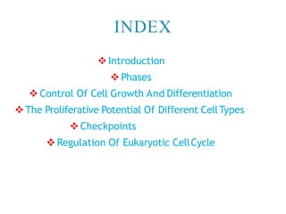
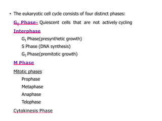
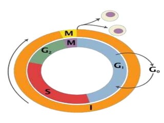
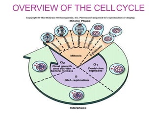
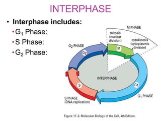
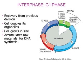
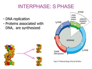
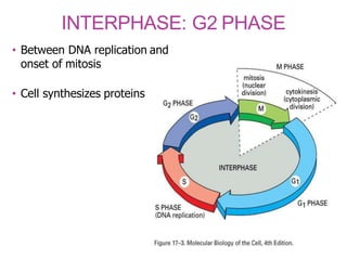
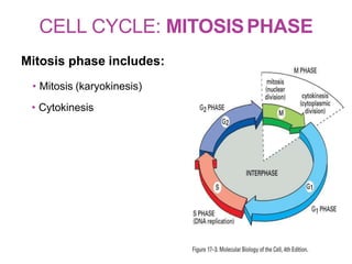
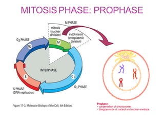
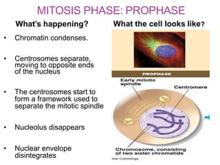
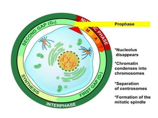
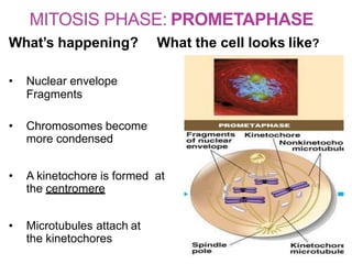
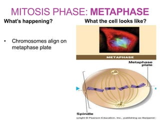
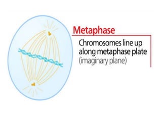
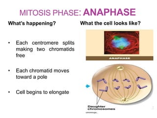
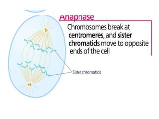
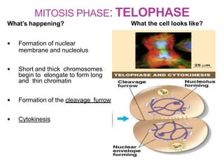
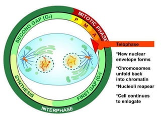
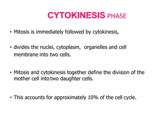
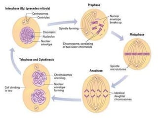
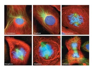
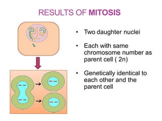
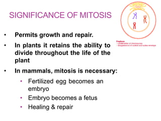
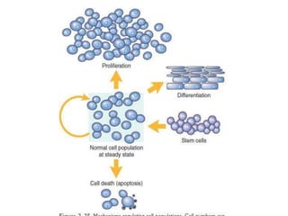
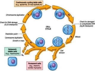
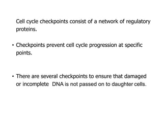
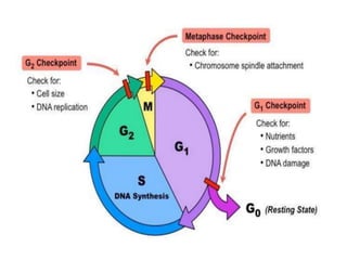
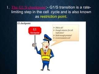
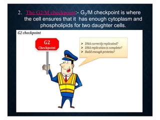
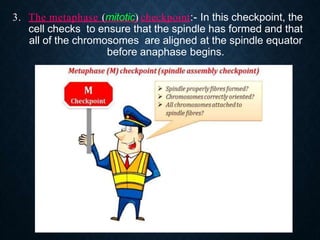
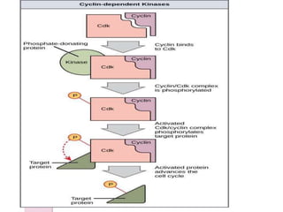
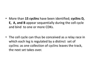
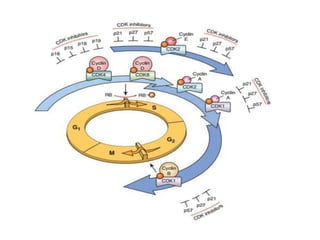
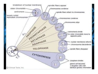
CELL CYCLE & CELL THERAPY
DR. ROOPAM JAIN
PROFESSOR & HEAD
INDEX
 Introduction
 Phases
 Control Of Cell Growth And Differentiation
The Proliferative Potential Of Different CellTypes
Checkpoints
Regulation Of Eukaryotic CellCycle
• The sequence of events that results in cell
division is called the cell cycle.
• The cell cycle, or cell-division cycle
duplication of its DNA (DNA replication)
division of cytoplasm
organelles to produce two daughter cells.
• The cell-division cycle is a vital process by which a single-
celled fertilized egg develops into a mature organism.
• Most cancers are in essence caused by deregulation of
the cell cycle (oncogenes & tumor suppressor genes -
influence its progression).
• In bacteria, which lack a cell nucleus, as in prokaryotes,
the cell cycle extends from the end of cell division to the
beginning of DNA replication and then the splitting of the
bacterial cell into two daughter cells.
• In cells with a nucleus, as in eukaryotes, the cell cycle is
also divided into 2 main stages:
During interphase, the cell grows, accumulating nutrients
needed for mitosis, and undergoes DNA replication
preparing it for cell division.
During the mitotic phase (including mitosis and
cytokinesis), the replicated chromosomes and
cytoplasm separate into two new daughter cells.
• To ensure the proper division of the cell, there are control
mechanisms known as cell cycle checkpoints.
• The eukaryotic cell cycle consists of four distinct phases:
G0 Phase- Quiescent cells that are not actively cycling
Interphase
o G1 Phase(presynthetic growth)
o S Phase (DNA synthesis)
o G2 Phase(premitotic growth)
M Phase
Mitotic phases
o Prophase
o Metaphase
o Anaphase
o Telophase
Cytokinesis Phase
OVERVIEW OF THE CELLCYCLE
INTERPHASE
• Interphase includes:
•G1 Phase:
•S Phase:
•G2 Phase:
INTERPHASE: G1 PHASE
• Recovery from previous
division
• Cell doubles its
organelles
• Cell grows in size
• Accumulates raw
materials for DNA
synthesis
INTERPHASE: S PHASE
• DNA replication
• Proteins associated with
DNA, are synthesized
INTERPHASE: G2 PHASE
• Between DNA replication and
onset of mitosis
• Cell synthesizes proteins
CELL CYCLE: MITOSISPHASE
Mitosis phase includes:
• Mitosis (karyokinesis)
• Cytokinesis
MITOSISPHASE: PROPHASE
MITOSIS PHASE: PROPHASE
What’s happening? What the cell looks like?
• Chromatin condenses.
• Centrosomes separate,
moving to opposite ends
of the nucleus
• The centrosomes start to
form a framework used to
separate the mitotic spindle
• Nucleolus disappears
• Nuclear envelope
disintegrates
MITOSIS PHASE: PROMETAPHASE
What’s happening? What the cell looks like?
• Nuclear envelope
Fragments
• Chromosomes become
more condensed
• A kinetochore is formed at
the centromere
• Microtubules attach at
the kinetochores
MITOSIS PHASE: METAPHASE
What’s happening? What the cell looks like?
• Chromosomes align on
metaphase plate
MITOSIS PHASE: ANAPHASE
What’s happening? What the cell looks like?
• Each centromere splits
making two chromatids
free
• Each chromatid moves
toward a pole
• Cell begins to elongate
MITOSIS PHASE: TELOPHASE
What’s happening? What the cell looks like?
• Formation of nuclear
membrane and nucleolus
• Short and thick chromosomes
begin to elongate to form long
and thin chromatin
• Formation of the cleavage furrow
• Cytokinesis
• Mitosis is immediately followed by cytokinesis,
• divides the nuclei, cytoplasm, organelles and cell
membrane into two cells.
• Mitosis and cytokinesis together define the division of the
mother cell into two daughter cells.
• This accounts for approximately 10% of the cell cycle.
CYTOKINESIS PHASE
RESULTS OF MITOSIS
• Two daughter nuclei
• Each with same
chromosome number as
parent cell ( 2n)
• Genetically identical to
each other and the
parent cell
SIGNIFICANCE OF MITOSIS
• Permits growth and repair.
• In plants it retains the ability to
divide throughout the life of the
plant
• In mammals, mitosis is necessary:
• Fertilized egg becomes an
embryo
• Embryo becomes a fetus
• Healing & repair
CONTROL OF CELL GROWTH
AND DIFFERENTIATION
• Entry of new cells into a tissue population is largely
determined by their proliferation rate
• Cell proliferation can be stimulated by intrinsic growth
factor, injury, cell death, or even mechanical deformation
of tissues.
• The biochemical mediators and /or mechanical stressors
present in the local micro-environment can either
stimulate or inhibit cell growth.
• The excess or deficiency of these can result in net cell
growth.
THE PROLIFERATIVE
POTENTIAL OF DIFFERENT
CELL TYPES
• Regenerative capacity:
Labile cells - Cells of these tissues are continuously being lost and
replaced by maturation from stem cells.
 E.g.- epidermis and GI Tract.
Stable cells - Cells of these tissues are quiescent and have
only minimal replicative activity.
 E.g.- hepatocytes.
Permanent cells - Cells of these tissues are considered to be
terminally differentiated and non-proliferative in postnatal life.
CHECKPOINTS
To ensure the proper division of the cell, there
are control mechanisms known as cell cycle
checkpoints
• Cell cycle checkpoints consist of a network of regulatory
proteins.
• Checkpoints prevent cell cycle progression at specific
points.
• There are several checkpoints to ensure that damaged
or incomplete DNA is not passed on to daughter cells.
1. The G1/S checkpoint:- G1/S transition is a rate-
limiting step in the cell cycle and is also known
as restriction point.
2. The G2/M checkpoint:- G2/M checkpoint is where
the cell ensures that it has enough cytoplasm and
phospholipids for two daughter cells.
3. The metaphase (mitotic) checkpoint:- In this checkpoint, the
cell checks to ensure that the spindle has formed and that
all of the chromosomes are aligned at the spindle equator
before anaphase begins.
REGULATION OF
EUKARYOTIC CELL CYCLE
• The cell cycle is regulated by activators and
inhibitors.
• Cell cycle progression is driven by proteins
called cyclins— named for the cyclic nature
of their production and degradation—and
cyclin-associated enzymes called cyclin-
dependent kinases (CDKs).
• More than 15 cyclins have been identified; cyclins D,
E, A, and B appear sequentially during the cell cycle
and bind to one or more CDKs.
• The cell cycle can thus be conceived as a relay race in
which each leg is regulated by a distinct set of
cyclins: as one collection of cyclins leaves the track,
the next set takes over.
• Enforcing the cell cycle checkpoints is the job of CDK
inhibitors (CDKIs); they accomplish this by modulating
CDK-cyclin complex activity.
• There are several different CDKIs:
• One family—composed of three proteins called p21
(CDKN1A), p27 (CDKN1B), and p57 (CDKN1C)— broadly
inhibits multiple CDKs.
• Other family of CDKI proteins has selective effects on cyclin
CDK4 and cyclin CDK6; these proteins are called
p15(CDKN2B), p16(CDKN2A), p18(CDKN2C), and
p19(CDKN2D).
CELL CYCLE & A CONCEPT OF CELL THERAPY

CELL CYCLE & A CONCEPT OF CELL THERAPY

  • 1.
    CELL CYCLE &CELL THERAPY DR. ROOPAM JAIN PROFESSOR & HEAD
  • 3.
    INDEX  Introduction  Phases Control Of Cell Growth And Differentiation The Proliferative Potential Of Different CellTypes Checkpoints Regulation Of Eukaryotic CellCycle
  • 4.
    • The sequenceof events that results in cell division is called the cell cycle.
  • 5.
    • The cellcycle, or cell-division cycle duplication of its DNA (DNA replication) division of cytoplasm organelles to produce two daughter cells. • The cell-division cycle is a vital process by which a single- celled fertilized egg develops into a mature organism. • Most cancers are in essence caused by deregulation of the cell cycle (oncogenes & tumor suppressor genes - influence its progression).
  • 6.
    • In bacteria,which lack a cell nucleus, as in prokaryotes, the cell cycle extends from the end of cell division to the beginning of DNA replication and then the splitting of the bacterial cell into two daughter cells.
  • 7.
    • In cellswith a nucleus, as in eukaryotes, the cell cycle is also divided into 2 main stages: During interphase, the cell grows, accumulating nutrients needed for mitosis, and undergoes DNA replication preparing it for cell division. During the mitotic phase (including mitosis and cytokinesis), the replicated chromosomes and cytoplasm separate into two new daughter cells. • To ensure the proper division of the cell, there are control mechanisms known as cell cycle checkpoints.
  • 9.
    • The eukaryoticcell cycle consists of four distinct phases: G0 Phase- Quiescent cells that are not actively cycling Interphase o G1 Phase(presynthetic growth) o S Phase (DNA synthesis) o G2 Phase(premitotic growth) M Phase Mitotic phases o Prophase o Metaphase o Anaphase o Telophase Cytokinesis Phase
  • 11.
    OVERVIEW OF THECELLCYCLE
  • 12.
    INTERPHASE • Interphase includes: •G1Phase: •S Phase: •G2 Phase:
  • 13.
    INTERPHASE: G1 PHASE •Recovery from previous division • Cell doubles its organelles • Cell grows in size • Accumulates raw materials for DNA synthesis
  • 14.
    INTERPHASE: S PHASE •DNA replication • Proteins associated with DNA, are synthesized
  • 15.
    INTERPHASE: G2 PHASE •Between DNA replication and onset of mitosis • Cell synthesizes proteins
  • 16.
    CELL CYCLE: MITOSISPHASE Mitosisphase includes: • Mitosis (karyokinesis) • Cytokinesis
  • 17.
  • 18.
    MITOSIS PHASE: PROPHASE What’shappening? What the cell looks like? • Chromatin condenses. • Centrosomes separate, moving to opposite ends of the nucleus • The centrosomes start to form a framework used to separate the mitotic spindle • Nucleolus disappears • Nuclear envelope disintegrates
  • 20.
    MITOSIS PHASE: PROMETAPHASE What’shappening? What the cell looks like? • Nuclear envelope Fragments • Chromosomes become more condensed • A kinetochore is formed at the centromere • Microtubules attach at the kinetochores
  • 21.
    MITOSIS PHASE: METAPHASE What’shappening? What the cell looks like? • Chromosomes align on metaphase plate
  • 23.
    MITOSIS PHASE: ANAPHASE What’shappening? What the cell looks like? • Each centromere splits making two chromatids free • Each chromatid moves toward a pole • Cell begins to elongate
  • 25.
    MITOSIS PHASE: TELOPHASE What’shappening? What the cell looks like? • Formation of nuclear membrane and nucleolus • Short and thick chromosomes begin to elongate to form long and thin chromatin • Formation of the cleavage furrow • Cytokinesis
  • 27.
    • Mitosis isimmediately followed by cytokinesis, • divides the nuclei, cytoplasm, organelles and cell membrane into two cells. • Mitosis and cytokinesis together define the division of the mother cell into two daughter cells. • This accounts for approximately 10% of the cell cycle. CYTOKINESIS PHASE
  • 30.
    RESULTS OF MITOSIS •Two daughter nuclei • Each with same chromosome number as parent cell ( 2n) • Genetically identical to each other and the parent cell
  • 31.
    SIGNIFICANCE OF MITOSIS •Permits growth and repair. • In plants it retains the ability to divide throughout the life of the plant • In mammals, mitosis is necessary: • Fertilized egg becomes an embryo • Embryo becomes a fetus • Healing & repair
  • 32.
    CONTROL OF CELLGROWTH AND DIFFERENTIATION
  • 33.
    • Entry ofnew cells into a tissue population is largely determined by their proliferation rate • Cell proliferation can be stimulated by intrinsic growth factor, injury, cell death, or even mechanical deformation of tissues. • The biochemical mediators and /or mechanical stressors present in the local micro-environment can either stimulate or inhibit cell growth. • The excess or deficiency of these can result in net cell growth.
  • 35.
    THE PROLIFERATIVE POTENTIAL OFDIFFERENT CELL TYPES
  • 36.
    • Regenerative capacity: Labilecells - Cells of these tissues are continuously being lost and replaced by maturation from stem cells.  E.g.- epidermis and GI Tract. Stable cells - Cells of these tissues are quiescent and have only minimal replicative activity.  E.g.- hepatocytes. Permanent cells - Cells of these tissues are considered to be terminally differentiated and non-proliferative in postnatal life.
  • 38.
  • 39.
    To ensure theproper division of the cell, there are control mechanisms known as cell cycle checkpoints
  • 40.
    • Cell cyclecheckpoints consist of a network of regulatory proteins. • Checkpoints prevent cell cycle progression at specific points. • There are several checkpoints to ensure that damaged or incomplete DNA is not passed on to daughter cells.
  • 42.
    1. The G1/Scheckpoint:- G1/S transition is a rate- limiting step in the cell cycle and is also known as restriction point.
  • 43.
    2. The G2/Mcheckpoint:- G2/M checkpoint is where the cell ensures that it has enough cytoplasm and phospholipids for two daughter cells.
  • 44.
    3. The metaphase(mitotic) checkpoint:- In this checkpoint, the cell checks to ensure that the spindle has formed and that all of the chromosomes are aligned at the spindle equator before anaphase begins.
  • 45.
  • 46.
    • The cellcycle is regulated by activators and inhibitors. • Cell cycle progression is driven by proteins called cyclins— named for the cyclic nature of their production and degradation—and cyclin-associated enzymes called cyclin- dependent kinases (CDKs).
  • 48.
    • More than15 cyclins have been identified; cyclins D, E, A, and B appear sequentially during the cell cycle and bind to one or more CDKs. • The cell cycle can thus be conceived as a relay race in which each leg is regulated by a distinct set of cyclins: as one collection of cyclins leaves the track, the next set takes over.
  • 49.
    • Enforcing thecell cycle checkpoints is the job of CDK inhibitors (CDKIs); they accomplish this by modulating CDK-cyclin complex activity. • There are several different CDKIs: • One family—composed of three proteins called p21 (CDKN1A), p27 (CDKN1B), and p57 (CDKN1C)— broadly inhibits multiple CDKs. • Other family of CDKI proteins has selective effects on cyclin CDK4 and cyclin CDK6; these proteins are called p15(CDKN2B), p16(CDKN2A), p18(CDKN2C), and p19(CDKN2D).