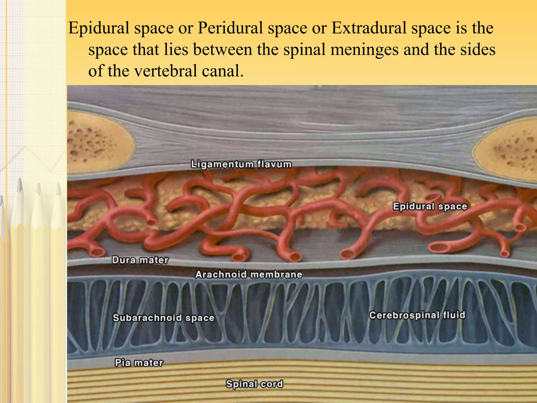
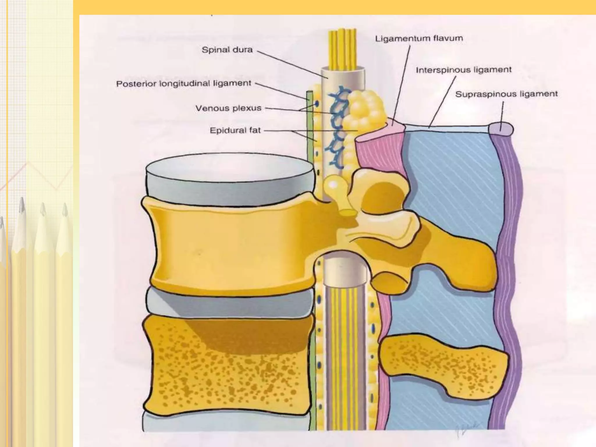
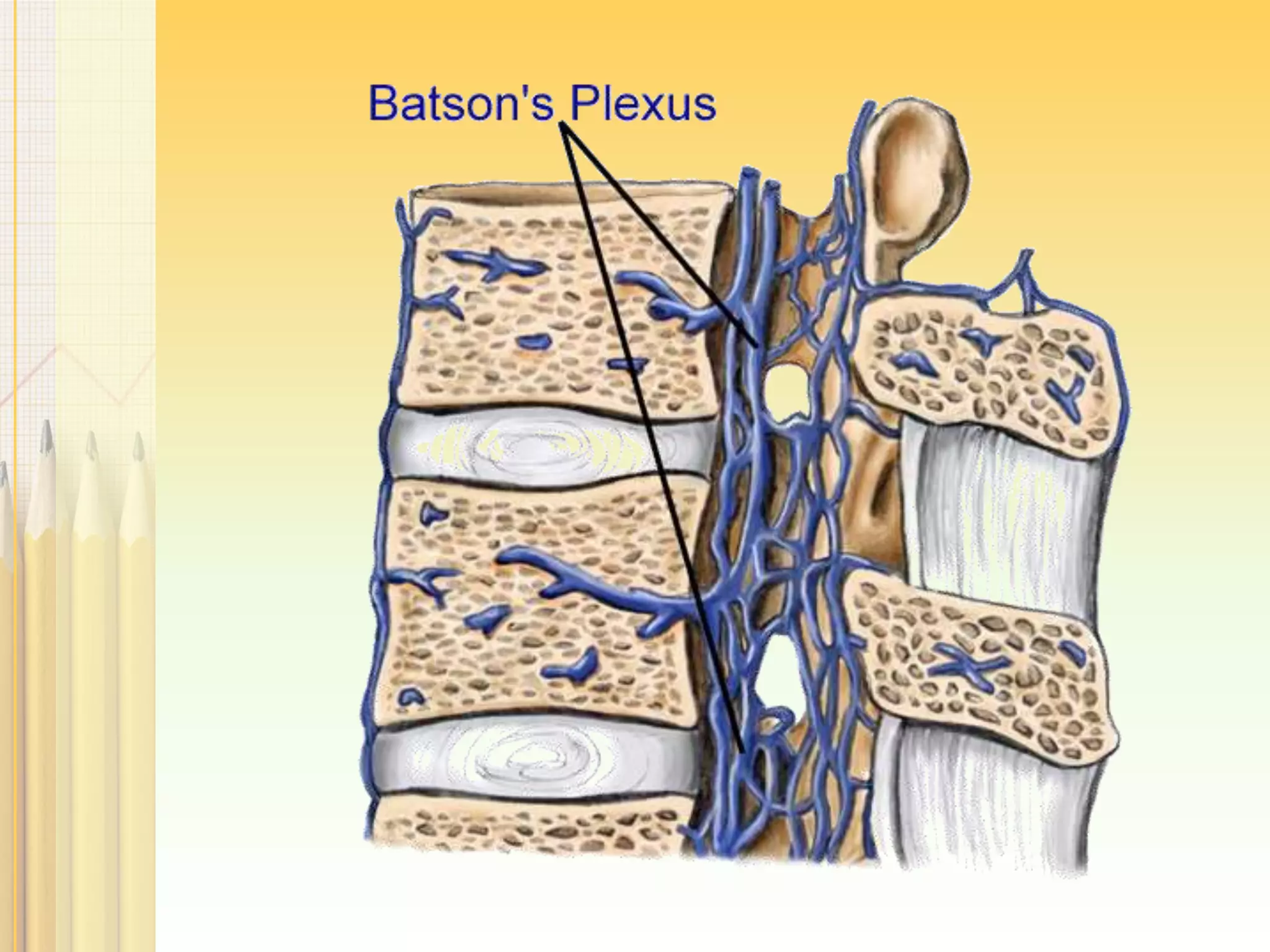
This document provides an overview of the anatomy of the epidural space. It discusses the boundaries, contents, size, and structures that must be penetrated to access the epidural space. Key points include that the epidural space lies between the spinal meninges and vertebral canal, contains connective tissue, fat, blood vessels and spinal nerves. It varies in size from 1-6mm depending on the spinal region. To access it requires penetrating the skin, ligaments and ligamentum flavum in the midline.
Neuromuscular monitoring, also known as train of four monitoring, is a technique used during recovery from the application of general anesthesia to objectively determine how well a patient's muscles are able to function. It involves the application of electrical stimulation to nerves and recording of muscle response using, for example, an acceleromyograph. Neuromuscular monitoring is typically used when neuromuscular-blocking drugs have been part of the general anesthesia and the doctor wishes to avoid postoperative residual curarization (PORC) in the patient, that is, the residual paralysis of muscles stemming from these drugs.
new technique for pain management ,described by dr forero ,it can replace epidural anesthesia,paravertebral anesthesia and other regional blocks.it can be used for both acute and chronic painful conditions
Intro to Hypoxic pulmonary vasoconstriction Arun Shetty
Hypoxic pulmonary vasoconstriction, a seldom heard phenomenon but very effective physiologic property which helps lungs utilise ventilation to the maximum
In critical care medicine the invasive life saving techniques are often employed and when all goes well such interventions will be withdrawn to all for normal physiology to resume. Identifying this point for safe withdrawal for the resumption of normal respiratory function is of utmost importance.
mapleson circuits used in anesthesia practice, are in their way out but it is as important to know the mechanism with which the gases flow to and fro through them.
At the end of the presentation we should be able to:
Understand the perineum
Know the perineal pouches, Ischiorectal fossa and their contents
Identify the boundaries and recessess of ischiorectal fossa
Understand the Vasculature of the perineum
Diamond-shaped area between
Pubic symphysis (anteriorly)
Coccyx (posteriorly)
Ischial tuberosities (laterally)
Males contain
Scrotum, root of penis, anus
Females contain
External genitalia, anus
New Directions in Targeted Therapeutic Approaches for Older Adults With Mantl...i3 Health
i3 Health is pleased to make the speaker slides from this activity available for use as a non-accredited self-study or teaching resource.
This slide deck presented by Dr. Kami Maddocks, Professor-Clinical in the Division of Hematology and
Associate Division Director for Ambulatory Operations
The Ohio State University Comprehensive Cancer Center, will provide insight into new directions in targeted therapeutic approaches for older adults with mantle cell lymphoma.
STATEMENT OF NEED
Mantle cell lymphoma (MCL) is a rare, aggressive B-cell non-Hodgkin lymphoma (NHL) accounting for 5% to 7% of all lymphomas. Its prognosis ranges from indolent disease that does not require treatment for years to very aggressive disease, which is associated with poor survival (Silkenstedt et al, 2021). Typically, MCL is diagnosed at advanced stage and in older patients who cannot tolerate intensive therapy (NCCN, 2022). Although recent advances have slightly increased remission rates, recurrence and relapse remain very common, leading to a median overall survival between 3 and 6 years (LLS, 2021). Though there are several effective options, progress is still needed towards establishing an accepted frontline approach for MCL (Castellino et al, 2022). Treatment selection and management of MCL are complicated by the heterogeneity of prognosis, advanced age and comorbidities of patients, and lack of an established standard approach for treatment, making it vital that clinicians be familiar with the latest research and advances in this area. In this activity chaired by Michael Wang, MD, Professor in the Department of Lymphoma & Myeloma at MD Anderson Cancer Center, expert faculty will discuss prognostic factors informing treatment, the promising results of recent trials in new therapeutic approaches, and the implications of treatment resistance in therapeutic selection for MCL.
Target Audience
Hematology/oncology fellows, attending faculty, and other health care professionals involved in the treatment of patients with mantle cell lymphoma (MCL).
Learning Objectives
1.) Identify clinical and biological prognostic factors that can guide treatment decision making for older adults with MCL
2.) Evaluate emerging data on targeted therapeutic approaches for treatment-naive and relapsed/refractory MCL and their applicability to older adults
3.) Assess mechanisms of resistance to targeted therapies for MCL and their implications for treatment selection
TEST BANK for Operations Management, 14th Edition by William J. Stevenson, Ve...kevinkariuki227
TEST BANK for Operations Management, 14th Edition by William J. Stevenson, Verified Chapters 1 - 19, Complete Newest Version.pdf
Report Back from SGO 2024: What’s the Latest in Cervical Cancer?bkling
Are you curious about what’s new in cervical cancer research or unsure what the findings mean? Join Dr. Emily Ko, a gynecologic oncologist at Penn Medicine, to learn about the latest updates from the Society of Gynecologic Oncology (SGO) 2024 Annual Meeting on Women’s Cancer. Dr. Ko will discuss what the research presented at the conference means for you and answer your questions about the new developments.
These simplified slides by Dr. Sidra Arshad present an overview of the non-respiratory functions of the respiratory tract.
Learning objectives:
1. Enlist the non-respiratory functions of the respiratory tract
2. Briefly explain how these functions are carried out
3. Discuss the significance of dead space
4. Differentiate between minute ventilation and alveolar ventilation
5. Describe the cough and sneeze reflexes
Study Resources:
1. Chapter 39, Guyton and Hall Textbook of Medical Physiology, 14th edition
2. Chapter 34, Ganong’s Review of Medical Physiology, 26th edition
3. Chapter 17, Human Physiology by Lauralee Sherwood, 9th edition
4. Non-respiratory functions of the lungs https://academic.oup.com/bjaed/article/13/3/98/278874
ARTIFICIAL INTELLIGENCE IN HEALTHCARE.pdfAnujkumaranit
Artificial intelligence (AI) refers to the simulation of human intelligence processes by machines, especially computer systems. It encompasses tasks such as learning, reasoning, problem-solving, perception, and language understanding. AI technologies are revolutionizing various fields, from healthcare to finance, by enabling machines to perform tasks that typically require human intelligence.
Flu Vaccine Alert in Bangalore Karnatakaaddon Scans
As flu season approaches, health officials in Bangalore, Karnataka, are urging residents to get their flu vaccinations. The seasonal flu, while common, can lead to severe health complications, particularly for vulnerable populations such as young children, the elderly, and those with underlying health conditions.
Dr. Vidisha Kumari, a leading epidemiologist in Bangalore, emphasizes the importance of getting vaccinated. "The flu vaccine is our best defense against the influenza virus. It not only protects individuals but also helps prevent the spread of the virus in our communities," he says.
This year, the flu season is expected to coincide with a potential increase in other respiratory illnesses. The Karnataka Health Department has launched an awareness campaign highlighting the significance of flu vaccinations. They have set up multiple vaccination centers across Bangalore, making it convenient for residents to receive their shots.
To encourage widespread vaccination, the government is also collaborating with local schools, workplaces, and community centers to facilitate vaccination drives. Special attention is being given to ensuring that the vaccine is accessible to all, including marginalized communities who may have limited access to healthcare.
Residents are reminded that the flu vaccine is safe and effective. Common side effects are mild and may include soreness at the injection site, mild fever, or muscle aches. These side effects are generally short-lived and far less severe than the flu itself.
Healthcare providers are also stressing the importance of continuing COVID-19 precautions. Wearing masks, practicing good hand hygiene, and maintaining social distancing are still crucial, especially in crowded places.
Protect yourself and your loved ones by getting vaccinated. Together, we can help keep Bangalore healthy and safe this flu season. For more information on vaccination centers and schedules, residents can visit the Karnataka Health Department’s official website or follow their social media pages.
Stay informed, stay safe, and get your flu shot today!
- Video recording of this lecture in English language: https://youtu.be/lK81BzxMqdo
- Video recording of this lecture in Arabic language: https://youtu.be/Ve4P0COk9OI
- Link to download the book free: https://nephrotube.blogspot.com/p/nephrotube-nephrology-books.html
- Link to NephroTube website: www.NephroTube.com
- Link to NephroTube social media accounts: https://nephrotube.blogspot.com/p/join-nephrotube-on-social-media.html
Factory Supply Best Quality Pmk Oil CAS 28578–16–7 PMK Powder in Stockrebeccabio
Factory Supply Best Quality Pmk Oil CAS 28578–16–7 PMK Powder in Stock
Telegram: bmksupplier
signal: +85264872720
threema: TUD4A6YC
You can contact me on Telegram or Threema
Communicate promptly and reply
Free of customs clearance, Double Clearance 100% pass delivery to USA, Canada, Spain, Germany, Netherland, Poland, Italy, Sweden, UK, Czech Republic, Australia, Mexico, Russia, Ukraine, Kazakhstan.Door to door service
Hot Selling Organic intermediates
Ozempic: Preoperative Management of Patients on GLP-1 Receptor Agonists Saeid Safari
Preoperative Management of Patients on GLP-1 Receptor Agonists like Ozempic and Semiglutide
ASA GUIDELINE
NYSORA Guideline
2 Case Reports of Gastric Ultrasound
Ozempic: Preoperative Management of Patients on GLP-1 Receptor Agonists
Anatomy of epidural space
1. Anatomy of Epidural Space
Chairperson: Dr.Vinayak Jannu
Presenters: Dr.Niharika R & Dr. Karthavya S L
2. Epidural space or Peridural space or Extradural space is the
space that lies between the spinal meninges and the sides
of the vertebral canal.
3. History
1885: Corning first performed epidural anesthesia with
cociane for pain relief in extremity but it was apparently
accidental.
1895: Cathelin first used epidural in sacral region which is
now called caudal analgesia.
1921: Fidel Pages first used epidural anaesthesia in his
surgical practice.
Tuohy introduced the needle used in Epidural Anaesthesia
in 1945.
1951: Crawford used epidural anesthesia for thoracic
surgery.
4. The epidural space is the potential space between periosteum
lining the vertebral canal and the spinal dura mater.
It extends from the foramen magnum to the sacral hiatus, and
surrounds the dura mater anteriorly, laterally, and posteriorly
5. Boundaries
Cranially by foramen magnum.
Caudally by sacrococcygeal ligament. (sacral hiatus)
Anteriorly by posterior longitudinal ligament
Laterally by vertebral pedicles & intervertebral foramina.
Posteriorly by ligamentum flavum and the laminae.
6. Boundaries
At foramen magnum the vertebral periosteum fuses with
the periosteal layer of the skull
This periosteal layer is the extracranial extension of the
endosteal layer of cranial dura,the spinal dura at the same
point fuses with the meningeal layer of cranial dura.
The lower limit is the sacrococcygeal ligament.
7. Boundaries
The space is more extensive and easily distensible
posteriorly while anteriorly the dura adheres closely to the
periosteum of vertebral bodies.
Laterally the space accompany the spinal nerves through
the intervertebral foramina into the paravertebral tissue up
to the angle of ribs.
8. Contents
Areolar connective tissue
Fat
Spinal nerve roots with their dural sleeves
Blood vessels-spinal arteries and venous plexus (Batson’s
plexus)
Lymphatics
9. Areolar connective tissue
It is present in significant amounts ventrally forming
strong connections between duramater and anterior
longitudinal ligament in the vertebral canal.
The existence of fibrous connections in the posterior
epidural space called as PLICA MEDIANA DORSALIS of
the duramater extending longitudinally in the midline
connecting the dura and ligamentum flavum is also noted.
10. PLICA MEDIANA DORSALIS
This is in form of strands connecting flaval ligament to the
dura.
It is present in the midline and these bands divide the
epidural space into right and left sides and narrow the
space.
The connection is well developed in region of vertebral
arches.
In some instances, it forms a complete membrane in a
dorsomedian sagittal plane.
11. Epidural Fat
It is principally present in posterior and lateral space.
It has effects on pharmacology of drugs injected
intrathecally.
There is a linear relationship between opiod’s lipid
solubility and its terminal elimination halftime in the
epidural space.
Increased lipid solubility leads to sequestration of drugs in
fat, thereby reducing bioavailability of drug.
12.
13. Lymphatics
The lymphatics of the epidural space are concentrated in the
region of the dural roots where they remove foreign
materials including microorganisms from the
subarachnoid and epidural spaces.
14. Blood vessels
The epidural arteries located in the lumbar region of the
vertebral column are branches of the ilio-lumbar arteries.
These arteries are found in the lateral region of the space
and therefore not threatened by an advancing epidural
needle.
15. Blood vessels
The internal vertebral venous plexus consists of four
interconnecting longitudinal vessels, two anterior and two
posterior.
The external vertebral plexus (EVP) in contrast, lies
peripheral to the vertebrae and is made of the anterior and
posterior external vertebral plexuses.
The EVP is situated anterior to the vertebral bodies and in
relation to the laminae, spinous processes,transverse
processes and articular processes respectively.
16.
17. Blood vessels
These veins communicate with the segmental veins of the neck,
the intercostal, azygos and lumbar veins. With the veins of bones
of the vertebral column, the internal and external vertebral
plexuses form Batson’s plexus.
These veins are predominantly in the antero-lateral part of the
epidural space, and ultimately drain into the azygous system of
veins.
As the whole system is valveless, increased intrathoracic or intra-
abdominal pressure (e.g. ascites, pregnancy) can lead to major
congestion and vessel enlargement within the spinal canal.
18.
19. Size of epidural space
The distance across the semi or half moon circular
epidural space varies.
In the anterior region it is almost nonexistent while it can
be measured in posterior region, the average values are;
Cervical: 1mm-1.5mm
Upper thoracic: 2.5mm-3mm
Lower thoracic: 4mm-5mm
Lumbar: 5mm-6mm
20. How to reach?
To reach epidural space in midline these structures have to
be penetrated:
Skin and subcutaneous tissues
Supraspinous ligaments
Interspinous ligaments
Ligamentum flavum