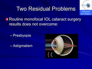
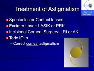
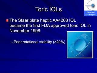
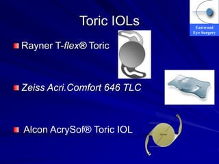
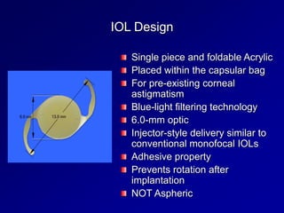
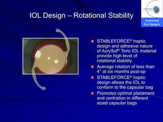
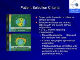
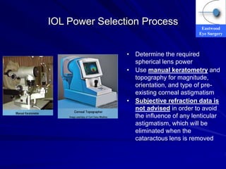
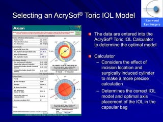
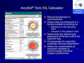
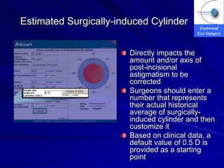
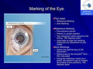
Eastwood
Eye Surgery
Toric and Modern IOL
Technology
Dr Gagan Khannah
Ophthalmic Surgeon
Eastwood Eye Surgery
Sydney Eye Hospital
Stamford Grand
3rd May 2009
Eastwood
Eye Surgery
Cataract and Refractive
Surgery
Cataract surgery and refractive surgery
are now seen as a surgical spectrum
Significant advances in safety, technology,
techniques and results
2006 200,000 Cataract operations
2006 50,000 Refractive operations
>10% of >60yo have IOLs
Cataract surgery is very cost effective
surgery
Eastwood
Eye Surgery
Two Residual Problems
Routine monofocal IOL cataract surgery
results does not overcome:
– Presbyopia
– Astigmatism
Eastwood
Eye Surgery
Treatment of Astigmatism
Spectacles or Contact lenses
Excimer Laser: LASIK or PRK
Incisional Corneal Surgery: LRI or AK
Toric IOLs
– Correct corneal astigmatism
Eastwood
Eye Surgery
Toric IOLs
The Staar plate haptic AA4203 IOL
became the first FDA approved toric IOL in
November 1998
– Poor rotational stability (>20%)
Eastwood
Eye Surgery
Toric IOLs
Rayner T-flex® Toric
Zeiss Acri.Comfort 646 TLC
Alcon AcrySof® Toric IOL
IOL Design
Single piece and foldable Acrylic
Placed within the capsular bag
For pre-existing corneal
astigmatism
Blue-light filtering technology
6.0-mm optic
Injector-style delivery similar to
conventional monofocal IOLs
Adhesive property
Prevents rotation after
implantation
NOT Aspheric
Eastwood
Eye Surgery
IOL Design – Optic Markings
Surgeons must
Choose the correct
AcrySof® Toric IOL power
Ensure precise alignment of
IOL within the eye relative
to the patient’s axis of
corneal astigmatism
Designed with axis marks
on the posterior surface
IOL placed and marks
aligned precisely with the
steep axis of the
postincisional cornea
Eastwood
Eye Surgery
IOL Design – Rotational Stability
Lens stability is important
Off-axis rotation reduces the
corrective cylinder power
For every 1° of rotation,
3.3% of the lens cylinder
power is lost
For 30° of rotation there is a
complete loss of astigmatic
correction
Additional astigmatism or
visual problems with greater
than 30° of rotation
Eastwood
Eye Surgery
IOL Design – Rotational Stability
STABLEFORCE® haptic
design and adhesive nature
of AcrySof® Toric IOL material
provide high level of
rotational stability
Average rotation of less than
4° at six months post-op
STABLEFORCE® haptic
design allows the IOL to
conform to the capsular bag
Promotes optimal placement
and centration in different
sized capsular bags
Eastwood
Eye Surgery
AcrySof® Toric IOL Models
• Three AcrySof® Toric IOL
models initially available
• Chart shows the model
numbers, the power at the IOL
and corneal planes, and the
recommended range of
astigmatism correction
• Additional power options will
be added in the future to
address a broader range of
astigmatic conditions
• Aspheric models to be
released in Australia
Eastwood
Eye Surgery
Patient Selection Criteria
Proper patient selection is critical to
achieve success
Suitable candidates are cataract
patients with pre-existing corneal
astigmatism
> 0.75 D with the following
characteristics
– Manual keratometry: steep and
flat meridians ~90° apart
– Corneal topography: symmetrical
astigmatism
– Intact capsular bag compatible with
continuous curvilinear capsulotomy
performed with in-the-bag
placement of the IOL
Eastwood
Eye Surgery
IOL Power Selection Process
• Determine the required
spherical lens power
• Use manual keratometry and
topography for magnitude,
orientation, and type of pre-
existing corneal astigmatism
• Subjective refraction data is
not advised in order to avoid
the influence of any lenticular
astigmatism, which will be
eliminated when the
cataractous lens is removed
Eastwood
Eye Surgery
Selecting an AcrySof® Toric IOL Model
The data are entered into the
AcrySof® Toric IOL Calculator
to determine the optimal model
Calculator
– Considers the effect of
incision location and
surgically induced cylinder
to make a more precise
calculation
– Determines the correct IOL
model and optimal axis
placement of the IOL in the
capsular bag
Eastwood
Eye Surgery
AcrySof® Toric IOL Calculator
Manual keratometry is
recommended
Output data are displayed in a
format suitable for printing for
– Reference in the operating
room
– Inclusion in the patient’s chart
Determines the optimal axis
placement of the lens within the
capsular bag
Compensates for expected
surgically induced astigmatism
Allows for customization of
important variables to
accommodate individual
surgeon preferences
Eastwood
Eye Surgery
Estimated Surgically-induced Cylinder
Directly impacts the
amount and/or axis of
post-incisional
astigmatism to be
corrected
Surgeons should enter a
number that represents
their actual historical
average of surgically-
induced cylinder and then
customize it
Based on clinical data, a
default value of 0.5 D is
provided as a starting
point
Eastwood
Eye Surgery
Marking of the Eye
Two steps
– Reference Marking
– Axis Marking
Reference Marking
– Pre-induction period
– Patient in upright position
– Two reference marks placed at the
limbus, 180 degrees apart
– Used later to align the marking
instrument for placement of axis
marks
Axis Markings
• Define the optimal axis of IOL
placement
• Determined by the AcrySof® Toric
IOL Calculator
• Using the reference marks as a
guide, the patient’s eye is marked
accurately at two positions, 180
degrees apart
Eastwood
Eye Surgery
Reference Marking
Eastwood
Eye Surgery
Axis Marking
Eastwood
Eye Surgery
Intraoperative IOL Alignment
Gross alignment
– Inject the IOL into the capsular bag
– Rotate the IOL clockwise, approximately 20° to 30°
short of the intended final axis location
Viscoelastic removal
– Ensure that the IOL does not rotate beyond the
intended final axis location
– Carefully remove viscoelastic from both the anterior
and posterior sides of the lens
Final alignment of the IOL
– Rotate the lens clockwise precisely to the intended
axis of alignment as previously marked
Eastwood
Eye Surgery
Summary
AcrySof® Toric IOL
– Good rotational stability
– The presence of an online calculator brings a high
level of precision and accuracy to the selection of
the correct IOL model and optimal axis placement
of the IOL. Provides flexibility in surgical planning
for precise correction of astigmatism
– Routine surgical technique except accurate marking
of the eye, and precise alignment of the IOL within
the capsular bag
Eastwood
Eye Surgery
Limitations
Not Aspheric
Limited cylinder power options
No combination of Toric Multifocal yet
Always under promise and over
deliver!!
Eastwood
Eye Surgery
Future: Super IOL
One piece
Acrylic
Aspheric
Accommodative or Multifocal
Toric
Preloaded
Centration will become vital
Thank You!

Cataract and Refractive Surgery.ppt

  • 1.
    Eastwood Eye Surgery Toric andModern IOL Technology Dr Gagan Khannah Ophthalmic Surgeon Eastwood Eye Surgery Sydney Eye Hospital Stamford Grand 3rd May 2009
  • 2.
    Eastwood Eye Surgery Cataract andRefractive Surgery Cataract surgery and refractive surgery are now seen as a surgical spectrum Significant advances in safety, technology, techniques and results 2006 200,000 Cataract operations 2006 50,000 Refractive operations >10% of >60yo have IOLs Cataract surgery is very cost effective surgery
  • 3.
    Eastwood Eye Surgery Two ResidualProblems Routine monofocal IOL cataract surgery results does not overcome: – Presbyopia – Astigmatism
  • 4.
    Eastwood Eye Surgery Treatment ofAstigmatism Spectacles or Contact lenses Excimer Laser: LASIK or PRK Incisional Corneal Surgery: LRI or AK Toric IOLs – Correct corneal astigmatism
  • 5.
    Eastwood Eye Surgery Toric IOLs TheStaar plate haptic AA4203 IOL became the first FDA approved toric IOL in November 1998 – Poor rotational stability (>20%)
  • 6.
    Eastwood Eye Surgery Toric IOLs RaynerT-flex® Toric Zeiss Acri.Comfort 646 TLC Alcon AcrySof® Toric IOL
  • 7.
    IOL Design Single pieceand foldable Acrylic Placed within the capsular bag For pre-existing corneal astigmatism Blue-light filtering technology 6.0-mm optic Injector-style delivery similar to conventional monofocal IOLs Adhesive property Prevents rotation after implantation NOT Aspheric
  • 8.
    Eastwood Eye Surgery IOL Design– Optic Markings Surgeons must Choose the correct AcrySof® Toric IOL power Ensure precise alignment of IOL within the eye relative to the patient’s axis of corneal astigmatism Designed with axis marks on the posterior surface IOL placed and marks aligned precisely with the steep axis of the postincisional cornea
  • 9.
    Eastwood Eye Surgery IOL Design– Rotational Stability Lens stability is important Off-axis rotation reduces the corrective cylinder power For every 1° of rotation, 3.3% of the lens cylinder power is lost For 30° of rotation there is a complete loss of astigmatic correction Additional astigmatism or visual problems with greater than 30° of rotation
  • 10.
    Eastwood Eye Surgery IOL Design– Rotational Stability STABLEFORCE® haptic design and adhesive nature of AcrySof® Toric IOL material provide high level of rotational stability Average rotation of less than 4° at six months post-op STABLEFORCE® haptic design allows the IOL to conform to the capsular bag Promotes optimal placement and centration in different sized capsular bags
  • 11.
    Eastwood Eye Surgery AcrySof® ToricIOL Models • Three AcrySof® Toric IOL models initially available • Chart shows the model numbers, the power at the IOL and corneal planes, and the recommended range of astigmatism correction • Additional power options will be added in the future to address a broader range of astigmatic conditions • Aspheric models to be released in Australia
  • 12.
    Eastwood Eye Surgery Patient SelectionCriteria Proper patient selection is critical to achieve success Suitable candidates are cataract patients with pre-existing corneal astigmatism > 0.75 D with the following characteristics – Manual keratometry: steep and flat meridians ~90° apart – Corneal topography: symmetrical astigmatism – Intact capsular bag compatible with continuous curvilinear capsulotomy performed with in-the-bag placement of the IOL
  • 13.
    Eastwood Eye Surgery IOL PowerSelection Process • Determine the required spherical lens power • Use manual keratometry and topography for magnitude, orientation, and type of pre- existing corneal astigmatism • Subjective refraction data is not advised in order to avoid the influence of any lenticular astigmatism, which will be eliminated when the cataractous lens is removed
  • 14.
    Eastwood Eye Surgery Selecting anAcrySof® Toric IOL Model The data are entered into the AcrySof® Toric IOL Calculator to determine the optimal model Calculator – Considers the effect of incision location and surgically induced cylinder to make a more precise calculation – Determines the correct IOL model and optimal axis placement of the IOL in the capsular bag
  • 15.
    Eastwood Eye Surgery AcrySof® ToricIOL Calculator Manual keratometry is recommended Output data are displayed in a format suitable for printing for – Reference in the operating room – Inclusion in the patient’s chart Determines the optimal axis placement of the lens within the capsular bag Compensates for expected surgically induced astigmatism Allows for customization of important variables to accommodate individual surgeon preferences
  • 16.
    Eastwood Eye Surgery Estimated Surgically-inducedCylinder Directly impacts the amount and/or axis of post-incisional astigmatism to be corrected Surgeons should enter a number that represents their actual historical average of surgically- induced cylinder and then customize it Based on clinical data, a default value of 0.5 D is provided as a starting point
  • 17.
    Eastwood Eye Surgery Marking ofthe Eye Two steps – Reference Marking – Axis Marking Reference Marking – Pre-induction period – Patient in upright position – Two reference marks placed at the limbus, 180 degrees apart – Used later to align the marking instrument for placement of axis marks Axis Markings • Define the optimal axis of IOL placement • Determined by the AcrySof® Toric IOL Calculator • Using the reference marks as a guide, the patient’s eye is marked accurately at two positions, 180 degrees apart
  • 18.
  • 19.
  • 20.
    Eastwood Eye Surgery Intraoperative IOLAlignment Gross alignment – Inject the IOL into the capsular bag – Rotate the IOL clockwise, approximately 20° to 30° short of the intended final axis location Viscoelastic removal – Ensure that the IOL does not rotate beyond the intended final axis location – Carefully remove viscoelastic from both the anterior and posterior sides of the lens Final alignment of the IOL – Rotate the lens clockwise precisely to the intended axis of alignment as previously marked
  • 21.
    Eastwood Eye Surgery Summary AcrySof® ToricIOL – Good rotational stability – The presence of an online calculator brings a high level of precision and accuracy to the selection of the correct IOL model and optimal axis placement of the IOL. Provides flexibility in surgical planning for precise correction of astigmatism – Routine surgical technique except accurate marking of the eye, and precise alignment of the IOL within the capsular bag
  • 22.
    Eastwood Eye Surgery Limitations Not Aspheric Limitedcylinder power options No combination of Toric Multifocal yet Always under promise and over deliver!!
  • 23.
    Eastwood Eye Surgery Future: SuperIOL One piece Acrylic Aspheric Accommodative or Multifocal Toric Preloaded Centration will become vital
  • 24.