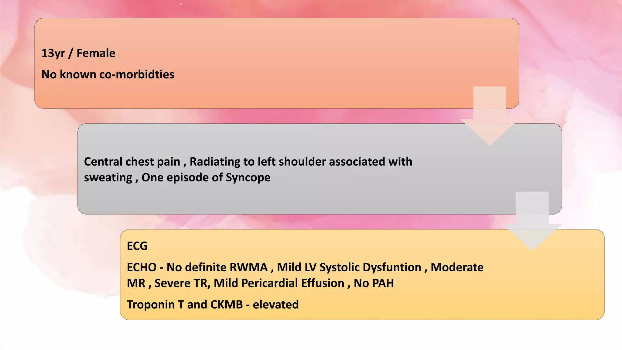
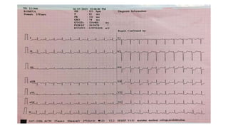
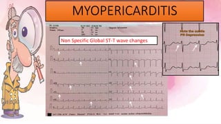
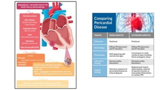
13yr / Female
No known co-morbidties
Central chest pain , Radiating to left shoulder associated with
sweating , One episode of Syncope
ECG
ECHO - No definite RWMA , Mild LV Systolic Dysfuntion , Moderate
MR , Severe TR, Mild Pericardial Effusion , No PAH
Troponin T and CKMB - elevated
MI
MYOPERICARDITIS
MYOPERICARDITIS
Non Specific Global ST-T wave changes
ECG changes in Myocarditis
The most common ECG abnormalities seen in myocarditis are:
• Sinus tachycardia
• Non-specific ST segment and T waves changes
Other ECG changes are variable, and may include:
• Prolonged QRS
• QT prolongation
• Diffuse T wave inversion
• Ventricular arrhythmias
• AV conduction defects
ECG changes
in Pericarditis
Pericarditis is classically associated with ECG changes that evolve through four stages.
Stage 1 – widespread STE and PR depression with reciprocal changes in aVR (occurs during the first two weeks)
Stage 2 – normalisation of ST changes; generalised T wave flattening (1 to 3 weeks)
Stage 3 – flattened T waves become inverted (3 to several weeks)
Stage 4 – ECG returns to normal (several weeks onwards)
Note: Less than 50% of patients progress through all four classical stages and evolution of changes may not follow
this typical pattern.
Summary
• This 13 year old patient admitted with above complaints of acute onset. With ECG and
ECHO changes supported with elevated Cardiac markers suggestive of Probable
myopericarditis with viral etiology.
• She initially worsened with deterioration from mild to severe LV systolic dysfunction and
respiratory distress requiring CPAP and IV inotropic support.
• Gradually recovered and tapered off inotropic and CPAP supports.
• No arrythmia documented during hospital stay.
• Patient was discharged with Steroids on tapering dose.
THANK YOU

case presentation.pptx

  • 1.
    13yr / Female Noknown co-morbidties Central chest pain , Radiating to left shoulder associated with sweating , One episode of Syncope ECG ECHO - No definite RWMA , Mild LV Systolic Dysfuntion , Moderate MR , Severe TR, Mild Pericardial Effusion , No PAH Troponin T and CKMB - elevated
  • 4.
  • 5.
  • 8.
    ECG changes inMyocarditis The most common ECG abnormalities seen in myocarditis are: • Sinus tachycardia • Non-specific ST segment and T waves changes Other ECG changes are variable, and may include: • Prolonged QRS • QT prolongation • Diffuse T wave inversion • Ventricular arrhythmias • AV conduction defects
  • 9.
    ECG changes in Pericarditis Pericarditisis classically associated with ECG changes that evolve through four stages. Stage 1 – widespread STE and PR depression with reciprocal changes in aVR (occurs during the first two weeks) Stage 2 – normalisation of ST changes; generalised T wave flattening (1 to 3 weeks) Stage 3 – flattened T waves become inverted (3 to several weeks) Stage 4 – ECG returns to normal (several weeks onwards) Note: Less than 50% of patients progress through all four classical stages and evolution of changes may not follow this typical pattern.
  • 10.
    Summary • This 13year old patient admitted with above complaints of acute onset. With ECG and ECHO changes supported with elevated Cardiac markers suggestive of Probable myopericarditis with viral etiology. • She initially worsened with deterioration from mild to severe LV systolic dysfunction and respiratory distress requiring CPAP and IV inotropic support. • Gradually recovered and tapered off inotropic and CPAP supports. • No arrythmia documented during hospital stay. • Patient was discharged with Steroids on tapering dose.
  • 11.