Pericarditis and Myocarditis
Dr. Nagendra Chaudhary
MD Pediatrics (AIIMS, New Delhi)
Professor and Head
Department of Pediatrics, UCMS-TH
Pericarditis
Introduction
Pericardium
 Fibro-elastic sac
 Visceral and parietal layers
 Separated by a space called pericardial cavity
Diseases of the pericardium
• Acute and recurrent pericarditis
• Pericardial effusion
• Cardiac tamponade
• Constrictive pericarditis
Definition
Acute pericarditis refers to inflammation of
the pericardial sac
Etiology
A. Idiopathic
• Presumed to be viral, post viral, or immune
mediated
• Majority of patients are not found to have an
identifiable cause
Etiology
B. Infectious causes
• Viral (common): Coxsackie virus, coronavirus,
influenza, herpes viruses, varicella, mumps, rubella,
parvovirus B19, HIV
• Bacterial: Mycobacterium tuberculosis
• Fungal (very rare): Aspergillus, Candida, Histoplasma
• Parasitic (very rare): Toxoplasma, Echinococcus,
Trypanosoma cruzi
Etiology
C. Non-infectious causes
• Autoimmune (common): SLE, rheumatoid arthritis,
scleroderma, MCTD
• Neoplastic: Pericardial tumors
• Metabolic: Uremia
• Trauma
• Drugs- Penicillin, isoniazid, methyl dopa
• Radiation
Common etiology
Viral in origin in developed countries
whereas in developing world
Tuberculosis is the most frequent etiology
Classification
Clinical features
• Chest pain and/or dyspnea : Sharp and pleuritic,
worsened when supine and improved by sitting up
and leaning forward
• Pericardial friction rub: Superficial scratchy or
squeaking sound
• ECG changes: New widespread ST elevation and PR
depression
• Pericardial effusion: Common feature of
pericarditis
Presence of at least 2 of the above 4 criteria
Clinical features
Non specific symptoms
• Infectious etiology- fever and leukocytosis
• Viral etiologies- flu-like" respiratory or
gastrointestinal symptoms
• Autoimmune disorder or malignancy may present
with signs or symptoms specific to their
underlying disorder
Clinical signs
• Pericardial friction rub
• Tachycardia
• Signs of pericardial effusion
– Muffled heart sounds
– Hypotension
– Raised JVP if tamponade
Investigations
Echocardiography
• Can be normal
• Detects pericardial effusion
Chest X-ray
• Can be normal
• Detects pericardial effusion
Cardiac biomarkers
• May be associated with in serum biomarkers of
myocardial injury (Troponin I or T)
Investigations
Signs of inflammation
• Increase in WBC count, ESR and CRP
Cardiac MR and/or CT
• thickness of the pericardium
Pericardiocentesis and pericardial biopsy
ECG changes in Pericarditis
Up-sloping (concave up) ST-segment elevations in leads II, III, aVF, and V2 to V6
Management
Hospital admission
Urgent pericardiocentesis
• for cardiac tamponade
Drugs
• NSAIDs- Ibuprofen
• Colchicine
• Steroids
• Antibiotics/anti-TB drugs
Dialysis
• for uremic patients
Myocarditis
Definition
Acute myocarditis
• Inflammation of the heart muscle
(myocardium) leading to myocardial injury,
necrosis and impaired contractility
• Incidence- 0.2 to 2 cases per 100,000; aged
<18 years
Pathogenesis
• Myocardial inflammation, necrosis and fibrosis
• Myocardial damage leading to decreased
systolic function and cardiomegaly
• Features of shock and hyoptension leading to
death
Etiology
A. Infectious
• Viral (most common)- Coxsackie B, Adenovirus,
Influenza, Parvovirus B19, SARS-CoV-2
• Bacterial: Diptheria, mycoplasma. Lyme disease
• Protozoal- Trypanosoma cruzi (chagas disease)
• Fungal- Candida (rare)
Etiology
B. Non-infectious
• Autoimmune- SLE, Kawasaki disease
• Hypersensitivity/drug induced
• Toxins- alcohol
• Post vaccination (rare)
Clinical features
• Variable
• May range from subclinical disease to
Cardiogenic shock, arrhythmias, and
sudden death
Clinical features
Symptoms
• Viral prodrome- fever, malaise, sore throat, body
ache
• Dyspnea- on exertion
• Palpitation
• Chest pain
• Syncope
Clinical features
Signs
• Tachycardia
• Signs of heart failure- tachypnea, fine crepts, S3
gallop, hepatomegaly, peripheral edema
• Arrhythmia
• Hypotension
• Shock
Fulminant myocarditis
• Most severe form of myocarditis
• Leads to sudden and severe myocardial
inflammation, myocyte necrosis, edema,
and Cardiogenic shock
• May cause death in 68.8 % of affected
children within 7 days of hospital admission
Investigations
• ECG: sinus tachycardia, ST-T changes,
conduction blocks
• Chest x-ray: Cardiomegaly, pulmonary
congestion
• Echocardiography: Dilated ventricles, global
hypokinesia, reduced ejection fraction
Investigations
• Cardiac enzymes- Increased Troponin and CK-
MB
• Blood tests- Increased CRP, ESR, TLC
• Cardiac MRI- myocardial edema
• Endomyocardial biopsy (gold standard)
Diagnostic clues
Recent viral illness
+
New onset heart failure/ arrythmia
+
Tachycardia (out of proportion)
+
Elevated cardiac enzymes
+
ECHO findings s/o cardiac dysfunction
+
ECG changes
Treatment
General
• Hospital admission
• Bed rest
• Restrict physical activity
• Supplemental oxygen
• IV fluids
Treatment
Medical
• Treat heart failure- Diuretics, ACE inhibitors, B-
blockers
• Manage arrhythmia
• Avoid NSAIDs
• IVIG or corticosteroids (in auto-immune cases)
• Antibiotics- in bacterial causes
Prognosis
• Poor in newborns (75% mortality)
• Better in children and adolescents
• 5 -15% patients require cardiac transplantation
• A sub-set of patients develop dilated
cardiomyopathy
Thank You

Pericarditis and myocarditis in children.pptx

  • 1.
    Pericarditis and Myocarditis Dr.Nagendra Chaudhary MD Pediatrics (AIIMS, New Delhi) Professor and Head Department of Pediatrics, UCMS-TH
  • 2.
  • 3.
    Introduction Pericardium  Fibro-elastic sac Visceral and parietal layers  Separated by a space called pericardial cavity
  • 4.
    Diseases of thepericardium • Acute and recurrent pericarditis • Pericardial effusion • Cardiac tamponade • Constrictive pericarditis
  • 5.
    Definition Acute pericarditis refersto inflammation of the pericardial sac
  • 6.
    Etiology A. Idiopathic • Presumedto be viral, post viral, or immune mediated • Majority of patients are not found to have an identifiable cause
  • 7.
    Etiology B. Infectious causes •Viral (common): Coxsackie virus, coronavirus, influenza, herpes viruses, varicella, mumps, rubella, parvovirus B19, HIV • Bacterial: Mycobacterium tuberculosis • Fungal (very rare): Aspergillus, Candida, Histoplasma • Parasitic (very rare): Toxoplasma, Echinococcus, Trypanosoma cruzi
  • 8.
    Etiology C. Non-infectious causes •Autoimmune (common): SLE, rheumatoid arthritis, scleroderma, MCTD • Neoplastic: Pericardial tumors • Metabolic: Uremia • Trauma • Drugs- Penicillin, isoniazid, methyl dopa • Radiation
  • 9.
    Common etiology Viral inorigin in developed countries whereas in developing world Tuberculosis is the most frequent etiology
  • 10.
  • 11.
    Clinical features 
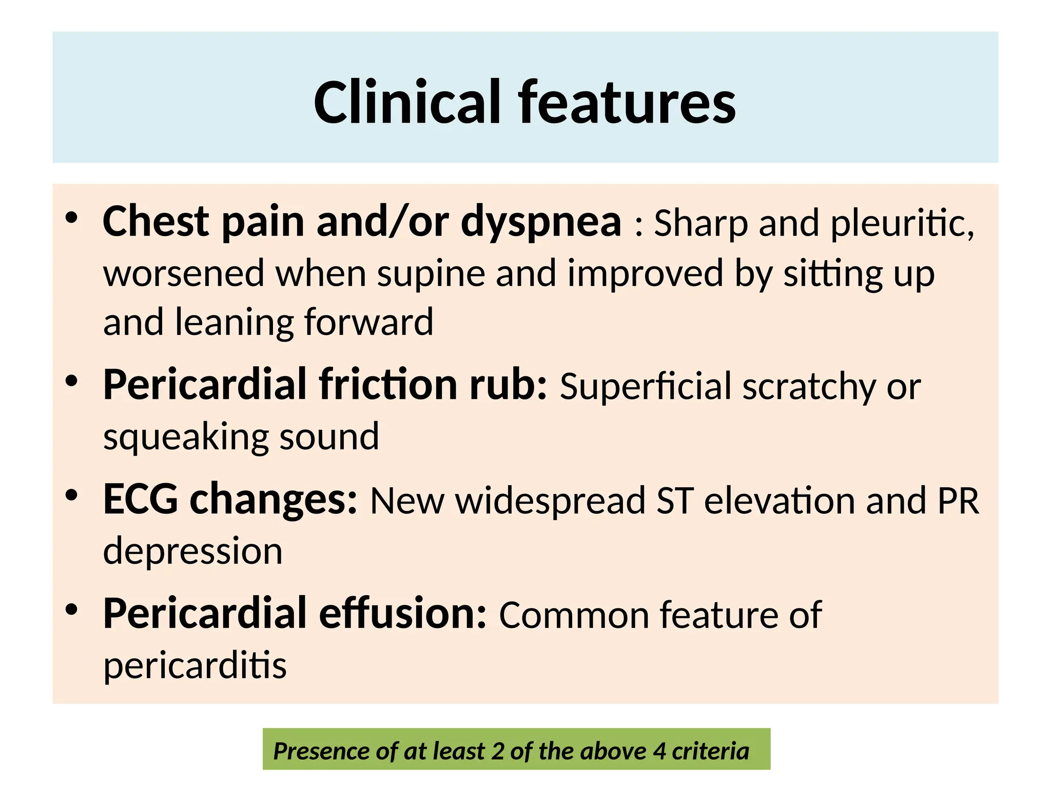
• Chestpain and/or dyspnea : Sharp and pleuritic, worsened when supine and improved by sitting up and leaning forward • Pericardial friction rub: Superficial scratchy or squeaking sound • ECG changes: New widespread ST elevation and PR depression • Pericardial effusion: Common feature of pericarditis Presence of at least 2 of the above 4 criteria
  • 12.
    Clinical features Non specificsymptoms • Infectious etiology- fever and leukocytosis • Viral etiologies- flu-like" respiratory or gastrointestinal symptoms • Autoimmune disorder or malignancy may present with signs or symptoms specific to their underlying disorder
  • 13.
    Clinical signs • Pericardialfriction rub • Tachycardia • Signs of pericardial effusion – Muffled heart sounds – Hypotension – Raised JVP if tamponade
  • 14.
    Investigations Echocardiography • Can benormal • Detects pericardial effusion Chest X-ray • Can be normal • Detects pericardial effusion Cardiac biomarkers • May be associated with in serum biomarkers of myocardial injury (Troponin I or T)
  • 15.
    Investigations Signs of inflammation •Increase in WBC count, ESR and CRP Cardiac MR and/or CT • thickness of the pericardium Pericardiocentesis and pericardial biopsy
  • 16.
    ECG changes inPericarditis Up-sloping (concave up) ST-segment elevations in leads II, III, aVF, and V2 to V6
  • 17.
    Management Hospital admission Urgent pericardiocentesis •for cardiac tamponade Drugs • NSAIDs- Ibuprofen • Colchicine • Steroids • Antibiotics/anti-TB drugs Dialysis • for uremic patients
  • 18.
  • 19.
    Definition Acute myocarditis • Inflammationof the heart muscle (myocardium) leading to myocardial injury, necrosis and impaired contractility • Incidence- 0.2 to 2 cases per 100,000; aged <18 years
  • 20.
    Pathogenesis • Myocardial inflammation,necrosis and fibrosis • Myocardial damage leading to decreased systolic function and cardiomegaly • Features of shock and hyoptension leading to death
  • 21.
    Etiology A. Infectious • Viral(most common)- Coxsackie B, Adenovirus, Influenza, Parvovirus B19, SARS-CoV-2 • Bacterial: Diptheria, mycoplasma. Lyme disease • Protozoal- Trypanosoma cruzi (chagas disease) • Fungal- Candida (rare)
  • 22.
    Etiology B. Non-infectious • Autoimmune-SLE, Kawasaki disease • Hypersensitivity/drug induced • Toxins- alcohol • Post vaccination (rare)
  • 23.
    Clinical features • Variable •May range from subclinical disease to Cardiogenic shock, arrhythmias, and sudden death
  • 24.
    Clinical features Symptoms • Viralprodrome- fever, malaise, sore throat, body ache • Dyspnea- on exertion • Palpitation • Chest pain • Syncope
  • 25.
    Clinical features Signs • Tachycardia •Signs of heart failure- tachypnea, fine crepts, S3 gallop, hepatomegaly, peripheral edema • Arrhythmia • Hypotension • Shock
  • 26.
    Fulminant myocarditis • Mostsevere form of myocarditis • Leads to sudden and severe myocardial inflammation, myocyte necrosis, edema, and Cardiogenic shock • May cause death in 68.8 % of affected children within 7 days of hospital admission
  • 27.
    Investigations • ECG: sinustachycardia, ST-T changes, conduction blocks • Chest x-ray: Cardiomegaly, pulmonary congestion • Echocardiography: Dilated ventricles, global hypokinesia, reduced ejection fraction
  • 28.
    Investigations • Cardiac enzymes-Increased Troponin and CK- MB • Blood tests- Increased CRP, ESR, TLC • Cardiac MRI- myocardial edema • Endomyocardial biopsy (gold standard)
  • 29.
    Diagnostic clues Recent viralillness + New onset heart failure/ arrythmia + Tachycardia (out of proportion) + Elevated cardiac enzymes + ECHO findings s/o cardiac dysfunction + ECG changes
  • 30.
    Treatment General • Hospital admission •Bed rest • Restrict physical activity • Supplemental oxygen • IV fluids
  • 31.
    Treatment Medical • Treat heartfailure- Diuretics, ACE inhibitors, B- blockers • Manage arrhythmia • Avoid NSAIDs • IVIG or corticosteroids (in auto-immune cases) • Antibiotics- in bacterial causes
  • 32.
    Prognosis • Poor innewborns (75% mortality) • Better in children and adolescents • 5 -15% patients require cardiac transplantation • A sub-set of patients develop dilated cardiomyopathy
  • 33.