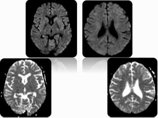
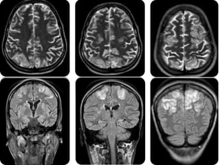
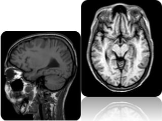
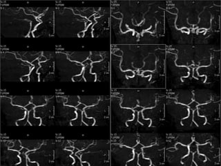
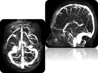
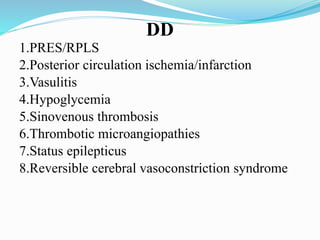
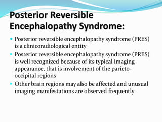
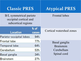
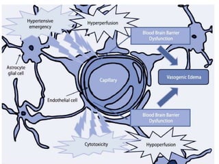
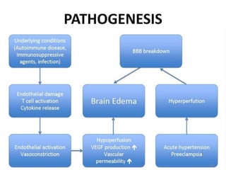
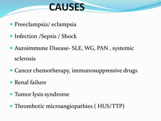
Dr. Sethu Sadanadnan
Registrar
Radiology
 21yr old female patient
 k/c/o SLE on steroids
 Presented with GTCS and Altered sensorium
DD
1.PRES/RPLS
2.Posterior circulation ischemia/infarction
3.Vasulitis
4.Hypoglycemia
5.Sinovenous thrombosis
6.Thrombotic microangiopathies
7.Status epilepticus
8.Reversible cerebral vasoconstriction syndrome
DD
1.P R E S / R P L S
2.Posterior circulation ischemia/infarction
3.Vasulitis
4.Hypoglycemia
5.Sinovenous thrombosis
6.Thrombotic microangiopathies
7.Status epilepticus
8.Reversible cerebral vasoconstriction syndrome
Posterior Reversible
Encephalopathy Syndrome:
 Posterior reversible encephalopathy syndrome (PRES)
is a clinicoradiological entity
 Posterior reversible encephalopathy syndrome (PRES)
is well recognized because of its typical imaging
appearance, that is involvement of the parieto-
occipital regions
 Other brain regions may also be affected and unusual
imaging manifestations are observed frequently
Classic PRES Atypical PRES
B/L symmetrical parieto
occipital cortical and
subcortical regions
Frontal lobes
Cortical watershed zones
Basal ganglia
Brainstem
Cerebellum
Spinal cord
PATHOGENESIS
CAUSES
 Preeclampsia/ eclampsia
 Infection /Sepsis / Shock
 Autoimmune Disease- SLE, WG, PAN , systemic
sclerosis
 Cancer chemotherapy, immunosuppressive drugs
 Renal failure
 Tumor lysis syndrome
 Thrombotic microangiopathies ( HUS/TTP)
 Enhancement (up to 37%) : cortical, leptomeningeal,
parenchymal or pachymeningeal
 Restricted diffusion (11-26%)
 Hemorrhage (10.5-17.1%) : parenchymal or
subarachnoid
 Altered brain perfusion : regional decreased or
increased, depends on disease time course
 Unilateral hemispheric involvement (2.6%)
Altered sensorium Case 2014

Altered sensorium Case 2014

  • 1.
  • 2.
     21yr oldfemale patient  k/c/o SLE on steroids  Presented with GTCS and Altered sensorium
  • 8.
    DD 1.PRES/RPLS 2.Posterior circulation ischemia/infarction 3.Vasulitis 4.Hypoglycemia 5.Sinovenousthrombosis 6.Thrombotic microangiopathies 7.Status epilepticus 8.Reversible cerebral vasoconstriction syndrome
  • 9.
    DD 1.P R ES / R P L S 2.Posterior circulation ischemia/infarction 3.Vasulitis 4.Hypoglycemia 5.Sinovenous thrombosis 6.Thrombotic microangiopathies 7.Status epilepticus 8.Reversible cerebral vasoconstriction syndrome
  • 10.
    Posterior Reversible Encephalopathy Syndrome: Posterior reversible encephalopathy syndrome (PRES) is a clinicoradiological entity  Posterior reversible encephalopathy syndrome (PRES) is well recognized because of its typical imaging appearance, that is involvement of the parieto- occipital regions  Other brain regions may also be affected and unusual imaging manifestations are observed frequently
  • 11.
    Classic PRES AtypicalPRES B/L symmetrical parieto occipital cortical and subcortical regions Frontal lobes Cortical watershed zones Basal ganglia Brainstem Cerebellum Spinal cord
  • 14.
  • 15.
    CAUSES  Preeclampsia/ eclampsia Infection /Sepsis / Shock  Autoimmune Disease- SLE, WG, PAN , systemic sclerosis  Cancer chemotherapy, immunosuppressive drugs  Renal failure  Tumor lysis syndrome  Thrombotic microangiopathies ( HUS/TTP)
  • 16.
     Enhancement (upto 37%) : cortical, leptomeningeal, parenchymal or pachymeningeal  Restricted diffusion (11-26%)  Hemorrhage (10.5-17.1%) : parenchymal or subarachnoid  Altered brain perfusion : regional decreased or increased, depends on disease time course  Unilateral hemispheric involvement (2.6%)

Editor's Notes

  • #14 Hydrostatic leakage with extravasation / transudation of fluid and macromolecules through arteriolar walls with damaged endothelium